Validating Autonomous Synthesis Outcomes: A Modern Guide to XRD and AI-Enhanced Rietveld Refinement
This article provides a comprehensive framework for researchers and drug development professionals to validate crystalline materials from autonomous synthesis platforms.
Validating Autonomous Synthesis Outcomes: A Modern Guide to XRD and AI-Enhanced Rietveld Refinement
Abstract
This article provides a comprehensive framework for researchers and drug development professionals to validate crystalline materials from autonomous synthesis platforms. It covers the foundational principles of X-ray diffraction (XRD), details robust methodological workflows for laboratory data collection and analysis, addresses common troubleshooting and optimization challenges, and explores advanced validation techniques incorporating machine learning. By integrating traditional Rietveld refinement with emerging AI-driven tools like Spotlight and PXRDGen, this guide enables accurate, efficient structural verification to accelerate materials discovery and pharmaceutical development.
XRD and Rietveld Fundamentals: Core Principles for Autonomous Synthesis Validation
In the evolving landscape of materials science and drug development, autonomous workflows are rapidly transforming how researchers discover and characterize new compounds. Central to this transformation is X-ray diffraction (XRD), which serves as the fundamental bridge between experimental synthesis and structural validation. Unlike traditional approaches that rely heavily on human expertise and intermittent analysis, modern autonomous systems integrate XRD as an embedded analytical sensor that provides real-time feedback on synthesis outcomes. This integration enables a closed-loop workflow where robotic systems can plan, execute, and interpret experiments with minimal human intervention. The emergence of this capability marks a paradigm shift in materials research, accelerating the journey from powder patterns to precise structural fingerprints.
The critical importance of XRD in these workflows stems from its unique ability to provide non-destructive, atomic-level insights into crystalline materials. As battery electrodes, pharmaceutical compounds, and other functional materials are synthesized autonomously, XRD data serves as the ground truth for identifying crystalline phases, determining structural parameters, and quantifying reaction products [1]. This review examines how XRD technologies, combined with advanced analysis methods including Rietveld refinement and machine learning, are enabling fully autonomous characterization pipelines. We compare the performance of different computational approaches and experimental platforms, providing researchers with a comprehensive framework for validating autonomous synthesis outcomes.
Comparative Analysis of XRD Analysis Techniques for Autonomous Workflows
The transition from traditional XRD analysis to fully autonomous workflows requires sophisticated computational approaches that can rapidly interpret diffraction data. Below, we compare the principal techniques that enable this automation, evaluating their methodologies, performance, and suitability for different applications.
Table 1: Performance Comparison of Machine Learning Models for Crystal System Classification from XRD Patterns
| Model/Approach | Classification Accuracy | Data Requirements | Key Advantages | Limitations |
|---|---|---|---|---|
| Extremely Randomized Trees (ExRT) [2] | ~90% for most crystal systems (except triclinic: lower accuracy) | 199,391 simulated patterns from ICSD | Fast training (minutes), high interpretability, robustness to missing peaks | Lower accuracy for low-symmetry systems, requires manual feature engineering |
| Contrastive Learning (EACNN) [3] | Reveals gradual symmetry-breaking patterns | Not specified | Reduces database dependency, enables continuous embedding space | Complex architecture, computationally intensive |
| Computer Vision Models (ResNet, Swin Transformer) [4] | Highest accuracy with radial images | 467,861 structures from COD | Leverages state-of-the-art image recognition, benefits from transfer learning | Requires 2D image conversion, high computational cost |
| Autoregressive Language Model (deCIFer) [5] | 94% match rate on unseen data | ~2.3M crystal structures | Generates full CIF files, conditions on experimental PXRD data | Requires extensive training data, complex implementation |
Table 2: Comparison of Autonomous XRD Experimental Platforms
| Platform/System | Automation Scope | Sample Throughput | Key Innovations | Demonstrated Applications |
|---|---|---|---|---|
| A-Lab [6] | End-to-end: synthesis to characterization | 41 novel compounds in 17 days | Active learning integration, literature-mining for recipes | Solid-state synthesis of inorganic powders (oxides, phosphates) |
| Autonomous Robotic Experimentation (ARE) System [7] | Sample preparation to data analysis | 40 samples per batch | Robotic powder handling, low-background sample preparation | Quantitative phase analysis with minimal sample amounts |
| Traditional Lab with Manual Operation | Limited or no automation | Varies with human operator | N/A | Established benchmark for comparison |
Insights from Comparative Analysis
The comparative data reveals several critical trends in autonomous XRD analysis. First, machine learning approaches consistently outperform traditional methods in classification tasks, with accuracy exceeding 90% for most crystal systems [2]. The evolution from feature-based models like ExRT to deep learning architectures demonstrates a clear trajectory toward higher accuracy, albeit with increased computational requirements. Second, the integration of robotics with XRD instrumentation addresses fundamental challenges in reproducibility and sample preparation quality. The ARE system's ability to achieve low-background patterns through precise robotic powder handling represents a significant advancement for applications requiring high data quality in the low-angle region [7].
Most importantly, the emergence of end-to-end autonomous laboratories like the A-Lab demonstrates the powerful synergy between computational prediction, robotic experimentation, and XRD characterization. By achieving a 71% success rate in synthesizing novel compounds identified through computational screening, this platform validates the critical role of XRD in bridging computational materials design with experimental realization [6]. The system's use of XRD not just for verification but as a feedback mechanism for active learning-based synthesis optimization represents the cutting edge in autonomous materials development.
Experimental Protocols: Methodologies for Autonomous XRD Analysis
Autonomous Synthesis and Characterization Protocol (A-Lab)
The A-Lab operates through a tightly integrated workflow that combines computational prediction, robotic synthesis, and XRD characterization [6]:
Target Identification: Compounds are first identified through large-scale ab initio phase-stability calculations from the Materials Project and Google DeepMind. Only air-stable targets predicted to be on or near (<10 meV per atom) the convex hull are selected.
Recipe Generation: Initial synthesis recipes are proposed using natural language models trained on historical literature data. The system assesses target similarity to known materials to identify potential precursor combinations.
Robotic Synthesis: Robotic arms handle all sample preparation, including powder dispensing, mixing in alumina crucibles, and transfer to box furnaces for heating. The system can process multiple samples simultaneously.
XRD Characterization and Analysis: After synthesis, samples are automatically transferred to XRD instruments for measurement. Two machine learning models work in tandem to analyze the patterns:
- A probabilistic model extracts phase and weight fractions by comparing against the Inorganic Crystal Structure Database (ICSD)
- Automated Rietveld refinement confirms the identified phases and provides quantitative composition data
Active Learning Optimization: If the initial synthesis fails to produce >50% target yield, an active learning algorithm (ARROWS3) proposes improved recipes based on observed reaction pathways and thermodynamic driving forces computed using formation energies.
This protocol successfully synthesized 41 of 58 target compounds, demonstrating the effectiveness of XRD-driven autonomous discovery. The integration of computational guidance with experimental validation creates a recursive improvement cycle where each failed synthesis provides data to enhance subsequent attempts.
Automated XRD Data Analysis Protocol
For autonomous XRD data interpretation, researchers have developed standardized protocols that combine traditional methods with machine learning:
Data Preprocessing: Raw XRD patterns are normalized to maximum intensity, constraining values to the [0,1] interval. For 2D deep learning models, 1D diffractograms are mathematically transformed into radial images using coordinate transformations that emphasize peak positions and relative intensities [4].
Feature Extraction: In interpretable machine learning approaches, eleven key features are extracted from each pattern: the positions of the first ten low-angle peaks and the total number of peaks between 5° and 90° 2θ. This feature reduction addresses the "curse of dimensionality" while preserving essential structural information [2].
Model Training and Validation: Models are trained using k-fold cross-validation (typically 2-fold) on large datasets of simulated XRD patterns. For example, the SIMPOD benchmark uses 467,861 crystal structures from the Crystallography Open Database, ensuring structural diversity across inorganic, organic, metal-organic, and mineral categories [4].
Phase Identification and Quantification: Probabilistic ML models compare experimental patterns to simulated references, followed by automated Rietveld refinement to determine precise phase fractions and structural parameters. This combination achieves accuracy comparable to human experts while operating at significantly higher throughput.
Visualization of Autonomous XRD Workflows
The following diagrams illustrate key workflows and logical relationships in autonomous XRD analysis, providing visual guidance for implementation.
Autonomous Synthesis Workflow with XRD Validation
Machine Learning Approaches for XRD Analysis
Implementing autonomous XRD workflows requires both hardware infrastructure and computational tools. The following table details essential resources referenced in recent literature.
Table 3: Essential Research Reagents and Tools for Autonomous XRD Workflows
| Tool/Resource | Type | Function in Autonomous Workflows | Example Implementations |
|---|---|---|---|
| Robotic Arm Systems | Hardware | Precise powder sample handling and preparation | DENSO COBOTTA (6-axis arm with custom end effector) [7] |
| Specialized Sample Holders | Hardware | Enables automated loading/unloading, reduces background noise | Frosted glass holders with embedded magnets [7] |
| Rietveld Refinement Software | Software | Quantitative phase analysis, structure refinement | FullProf Suite [8], BRASS software package |
| Machine Learning Frameworks | Software | Automated phase identification, crystal system classification | PyTorch [4], H2O AutoML [4] |
| Diffraction Simulation Tools | Software | Generates training data for ML models, theoretical patterns | Dans Diffraction package [4], Pymatgen [2] |
| Crystallographic Databases | Data | Reference patterns for phase identification, ML training | ICSD [2], Crystallography Open Database (COD) [4], Materials Project [6] |
| Autoregressive Language Models | Software | Crystal structure prediction from diffraction data | deCIFer transformer model [5] |
The integration of XRD into autonomous workflows represents a fundamental shift in how materials and pharmaceutical research is conducted. The comparative analysis presented here demonstrates that machine learning-enhanced XRD analysis now achieves accuracy levels comparable to human experts while operating at significantly higher throughput. The emergence of end-to-end autonomous systems like the A-Lab validates the potential for completely unsupervised materials discovery and characterization.
Looking forward, several trends are poised to further enhance the role of XRD in autonomous workflows. The development of specialized transformer models like deCIFer, which can generate complete crystal structures directly from diffraction patterns, points toward a future where structural solution becomes increasingly automated [5]. Similarly, the creation of large, diverse benchmark datasets like SIMPOD addresses the critical need for training data that spans the full chemical space of interest to researchers [4]. In the pharmaceutical sector, where polymorph identification and characterization are critical, these advancements will enable rapid screening of crystal forms and detection of subtle structural variations that impact drug performance and stability.
As these technologies mature, autonomous XRD workflows will become increasingly accessible to research laboratories beyond specialized facilities. The convergence of robust robotic sample handling, advanced detector technology, and interpretable machine learning models creates a powerful ecosystem for accelerated materials discovery and pharmaceutical development. Through continued refinement and validation, these systems will cement XRD's role as the indispensable fingerprinting technology for autonomous characterization pipelines across scientific disciplines.
Crystallography forms the foundational framework for understanding the atomic-scale structure of materials, directly linking structural arrangement to physical properties and functional performance. For researchers validating autonomous synthesis outcomes with X-ray diffraction (X-ray diffraction) and Rietveld refinement, three conceptual pillars are indispensable: unit cells, space groups, and structure factors. These elements work in concert to define the architecture of crystalline materials, from the simplest elements to complex pharmaceutical compounds. The unit cell provides the basic repeating building block, the space group defines the symmetry operations that describe how these blocks repeat in space, and the structure factor determines how these arrangements interact with X-rays to produce the characteristic diffraction patterns used for materials identification and characterization [9] [10]. Mastery of these concepts enables researchers to decode diffraction data into meaningful structural information, facilitating the transition from experimental patterns to atomic-level understanding—a critical capability in high-throughput materials development and automated synthesis validation.
The following diagram illustrates the logical relationship and workflow between these three core concepts in crystallographic analysis:
Figure 1: The interrelationship between core crystallographic concepts shows how unit cells and space groups define crystal structure, which determines structure factors that ultimately produce experimental XRD patterns.
Theoretical Framework: Definitions and Fundamental Relationships
Unit Cells: The Basic Building Blocks
The unit cell represents the fundamental repeating unit that defines the crystal structure through periodic translation in three dimensions. It is characterized by both lattice parameters (the lengths of cell edges a, b, c and the angles between them α, β, γ) and the arrangement of atoms within the cell [9]. Crystallography recognizes seven distinct crystal systems that categorize unit cells based on their symmetry elements, which further combine to form 14 possible Bravais lattices when centering positions (primitive, body-centered, face-centered, etc.) are considered [9]. The unit cell's dimensions and geometry directly determine the positions of diffraction peaks in X-ray diffraction patterns through Bragg's law (nλ = 2d sinθ), where the d-spacings correspond to distances between crystallographic planes within the unit cell [11].
Space Groups: The Symmetry Operators
Space groups represent the complete set of symmetry operations that describe how a crystal structure repeats infinitely in three-dimensional space. The 230 unique space groups in three dimensions arise from combinations of the 32 crystallographic point groups with the 14 Bravais lattices, along with additional compound symmetry operations including screw axes and glide planes [10]. Each space group defines possible atomic positions through Wyckoff positions and governs systematic absences in diffraction patterns—critical clues for structure determination [10]. For example, the space group Pnma (No. 62) indicates a primitive orthorhombic lattice with n, m, and a glide planes [9]. Determining the correct space group is an essential step in solving unknown crystal structures from diffraction data [2].
Structure Factors: The Intensity Determinants
Structure factors (F) are mathematical functions that describe how atoms in a unit cell scatter X-rays, thereby determining the intensity of each diffraction peak. The structure factor for a given Bragg reflection (hkl) depends on the types and positions of all atoms in the unit cell and can be expressed as F(hkl) = Σ fᵢ exp[2πi(hxᵢ + kyᵢ + lzᵢ)], where fᵢ is the atomic scattering factor of atom i, and (xᵢ, yᵢ, zᵢ) are its fractional coordinates within the unit cell [11]. Heavier atoms with more electrons scatter X-rays more strongly and therefore contribute more significantly to structure factor magnitudes [11]. The structure factor encapsulates both the amplitude and phase of the scattered wave, though the phase information is lost in conventional diffraction experiments—creating the fundamental "phase problem" in crystallography.
Comparative Analysis: Quantitative Relationships and Characteristic Features
Table 1: Characteristic Features of the Seven Crystal Systems and Their Impact on Diffraction Patterns
| Crystal System | Lattice Parameters | Bravais Lattices | Characteristic XRD Features | Common Space Group Examples |
|---|---|---|---|---|
| Cubic | a = b = c, α = β = γ = 90° | P, I, F | Simple pattern with systematic absences depending on lattice type | Pm-3m, Fd-3m, Ia-3d |
| Tetragonal | a = b ≠ c, α = β = γ = 90° | P, I | Peak splitting in high-angle reflections | P4/mmm, I4/mcm |
| Orthorhombic | a ≠ b ≠ c, α = β = γ = 90° | P, C, I, F | Complex pattern with many peaks | Pnma, Cmcm, Pbca |
| Hexagonal | a = b ≠ c, α = β = 90°, γ = 120° | P | Distinct peak position relationships | P6₃/mmc |
| Trigonal | a = b = c, α = β = γ ≠ 90° | R | Similar to hexagonal but with additional reflections | R-3c, R-3m |
| Monoclinic | a ≠ b ≠ c, α = γ = 90° ≠ β | P, C | Complex pattern with variable peak broadening | P2₁/c, C2/c |
| Triclinic | a ≠ b ≠ c, α ≠ β ≠ γ ≠ 90° | P | Most complex pattern with highest peak density | P-1 |
Table 2: Comparison of Crystallographic Analysis Methods for Structure Determination
| Method | Key Principles | Information Obtained | Sample Requirements | Limitations |
|---|---|---|---|---|
| Single Crystal XRD | Direct measurement of 3D diffraction data using crystal rotation | Complete 3D atomic structure with highest accuracy | Single crystal of sufficient size (>0.1 mm) | Requires high-quality single crystals |
| Powder XRD with Rietveld Refinement | Whole-pattern fitting of 1D diffraction data [12] | Crystal structure refinement, quantitative phase analysis, microstructural parameters | Polycrystalline powder | Overlapping peaks complicate analysis [13] |
| Machine Learning Classification | Pattern recognition using trained algorithms on XRD data [2] | Crystal system, space group prediction, phase identification | Powder or polycrystalline sample | Limited by training data quality and scope |
| PDF (Pair Distribution Function) Analysis | Fourier transform of total scattering data including diffuse scattering | Local structure, disorder, nanocrystalline materials | Powder, amorphous, or nanocrystalline samples | Requires high-quality data to high Q-max |
Experimental Protocols: Methodologies for Structural Analysis
Powder XRD with Rietveld Refinement Protocol
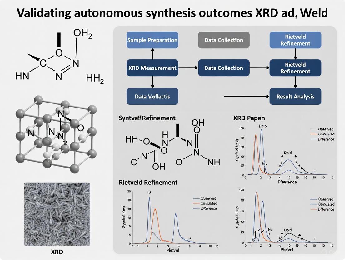
The Rietveld method represents the gold standard for refining crystal structures from powder diffraction data. This powerful whole-pattern fitting technique uses a non-linear least squares approach to refine a theoretical line profile until it matches the measured profile [12] [14]. The fundamental equation for Rietveld refinement models the calculated intensity at each point i in the pattern as:
Y(i) = b(i) + Σ Iₖ [yₖ(xₖ)]
where b(i) is the background intensity, Iₖ is the intensity of the k-th Bragg reflection, and yₖ is the peak shape function [12]. The peak shape is typically modeled using a pseudo-Voigt function that combines Gaussian and Lorentzian contributions to account for both instrumental and sample-induced broadening [12]. Modern implementations can simultaneously refine numerous parameters including lattice constants, atomic coordinates, thermal displacement parameters, phase fractions, crystallite size, and microstrain [14]. Successful application requires high-quality diffraction data, reasonable starting structural models, and careful sequential refinement of parameters to avoid correlations that can trap the refinement in false minima [12].
Machine Learning Approaches for Symmetry Classification
Recent advances have demonstrated the effectiveness of machine learning for classifying crystal systems and space groups directly from powder XRD patterns. One established protocol involves feature extraction from diffraction patterns followed by classification using ensemble methods like Extremely Randomized Trees (ExRT) [2]. The workflow begins with calculating feature vectors comprising (1) the positions of the first ten low-angle peaks and (2) the total number of peaks between 2θ = 0° and 90° [2]. These features capture essential pattern characteristics while avoiding the curse of dimensionality. The ExRT model then classifies crystal systems with approximately 90% accuracy for most systems (except triclinic) and achieves 80.46% accuracy for space group classification with a single candidate, rising to 92.42% when considering the five most likely candidates [2]. This approach significantly accelerates the initial stages of structure analysis compared to manual methods.
Autonomous Structure Determination with PXRDGen
Cutting-edge research has yielded fully automated structure determination pipelines such as PXRDGen, which integrates deep learning with traditional refinement. This end-to-end neural network combines three modules: a pre-trained XRD encoder that aligns diffraction patterns with crystal structures in latent space, a crystal structure generator based on diffusion or flow models conditioned on chemical formulas and XRD features, and a Rietveld refinement module that ensures optimal agreement with experimental data [13]. When evaluated on the MP-20 dataset of inorganic materials, PXRDGen achieved remarkable success rates of 82% with a single generated sample and 96% with 20 samples for valid compounds [13]. The system demonstrates particular effectiveness in resolving challenging cases involving overlapping peaks, localization of light atoms, and differentiation of neighboring elements [13].
Research Reagent Solutions: Essential Materials for Crystallographic Analysis
Table 3: Essential Research Reagents and Materials for Crystallographic Analysis
| Reagent/Material | Function/Application | Technical Specifications | Usage Notes |
|---|---|---|---|
| Silicon Standard (NIST) | Instrument alignment and peak position calibration | Powder, 99.9% purity, 1-10 μm particle size | Use for instrumental broadening determination before Rietveld analysis [14] |
| LaB₆ Standard (NIST) | Line profile and resolution calibration | Powder, certified reference material | Provides well-characterized peak shapes for profile fitting |
| Capillary Tubes (Borosilicate) | Sample containment for high-quality XRD | 0.5 mm inner diameter, wall thickness 0.01 mm | Minimizes background scattering and preferred orientation [15] |
| Crystallographic Databases (ICSD, COD) | Reference structures for phase identification and starting models | Millions of entries with refined atomic coordinates | Essential for Rietveld refinement starting models [14] |
| Rietveld Refinement Software | Structure refinement from powder data | GSAS, FullProf, TOPAS, MAUD | Implement Thompson-Cox-Hastings pseudo-Voigt profile function [14] |
Advanced Applications: Integrating Techniques for Comprehensive Characterization
Coupled Rietveld-EXAFS Analysis for Local Structure Determination
A novel methodological advancement couples Rietveld refinement of XRD data with Reverse Monte Carlo (RMC) analysis of Extended X-ray Absorption Fine Structure (EXAFS) spectra, enabling simultaneous determination of long-range periodic structure and local atomic environments [16]. This approach is particularly valuable for nanocrystalline materials where significant differences may exist between the average crystal structure and local coordination environments. The coupled method employs a feedback algorithm that exchanges information between consecutive refinements, with EXAFS spectra computed from partial pair distribution functions according to:
χᵢ(k) = Σ 4πnⱼ ∫ gᵢⱼ(r) · γ(r,k) · r² dr
where gᵢⱼ(r) is the partial pair distribution function, nⱼ is the number density of scattering atom j, and γ(r,k) is the EXAFS kernel containing amplitude and phase functions [16]. This coupled analysis provides a more complete structural description than either technique alone, as demonstrated in studies of nanocrystalline SnO₂ where it revealed subtle structural distortions not apparent from standalone analyses [16].
In Situ and Operando Crystallography for Process Monitoring
The combination of crystallographic fundamentals with advanced detection capabilities enables real-time monitoring of structural transformations during synthesis or under operational conditions. Modern synchrotron facilities and laboratory X-ray instruments increasingly support high-throughput measurements, in situ studies, and operando characterization during material function [11]. These applications require robust automated analysis pipelines where machine learning algorithms can rapidly process large datasets to identify structural changes, phase transitions, or reaction intermediates [11]. For example, in situ XRD has been employed to track the evolution of bone mineral (bioapatite) during fetal development, with Rietveld refinement quantifying structural parameters as a function of gestational age [15]. Such studies reveal how crystallographic characteristics evolve during natural processes and synthetic pathways, providing critical insights for optimizing materials synthesis and performance.
The workflow for these advanced applications integrates multiple characterization methods, as shown in the following diagram:
Figure 2: Integrated analysis workflow coupling Rietveld refinement of XRD data with Reverse Monte Carlo analysis of EXAFS data enables comprehensive structural characterization spanning both long-range periodicity and local atomic environments [16].
The integrated understanding of unit cells, space groups, and structure factors provides researchers with a powerful framework for materials characterization, particularly when deploying X-ray diffraction and Rietveld refinement to validate autonomous synthesis outcomes. The comparative data presented in this guide demonstrates that while traditional Rietveld refinement remains the most comprehensive method for full structure determination, emerging machine learning approaches offer compelling advantages for rapid classification and initial structure solution. Strategic implementation involves selecting the appropriate methodology based on research goals: ML classification for high-throughput screening, traditional Rietveld refinement for detailed structural analysis, and coupled techniques for materials exhibiting complex local structures. As autonomous synthesis platforms continue to generate increasingly complex materials, the fundamental crystallographic principles outlined in this guide will remain essential for extracting meaningful structural insights from diffraction data and accelerating materials discovery across pharmaceutical, energy, and electronic applications.
Rietveld refinement has emerged as a powerful whole-pattern fitting technique for quantitative phase analysis, particularly valuable in the era of autonomous materials discovery. Unlike traditional single-peak methods, this approach uses the entire X-ray diffraction (XRD) pattern, making it indispensable for validating synthesis outcomes in high-throughput experimental platforms [6] [17]. As autonomous laboratories like the A-Lab demonstrate the ability to synthesize 41 novel compounds in just 17 days [6], robust analytical methods like Rietveld refinement become crucial for accurately characterizing newly discovered materials. This guide examines how Rietveld refinement compares with other quantitative XRD methods and details its implementation for reliable phase analysis.
Understanding Rietveld Refinement
The Rietveld method represents a significant advancement in powder diffraction analysis. Developed by Hugo Rietveld, it uses a whole-pattern fitting approach where a calculated diffraction pattern is fitted to the observed experimental data through least-squares refinement [14]. Unlike single-peak methods that rely on the intensity of individual reflections, Rietveld refinement utilizes the entire diffraction pattern, effectively minimizing problems associated with peak overlap and preferred orientation [17].
The fundamental equation for quantitative analysis using the Rietveld method is:
wk = (skk) / Σ(siZMVi)
Where:
- wk = weight fraction of phase k
- s = Rietveld scale factor
- Z = number of formula units per unit cell
- M = mass of the formula unit
- V = unit cell volume [14]
This approach allows researchers to determine not only phase composition but also structural parameters, crystallite size, strain, and atomic displacements from the same dataset [14].
Method Comparison: Accuracy and Applications
Different quantitative XRD methods offer varying advantages depending on the sample characteristics and analytical requirements. A 2023 systematic comparison of three primary methods revealed distinct performance profiles.
Table 1: Comparison of Quantitative XRD Analysis Methods
| Method | Key Principle | Accuracy with Clay Minerals | Accuracy without Clay Minerals | Best Use Cases |
|---|---|---|---|---|
| Rietveld Refinement | Whole-pattern fitting using crystal structure models [14] | Lower accuracy [18] | High accuracy [18] | Crystalline phases with known structures; non-clay samples [18] |
| Full Pattern Summation (FPS) | Pattern fitting using measured reference patterns [18] [17] | Higher accuracy for sediments [18] | High accuracy [18] | Clay-containing samples; sediments; phases with unknown structures [18] |
| Reference Intensity Ratio (RIR) | Single peak intensity with reference values [18] | Lower accuracy [18] | Lower accuracy [18] | Quick estimates; simple mixtures [18] |
The Rietveld method excels with crystalline materials where accurate structural models are available, while FPS shows wider applicability for sediment analysis and samples containing clay minerals [18]. The RIR method, while handy for rapid assessment, generally provides lower analytical accuracy across sample types [18].
Table 2: Quantitative Accuracy Assessment from Artificial Mixtures
| Method | Software Tools | Absolute Error Range | Key Limitations |
|---|---|---|---|
| Rietveld Refinement | HighScore, TOPAS, GSAS, MAUD [18] [14] | Varies by sample type [18] | Requires known crystal structures; struggles with disordered/unknown structures [18] |
| Full Pattern Summation (FPS) | FULLPAT, ROCKJOCK [18] | Generally low for diverse samples [18] | Requires comprehensive reference pattern library [18] |
| Reference Intensity Ratio (RIR) | JADE [18] | Generally higher than whole-pattern methods [18] | Susceptible to preferred orientation and peak overlap [18] |
Experimental Protocols for Reliable Results
Sample Preparation Methodology
Proper sample preparation is critical for high-quality Rietveld analysis. The autonomous robotic experimentation system demonstrates optimal protocols:
- Particle Size Control: Grind samples to <45 μm (325 mesh) to minimize micro-absorption effects and ensure reproducible peak intensities [18]
- Homogenization: Mix powders thoroughly for 30 minutes in an agate mortar [18]
- Surface Preparation: Use gentle compression with soft gel attachments to create smooth, even surfaces that minimize background noise, particularly in low-angle regions [7]
- Consistent Mounting: Employ specialized holders with frosted glass centers to support powder samples while reducing background contribution [7]
Data Collection Parameters
Standardized measurement conditions ensure reproducible results:
- Radiation: Cu Kα (λ = 1.5418 Å) [18]
- Scan Range: 3° to 70° (2θ) for comprehensive pattern capture [18]
- Step Size: 0.0167° for detailed profile definition [18]
- Scan Speed: 2°/min balances resolution with measurement time [18]
- Instrument Settings: 40 mA, 40 kV generator settings under controlled temperature (25 ± 3°C) and humidity (60%) conditions [18]
Refinement Strategy
Successful Rietveld refinement follows a systematic parameter adjustment process:
- Initialization: Begin with crystal structure models from validated databases (ICSD, COD, Materials Project) [18] [6]
- Parameter Refinement: Sequentially refine scale factors, zero-shift, background coefficients, unit cell parameters, peak width parameters, atomic positions, and preferred orientation [18] [14]
- Quality Assessment: Monitor agreement indices (Rp, Rwp, GOF) to evaluate refinement quality [14]
Rietveld Refinement Workflow
The Scientist's Toolkit: Essential Research Reagents and Materials
Table 3: Essential Materials for Rietveld Analysis Experiments
| Material/Reagent | Function | Application Notes |
|---|---|---|
| High-Purity Standards | Reference materials for instrument calibration and method validation [18] | Corundum (Al2O3) or silicon for instrumental broadening correction [14] |
| Crystal Structure Databases | Sources of structural models for Rietveld refinement [18] [6] | ICSD, COD, Materials Project provide CIF files for calculation [18] [14] |
| Specialized Sample Holders | Precise presentation of powder samples for XRD measurement [7] | Frosted glass centers with magnetic embedding for automated systems [7] |
| Software Platforms | Implementation of refinement algorithms and quantitative analysis [18] [14] | HighScore, TOPAS, GSAS, MAUD, FullProf offer Rietveld capabilities [18] [14] |
Autonomous Material Discovery Workflow
Rietveld refinement plays a crucial role in autonomous materials discovery pipelines, as demonstrated by the A-Lab platform:
Role of Rietveld in Autonomous Discovery
This integrated approach enables high-throughput validation of novel compounds. The A-Lab successfully realized 41 of 58 target compounds using this workflow, with Rietveld refinement providing essential phase quantification and structural validation [6]. The autonomous system combines computational predictions, historical literature data, machine learning, and robotics, with Rietveld analysis serving as the critical validation step [6].
Practical Considerations and Limitations
While powerful, Rietveld refinement has specific limitations that researchers must consider:
- Structure Dependency: Requires known crystal structures; cannot analyze phases with completely unknown atomic arrangements [18] [14]
- Clay Mineral Challenges: Demonstrates lower accuracy for clay-containing samples compared to FPS methods [18]
- Kinetic Limitations: Cannot detect phases with concentrations below approximately 0.5-1 wt% in typical laboratory conditions [18]
- Preferred Orientation: Requires correction models (March-Dollase, spherical harmonics) for accurate intensity calculation in oriented samples [19]
For complex samples containing disordered phases or unknown structures, combined approaches using both structure-based refinement and pattern-fitting methods may provide optimal results [17].
Rietveld refinement represents the gold standard for quantitative phase analysis of crystalline materials with known structures, particularly in automated research environments. Its whole-pattern approach provides significant advantages over single-peak methods for complex mixtures, while its integration with computational databases and machine learning platforms positions it as an essential tool for accelerating materials discovery. As autonomous laboratories continue to expand their capabilities, Rietveld refinement will remain indispensable for validating synthesis outcomes and extracting precise structural information from powder diffraction data.
Within the framework of autonomous materials synthesis, the reliability of experimental outcomes hinges on robust and unambiguous characterization data. Powder X-ray diffraction (XRD) serves as a critical validation tool in these self-driving laboratories, providing definitive evidence of successful phase formation [20]. However, the integrity of this validation is directly contingent upon the quality of the underlying XRD data collection parameters. Optimizing incident wavelength, instrument geometry, and particle size is not merely a preparatory step but a fundamental requirement for generating data that can withstand automated analysis algorithms and Rietveld refinement protocols. This guide objectively compares the performance of different configuration choices, providing the experimental data and methodologies necessary to inform their selection in high-throughput research environments.
Core XRD Principles and the Autonomous Workflow
X-ray diffraction operates on the principle of constructive interference of monochromatic X-rays scattered by the periodic lattice of a crystalline material. This interaction is governed by Bragg's Law: ( nλ = 2d \sinθ ), where ( λ ) is the incident X-ray wavelength, ( d ) is the interplanar spacing, and ( θ ) is the Bragg angle [21] [22]. The resulting diffraction pattern acts as a unique fingerprint for a material's crystal structure [21].
In autonomous research, this pattern is the primary data used by automated analysis pipelines. The clarity, signal-to-noise ratio, and resolution of the pattern directly influence the performance of deep learning models and the accuracy of subsequent Rietveld refinement, a powerful method for whole-pattern fitting used in structural analysis [23] [24]. Suboptimal data collection can lead to false positives or negatives in phase identification, misclassification of crystal systems, and unreliable refinement metrics, thereby invalidating the synthesis outcome.
The following diagram illustrates how optimized data collection integrates into a generalized autonomous synthesis and validation workflow.
Optimizing Incident X-ray Wavelength
The choice of incident X-ray wavelength is a critical determinant of a diffractogram's angular range, resolution, and susceptibility to fluorescence artifacts. The most common sources are Copper (Cu) and Molybdenum (Mo) anodes, each with distinct performance characteristics suited to different material classes [21].
Copper Kα radiation (λ = 1.5418 Å) is the workhorse for most routine analyses, particularly for materials containing light elements. Its longer wavelength provides good angular separation of diffraction peaks (higher dispersion), which is beneficial for resolving complex patterns. However, its energy is sufficient to excite fluorescence in samples containing elements like Fe, Co, and Mn, leading to a high, noisy background that can obscure weaker diffraction peaks [21].
Molybdenum Kα radiation (λ = 0.7107 Å) offers a solution to fluorescence problems. Its higher energy penetrates samples more effectively and minimizes fluorescence for heavier elements. The shorter wavelength also compresses the diffraction pattern into a smaller 2θ range, which can be advantageous for certain detectors. The primary trade-off is reduced dispersion, which may lead to peak overlap in low-symmetry crystal systems [21] [22].
Table 1: Performance Comparison of Common X-ray Anode Materials
| Anode Material | Wavelength (Å) | Key Advantages | Key Limitations | Ideal Use Cases |
|---|---|---|---|---|
| Copper (Cu) | 1.5418 | High angular dispersion for better peak separation; high intensity for faster data collection. | Can cause high fluorescence with Fe, Co, Mn, etc., increasing background noise. | Routine analysis of organic compounds, pharmaceuticals, and most inorganic materials without fluorescent elements. |
| Molybdenum (Mo) | 0.7107 | Minimizes fluorescence from heavier elements; greater penetration depth. | Lower angular dispersion can cause peak overlap. | Materials containing heavy or transition metals (e.g., catalysts, intermetallics), single-crystal diffraction. |
Experimental Protocol: Wavelength Selection for Iron-Containing Sample
Objective: To compare the signal quality from an iron oxide (Fe₂O₃) sample using Cu and Mo Kα radiation sources. Methodology:
- Prepare a finely ground and homogeneous powder sample of Fe₂O₃ using a mortar and pestle.
- Load the sample into a standard powder diffractometer equipped with a Cu anode tube.
- Collect a diffraction pattern over a 2θ range of 20° to 80° with a step size of 0.02° and a counting time of 2 seconds per step.
- Without moving the sample, switch the X-ray source to a Mo anode tube (or use a separate instrument with a Mo source).
- Collect a second diffraction pattern over an equivalent d-spacing range (approximately 12° to 35° 2θ for Mo) using the same counting statistics. Supporting Data: Analysis of the two patterns will reveal a significantly elevated background in the data collected with Cu Kα radiation due to fluorescence, whereas the pattern collected with Mo Kα will show a flatter background and clearer peak definition, facilitating more accurate phase identification and refinement [21].
Instrument Geometry and Data Collection Parameters
The geometric configuration of the diffractometer and the selection of scan parameters directly control the resolution, intensity, and statistical quality of the data.
Instrument Geometry
Modern X-ray diffractometers consist of several key components: an X-ray source, incident beam optics (e.g., slits, monochromators), a sample stage, and a detector system, all precisely aligned on a goniometer [21]. The two primary geometries for powder diffraction are:
- Bragg-Brentano (θ-2θ): The most common geometry for flat powder samples. The X-ray source is fixed, while the sample and detector rotate in a coupled manner (θ and 2θ, respectively). This configuration focuses the diffracted beam from lattice planes parallel to the sample surface [21] [22].
- Parallel Beam: Uses specialized optics to produce a parallel, non-diverging beam. This geometry is less sensitive to sample surface imperfections, displacement errors, and is ideal for analyzing rough surfaces or thin films.
Optimizing Scan Parameters
The choice of scan parameters represents a practical trade-off between data quality, resolution, and collection time—a crucial consideration in high-throughput settings.
Table 2: Impact of XRD Scan Parameters on Data Quality
| Parameter | Effect on Data Quality | Recommended Values for Common Scenarios |
|---|---|---|
| Scan Speed (°/min) | Fast (>5°): Low intensity, noisy data, suitable for quick phase check.Slow (0.5-2°): High intensity, better signal-to-noise, reveals weak peaks. | Routine Phase ID: 5-10°/min [22]Rietveld Refinement: 0.5-2°/min [22]Trace Phase Detection: <0.5°/min [22] |
| Step Size (°) | Large (>0.05): May miss or poorly define narrow peaks.Small (0.01-0.02): Accurately defines peak shape and position. | Routine Phase ID: 0.02° [22]Fine Structure/Crystallite Size: 0.01° or smaller [22] |
| Counting Time (s/step) | Short: Poor counting statistics, noisy data.Long: Excellent statistics, reveals weak reflections, but time-consuming. | Scale with scan speed and step size. Longer times are essential for weak scatterers, trace phases, or high-quality refinements. |
Particle Size and Sample Preparation
The physical preparation of the powder sample is often the most variable and critical factor influencing data quality. Imperfect preparation can introduce preferred orientation (texture), micro-absorption, and peak broadening, which severely compromise quantitative phase analysis and Rietveld refinement [22] [25].
The Critical Role of Particle Size
Optimal particle size for XRD lies typically in the 1-10 micrometer range [22]. Particles that are too large (>10 μm) cause:
- Spottiness: A grainy diffraction pattern with inconsistent peak intensities due to an insufficient number of crystallites contributing to the diffraction signal.
- Preferred Orientation: Non-random alignment of crystallites, which skews intensity ratios away from their theoretical values.
Conversely, particles that are too small (<0.1 μm) lead to significant peak broadening due to the Scherrer effect, described by the Scherrer equation: ( D = \frac{K \lambda}{\beta \cos \theta} ), where ( D ) is the volume-weighted crystallite size, ( K ) is a shape factor (~0.9), ( \lambda ) is the wavelength, and ( \beta ) is the pure broadening of the peak at half its maximum intensity (FWHM) in radians [25]. This broadening can obscure closely spaced peaks.
Experimental Protocol: Standard Powder Sample Preparation
Objective: To prepare a powder sample that minimizes preferred orientation and ensures a representative number of crystallites. Methodology:
- Grinding: Use an agate mortar and pestle to grind the sample gently until it feels smooth and exhibits no grittiness. The goal is a fine powder where the particle size is consistently below 10 micrometers [22].
- Loading (Side-Loading Method):
- Place a glass microscope slide or a specialized sample holder on a flat surface.
- Create a shallow well in the holder if necessary.
- Place the powdered sample into the well. Holding a second slide perpendicular to the holder, gently sweep the powder across the cavity. This "side-loading" technique helps randomize crystal orientations rather than pressing them into alignment.
- Leveling: Use the edge of the second slide to level the excess powder, creating a smooth, flat surface flush with the holder edge. Avoid any pressing or tamping motion. Supporting Data: A well-prepared sample will yield a diffraction pattern where the relative intensities of peaks match the reference pattern from the ICDD database. A poorly prepared, textured sample will show significant deviations in these intensities, which can be quantified during Rietveld refinement by a high texture coefficient or a poor goodness-of-fit if texture is not modeled [22] [24].
The Scientist's Toolkit: Essential Research Reagents and Materials
Table 3: Key Materials and Reagents for XRD Sample Preparation and Analysis
| Item | Function/Benefit | Application Notes |
|---|---|---|
| Agate Mortar & Pestle | Provides a very hard, non-porous surface for grinding. Chemically inert, minimizing sample contamination. | The standard tool for achieving a homogeneous fine powder. Gentle grinding is key to reducing crystallite size without inducing excessive strain. |
| Zero-Background Holder (ZBH) | Made from a single crystal of silicon cut off-axis. Produces a negligible diffraction background, improving signal-to-noise for weak peaks. | Ideal for analyzing small quantities of sample or materials with very weak scattering power. |
| Standard Reference Material (e.g., NIST SRM 674b) | A crystalline material with certified lattice parameters and peak positions. Used for instrument alignment and calibration. | Essential for correcting systematic errors in peak position for residual stress or precise lattice parameter determination. |
| Internal Standard (e.g., ZnO, Al₂O₃) | A well-characterized powder mixed with the unknown sample in a known proportion. | Used in quantitative phase analysis to correct for absorption effects and to determine absolute phase abundances. |
The validation of autonomous synthesis outcomes depends on the generation of high-fidelity XRD data. As demonstrated, the strategic selection of incident wavelength, the careful optimization of instrument geometry and scan parameters, and the meticulous preparation of powder samples are not optional but foundational practices. The comparative data and protocols outlined herein provide a framework for researchers to make informed decisions, ensuring that the diffraction patterns fed into automated analysis pipelines and Rietveld refinement software are of sufficient quality to yield reliable, conclusive, and reproducible results. By adhering to these best practices, the promise of autonomous discovery—to bridge the gap between computational prediction and experimental realization—can be robustly and efficiently achieved.
X-ray diffraction (XRD) is a cornerstone analytical technique for determining the atomic and molecular structure of crystalline materials. For researchers validating autonomous synthesis outcomes, selecting the appropriate diffraction method is crucial for obtaining reliable structural validation. Single-crystal X-ray diffraction (SCXRD) and powder X-ray diffraction (PXRD) represent the two primary approaches, each with distinct capabilities, limitations, and applications. This guide provides an objective comparison of these techniques to help researchers select the optimal validation tool for their specific materials and research objectives, particularly within the context of autonomous synthesis validation and Rietveld refinement research.
Core Principles and Technical Differentiation
Fundamental Technical Differences
SCXRD and PXRD, while based on the same fundamental principles of Bragg's Law, differ significantly in their implementation and data output. SCXRD analyzes a single, well-formed crystal, producing a diffraction pattern consisting of discrete spots that provide three-dimensional structural information [26]. In contrast, PXRD analyzes a collection of randomly oriented microcrystals (crystallites), producing a diffraction pattern characterized by concentric rings that are presented as a plot of intensity versus diffraction angle (2θ) [26] [27]. This fundamental difference in sample form leads to significant variations in the type and quality of structural information obtainable.
The data collection processes also differ substantially. SCXRD involves systematically rotating a single crystal within the X-ray beam while recording diffraction intensities at numerous orientations, creating a comprehensive three-dimensional dataset [26]. PXRD, however, requires no crystal rotation during measurement, as the random orientation of crystallites in the powder ensures that all possible diffraction orientations are sampled simultaneously, though this comes at the cost of collapsing three-dimensional information into a one-dimensional diffractogram [26].
Sample Requirements and Preparation
The sample requirements for these techniques represent one of the most significant practical differentiators:
SCXRD requires high-quality single crystals with well-defined faces and minimal defects. The crystal must be sufficiently large (typically ≥ 0.1 mm in one dimension) and well-ordered [26] [28]. Preparing suitable single crystals often demands optimized crystallization conditions and can be time-consuming, making SCXRD less practical for materials that do not readily form large crystals [26].
PXRD works with finely powdered samples, eliminating the need for large single crystals. The powder consists of numerous randomly oriented microcrystals, ensuring all possible crystal orientations contribute to the diffraction pattern. Sample preparation is minimal, usually involving simple grinding and packing, making PXRD more accessible and faster than SCXRD for many applications [26].
Table 1: Sample Requirements and Preparation Comparison
| Parameter | Single-Crystal XRD | Powder XRD |
|---|---|---|
| Sample Form | Single, well-formed crystal | Finely powdered microcrystals |
| Minimum Crystal Size | ≥ 0.1 mm (typically >50 μm) | Micrometer-scale particles |
| Preparation Complexity | High (requires optimized crystallization) | Low (grinding and packing) |
| Crystal Quality Demand | Very high (minimal defects) | Moderate (random orientation critical) |
| Typical Preparation Time | Days to weeks | Minutes to hours |
Analytical Capabilities and Limitations
Structural Information Resolution
The resolution and type of structural information obtainable from SCXRD versus PXRD represents the most significant technical differentiator:
SCXRD provides atomic-level resolution, enabling direct determination of bond lengths, angles, electron density distributions, and molecular conformations with exceptional precision (often better than a few thousandths of a nanometer) [27]. This high resolution makes SCXRD the gold standard for solving complex crystal structures, including proteins, small organic molecules, and inorganic materials [26]. SCXRD can definitively identify polymorphs by revealing precise molecular conformations and packing arrangements, as demonstrated in classic studies of ROY (red, orange, yellow) polymorphs where different colors arise from changes in molecular conformation [29]. For hydrates, SCXRD can categorize structures into channel hydrates, isolated-site hydrates, and metal-coordinated hydrates, precisely determining water positions and interactions within the host lattice [29].
PXRD, while excellent for phase identification and crystallinity analysis, does not provide direct atomic positions with SCXRD's precision [26]. However, with advanced computational techniques like Rietveld refinement, PXRD can provide valuable structural insights, including unit cell parameters and, in favorable cases, atomic coordinates [26]. Modern approaches are addressing traditional PXRD limitations; for instance, the PXRDGen neural network integrates a pretrained XRD encoder, a diffusion/flow-based structure generator, and a Rietveld refinement module to solve structures with significantly improved accuracy [30].
Applications in Research and Validation Contexts
The applications of SCXRD and PXRD diverge according to their analytical strengths:
SCXRD is widely used in molecular chemistry, drug discovery, materials science, and crystallography research due to its unparalleled structural resolution [26]. It is particularly valuable for determining precise 3D atomic arrangements in proteins, catalysts, pharmaceuticals, and novel materials, making it essential for understanding molecular interactions and reaction mechanisms [26]. In pharmaceutical development, SCXRD provides critical information for regulatory and intellectual property strategies by unambiguously defining polymorphs, hydrates, solvation states, stoichiometry, and absolute configuration [29].
PXRD excels in phase identification, quality control, and bulk material characterization [26]. It is widely used in pharmaceutical development to identify drug polymorphs, in materials science to study crystallinity and stress-strain behavior, and in geology to identify minerals [26]. PXRD is particularly valuable for high-throughput screening and analyzing multiphase mixtures, making it ideal for routine analysis in industrial settings [26] [27].
Table 2: Analytical Capabilities and Typical Applications
| Aspect | Single-Crystal XRD | Powder XRD |
|---|---|---|
| Structural Resolution | Atomic-level (sub-Ångström) | Phase-level |
| Primary Applications | Complete structure determination, Absolute configuration analysis, Conformational analysis | Phase identification, Quantitative phase analysis, Crystallinity assessment |
| Polymorph Characterization | Definitive identification via atomic coordinates | Pattern matching against references |
| Hydrate/Solvate Analysis | Precise solvent position and occupancy | Indication through pattern changes |
| Throughput Capability | Low to moderate | High |
| Mixture Analysis | Challenging (requires separation) | Excellent (multiphase capability) |
Experimental Protocols and Methodologies
Data Collection Workflows
The experimental workflows for SCXRD and PXRD involve distinct protocols optimized for their respective sample types and information goals:
SCXRD Protocol:
- Crystal Selection: A suitable single crystal is selected under a microscope and mounted on a goniometer, often using a fiber optic or loop of cryoprotective oil [26].
- Data Collection: The crystal is systematically rotated within the X-ray beam while recording diffraction intensities at numerous orientations. This produces a series of discrete diffraction spots, each corresponding to specific atomic planes [26].
- Data Processing: The collected dataset undergoes complex processing, including correction for absorption and other artifacts, to generate a set of structure factors [28].
- Structure Solution: Computational methods (direct methods, Patterson synthesis, or intrinsic phasing) are used to determine initial phase estimates and generate an electron density map [28].
- Model Building and Refinement: An atomic model is built into the electron density and iteratively refined against the diffraction data to optimize agreement factors [28].
PXRD Protocol:
- Sample Preparation: The material is ground into a fine powder to ensure random orientation of crystallites and packed into a sample holder [26].
- Data Collection: The sample is exposed to monochromatic X-rays, and a detector records the intensity of scattered X-rays as a function of the 2θ angle [26].
- Phase Identification: The resulting diffractogram is compared with reference patterns in databases (e.g., ICDD) for compound identification [26].
- Rietveld Refinement: For quantitative analysis, the crystal structure model is refined against the entire powder pattern rather than individual peaks, enabling extraction of structural parameters, phase abundances, and microstructural information [30] [31].
Workflow Visualization
Performance Comparison and Experimental Data
Quantitative Performance Metrics
The performance characteristics of SCXRD and PXRD vary significantly across multiple parameters critical for experimental planning and validation strategy development:
Table 3: Performance Metrics and Practical Considerations
| Parameter | Single-Crystal XRD | Powder XRD |
|---|---|---|
| Data Collection Time | Hours to days | Minutes to hours |
| Structure Solution Time | Hours to days (after data collection) | Minutes to hours (with modern computational methods) |
| Detection Limits | Not applicable (single component) | ~0.2-0.3 wt% for crystalline phases [31] |
| Light Element Sensitivity | Moderate (H atoms often locatable) | Low (challenging for H, Li) [32] |
| Element Differentiation | Excellent (precise electron density) | Limited for neighboring elements [30] |
| Peak Overlap Issues | Minimal (discrete reflections) | Significant (especially at high angles) [27] |
| Preferred Orientation Effects | Minimal | Can significantly affect intensities [27] |
Recent advances in artificial intelligence are dramatically improving PXRD capabilities. The PXRDGen neural network demonstrates how machine learning can address traditional PXRD limitations, achieving record high matching rates of 82% (1-sample) and 96% (20-samples) for valid compounds on the MP-20 inorganic dataset, with Root Mean Square Error (RMSE) approaching the precision limits of Rietveld refinement [30]. This approach effectively tackles key challenges in PXRD, including resolving overlapping peaks, localizing light atoms, and differentiating neighboring elements [30].
Autonomous Synthesis Validation Considerations
For researchers validating autonomous synthesis outcomes, several specific considerations should guide technique selection:
Throughput Requirements: Autonomous systems often generate numerous samples requiring characterization. PXRD provides significantly higher throughput for initial screening, while SCXRD delivers definitive structural validation for selected targets [26].
Sample Limitations: Many materials synthesized autonomously may not form suitable single crystals, necessitating PXRD analysis. However, microcrystal electron diffraction (microED) is emerging as a complementary technique for structural analysis of microcrystals, bridging the gap between SCXRD and PXRD [33].
Quantitative Analysis: For mixture analysis common in reaction optimization, PXRD with Rietveld refinement enables quantitative phase analysis, with Mo Kα1 radiation providing slightly better accuracy than Cu Kα1 for challenging mixtures including amorphous content [31].
Structure Prediction Integration: PXRD data can be effectively integrated with crystal structure prediction (CSP) algorithms through multi-objective evolutionary searches that use both a structure's enthalpy and similarity to a reference PXRD pattern, facilitating structure solution of complex systems [32].
Essential Research Reagent Solutions
Successful diffraction analysis requires appropriate materials and computational tools. The following table outlines key resources for implementing the experimental protocols discussed:
Table 4: Essential Research Reagents and Computational Tools
| Resource Category | Specific Examples | Function and Application |
|---|---|---|
| Sample Preparation | Cryoprotective oils, Glass/Kapton capillaries, Sample holders | Mounting and protection of sensitive crystals (SCXRD), Uniform packing of powders (PXRD) |
| Reference Databases | Cambridge Structural Database (CSD), Powder Diffraction File (PDF) | Reference patterns for phase identification, Structural models for refinement |
| Computational Tools | PXRDGen neural network [30], DMCpy [34], Rietveld refinement software | Structure solution from PXRD data, Data reduction and visualization, Quantitative phase analysis |
| Analysis Methodologies | Multi-objective evolutionary algorithms [32], Dynamic diffraction theory refinements [33] | Integration of computational and experimental data, Accurate refinement of electron diffraction data |
| Specialized Equipment | Cryogenic coolers, Environmental sample holders, 2D detectors | Temperature control for sensitive samples, Analysis under controlled atmospheres, Efficient data collection |
SCXRD and PXRD offer complementary capabilities for validating autonomous synthesis outcomes. SCXRD remains the gold standard for definitive structural determination when suitable crystals are available, providing atomic-level resolution essential for understanding molecular interactions, confirming polymorph identities, and supporting intellectual property claims. PXRD offers practical advantages for high-throughput analysis, mixture characterization, and routine quality control, with ongoing advancements in computational methods and artificial intelligence continually expanding its capabilities.
The optimal technique selection depends on specific research goals, sample characteristics, and practical constraints. For comprehensive materials characterization programs, a combined approach utilizing PXRD for initial screening and SCXRD for definitive structural validation of selected targets represents the most effective strategy. As autonomous synthesis platforms continue to advance, integration of these diffraction methods with computational prediction and machine learning algorithms will be essential for achieving rapid, accurate structural validation of novel materials.
From Data to Structure: Practical Workflows for Laboratory XRD Analysis
The accurate characterization of crystalline materials is fundamental to advancements in pharmaceutical development and materials science. For systems where single crystals are unavailable, powder X-ray diffraction (PXRD) is the definitive technique for structure determination. The push towards autonomous synthesis and high-throughput experimentation creates a pressing need for highly reliable, automated analytical workflows. This guide objectively compares PXRD data collection strategies, focusing on the synergistic use of capillary transmission geometry and variable count time (VCT) protocols. When deployed together, these methods provide the high-fidelity data required to validate autonomous synthesis outcomes through robust Rietveld refinement, forming a critical link in a closed-loop, materials-discovery pipeline.
Comparative Analysis of PXRD Geometries
The choice of diffraction geometry directly impacts data quality by influencing how the X-ray beam interacts with the powdered sample. The following sections compare the most common geometries.
Capillary Transmission Geometry
This method involves packing the powdered sample into a thin, rotating glass capillary, typically 0.3–0.7 mm in diameter, and measuring diffraction in transmission mode [35] [36].
- Mechanism: A monochromatic X-ray beam passes through the capillary. Rotating the capillary during measurement ensures that a statistically representative number of crystallites contribute to the diffraction pattern, averaging out their orientations [35] [36].
- Key Advantages:
- Minimizes Preferred Orientation: Spinning the capillary is the most effective method for reducing peak intensity artifacts caused by non-random crystallite orientation, a common issue with platy or needle-like crystals [37] [36].
- Controlled Atmosphere: Capillaries can be sealed, making this geometry ideal for air- or moisture-sensitive samples common in pharmaceutical development [36].
- Clean Background: Borosilicate glass capillaries contribute minimal background interference compared to flat-plate holders, leading to a better signal-to-noise ratio [36].
- Small Sample Requirement: Requires only a few milligrams of material, which is advantageous when sample quantity is limited [36].
Reflection Geometries
Reflection geometries, such as the common Bragg-Brentano para-focusing arrangement, involve mounting the sample on a flat surface and measuring the diffraction pattern from the same side as the incident beam.
- Mechanism: The X-ray beam is directed onto a flat sample surface, and the detector moves to capture the diffracted rays. Sample spinning may be used, but it is less effective at averaging orientations than in capillary transmission [37].
- Key Disadvantages:
- Pronounced Preferred Orientation: This geometry is highly susceptible to peak intensity inaccuracies from preferred orientation, as crystallites tend to align with their plate-like faces parallel to the sample surface [37].
- Sample Surface Effects: Data quality is sensitive to surface flatness and packing density, introducing potential for error and poor reproducibility [37].
- Larger Sample Consumption: Typically requires more material than capillary mounting to ensure infinite thickness and a representative surface.
Foil Transmission Geometry
A hybrid approach where the sample is contained between thin foils or films, combining aspects of both transmission and reflection methods.
- Mechanism: The powder is sandwiched between low-absorption polymer foils, and diffraction is measured in transmission mode. The sample holder may or may not rotate [37].
- Comparative Performance: A 2025 study on metformin embonate polymorphs found that foil transmission geometry yielded symmetric, well-resolved peaks and a Rietveld refinement profile fit superior to reflection geometry, though capillary transmission still provided the best overall fit with the lowest residual factors [37]. The study positioned foil transmission as a "bridge-configuration" that mitigates several challenges of reflection geometry while being easier to prepare than capillaries.
Experimental Comparison of Geometries
A direct, experimental comparison of these geometries using a pharmaceutical model system clearly demonstrates their performance differences.
Table 1: Experimental Comparison of PXRD Geometries Using Metformin Embonate Polymorphs [37]
| Geometry Type | Peak Shape | Resolution | Preferred Orientation Mitigation | Rietveld Refinement Goodness-of-Fit (Relative Performance) |
|---|---|---|---|---|
| Capillary Transmission | Symmetric, well-resolved | High | Excellent | Best (Lowest Rwp and GOF) |
| Foil Transmission | Symmetric, well-resolved | High | Very Good | Intermediate |
| Reflection (Bragg-Brentano) | Broader, merged, inherent asymmetry | Lower | Poor | Worst |
The data show that capillary transmission geometry delivers superior data quality, which translates directly into more reliable and conclusive Rietveld refinement outcomes [37]. This makes it the gold standard for critical applications like crystal structure determination from powder data (SDPD) and the validation of autonomous synthesis products [35].
Optimizing Data Collection with Variable Count Times
While geometry determines the fundamental quality of diffraction data, the data collection strategy determines how effectively that quality is realized. A key optimization is the use of Variable Count Times (VCT).
The Rationale for VCT
Diffracted intensity decreases significantly at higher 2θ angles due to the atomic form factor and other geometric effects. A fixed count time throughout the scan range forces a trade-off: short times result in poor signal-to-noise at high angles, while long times waste instrument time on the already strong low-angle peaks [35]. A VCT scheme dynamically addresses this by allocating measurement time based on diffraction angle.
- Principle: Systematically increase the count time per step as the scan progresses to higher 2θ angles. This compensates for the natural fall-off in diffracted intensity, ensuring sufficient counting statistics across the entire pattern [35].
- Impact on Data Quality: The primary benefit is a significant improvement in the signal-to-noise ratio for high-angle reflections. This is critical because high-angle data provides the fine detail necessary for accurate lattice parameter determination, atomic coordinate refinement, and the modeling of thermal parameters during Rietveld analysis [35].
Standardized VCT Protocol
A generic, scalable VCT scheme for laboratory PXRD is recommended for collecting Rietveld-quality data [35]. This protocol can be adjusted based on sample scattering power and instrument intensity.
Table 2: A Generic Variable Count Time Scheme for Rietveld-Quality Data [35]
| Start 2θ (°) | End 2θ (°) | Step Size (°) | Count Time per Step (seconds) |
|---|---|---|---|
| 2.5 | 22 | 0.017 | 2 |
| 22 | 40 | 0.017 | 4 |
| 40 | 55 | 0.017 | 15 |
| 55 | 70 | 0.017 | 24 |
This protocol prioritizes speed at low angles where intensity is high and invests more time at high angles where signal is weak. For a typical laboratory diffractometer, implementing this VCT scheme over a 12-hour collection for a molecular organic sample irradiated with Cu Kα1 radiation can achieve a real-space resolution of about 1.35 Å, which is desirable for final Rietveld refinement [35].
An Integrated Workflow for Autonomous Synthesis Validation
The combination of capillary transmission geometry and a VCT protocol forms the core of a robust data collection strategy suitable for validating the output of an autonomous synthesis platform. The workflow below integrates these elements into a complete validation pipeline.
Diagram Title: Autonomous Synthesis XRD Validation Workflow
This workflow ensures that the data fed into the Rietveld refinement module is of the highest possible quality, minimizing artifacts and maximizing signal. This is crucial for automated systems, which rely on unambiguous, high-fidelity data to make correct decisions about synthesis outcomes without requiring human intervention to diagnose data quality issues.
The Scientist's Toolkit: Essential Research Reagents and Materials
Successful implementation of the optimal strategy requires specific materials and software.
Table 3: Essential Research Reagents and Software for High-Quality PXRD
| Item Name | Function / Application | Key Specifications |
|---|---|---|
| Borosilicate Glass Capillaries | Sample containment for transmission geometry [35]. | Diameter: 0.3 mm (heavy elements), 0.7 mm (standard, organic samples) [35] [36]. |
| McCrone Micronising Mill | Sample grinding to achieve optimal particle size [38]. | Produces particles in the 5-20 µm range to ensure homogeneous packing and mitigate microabsorption [38]. |
| Open-Flow N₂ Gas Cooler | Low-temperature data collection [35]. | ~150 K operation; reduces thermal motion, improving signal-to-noise at high 2θ [35]. |
| DASH | Crystal structure solution from PXRD data via global optimization [35]. | Performs indexing, space-group determination, and Pawley refinement. |
| TOPAS-Academic | Rietveld refinement software [35]. | Features robust refinement capabilities for complex multiphase samples. |
| PXRDGen | AI-driven crystal structure determination from PXRD patterns [13]. | An end-to-end neural network that solves and refines structures with high accuracy, promising automation. |
The objective comparison presented in this guide demonstrates that capillary transmission geometry is unequivocally superior to reflection and foil transmission methods for obtaining quantitative, artifact-free PXRD data. When coupled with a variable count time data collection protocol, it produces a diffraction pattern with excellent signal-to-noise across the entire angular range. This combination directly addresses the core requirement for validating autonomous synthesis: providing a stream of high-fidelity, reliable data that enables robust, automated Rietveld refinement and confident structural validation. By adopting this optimized data collection strategy, researchers can build a more dependable and effective bridge between autonomous synthesis and conclusive materials characterization.
Within autonomous materials discovery and pharmaceutical development pipelines, the integrity of X-ray diffraction (XRD) data is paramount for validating synthesis outcomes. High-throughput and autonomous experiments rely on robust, automated data analysis, including Rietveld refinement, to accurately identify phases and determine crystal structures. The fidelity of these analytical conclusions is fundamentally contingent upon the quality of the initial powder sample preparation. Inadequate preparation can introduce significant biases, namely preferred orientation and suboptimal particle size effects, which distort diffraction intensity ratios and compromise quantitative analysis [39]. This guide objectively compares prevalent sample preparation protocols, evaluating their efficacy in mitigating these artifacts to ensure data reliability in autonomous research workflows.
Fundamentals of Sample-Induced Artifacts
Preferred Orientation
In powder XRD, the ideal sample comprises randomly oriented crystallites, ensuring that the relative intensities of diffraction peaks accurately represent the true crystal structure. Preferred orientation occurs when crystalline grains with anisotropic shapes (e.g., needle-like or plate-like structures) align preferentially along certain directions during sample packing [39]. This non-random alignment causes specific diffraction peaks to be artificially enhanced or suppressed in intensity.
- Impact on Analysis: Distorted intensity ratios severely impact both qualitative phase identification and, more critically, quantitative phase analysis (QPA). While Rietveld refinement software can incorporate functions to correct for this intensity bias, the accuracy of such corrections is limited, making physical mitigation during preparation essential [39].
- Detection Methods: The presence of preferred orientation can be evaluated using:
- Two-Dimensional Detectors: Debye rings with non-uniform intensity distributions indicate preferred orientation, whereas randomly oriented samples show uniform rings [39].
- Rocking Curve Measurements: While less sensitive, these measurements can reveal orientation by showing sharp intensity increases at specific incident angles (ω) [39].
Particle Size and Morphology
The size and morphology of powder particles are critical for achieving a representative diffraction pattern.
- Optimal Particle Size: A carefully controlled particle size distribution, typically in the range of 20–50 µm, is recommended to balance several requirements: ensuring homogeneous packing, obtaining a true powder average, and mitigating preferred orientation [35].
- Consequences of Improper Sizing:
- Excessively Fine Particles: Over-grinding can induce peak broadening due to crystallite size reduction and may even cause unintended phase transitions [35].
- Excessively Coarse or Anisotropic Particles: Increase the susceptibility to preferred orientation and complicate the achievement of a homogeneous, random sample [39] [35].
Table 1: Consequences of Sample Preparation Artifacts on XRD Analysis
| Artifact | Effect on Diffraction Pattern | Impact on Quantitative Analysis |
|---|---|---|
| Preferred Orientation | Deviation of peak intensity ratios from theoretical values | Reduced accuracy of phase quantification in Rietveld refinement [39] |
| Overly Fine Particles | Broadening of diffraction peaks | Increased peak overlap, complicating indexing, structure solution, and refinement [35] |
| Anisotropic Particle Morphology | Exacerbates preferred orientation | Introduces strong intensity bias, requiring sophisticated correction models [39] |
Experimental Protocols for Sample Preparation
A variety of protocols exist for mounting powder samples for XRD analysis. The following section details the methodologies cited in current literature and practice.
Capillary Transmission Geometry
Widely regarded as the gold standard for high-quality powder XRD, particularly for crystal structure determination from powder data (SDPD) [35].
- Methodology: The powder sample is filled into a thin-walled borosilicate glass capillary, typically 0.7 mm in diameter. The capillary is then rotated during data collection [35].
- Mechanism of Mitigation: The rotation of a small-diameter capillary in transmission geometry maximizes the number of particle orientations presented to the X-ray beam, effectively averaging out preferential alignment and providing diffraction data that closely approximates the ideal random distribution.
- Considerations: While 0.3 mm capillaries are an option for highly absorbing samples, they are more challenging to fill. A 1.0 mm capillary requires more sample material and offers a less optimal geometry for reducing orientation [35].
Standard Flat-Plate Back-Loading
A common and practical method used in many laboratory settings for reflection geometry measurements.
- Methodology: A cavity sample holder is filled with the powder, and the excess is scraped off to create a level surface. The "back-loading" variation involves filling the holder from the rear, which can help reduce orientation by minimizing the shearing forces that cause plate-like crystals to align parallel to the surface.
- Mechanism of Mitigation: Aims to create a packed surface with minimal directional force during packing. It is less effective than capillary rotation but generally better than simple front-loading and scraping.
- Typical Use: A standard technique for routine phase identification where the highest accuracy in intensity is not critical.
Side-Filling and Gentle Packing
Emphasizes a minimalist approach to disturbing the powder.
- Methodology: The powder is carefully introduced into the holder from the side using a spatula or scoop, with avoidance of any pressing or scraping on the front surface. The goal is to let the powder settle under gravity alone.
- Mechanism of Mitigation: Avoids the application of directional pressure that is the primary cause of particle alignment in flat-plate methods.
- Typical Use: A simple step that can be employed to reduce orientation in reflection geometry measurements.
The logical relationship between the choice of preparation method and the resulting data quality can be summarized in the following workflow, which is crucial for autonomous research pipelines where decision-making must be automated.
Comparative Performance Analysis
The following table synthesizes data from experimental protocols to provide a direct comparison of the preparation methods discussed.
Table 2: Objective Comparison of Sample Preparation Protocols
| Preparation Protocol | Efficacy Against Preferred Orientation | Particle Size Control | Ease of Implementation | Recommended Use Case |
|---|---|---|---|---|
| Capillary Transmission with Rotation | Excellent [35] | High (requires 20-50 µm for 0.7 mm capillary) [35] | Moderate (requires specific hardware) | Crystal structure solution & refinement (SDPD), high-accuracy QPA [35] |
| Flat-Plate Back-Loading | Good | Moderate | High | Routine phase analysis, quality control |
| Side-Filling / Gentle Packing | Fair | Moderate | High | Quick screening, highly oriented materials |
The quantitative impact of proper preparation is evident in analytical results. For instance, in quantitative phase analysis via Rietveld refinement, the accuracy of the reported phase percentages is directly linked to the quality of the scale factors derived from the fit between the calculated and experimental pattern. Better physical preparation leads to a better fit and, consequently, more accurate scale factors and quantitative results [40]. Protocols that minimize preferred orientation effectively reduce systematic intensity errors, leading to more reliable scale factors during refinement.
The Scientist's Toolkit: Essential Research Reagents and Materials
Successful sample preparation requires the use of specific materials and tools to implement the protocols effectively.
Table 3: Essential Materials for Powder XRD Sample Preparation
| Item | Specification/Function |
|---|---|
| Borosilicate Glass Capillaries | Typically 0.7 mm diameter; used in transmission geometry to minimize preferred orientation via sample rotation [35]. |
| Micron-Mesh Sieves | For selecting and controlling particle size distribution (target 20-50 µm) to ensure a true powder average and reduce orientation [35]. |
| Agar Mortar and Pestle | For gentle grinding of samples to achieve the target particle size without introducing excessive strain or phase transitions. |
| Sample Holders (Flat-Plate) | Cavity holders designed for back-loading or side-filling to reduce shearing forces during packing in reflection geometry. |
| Open-Flow N₂ Gas Cooler | For low-temperature (~150 K) data collection; mitigates form-factor fall-off and improves signal-to-noise at high 2θ angles [35]. |
The choice of sample preparation protocol has a direct and measurable impact on the quality and reliability of XRD data. For autonomous research workflows aimed at rigorous validation of synthesis outcomes, capillary transmission geometry with rotation represents the benchmark for mitigating preferred orientation and is strongly recommended for critical applications like crystal structure determination and high-accuracy quantitative analysis [35]. While flat-plate methods like back-loading offer practical convenience for routine phase identification, their limitations in controlling intensity bias must be acknowledged. As the field moves toward increasingly automated and high-throughput discovery, integrating robust, physics-based sample preparation protocols—not just advanced algorithms—will be fundamental to ensuring the validity of autonomous scientific conclusions.
For researchers validating autonomous synthesis outcomes, the automated determination of crystal structures from powder X-ray diffraction (PXRD) data is a critical step. This guide objectively compares the performance, methodologies, and experimental data of modern software approaches for indexing and space group determination, providing a framework for integrating these tools into your research workflow.
Software Performance and Quantitative Comparison
The effectiveness of automated software is primarily measured by its accuracy in determining crystal structures and its speed in delivering reliable results. The table below summarizes the key performance metrics for leading tools as reported in recent literature.
Table 1: Performance Comparison of Automated Indexing and Structure Determination Software
| Software / Approach | Reported Match Rate (Valid Compounds) | Key Strength | Primary Data Type | Notable Experimental Result |
|---|---|---|---|---|
| PXRDGen (AI Model) | 82% (1-sample), 96% (20-samples) [30] | End-to-end neural network; atomic-level accuracy [30] | PXRD Data [30] | RMSE generally < 0.01, approaching Rietveld refinement precision limits [30] |
| EXPO2014 | "Good results" across crystal systems [41] | Probabilistic determination of extinction symbols [41] | PXRD Data [41] | Identifies most plausible space groups with Fig. of Merit (FoM); uses CSD frequency for ambiguity resolution [41] |
| POINTLESS (CCP4 Suite) | High confidence in Laue group assignment [42] | Scores potential symmetry elements via correlation coefficients [42] | Single-crystal Intensity Data [42] | Example: Assigned space group P2₁2₁2₁, though confidence was limited by few axial reflections [42] |
Core Experimental Protocols and Methodologies
The PXRDGen End-to-End Neural Workflow
PXRDGen represents a paradigm shift by using a conditional generative model to solve and refine crystal structures directly from a powder pattern and a chemical formula [30]. Its protocol involves three integrated modules:
- Pre-trained XRD Encoder (PXE) Module: This module uses contrastive learning to align the latent space of PXRD patterns with crystal structures. It extracts features from the PXRD data, with studies showing a Transformer-based encoder achieves a top-10 hit rate of 92.42% for retrieving correct crystal structures [30].
- Crystal Structure Generation (CSG) Module: Conditioned on the PXRD features and chemical formula, this module generates candidate crystal structures. It employs either diffusion or flow-based generative frameworks. Research indicates that a flow-based CSG module can achieve state-of-the-art match rates and speed [30].
- Rietveld Refinement (RR) Module: The generated structures are automatically fed into a Rietveld refinement module to optimize the alignment between the predicted crystal structure and the experimental PXRD data, ensuring atomic-level accuracy [30].
The EXPO2014 Probabilistic Workflow
EXPO2014 employs a probabilistic procedure to determine the space group from indexed PXRD data, which is critical when working with one-dimensional data prone to peak overlap [41]. The standard protocol is as follows:
- Full Pattern Decomposition: The experimental pattern is decomposed using the Le Bail algorithm to extract integrated intensities. This is initially performed in the space group with the largest Laue symmetry and no extinction conditions for the given crystal system (e.g., P 2/m 2/m 2/m for orthorhombic) [41].
- Intensity Normalization: The extracted intensities are normalized using the classical Wilson method [41].
- Probability Calculation: The statistics of the weighted, normalized intensities are analyzed to calculate the probability for each possible extinction symbol compatible with the crystal system suggested by the initial indexing [41].
- Result Interpretation: The software outputs a list of the most plausible space groups with a Fig. of Merit (FoM). In cases of ambiguity, it provides additional data such as the number of non-overlapped, systematically absent reflections and the space group's frequency in the Cambridge Structural Database (CSD) to guide the user's choice [41].
The POINTLESS Symmetry Analysis Workflow
Designed for single-crystal data but relevant for its robust symmetry analysis, POINTLESS determines the Laue and space group by scoring symmetry elements [42]. Its protocol is:
- Lattice Symmetry Identification: From the unit-cell dimensions and lattice centring, the highest compatible lattice symmetry is identified [42].
- Scoring Symmetry Elements: Each potential rotational symmetry element belonging to the lattice symmetry is scored using the correlation coefficients between all pairs of symmetry-related observations. This method is preferred over R factors as it is less dependent on unknown scales [42].
- Combining Elements: Combinations of symmetry elements for all possible subgroups of the lattice-symmetry group (Laue or Patterson groups) are scored [42].
- Space Group Assignment: Finally, possible space groups are scored based on axial systematic absences, though the confidence in this step can be limited by the number of recorded axial reflections [42].
Workflow Visualization
The following diagram synthesizes the general workflow for automated crystal structure determination, integrating the key stages from data input to final validation, as described across the software tools.
Automated Crystal Structure Workflow
This workflow highlights the critical path from raw data to a refined structure. Modern AI tools like PXRDGen integrate and automate several of these stages (B through E) into a single, end-to-end process [30].
The Scientist's Toolkit: Essential Research Reagents and Software
A reliable toolkit is fundamental for conducting these analyses. The following table lists key software solutions and their primary functions in the context of indexing and space group determination.
Table 2: Essential Software Tools for XRD Analysis
| Tool Name | Primary Function in Research | Application Context |
|---|---|---|
| FullProf Suite [8] | Performs Rietveld refinement for structure validation, phase identification, and microstructural analysis (crystallite size/strain). | Essential for the final step of structure validation and quantitative analysis of powder diffraction data. |
| CCP4 Suite (POINTLESS) [42] | Determines Laue group and space group from single-crystal diffraction data by scoring symmetry elements and systematic absences. | A standard in single-crystal structural biology and chemistry; provides a robust method for symmetry analysis. |
| EXPO2014 [41] | Determines space group from PXRD data via a probabilistic analysis of extinction conditions and integrated intensities. | Critical for ab initio structure solution from powder data where single crystals are unavailable. |
| PXRDGen [30] | End-to-end AI model that solves and refines crystal structures directly from PXRD data and a chemical formula. | Represents a cutting-edge approach for high-throughput, automated structure determination from powders. |
Structure solution from powder diffraction data (SDPD) presents significant challenges due to peak overlap and the loss of three-dimensional information when data is projected onto a one-dimensional pattern. Direct-space methods have emerged as a powerful solution to this problem, recasting structure determination as a global optimization problem. Unlike traditional reciprocal-space approaches that require extracted structure factor moduli, direct-space techniques compare entire calculated and experimental patterns, avoiding the pitfalls of intensity extraction. The core of this methodology involves navigating a complex hypersurface R(Γ), where R is a function quantifying the agreement between patterns, and Γ represents the structural variables defining a trial crystal structure. The objective is to locate the global minimum on this surface, which corresponds to the correct crystal structure.
These methods are particularly vital for analyzing complex pharmaceutical compounds, nanocrystalline materials, and products from autonomous synthesis laboratories where single crystals are often unavailable. The development of robust global optimization algorithms has enabled researchers to solve increasingly complex structures from powder data alone, accelerating materials discovery and validation in high-throughput research environments. Their implementation has become more efficient through parallel computing, machine learning surrogates, and sophisticated similarity metrics that guide the search process, making them indispensable tools for modern materials characterization.
Comparative Analysis of Methodologies and Software
Key Software Platforms and Algorithms
Table 1: Software Solutions for Direct-Space Structure Determination
| Software/Algorithm | Methodology | Key Features | Applicability |
|---|---|---|---|
| Spotlight [43] | Global optimization with ensemble optimizers | Hierarchical parallel execution, machine-learned surrogate models, compatible with GSAS, GSAS-II, MAUD | High-temperature studies, parametric experiments, phase transformations |
| FIDEL-GO [44] | Global optimization without prior indexing | Cross-correlation similarity (S12), tolerates large unit-cell deviations, multi-step optimization with clustering | Organic and metal-organic compounds, low-crystallinity powders, unindexable patterns |
| XtalOpt-VC-GPWDF [32] | Evolutionary algorithm with multi-objective search | Combines enthalpy minimization and PXRD similarity, variable-cell Gaussian similarity index | Inorganic systems, high-pressure phases, metastable structures |
| DASH [35] | Real-space global optimization | Genetic algorithm, integrated with TOPAS for refinement, molecular geometry optimization | Molecular organic crystals, pharmaceutical polymorphs |
| FPASS [32] | Genetic algorithm combining DFT and XRD | Uses statistical symmetry information, integrates computational and experimental data | Complex inorganic structures, combined computational-experimental approaches |
| Eager [45] | Genetic algorithm with whole-profile R-factor | Optimized Rwp calculation, accelerated pattern comparison | General inorganic and organic materials |
Performance Comparison and Selection Criteria
Table 2: Quantitative Performance Comparison Across Methodologies
| Method | Similarity Metric | Search Parameters | Computational Efficiency | Success Demons tration |
|---|---|---|---|---|
| Spotlight | R-factor (Rwp) | Lattice parameters, phase fractions, texture | Parallel HPC scaling, surrogate model acceleration | U-Mo alloys, Ti-6Al-4V, Al2O3, PbSO4 [43] |
| FIDEL-GO | Cross-correlation (S12) | Unit cell, molecular position/orientation, internal coordinates | Multi-step optimization with range adaptation | Difluoro-quinacridone (14 peaks), dichloro-bis(pyridine-N)copper(II) [44] |
| XtalOpt-VC-GPWDF | VC-GPWDF similarity + Enthalpy | Atomic coordinates, unit cell parameters | Multi-objective evolutionary algorithm | Ramp-compressed elements, inorganic minerals [32] |
| Traditional GA | Rwp | Molecular position/orientation, torsion angles | Standard optimization, no parallelization | Various molecular crystals [35] |
| FPASS | Weighted profile similarity | Atomic coordinates, symmetry-constrained | DFT-guided genetic algorithm | ZnO, TiO2 phases [32] |
The selection of an appropriate structure solution strategy depends on multiple factors including crystal system complexity, data quality, and available computational resources. For organic molecular crystals with reasonable powder patterns, established tools like DASH and TOPAS provide robust solutions through well-integrated workflows [35]. For particularly challenging patterns with low crystallinity or preferred orientation, FIDEL-GO's cross-correlation approach offers advantages as it doesn't require prior indexing and can handle significant peak shifts [44]. In autonomous research pipelines where high-throughput is essential, Spotlight's parallel implementation and machine learning components provide significant efficiency gains [43]. For systems where energy calculations can provide complementary guidance, hybrid methods like XtalOpt-VC-GPWDF and FPASS that combine experimental data with computational chemistry offer enhanced reliability in identifying the correct structure among polymorphic possibilities [32].
Experimental Protocols and Workflows
Data Collection Best Practices for Global Optimization
Successful structure solution via global optimization methods requires high-quality powder X-ray diffraction data collected with appropriate parameters. The recommended setup uses monochromatic Cu Kα1 radiation (λ = 1.54056 Å) which provides stronger diffraction intensity compared to Mo sources due to the λ³ dependence of scattering intensity [35]. Capillary transmission geometry with sample rotation is preferred as it minimizes preferred orientation effects and ensures optimal beam-sample interaction. For molecular organic crystals, particle size of 20-50 μm in a 0.7 mm diameter capillary provides a balance between homogeneous packing and true powder averaging [35].
Data collection should employ a two-stage approach with an initial shorter scan (e.g., 2 hours, 2.5-40° 2θ) for indexing and global optimization, followed by a longer variable count time (VCT) scheme (e.g., 12 hours extending to 70° 2θ) for Rietveld refinement [35]. The VCT approach, with increasing count times at higher angles (e.g., 2s up to 22°, 4s to 40°, 15s to 55°, and 24s to 70°), ensures adequate signal-to-noise ratio in the high-angle region where diffraction intensity falls off dramatically. Low-temperature data collection (approximately 150 K) using an open-flow N₂ gas cooler is highly advantageous as it mitigates thermal motion effects and improves data quality at higher 2θ values, though care must be taken to avoid temperature-induced phase transitions [35].
Global Optimization Workflow for Structure Solution
Implementation Protocols for Key Methods
Spotlight Protocol for Automated Analysis: The Spotlight workflow begins with defining a refinement plan and parameter space for global optimization. The spotlight_minimize executable launches multiple subprocesses (typically one per CPU) using MPI-based parallelization across high-performance computing resources. Each subprocess draws parameter sets from the defined space and initiates local optimization algorithms that minimize the R-factor through iterative refinement generations. Results are written to a shared database accessible to all processes, enabling collective learning. The system employs machine-learning surrogates that are continuously updated as more refinements are performed, progressively improving the accuracy of the R-factor surface prediction until the surrogate converges to the true response surface [43].
FIDEL-GO Protocol for Unindexed Data: For challenging patterns that resist conventional indexing, FIDEL-GO starts with large sets of random structures across multiple space groups without prior unit cell knowledge. The optimization employs a multi-step procedure with built-in clustering to eliminate duplicate structures and iterative adaptation of parameter ranges. The key innovation is the use of cross-correlation functions for pattern comparison, specifically the generalized similarity measure S12, which correlates data points within a defined 2θ neighborhood range. This approach allows comparison of simulated and experimental patterns even with strongly deviating unit-cell parameters. The algorithm simultaneously fits unit-cell parameters, molecular position and orientation, and selected internal degrees of freedom, with the best structures proceeding to automated Rietveld refinement [44].
XtalOpt-VC-GPWDF Multi-Objective Search: This approach integrates crystal structure prediction with experimental validation through a multi-objective evolutionary algorithm. The fitness function combines both enthalpy (H) from DFT calculations and PXRD similarity (S) using the variable-cell Gaussian powder-based similarity index: fitness = w·(S_s - S_min)/(S_max - S_min) + (1-w)·(H_s - H_min)/(H_max - H_min), where w is a weight between 0 and 1. The algorithm performs local optimization of candidate structures followed by deliberate distortion to find the best match with reference PXRD patterns. This methodology transcends both computational limitations (theoretical method choices, 0 K approximation) and experimental constraints (external stimuli, metastability) to identify the correct structure [32].
Table 3: Essential Research Tools for Direct-Space Structure Solution
| Resource Category | Specific Tools | Function and Application |
|---|---|---|
| Software Platforms | Spotlight [43], FIDEL-GO [44], XtalOpt [32], DASH [35], TOPAS [35] | Implement global optimization algorithms, structure solution, and refinement |
| Computational Resources | High-performance computing clusters, MPI parallelization [43] | Enable computationally intensive searches and surrogate model training |
| Reference Databases | ICSD, COD [11], Cambridge Structural Database [35], Materials Project [6] | Provide reference structures for validation and machine learning training |
| Laboratory Equipment | Capillary diffractometers, monochromatic Cu Kα1 sources, sample cooling devices [35] | Produce high-quality powder data essential for successful structure solution |
| Validation Tools | ORCA (molecular geometry) [35], Mercury (visualization) [35], PLATON (validation) [35] | Verify solution correctness and geometric合理性 |
Global optimization and direct-space methods have fundamentally transformed structure solution from powder diffraction data, enabling researchers to tackle increasingly complex materials that defy traditional characterization approaches. The continuing evolution of these methodologies—through enhanced parallelization, machine learning acceleration, and sophisticated similarity metrics—promises to further expand the boundaries of solvable structures. For autonomous synthesis validation, these approaches provide the essential bridge between computational predictions and experimental verification, enabling high-confidence identification of novel phases from complex multiphase products.
The integration of multi-objective optimization that combines energy calculations with experimental pattern matching represents a particularly promising direction, as it leverages the complementary strengths of both computational and experimental approaches. As autonomous laboratories continue to generate vast arrays of novel materials, the role of robust, automated structure solution tools will only grow in importance. The methodologies described in this review provide the foundational framework for this exciting future of accelerated materials discovery and characterization.
The validation of autonomous synthesis outcomes in materials science increasingly relies on X-ray diffraction (XRD) paired with Rietveld refinement, a powerful method for extracting precise structural and microstructural information from polycrystalline materials. This technique refines a theoretical line profile to match experimental diffraction data, enabling the determination of critical parameters including lattice constants, atomic positions, and microstructural features such as crystallite size and microstrain [12]. The accuracy of these parameters directly influences the interpretation of material properties and the validation of synthesis protocols.
However, traditional Rietveld refinement faces significant challenges, including a strong dependence on expert intuition for selecting initial parameters and the inherent difficulty of analyzing materials with complex disorders or multiphase compositions [46] [43]. Recent advancements are addressing these limitations through the integration of pair distribution function (PDF) analysis, artificial intelligence (AI), and automated global optimization algorithms, enhancing both the accuracy and accessibility of comprehensive structural analysis [47] [13] [43]. This guide objectively compares these evolving methodologies, providing researchers with a framework for selecting the optimal approach for validating autonomously synthesized materials.
Comparative Analysis of Refinement Techniques and Performance
The table below summarizes the core characteristics, strengths, and limitations of contemporary refinement methodologies.
Table 1: Comparison of Modern Refinement Techniques for Powder XRD Analysis
| Technique | Core Function | Key Measurable Parameters | Typical Rwp Values/Accuracy | Advantages | Limitations |
|---|---|---|---|---|---|
| Traditional Rietveld Refinement [12] | Least-squares fitting of a full-pattern model to powder XRD data. | Lattice parameters (a, b, c, α, β, γ), atomic coordinates (x, y, z), isotropic/anisotropic displacement parameters (Uiso, Uaniso), phase fractions, texture. | Rwp ~8-9% [48]; Lattice parameter accuracy can be ~0.01 Å without proper peak-shift control [48]. | Well-established, high reproducibility with good initial models; extensive software support (GSAS, TOPAS). | Heavy reliance on user expertise; prone to inaccuracies from poor initial models; struggles with severe peak overlap and nanostructured materials. |
| Combined Rietveld & PDF Analysis [47] | Couples long-range (Bragg scattering) and local structure (diffuse scattering) analysis. | Uaniso parameters, local atomic displacements, nanocrystalline structure. | Provides crucial Uaniso values for illite, enabling refined structure-property understanding [47]. | Sensitive to local disorder and nanocrystalline environments; provides a more complete structural picture. | Requires high-energy, high-quality synchrotron data; complex data analysis. |
| AI-Driven Structure Solution (PXRDGen) [13] | End-to-end neural network for solving and refining crystal structures directly from PXRD data. | Atomic coordinates, lattice parameters, space group. | Matching rate to ground truth: 82% (1-sample) to 96% (20-samples); RMSE < 0.01 [13]. | High speed (seconds); minimal human intervention; effectively resolves peak overlap and light atom localization. | "Black box" nature; requires large, high-quality training datasets; physical interpretability can be limited. |
| Automated Global Optimization (Spotlight) [43] | Uses machine learning and parallel computing to find optimal starting parameters for Rietveld refinement. | Lattice parameters, phase fractions. | Identifies global minima in R-factor surfaces, enabling convergence to accurate lattice parameters [43]. | Reduces expert bias and trial-and-error; efficiently handles parametric studies and multiphase mixtures. | Computationally intensive; requires access to high-performance computing (HPC) resources for complex problems. |
Quantitative data demonstrates the progressive enhancement in refinement capabilities. AI-driven solutions like PXRDGen achieve a remarkable 96% structure matching rate by leveraging 20 generated samples, with a root mean square error (RMSE) approaching the precision limits of traditional Rietveld refinement [13]. Furthermore, integrated approaches have successfully determined anisotropic atomic displacement parameters (Uaniso) for challenging nanocrystalline systems like the 1M illite polytype, a feat difficult to accomplish with conventional methods [47].
Experimental Protocols for Key Refinement Methodologies
Integrated Synchrotron XRD and PDF Analysis
This protocol, used for illite characterization, is designed for materials exhibiting nanocrystallinity and structural disorder [47].
- Sample Preparation: The <2 µm size fraction was separated via sedimentation. Carbonates and iron oxides were removed using sodium acetate buffer and sodium dithionite treatments, respectively. The sample was packed into 1 mm diameter polyimide capillaries.
- Data Collection: Synchrotron XRD data was collected at Beamline 17-BM of the Advanced Photon Source (APS) using a monochromatic X-ray wavelength of 0.24152 Å. Data for PDF analysis was collected in transmission geometry with an area detector, achieving a maximum momentum transfer (Qmax) of 19.6 Å⁻¹.
- Data Processing & Refinement:
- Rietveld Refinement: An initial structural model was refined using TOPAS software, adjusting unit-cell dimensions, fractional atomic coordinates, and isotropic displacement parameters (Uiso) [47].
- PDF Transformation: 1D intensity profiles were converted to real-space PDFs using PDFgetX3.
- PDF Refinement: Local structural features, including Uaniso parameters, were refined against the PDF data using PDFgui [47].
AI-Driven Structure Determination with PXRDGen
This protocol automates crystal structure solution and refinement for high-throughput validation [13].
- Data Preprocessing: The system is pre-trained on a large corpus of stable crystal structures and their corresponding theoretical PXRD patterns (e.g., from the MP-20 dataset) using contrastive learning to align PXRD and crystal structure latent spaces.
- Structure Generation:
- Encoding: The experimental PXRD pattern is fed into a pre-trained XRD encoder (based on CNN or Transformer architectures).
- Generation: A conditional crystal structure generator (using a diffusion or flow-based model) produces candidate atomic structures based on the encoded PXRD features and the chemical formula.
- Unit Cell Handling: Lattice parameters can be independently extracted from the PXRD data via a dedicated network (CellNet) or conventional indexing.
- Validation & Refinement: The generated structures are automatically validated and refined against the experimental PXRD data using an integrated Rietveld refinement module [13].
Automated Parameter Optimization with Spotlight
This protocol leverages high-performance computing to overcome the initial parameter problem in Rietveld refinement [43].
- Initial Setup: The user defines a refinement script (e.g., using gsaslanguage or MILK) and specifies the parameter space to explore (e.g., ranges for lattice parameters and phase fractions).
- Global Optimization:
- Parallel Sampling: Spotlight launches an ensemble of local optimizers in parallel across distributed computing resources, each evaluating a different starting point in the parameter space.
- Surrogate Modeling: The results (e.g., R-factors) from these parallel refinements are used to train a machine-learning surrogate model of the refinement's response surface.
- Convergence: The process iterates until the surrogate model accurately identifies the global minimum, which provides the optimal starting parameters.
- Full Refinement: The identified optimal parameters are then used as the starting point for a final, comprehensive Rietveld refinement.
Workflow Visualization for Comparative Analysis
The following diagram illustrates the logical relationship and data flow between the different refinement methodologies discussed, highlighting their roles in a modern materials characterization pipeline.
The workflow demonstrates how AI and automation tools can either bypass traditional manual steps or significantly enhance their efficiency, leading to more reliable and accelerated structural validation.
The table below lists key software and computational tools essential for implementing the advanced refinement techniques described in this guide.
Table 2: Key Research Reagent Solutions for Advanced Refinement
| Tool/Resource Name | Type | Primary Function | Application Context |
|---|---|---|---|
| TOPAS [47] | Software | Non-linear least-squares refinement of powder diffraction data. | Traditional and combined Rietveld refinement; allows for flexible modeling of complex structures and microstructures. |
| GSAS-II/GSAS [43] | Software | Comprehensive package for powder diffraction data reduction and structure refinement. | Traditional Rietveld refinement; serves as a computational engine for automation tools like Spotlight. |
| PDFgui [47] | Software | Modeling and analysis of atomic pair distribution functions (PDF). | Local structure refinement as part of a combined Rietveld-PDF analysis strategy. |
| PXRDGen [13] | AI Model | End-to-end neural network for determining crystal structures from PXRD data. | Autonomous structure solution and refinement, particularly for high-throughput studies. |
| Spotlight [43] | Python Package | Automated global optimization of Rietveld starting parameters using machine learning. | Efficiently finding convergence points for complex refinements, especially in parametric studies. |
| Bilbao Crystallographic Server [46] | Web Resource | Database and tools for space group symmetry, Wyckoff positions, and group-subgroup relationships. | Determining initial atomic positions and constraints for building structural models in Rietveld refinement. |
Solving Common Challenges: Optimization Strategies for Reliable Refinement
Peak overlap in X-ray diffraction (XRD) presents a significant challenge in pharmaceutical development, particularly for the characterization of complex crystalline forms and the validation of autonomous synthesis outcomes. This guide compares the performance of advanced techniques and software solutions designed to overcome these analytical hurdles.
Comparison of Analytical Techniques for Addressing Peak Overlap
The following techniques are instrumental in deconvoluting overlapping signals in pharmaceutical XRD analysis.
Primary Techniques
| Technique | Key Principle | Best for Pharmaceutical Applications Such As | Reported Performance / Limitations |
|---|---|---|---|
| Rietveld Refinement [49] [50] [51] | Full-pattern fitting using crystal structure models; standardless quantification. | Quantitative analysis of polymorphic mixtures[e.g., carbamazepine forms [50]], crystallite size determination[milled azithromycin [49]], amorphous content quantification [31]. | - Accuracy: Relative error <±5% for analyte concentrations ≥20% w/w [50].- Detection Limit: Possible detection of analytes at <1% w/w [50]. |
| Full-Pattern Search-Matching [52] [53] | Compares entire measured pattern to a database of reference patterns for phase identification. | Initial phase identification in complex mixtures, excipient compatibility studies. | - Utility: Provides a quick first pass for phase identification, but may struggle with severe overlap or unknown phases not in the database. |
| Chemometric Deconvolution [54] | Uses statistical algorithms to resolve overlapping signals without requiring full structural models. | Analyzing marginal-quality samples where traditional methods fail, recovering information from severely overlapped data. | - Performance: Demonstrated ability to recover accurate peak-size ratios from severely overlapped simulated chromatograms, suggesting utility for XRD [54]. |
Complementary XRD Instrumentation and Data Collection Strategies
| Factor | Impact on Peak Overlap & Quantification | Recommendation |
|---|---|---|
| X-ray Radiation (Cu vs. Mo) [31] | - Cu Kα: Higher diffraction intensity, but smaller irradiated volume (~2 mm³), more susceptible to microabsorption and sample preparation effects.- Mo Kα: Lower intensity (factor of ~10.2) but larger irradiated volume (~100 mm³), minimizes absorption effects, squeezes pattern into smaller angular range. | Mo Kα radiation can yield more accurate Rietveld quantitative phase analysis (RQPA) for challenging samples, despite longer counting times being needed to compensate for lower intensity [31]. |
| Measurement Geometry (Reflection vs. Transmission) [49] | - Reflection (Bragg-Brentano): Susceptible to peak broadening from slight variations in sample preparation (e.g., density).- Transmission Geometry: Mitigates issues of sample preparation for low-absorbing organic pharmaceuticals. | Use transmission geometry for organic pharmaceutical materials to achieve more reproducible and reliable results for crystallite size and phase analysis [49]. |
Experimental Protocols for Pharmaceutical Analysis
This protocol uses carbamazepine polymorphs and dihydrate as a model system.
- 1. Sample Preparation:
- Internal Standard: Lithium fluoride (LiF) is mixed homogeneously with the pharmaceutical sample.
- Homogenization: Mixtures of various compositions are prepared by grinding weighed phases in an agate mortar for 20 minutes to ensure homogeneity.
- 2. Data Collection:
- Instrumentation: Standard X-ray powder diffractometer.
- Measurement: XRD patterns are collected for each prepared mixture.
- 3. Data Analysis:
- Refinement: The XRD pattern is analyzed by the Rietveld method. The refinement process adjusts the calculated pattern (based on crystal structure models of all crystalline phases and the internal standard) to match the observed pattern.
- Quantification: At the end of the refinement, the scale factors are used to determine the weight fractions of each phase, including the internal standard, which validates the quantification.
This protocol monitors process-induced changes, such as milling, in a drug compound.
- 1. Milling/Treatment: The model drug substance (e.g., azithromycin) is processed (e.g., milled in a ball mill for varying times from 1 to 4 minutes).
- 2. Sample Preparation:
- Prepared in back-loading holders for reflection geometry or in 0.7 mm glass capillaries for transmission geometry.
- A standard reference material (e.g., LaB₆ from NIST) with no size/strain broadening is measured to determine the instrument profile function.
- 3. Data Collection:
- Measurements are performed using a laboratory diffractometer, with transmission geometry being critical for organic materials to avoid preparation-induced broadening.
- A scan range of 6.5° to 40° 2θ is used with a small step size (e.g., 0.008°).
- 4. Data Analysis:
- Rietveld Refinement: The data are refined using software (e.g., HighScore Plus). The peak broadening in the drug sample, compared to the standard, is used to calculate the volume-weighted average crystallite size, assuming a Gaussian size distribution.
Workflow for Autonomous Synthesis and XRD Validation
The integration of automated synthesis and characterization is a powerful frontier in materials research. The following diagram illustrates a closed-loop workflow for autonomous discovery, with XRD and Rietveld refinement playing the central validation role.
The Scientist's Toolkit: Essential Research Reagents & Materials
| Item / Reagent | Function in Experiment |
|---|---|
| Standard Reference Material (e.g., LaB₆ from NIST) [49] | Used to determine the instrument profile function, allowing separation of instrument-induced broadening from sample-induced effects (crystallite size, strain). |
| Internal Standard (e.g., Lithium Fluoride - LiF) [50] | Added in a known amount to the sample to validate and improve the accuracy of quantitative phase analysis by the Rietveld method. |
| High-Purity Precursor Powders [6] | Starting materials for solid-state synthesis of target compounds; purity is critical to avoid impurity phases that complicate XRD patterns. |
| Capillaries for Transmission XRD [49] | Sample holders (e.g., 0.7 mm glass capillaries) for transmission geometry measurement, essential for reducing preparation errors in organic pharmaceutical analysis. |
| Automated Rietveld Refinement Software [52] [53] | Software (e.g., HighScore Plus, Profex) capable of processing and refining XRD patterns to extract quantitative phase information, crystallite size, etc., often essential for high-throughput analysis. |
The data and protocols presented herein demonstrate that while peak overlap is a formidable challenge, a combination of robust full-pattern analysis methods, careful experimental design, and emerging autonomous technologies provides a clear path toward reliable characterization of complex pharmaceutical materials.
Preferred orientation is a prevalent phenomenon in powder X-ray diffraction (PXRD) that occurs when crystalline grains with anisotropic shapes, such as needle-like or plate-like structures, align along specific directions during sample preparation [39]. This non-random alignment causes the intensity ratios of diffraction peaks to deviate from their true values, significantly compromising the accuracy of quantitative phase analysis [39]. The challenge is particularly acute for schistose minerals and materials with layered structures, where preferred orientation persists even after extensive grinding [55]. Effectively managing this artifact is crucial for validating synthesis outcomes in autonomous materials discovery platforms, where XRD serves as the primary characterization method [6] [7].
This guide systematically compares approaches for mitigating preferred orientation, from experimental sample preparation to computational corrections, providing researchers with a comprehensive toolkit for ensuring analytical accuracy in both traditional and automated laboratory environments.
Sample Preparation Techniques for Minimizing Preferred Orientation
Table 1: Sample Preparation Techniques for Reducing Preferred Orientation
| Technique | Key Procedure | Ideal Application | Key Advantages | Primary Limitations |
|---|---|---|---|---|
| Fine Grinding [56] | Grinding sample to a flour-like consistency (<44 μm) using mortar/pestle or mechanical mills. | Hard, brittle materials; general powder analysis. | Increases crystallite randomness; improves signal-to-background ratio. | Less effective for platy or fibrous minerals; can induce lattice strain. |
| Spray Drying [55] | Atomizing powder slurry to form spherical, randomly-oriented agglomerates. | Samples severely prone to orientation (e.g., micas, clays). | Produces highly randomized particles. | Complex setup; not universally practical; potential for amorphous content. |
| Side Loading [56] | Filling powder into a holder from the side, avoiding flattening of the surface. | Samples for qualitative analysis or where pressing is detrimental. | Simple; reduces alignment from pressing. | May not eliminate orientation for highly anisotropic crystals. |
| Additive Mixing [55] | Mixing sample with amorphous silica (50% wt.) and 2-3 drops of vegetable oil. | Platy minerals like clays and micas. | Disrupts particle alignment through physical separation and lubrication. | Introduces foreign material, diluting the sample; effectiveness varies. |
Proper sample preparation is the first and most crucial defense against preferred orientation. The fundamental goal is to achieve a state where crystallites are both finely powdered and randomly arranged [56].
The Role of Finely Powdering Samples
The inverse relationship between particle size and the degree of randomness cannot be overstated. A well-ground sample with particles of at most 44 microns (resembling flour) yields a diffraction pattern with a high signal-to-background ratio and accurate peak intensity ratios. In contrast, a poorly ground sample exhibits low signal-to-background, with smaller peaks disappearing into noise and peak ratios that are skewed due to insufficient crystallite randomness [56]. For powders that are difficult to handle, grinding under a liquid medium like ethanol or methanol minimizes sample loss and mitigates structural damage to the phases [56].
Advanced Preparation Strategies
For minerals with exceptionally strong shape anisotropy, such as clays and micas, standard grinding may be insufficient. The additive mixing method, which uses atomized silica and vegetable oil, aims to physically separate and lubricate particles to prevent alignment [55]. Furthermore, the advent of autonomous robotic laboratories (A-Labs) has introduced highly consistent sample preparation workflows. These systems use robotic arms with soft gel attachments to gently and uniformly flatten powder samples, resulting in smooth surfaces that minimize background noise and improve reproducibility [7].
Mathematical Correction Methods
When preferred orientation cannot be fully eliminated physically, mathematical corrections during data analysis become essential. These are typically implemented within the Rietveld refinement framework.
Rietveld Refinement and March-Dollase Corrections
The Rietveld method is a powerful whole powder pattern fitting (WPPF) technique that refines a structural model against the entire experimental diffraction pattern [39]. To account for intensity bias, it incorporates preferred orientation functions (denoted as ( P_K ) in the refinement model) [16]. A common approach is the March-Dollase function, which models the distribution of crystallite orientations to correct the intensities of affected peaks. This correction is particularly critical for obtaining accurate results in the quantitative analysis of highly oriented materials like cement and clay minerals [39].
A Novel Multiplicity-Factor Model for Schistose Minerals
A recent mathematical model developed specifically for schistose minerals offers a complementary approach to the Rietveld method [55]. This model corrects the intensity ( I{hkl} ) of a diffraction peak by incorporating the multiplicity factor ( m{hkl} ) of the corresponding crystal plane.
The correction model is described by the following equations: The probability of diffraction for a single grain in a non-preferred material is given by ( P{hkl} = \delta \cdot m{hkl} / 4\pi ), where ( \delta ) is the spatial angle of scattering [55]. In a preferred orientation scenario, the intensity is altered by a factor ( R ), leading to the relationship for the corrected intensity: ( I'{hkl} \propto 1/m{hkl} \cdot I{hkl} ) [55]. The final mass fraction ( Wi ) of a phase is calculated using these corrected intensities in conjunction with its Reference Intensity Ratio (RIR) value [55].
Table 2: Comparison of Mathematical Correction Methods
| Method | Core Principle | Implementation Context | Best For | Reported Accuracy |
|---|---|---|---|---|
| Rietveld Refinement [39] [16] | Whole-pattern fitting with orientation distribution functions. | WPPF software (e.g., Profex/BGMN). | Complex mixtures, polyphasic materials, general use. | High accuracy when model is correct; dependent on refinement skill. |
| Multiplicity-Factor Model [55] | Corrects individual peak intensities using crystallographic multiplicity. | Can be applied with traditional RIR analysis. | Schistose minerals (e.g., calcite, dolomite, micas). | Errors <1.56 wt% in calcite-dolomite mixtures. |
| Machine Learning Phase Quantification [57] | Neural networks trained on synthetic XRD patterns to identify/quantify phases. | Custom Python/GPU environments. | High-throughput analysis, very large datasets (e.g., XRD-CT). | ~6% error on experimental 4-phase mixtures. |
Experimental Validation and Comparative Performance
Validation of the Multiplicity-Factor Model
The multiplicity-factor model was rigorously tested on mixtures of calcite (CaCO₃) and dolomite (CaMg(CO₃)₂), minerals known for their pronounced preferred orientation [55]. The study involved preparing samples with known mass ratios ranging from 9:1 to 1:9 (dolomite to calcite). Quantitative analysis using the traditional RIR method without correction yielded significant errors due to abnormal peak intensities. After applying the mathematical correction, the quantitative results showed a dramatic improvement, with errors reduced to less than 1.56 wt% across all mixtures [55]. This demonstrates the model's efficacy in delivering high accuracy while simplifying sample preparation requirements.
Performance in Automated and High-Throughput Systems
The integration of autonomous robotic experimentation (ARE) systems has transformed XRD analysis. These systems, which combine robotic arms for precise sample preparation with machine learning for data analysis, consistently produce high-quality samples with reduced background noise [7]. This consistency is vital for reliable quantitative analysis. Furthermore, machine learning models, particularly deep neural networks (DNNs) trained on vast datasets of synthetic XRD patterns, are emerging as powerful tools for automated phase identification and quantification. These DNNs can achieve quantification errors as low as 0.5% on synthetic test data and ~6% on real experimental data for complex multi-phase mixtures, offering a promising alternative to traditional Rietveld refinement for high-throughput environments [57] [11].
Integrated Workflows for Autonomous Research
The management of preferred orientation is being seamlessly woven into next-generation, autonomous materials research pipelines. The workflow of an "A-Lab" exemplifies this integration.
This workflow highlights how automated sample preparation and ML-driven analysis with built-in corrections create a closed-loop system for rapid materials discovery and validation [6] [7]. The system's ability to learn from failed syntheses and propose new recipes relies on accurate quantification, which is safeguarded by protocols to manage preferred orientation.
The Scientist's Toolkit: Essential Research Reagents and Materials
Table 3: Key Research Reagent Solutions for Managing Preferred Orientation
| Item | Function/Benefit | Application Context |
|---|---|---|
| Agate Mortar & Pestle [56] | Hard, low-contamination grinding medium for manual powdering. | General sample preparation for a wide range of materials. |
| McCrone Micronizing Mill [56] | Uses agitated pellets to grind samples to ~1 μm with narrow size distribution. | High-quality quantitative analysis requiring ultra-fine powders. |
| Ethanol/Methanol [56] | Liquid grinding medium that reduces lattice strain and sample loss. | Grinding of sensitive or valuable samples. |
| Atomized Silica Powder [55] | Amorphous additive to disrupt alignment of platy particles. | Sample preparation for clays and micas using the additive mixing method. |
| Vegetable Oil [55] | Lubricating additive to reduce particle alignment during packing. | Used with atomized silica in the additive mixing method. |
| Robotic End-Effector (Soft Gel) [7] | Enables gentle, uniform flattening of powder surface in automated systems. | Autonomous robotic sample preparation for PXRD. |
| Frosted Glass Sample Holder [7] | Supports powder sample while minimizing background noise, especially at low angles. | Automated XRD systems, crucial for measuring low-angle peaks. |
Managing preferred orientation effectively requires a multi-pronged strategy. For many applications, rigorous fine grinding remains the foundational step. When physical preparation is insufficient, mathematical corrections like the March-Dollase function in Rietveld refinement or the novel multiplicity-factor model provide powerful solutions to recover accurate quantitative data. The emergence of autonomous robotic systems and machine learning models is setting a new standard for consistency and throughput, integrating robust protocols for handling preferred orientation directly into high-discovery pipelines. The choice of the optimal method depends on the specific material system, the required analytical precision, and the operational context of the laboratory.
In the evolving landscape of materials science and drug development, the validation of autonomous synthesis outcomes through techniques like X-ray diffraction (XRD) and Rietveld refinement hinges on two foundational pillars: rigorous instrument alignment and systematic data quality assessment. As autonomous laboratories, such as the A-Lab described in Nature, accelerate the discovery of novel inorganic materials, the integrity of their findings depends entirely on the reliability of the underlying data [6]. Proper machine alignment ensures that instruments measure accurately and consistently, while data quality assessment provides a framework for evaluating the fitness of the data for its intended purpose. For researchers and scientists, understanding and implementing these practices is not merely procedural but fundamental to ensuring that the high-throughput results from autonomous systems yield reproducible, trustworthy, and scientifically valid conclusions.
This guide objectively compares traditional methods with emerging AI-driven approaches for data validation in materials research, providing supporting experimental data and detailed methodologies to inform laboratory practices.
Data Quality Assessment: A Framework for Valid Data
A Data Quality Assessment (DQA) is a systematic process for evaluating the reliability of data based on specific dimensions such as accuracy, completeness, consistency, timeliness, and validity [58] [59]. In the context of research, it is a business- or research-driven effort that determines if data is "fit for purpose," ensuring it can support sound decision-making and robust scientific conclusions [60].
The Core Dimensions of Data Quality
Different frameworks propose slightly different dimensions, but the core concepts are consistent. The table below summarizes the key dimensions used to measure data quality.
Table 1: Core Data Quality Dimensions and Their Definitions
| Dimension | Definition | Application in Analytical Research |
|---|---|---|
| Accuracy [59] [60] | The affinity of data with the original intent or its veracity compared to an authoritative source. | Correct crystal structure determination from an XRD pattern when compared to a known standard. |
| Completeness [59] [60] | The availability of all required data attributes and records. | Ensuring all necessary diffraction angles in an XRD scan have been collected and are not missing. |
| Consistency [58] [59] | The uniformity of data across different systems and over time, complying with required patterns. | XRD data collected from the same sample on different days or instruments yields the same phase identification. |
| Timeliness [58] [59] | The data is sufficiently current and available when needed to influence decisions. | XRD data is available and up-to-date for rapid iteration in an autonomous synthesis feedback loop. |
| Validity/Conformity [58] [60] | Data adheres to a specific syntax, format, or range of values (e.g., a predefined standard). | The format of the XRD data file conforms to the requirements of the Rietveld refinement software. |
| Integrity [58] [59] | The accuracy of data relationships and protection from unauthorized manipulation. | The correct parent-child linkage in a database of crystal structures and their associated diffraction patterns. |
| Uniqueness [60] | Each record is unambiguous and not duplicated. | Ensuring no duplicate entries for the same crystal structure exist in a reference database. |
A Step-by-Step DQA Process
Performing a DQA is a multi-stage process. The following workflow, synthesized from best practices, outlines the key steps from planning to reporting [58] [61].
- Select Indicators: Begin by selecting a minimal set of high-priority indicators for assessment. These should be critical to the research goals, show unusual progress, or be suspected of having quality issues [61].
- Review Available Documents and Datasets: Review existing data governance policies, previous DQA reports, schemas, and metadata to understand the data's origins and transformations [58] [61].
- Assess Data Collection and Management System: Evaluate the technical infrastructure, data input methods (manual or automated), and system scalability to understand its capacity to maintain data quality [58] [61].
- Review System Implementation: Interview key stakeholders to understand data usage patterns and challenges. Review how data-related KPIs impact business decisions and how data quality issues are reported and addressed [58].
- Verify and Validate Data: This is a hands-on, experimental phase. Data is cross-referenced against trusted internal or external sources. In research, this involves using data visualization tools and statistical methods to identify patterns and anomalies [58] [61].
- Compile a DQA Report: Synthesize all findings into a comprehensive report. This document should include an executive summary, key findings, the process used, scores per issue, recommendations, and a conclusion outlining expected improvements [58] [59] [61].
Instrument Alignment and XRD Validation
The Critical Role of Machine Alignment
In mechanical terms, proper alignment is fundamental. Misalignment of components like rolls, rails, and motors leads to increased waste, poor product quality, unplanned downtime, and excessive component wear [62]. A basic but effective principle is to use the machine's centerline as a common reference for all measurements. Aligning components to each other instead of this common reference increases the risk of creating a parallelogram in the line, leading to significant tracking, tension, and product quality issues [62]. While this principle is stated in an industrial context, the conceptual parallel in analytical instrumentation is the requirement for precise, calibrated alignment to ensure that measurements are accurate and comparable.
XRD and Rietveld Refinement for Autonomous Validation
X-ray diffraction (XRD) is a cornerstone technique for characterizing crystalline materials. In powder XRD (PXRD), the three-dimensional atomic structure is compressed into a one-dimensional diffraction pattern, leading to challenges such as peak overlap, which obscures the relative intensities needed to determine unknown crystal structures [30].
Rietveld refinement is a powerful computational method used to overcome these challenges. It is a pattern-fitting technique for structure refinement that uses the entire profile of the diffraction pattern, not just integrated peak intensities [8]. This allows for the determination of positional and thermal parameters even when diffraction peaks are not well-separated [8]. The process involves refining a crystal structure model by adjusting parameters to achieve the best possible fit between the calculated and experimental diffraction patterns [16].
In modern research, particularly in autonomous workflows like the A-Lab, XRD and Rietveld refinement are critical for validation. The A-Lab uses XRD to characterize synthesis products, with two machine learning models working together to analyze the patterns. The phases identified by ML are then confirmed with automated Rietveld refinement to report weight fractions, which informs the autonomous system whether a synthesis was successful [6]. This creates a closed loop of synthesis and validation.
Comparative Analysis: Traditional vs. AI-Accelerated Methods
The field of materials characterization is undergoing a significant shift with the integration of artificial intelligence. The table below compares traditional approaches with emerging AI-driven methods for crystal structure determination and validation.
Table 2: Comparison of Traditional vs. AI-Accelerated Methods for Crystal Structure Determination
| Aspect | Traditional / Established Methods | AI-Accelerated / Emerging Methods | Supporting Data / Performance |
|---|---|---|---|
| Structure Solution Approach | Global optimization algorithms (e.g., simulated annealing, genetic algorithms) requiring knowledge of space group and structural units [30]. | End-to-end neural networks that learn joint structural distributions from crystals and their PXRD patterns [30]. | PXRDGen achieves 82% matching rate with a single sample on the MP-20 dataset [30]. |
| Reliance on Human Expertise | Rietveld refinement demands significant human participation, intuition, and good initial values for the target structure [30]. | Automated refinement integrated into a closed-loop system, minimizing human intervention after initial setup [6]. | The A-Lab operated for 17 days continuously with fully automated synthesis and validation [6]. |
| Analysis Speed | Labor-intensive and time-consuming, often taking hours to days for analysis and refinement [30]. | Extremely fast structure determination. PXRDGen solves structures in seconds [30]. | PXRDGen provides results in seconds versus hours/days for traditional methods [30]. |
| Handling of Complex Challenges | Struggles with locating light atoms and differentiating neighboring elements due to similar scattering factors [30]. | Effectively tackles key challenges like localization of light atoms (e.g., hydrogen, lithium) and differentiation of neighboring elements [30]. | PXRDGen's RMSE is generally less than 0.01, approaching the precision limits of Rietveld refinement [30]. |
| Throughput & Scalability | Limited by human expert availability, not easily scalable for high-throughput screening. | Designed for high-throughput; integrates computation, historical knowledge, and robotics for autonomous discovery [6]. | The A-Lab successfully synthesized 41 of 58 novel compounds in a single continuous run [6]. |
Experimental Protocols for Validation
Protocol for Traditional Rietveld Refinement [8]:
- Data Preparation: Convert experimental XRD data to a format suitable for refinement software (e.g., .dat for FullProf).
- Model Selection: Choose a reference Crystallographic Information File (.cif) as the initial structural model.
- Background Correction: Apply manual or automatic background correction to the diffraction pattern.
- Parameter Refinement: Calibrate atomic parameters (fractional coordinates, site occupancies, thermal parameters), FWHM parameters, and profile shape parameters iteratively.
- Phase Identification & Analysis: Use the refined model for space group determination, indexing of peaks, lattice parameter calculation, and microstructural analysis (crystallite size and strain via Williamson-Hall plot).
Protocol for AI-Driven Validation (as in A-Lab) [6]:
- Autonomous Synthesis: Robotics execute a synthesis recipe proposed by ML models trained on historical literature data.
- Automated Characterization: The product is ground into a fine powder and measured by XRD.
- ML-Powered Phase Analysis: Two probabilistic ML models, trained on experimental structures, work together to extract the phase and weight fractions of the synthesis products from the XRD pattern.
- Automated Rietveld Refinement: The ML-identified phases are confirmed with an automated Rietveld refinement, finalizing the weight fraction report.
- Decision & Iteration: If the target yield is insufficient (>50%), an active learning algorithm (ARROWS3) uses the observed outcome and ab initio reaction energies to propose an improved synthesis recipe, and the loop repeats.
The Scientist's Toolkit: Essential Research Reagent Solutions
The following table details key solutions and software tools essential for conducting rigorous instrument alignment, data quality assessment, and crystal structure validation.
Table 3: Essential Reagents and Tools for XRD and Data Quality Research
| Tool / Solution Name | Category | Primary Function | Key Application in Research |
|---|---|---|---|
| FullProf Suite [8] | Refinement Software | A comprehensive software package for Rietveld refinement of X-ray and neutron diffraction data. | Used for space group determination, profile fitting, lattice parameter calculation, and crystallite size-strain analysis. |
| Acceldata [58] [59] | Data Observability Platform | Provides tools for data reliability and pipeline observability, helping to predict and prevent data quality issues. | Monitors data pipelines for irregularities in complex data environments, ensuring data integrity for analysis. |
| PXRDGen [30] | AI Structure Solution | An end-to-end neural network that determines crystal structures from PXRD data using generative models. | Rapid, atomically accurate determination of crystal structures from powder diffraction data in seconds. |
| A-Lab Platform [6] | Autonomous Research | An integrated system using robotics, AI, and active learning to autonomously synthesize and characterize novel materials. | Accelerates materials discovery by closing the loop between computational prediction, synthesis, and XRD validation. |
| OpenRefine [59] | Data Cleansing Tool | A free, open-source tool for working with messy data, cleaning it, and transforming it into a structured format. | Cleansing and standardizing data before analysis to improve the accuracy and consistency of datasets. |
The convergence of rigorous data quality frameworks, precise instrument alignment principles, and powerful AI-driven analytical methods is setting a new standard for reproducible research. For scientists and drug development professionals, the choice between traditional and modern approaches is no longer binary. The experimental data shows that AI-enhanced methods like PXRDGen and autonomous labs like the A-Lab offer unprecedented speed and accuracy in structure determination and validation [30] [6]. However, their success is fundamentally built upon the foundational principles of data quality outlined in DQA processes [58] [60]. By integrating robust data assessment protocols with cutting-edge analytical tools, researchers can ensure that the accelerated pace of autonomous discovery does not come at the cost of reliability, ultimately fostering greater confidence in scientific outcomes.
Handling Disordered Structures and Light Elements in Organic Materials
The characterization of organic materials presents a unique set of challenges for researchers, particularly when dealing with disordered structures and the detection of light elements. These challenges become critically important in fields such as pharmaceutical development, where the precise structural understanding of active pharmaceutical ingredients (APIs) determines product efficacy and safety. Disordered structures, common in many organic systems, lack long-range periodicity and generate diffuse scattering patterns that complicate traditional crystallographic analysis. Meanwhile, light elements such as hydrogen, lithium, carbon, nitrogen, and oxygen exhibit weak X-ray scattering due to their low electron density, making them difficult to detect with conventional methods. Overcoming these limitations requires sophisticated analytical approaches, including advanced powder X-ray diffraction (PXRD) techniques, specialized sample preparation methods, and emerging artificial intelligence (AI)-driven solutions that together provide a comprehensive toolkit for modern materials scientists.
The core challenge with disordered organic materials lies in their structural complexity. Unlike ideal crystals with perfect periodicity, disordered systems exhibit variations in molecular orientation, positional randomness, and partial occupancy that manifest in diffraction patterns as broad peaks, high background signals, and diffuse scattering. These features often obscure the structural information needed for accurate phase identification and quantification. Simultaneously, light elements pose detection challenges because their scattering power scales approximately with the square of atomic number (Z²), resulting in significantly weaker diffraction signals compared to heavier elements. This combination of structural disorder and weak scattering creates a perfect storm of analytical difficulties that conventional laboratory techniques struggle to address effectively.
Comparative Performance of Analytical Methods
Methodologies and Technical Approaches
Traditional Laboratory PXRD methods for organic materials have evolved significantly to address these challenges. The recommended practice involves using monochromatic Cu Kα1 radiation (λ = 1.54056 Å) in capillary transmission geometry, which provides stronger diffraction signals crucial for organic compounds [35]. Sample preparation follows stringent protocols: gentle grinding to achieve optimal particle size distribution (20-50 μm) followed by packing into rotating borosilicate glass capillaries (typically 0.7 mm diameter) to minimize preferred orientation effects [35]. Data collection employs variable count time schemes to enhance signal-to-noise ratio at high angles, often combined with low-temperature measurements (~150 K) to mitigate form-factor fall-off and improve data quality at higher 2θ values where structural details are resolved [35].
AI-Enhanced Structure Determination represents a paradigm shift in addressing these challenges. The PXRDGen system exemplifies this approach, integrating three specialized modules: a pre-trained XRD encoder that uses contrastive learning to align PXRD patterns with crystal structures, a crystal structure generation module employing diffusion or flow-based generative frameworks, and a Rietveld refinement module that ensures optimal alignment between predicted structures and experimental data [13]. This neural network architecture learns joint structural distributions from experimentally stable crystals and their corresponding PXRD patterns, enabling it to resolve key challenges including overlapping peaks, localization of light atoms, and differentiation of neighboring elements [13].
Autonomous Robotic Experimentation (ARE) systems address reproducibility and efficiency challenges in PXRD analysis. These systems integrate robotic arms for precise powder sample preparation with machine learning techniques for automated data analysis [7]. Key innovations include specialized sample holders with frosted glass surfaces to minimize background noise, multifunctional end effectors for contamination-free handling, and automated workflows that reduce human intervention [7]. The robotic preparation consistently produces samples with smooth, even surfaces through gentle pressure application, significantly reducing background intensity particularly in the low-angle region (10-20° 2θ) critical for analyzing materials like organic compounds and perovskites [7].
Quantitative Performance Comparison
Table 1: Comparative Performance of PXRD Methods for Organic Materials
| Method | Structural Match Rate | Light Element Detection | Reproducibility | Analysis Time | Sample Requirement |
|---|---|---|---|---|---|
| Traditional PXRD | Manual interpretation dependent | Challenging; requires special corrections | Moderate; operator-dependent | Hours to days | ~100 mg |
| AI-Enhanced (PXRDGen) | 82-96% for valid compounds [13] | Enhanced localization of H, Li [13] | High; algorithm-driven | Seconds for structure generation [13] | Standard powder samples |
| Autonomous Robotic (ARE) | Comparable to manual preparation [7] | Improved low-angle data quality [7] | Excellent; robotic consistency | Fully automated workflow | Significantly reduced [7] |
Table 2: Specialized Capabilities for Disorder and Light Elements
| Method | Disordered Structure Handling | Low-Angle Peak Resolution | Background Reduction | Phase Quantification Accuracy |
|---|---|---|---|---|
| Traditional PXRD | Limited for complex disorders | Moderate | Standard | Variable; ~5-10% error |
| AI-Enhanced (PXRDGen) | Resolves overlapping peaks effectively [13] | Not specifically reported | Not specifically reported | RMSE <0.01 approaching Rietveld precision [13] |
| Autonomous Robotic (ARE) | Not specifically reported | Excellent; optimized sample preparation [7] | Significant reduction, especially at low angles [7] | High accuracy across mixture ratios [7] |
Experimental Protocols for Enhanced Analysis
Advanced Data Collection Methodology
Implementing optimal data collection protocols is fundamental for resolving disordered structures and light elements in organic materials. The recommended approach utilizes a two-tiered strategy with distinct parameters for initial structure solution versus refinement-quality analysis [35]. For the initial stages including indexing, Pawley refinement, space group determination, and global optimization, a 2-hour fixed-count scan with 0.017° step size from 2.5-40° 2θ provides sufficient data quality while maintaining efficiency. For Rietveld refinement purposes, an extended 12-hour variable count time scheme is essential, collecting data from 2.5-70° 2θ to achieve the 1.35 Å real-space resolution necessary for precise light element localization [35].
The variable count time protocol should be strategically designed to compensate for the rapid fall-off in diffracted intensity at high angles. An effective scheme progressively increases count times: 2 seconds per step from 2.5-22° 2θ, 4 seconds from 22-40° 2θ, 15 seconds from 40-55° 2θ, and 24 seconds from 55-70° 2θ [35]. This approach ensures adequate signal-to-noise ratio across the entire pattern without impractical collection times. Additionally, low-temperature data collection at approximately 150 K using an open-flow N₂ gas cooler is highly recommended to reduce thermal vibrations and improve high-angle data quality, though care must be taken to avoid temperature-induced phase transitions in sensitive organic compounds [35].
Sample Preparation Protocols
The critical importance of sample preparation cannot be overstated when working with disordered organic materials and light elements. The gold standard for SDPD (Structure Determination from Powder Diffraction) involves capillary transmission geometry with sample rotation to ensure optimal powder averaging and minimal preferred orientation [35]. For autonomous systems, specialized protocols have been developed that utilize robotic arms with soft gel attachments for gentle and uniform pressing of powder samples, resulting in smooth surfaces that significantly reduce background noise, particularly in the low-angle region below 20° 2θ where many organic materials exhibit crucial diffraction features [7].
Advanced sample holders play a pivotal role in data quality optimization. Designs featuring frosted glass central areas provide the ideal balance between sample retention and background minimization, effectively preventing powder from falling through while contributing negligible parasitic scattering [7]. Embedded magnets in the holder frame enable secure attachment during automated transfer and processing, maintaining sample integrity throughout the characterization workflow. These specialized preparation techniques enable reliable analysis with significantly reduced sample quantities compared to conventional manual methods, an important consideration for precious pharmaceutical compounds available only in limited quantities [7].
Workflow Visualization
The Scientist's Toolkit: Essential Research Reagents and Materials
Table 3: Essential Research Reagents and Materials for Advanced PXRD
| Item | Function/Purpose | Application Notes |
|---|---|---|
| Monochromatic Cu Kα1 Source | Provides strong diffraction intensity (∝ λ³) ideal for organic materials [35] | Essential for resolving weak scatterers; eliminates Kα2/Kβ stripping needs |
| Borosilicate Glass Capillaries (0.7 mm) | Optimal sample containment for transmission geometry [35] | Balances packing homogeneity with minimal absorption; rotating capability reduces preferred orientation |
| Open-Flow N₂ Gas Cooler | Low-temperature data collection (~150 K) [35] | Reduces thermal vibrations, improves high-angle data; prevents phase transitions |
| ζ-Factor Standards | Enables quantitative light element analysis [63] | Uses single-standard approach with built-in absorption correction; superior to Cliff-Lorimer for light elements |
| Specialized Sample Holders | Automated powder analysis with minimal background [7] | Frosted glass center prevents sample loss; embedded magnets enable robotic handling |
| Soft Gel Attachments | Robotic sample surface preparation [7] | Creates smooth, even surfaces through gentle pressure; reduces low-angle background |
| Reference Materials (e.g., L-glutamic acid) | Instrument alignment verification [35] | Checks instrument zero point (<0.017° 2θ); ensures data collection accuracy |
The comparative analysis of methods for handling disordered structures and light elements in organic materials reveals a rapidly evolving technological landscape where AI-enhanced and autonomous systems demonstrate significant advantages over traditional approaches. The integration of robotic sample preparation with machine learning-driven data interpretation creates a powerful synergy that addresses the fundamental challenges of reproducibility, sensitivity, and structural accuracy. The PXRDGen system's remarkable achievement of 96% match rates for valid compounds through its integrated neural network architecture represents a watershed moment in structural characterization, particularly for its ability to localize light elements and resolve overlapping peaks from disordered systems [13].
Looking forward, the convergence of autonomous experimentation platforms with advanced AI structure determination promises to revolutionize materials characterization, potentially creating fully closed-loop systems for autonomous materials discovery and optimization. These developments will be particularly transformative for pharmaceutical development, where the ability to rapidly and accurately characterize polymorphic forms, including those with structural disorder and dominant light element composition, directly impacts drug efficacy, safety, and intellectual property strategy. As these technologies mature and become more accessible, they will empower researchers to tackle increasingly complex structural challenges, ultimately accelerating the design and development of next-generation organic materials across diverse applications from medicine to energy storage and beyond.
This guide objectively compares the performance of the Spotlight package against other software in automating Rietveld refinement for X-ray diffraction (XRD) data analysis, crucial for validating outcomes in autonomous materials synthesis.
Rietveld refinement is the standard method for extracting crystallographic and micro-structural properties from powder diffraction datasets. However, performing reliable analysis on tens or hundreds of datasets from parametric experiments often creates a significant bottleneck. The process typically requires starting parameter values very close to the final solution—deviations of less than 1% in lattice parameters can prevent convergence. This makes the identification of phases and initial parameters a rate-limiting step, relying heavily on analyst experience and extensive trial-and-error [64].
Automating this process is particularly challenging in studies involving phase transformations or element repartitioning, where lattice parameters change. Traditional automation via sequential refinement or simple phase identification from databases often fails under these conditions. This guide evaluates software solutions designed to overcome these hurdles, focusing on their performance, methodologies, and applicability in advanced research settings [64].
Competitive Landscape: Rietveld Refinement Software
The drive for automation and efficiency in Rietveld refinement has led to several computational approaches. The following table summarizes the key tools and their characteristics.
Table 1: Software Tools for Automated Rietveld Refinement and Global Optimization
| Software | Primary Methodology | Automation Capability | Key Strengths |
|---|---|---|---|
| Spotlight [64] | Global optimization using ensemble optimizers & machine-learned surrogate models | High-throughput, automated starting parameter discovery | Hierarchical parallel execution on HPC clusters; minimal prior information required |
| BBO-Rietveld [64] | Bayesian Optimization (Hyperparameter framework via Optuna) | Global optimization of refinement parameters | Directly probes Rietveld parameter space; effective for optimization tasks |
| Traditional Packages (GSAS, GSAS-II, MAUD) [64] | Least-squares minimization | Sequential refinement from templates or previous parameters | Established, trusted algorithms; integrated scripting (gsaslanguage, MILK) for conditional refinement |
| PXRDGen [13] | End-to-end neural network integrating diffusion/flow models & Rietveld refinement | Full automated crystal structure solution from PXRD data | Rapid, atomically accurate structure determination; handles peak overlap and light atoms |
Performance Comparison: Spotlight vs. Alternative Approaches
Direct, like-for-like experimental comparisons between these tools are not extensively reported in the literature. However, their documented performance on specific tasks and datasets reveals distinct capabilities.
Table 2: Performance and Application Comparison
| Software | Reported Performance / Efficacy | Typical Application Context | Computational Scaling |
|---|---|---|---|
| Spotlight | Finds starting values for global optimum; demonstrated on U-Mo, Ti-6Al-4V, Al₂O₃, PbSO₄ [64] | Parametric/time-resolved studies with unknown starting points; phase transformations | Parallel execution on HPC clusters via mpi4py [64] |
| BBO-Rietveld | Effective global optimization [64] | Rietveld refinement parameter optimization | Limited to a single machine via Python multiprocessing [64] |
| PXRDGen | 82-96% structure matching rate on MP-20 dataset; RMSE <0.01 [13] | Solving unknown crystal structures directly from PXRD data | Seconds per structure on standard hardware [13] |
| Traditional Packages | Robust refinement, but success depends on user-provided starting parameters [64] | Standard refinement when a good initial model is known | Single-machine processing |
Analysis of Comparative Data
- Efficiency in High-Throughput Settings: Spotlight's architecture is specifically designed for complex, high-throughput scenarios where initial parameters are unknown. Its use of surrogate models and parallel execution makes it suited for processing large datasets from in-situ or operando studies, a task where traditional sequential refinement struggles [64].
- Accuracy in Structure Determination: PXRDGen demonstrates exceptional accuracy in solving crystal structures from powder data alone, achieving a 96% match rate with experimental structures. This represents a significant advancement for determining unknown structures, a process that is traditionally labor-intensive [13].
- Scalability and Resource Requirements: A key differentiator is computational scaling. Spotlight leverages high-performance computing (HPC) clusters via MPI, enabling it to tackle problems that are intractable on a single machine. In contrast, BBO-Rietveld is confined to a single node, limiting its problem size scope [64].
Experimental Protocols and Methodologies
Understanding the experimental setup is key to interpreting the performance data.
Spotlight's Workflow and Protocol
Spotlight replaces manual parameter selection with a machine-driven global optimization. The core protocol, executable via the spotlight_minimize command, involves [64]:
- Parameter Space Sampling: Initial parameters (e.g., lattice parameters, phase fractions) are drawn from a defined search space.
- Parallel Local Optimization: An ensemble of local optimizers runs in parallel on distributed computing resources, each minimizing a refinement cost function (e.g., R-factor) starting from a sampled point.
- Surrogate Model Training: A machine-learning model is iteratively trained on the refinement results to act as a surrogate for the expensive Rietveld refinement function.
- Convergence Check: The process repeats until the surrogate model's predictions converge with the actual refinement results. The global minimum of the surrogate model then provides the starting parameters for a final, full Rietveld refinement.
The following diagram visualizes this workflow and its logical structure.
PXRDGen's End-to-End AI Protocol
PXRDGen uses a different, AI-driven protocol for full crystal structure solution [13]:
- Data Encoding: A pre-trained XRD encoder (using Transformer or CNN architectures) extracts features from the input PXRD pattern using contrastive learning to align the pattern with crystal structures.
- Conditional Structure Generation: A crystal structure generation module (using diffusion or flow-based generative models) produces candidate crystal structures conditioned on the encoded PXRD features and the chemical formula.
- Automated Refinement: The best candidate structure is automatically fed into an integrated Rietveld refinement module to finalize the atomically accurate structure.
BBO-Rietveld's Bayesian Optimization Protocol
BBO-Rietveld employs a Bayesian Optimization loop [64] [65]:
- Surrogate Modeling: A probabilistic model (typically a Gaussian Process) is used to model the unknown function relating refinement parameters to a quality metric.
- Acquisition Function Maximization: An acquisition function (e.g., Expected Improvement, Upper Confidence Bound), which balances exploration and exploitation, is used to select the most promising next set of parameters to evaluate.
- Iterative Refinement: The selected parameters are evaluated by running a Rietveld refinement, and the result is used to update the surrogate model. This loop continues until a stopping criterion is met.
The Scientist's Toolkit: Essential Research Reagents and Software
This section details the key software and computational tools referenced in this guide, which form the essential "reagents" for modern, automated XRD analysis.
Table 3: Key Software Tools for Automated Rietveld Analysis
| Tool / Solution | Function in the Research Process |
|---|---|
| Spotlight [64] | Python package for automated starting parameter discovery via global optimization. |
| GSAS-II / MAUD [64] | Established, core Rietveld refinement software packages that Spotlight and other tools interface with. |
| FullProf Suite [8] | A standard software package used for traditional and manual Rietveld refinement. |
| PXRDGen [13] | End-to-end neural network for solving unknown crystal structures directly from PXRD data. |
| mpi4py [64] | Python library for parallel distributed computing, enabling Spotlight's HPC cluster use. |
The choice of software for autonomous synthesis validation depends heavily on the specific research problem. Spotlight is a powerful specialist for high-throughput parameter space exploration when the phases are known but obtaining convergent starting parameters is difficult. Its ability to leverage HPC resources makes it uniquely suited for large-scale parametric studies. PXRDGen represents a paradigm shift for solving unknown crystal structures, offering remarkable speed and accuracy where traditional methods would be prohibitively slow or difficult. BBO-Rietveld provides a robust solution for global optimization within a single machine's constraints. For well-behaved systems with known good starting models, traditional packages with their scripting capabilities remain effective. The trend is clearly toward greater integration of machine learning and high-performance computing to reduce expert intervention and accelerate scientific discovery.
Ensuring Accuracy: Validation Protocols and Emerging AI-Enhanced Methods
In modern materials science and drug development, determining the precise three-dimensional structure of a molecule is crucial for understanding its properties, function, and reactivity. No single analytical method can universally provide a complete structural picture; each technique possesses unique strengths and inherent limitations. X-ray crystallography reveals long-range order and atomic positions, Nuclear Magnetic Resonance (NMR) spectroscopy provides detailed local environmental and dynamic information, and computational methods offer predictive power and model refinement. The convergence of these techniques—cross-validation—is therefore essential for achieving definitive structural assignment, particularly for complex systems like polymorphic pharmaceuticals or novel materials emerging from autonomous discovery platforms.
This guide objectively compares the performance of Single-Crystal X-ray Diffraction (SCXRD), NMR spectroscopy, and computational crystal structure prediction (CSP), highlighting how their integration creates a robust validation framework. This is especially critical within research workflows that incorporate autonomous synthesis and Rietveld refinement, where automated decision-making requires high-fidelity structural confirmation [66] [6] [7].
Comparative Analysis of Key Structural Determination Techniques
Table 1: Performance comparison of primary structural determination techniques.
| Technique | Optimal Sample Type | Key Performance Metrics | Primary Applications | Major Limitations |
|---|---|---|---|---|
| SCXRD | Single crystals of sufficient size and quality [67] | Atomic resolution (~0.1 Å); Precision in atomic coordinates [68] | Definitive 3D structure determination; Absolute configuration [68] | Requires high-quality single crystals; Insensitive to disorder/dynamics [66] |
| NMR Crystallography | Microcrystalline powders, amorphous materials [66] | Chemical shift (ppm) accuracy; Resolution of distinct sites [69] [70] | Local structure and dynamics; Hydrogen bonding; Polymorph discrimination [69] [70] | Lower inherent sensitivity; Can require isotope labeling [66] |
| Computational CSP | In silico models (no physical sample) [66] [69] | Lattice energy prediction (kJ/mol); Rank-matching of experimental forms [66] | De novo structure prediction; Polymorph landscape mapping [66] [69] | Computationally expensive for flexible/flexible molecules [66] |
| MicroED | Nanocrystals (<1 µm) [66] | Resolution comparable to SCXRD [66] | Structure from nanocrystals; Protein structures [66] | Significant radiation damage; Requires data merging from multiple crystals [66] |
| Powder XRD (PXRD) | Polycrystalline powders [7] [16] | Pattern fitting (Rwp value); Phase quantification accuracy [7] [16] | Phase identification and quantification; In situ reaction monitoring [7] | Peak overlap; Structure solution can be ambiguous [66] |
Detailed Experimental Protocols for Cross-Validation
Protocol 1: NMR Crystallography-Guided Crystal Structure Prediction (CSP-NMRX)
The CSP-NMRX protocol is powerful for structural determination when only microcrystalline material is available, as demonstrated for polymorphs of the drug meloxicam and organic HCl salts [66] [69].
- Initial Structure Generation: Using the molecular formula and known space group (often from PXRD indexing), generate thousands of candidate crystal structures via Monte-Carlo simulated annealing (MC-SA) with a force field [69].
- DFT Geometry Optimization: Subject the low-energy candidate structures from Step 1 to dispersion-corrected Density Functional Theory (DFT-D2*) geometry optimization to obtain more accurate lattice energies and structural models [69].
- NMR Data Acquisition: Acquire high-resolution solid-state NMR spectra (e.g., ¹³C CPMAS, ¹⁵N CPMAS) of the experimental sample. For quadrupolar nuclei (e.g., ³⁵Cl in HCl salts), measure the quadrupolar coupling parameters [66] [69].
- Computational NMR Prediction: Calculate the corresponding NMR parameters (e.g., chemical shifts, electric field gradient tensors) for each DFT-optimized candidate structure using the gauge-including projector-augmented wave (GIPAW) method [69].
- Structure Selection and Validation: Identify the candidate structure whose computationally predicted NMR parameters show the best agreement with the experimental NMR spectrum. Validate the final selected structure by comparing its simulated PXRD pattern with the experimental one [66] [69].
Protocol 2: Autonomous Synthesis and Rietveld Refinement Workflow
This protocol is critical for high-throughput materials discovery and validation, as implemented in systems like the A-Lab [6] and autonomous PXRD platforms [7].
- Target Identification and Synthesis Proposal: Computational phase stability data (e.g., from the Materials Project) identifies target compounds. Machine learning models trained on historical literature propose initial solid-state synthesis recipes [6].
- Robotic Synthesis Execution: A robotic arm dispenses and mixes precursor powders, which are transferred to a furnace for heating according to the proposed recipe [6] [7].
- Automated Sample Preparation and PXRD: After heating, the sample is robotically ground and prepared for PXRD. The autonomous system ensures consistent, low-background sample presentation for high-quality data [7].
- ML-Powered Phase Analysis & Rietveld Refinement: The PXRD pattern is automatically analyzed. Machine learning models first identify present phases and their approximate weight fractions. This is followed by Rietveld refinement to extract precise structural and microstructural parameters (lattice parameters, phase fractions, etc.) [6] [7] [16].
- Active Learning Cycle: If the target yield is below a threshold (e.g., <50%), an active learning algorithm (e.g., ARROWS³) analyzes the outcome and proposes a modified synthesis recipe (e.g., new precursors, temperature, or heating profile). The loop (Steps 2-5) repeats until success or recipe exhaustion [6].
Workflow Visualization
Figure 1: The core cross-validation workflow for structural determination, integrating experimental data with computational models.
Essential Research Reagent Solutions
Table 2: Key reagents, materials, and software for cross-validation studies.
| Item Name | Category | Critical Function in Workflow |
|---|---|---|
| Crystallization Screening Kits | Chemical Reagent | Facilitate the growth of single crystals for SCXRD from various solvent/solute conditions [68]. |
| Isotopically Labeled Compounds (e.g., ¹³C, ¹⁵N) | Chemical Reagent | Enhance sensitivity and resolution in SSNMR spectroscopy for structural studies [70]. |
| High-Purity Precursor Powders | Material | Essential for reliable and reproducible solid-state synthesis in autonomous discovery platforms [6]. |
| Polymorph (e.g., in BIOVIA Materials Studio) | Software | Predicts possible crystal packing and generates candidate crystal structures for CSP [69]. |
| CASTEP/GIPAW | Software | Performs DFT calculations to optimize crystal structures and compute NMR parameters from them [69]. |
| Rietveld Refinement Software (e.g., TOPAS) | Software | Refines crystal structure and microstructural details against experimental powder diffraction data [16]. |
Case Study: Resolving Elusive Meloxicam Polymorphs
A landmark study on the anti-inflammatory drug meloxicam (MLX) perfectly illustrates the necessity of a multi-technique approach. Three polymorphs (MLX-II, MLX-III, and MLX-V) had eluded structure determination for two decades. Researchers successfully solved them by strategically applying different techniques, as no single method sufficed for all forms [66]:
- MLX-III: Solved by SCXRD, as this form could be grown into a single crystal of sufficient quality [66].
- MLX-II: This microcrystalline powder was solved using the CSP-NMRX protocol. The structure was determined by comparing experimental solid-state NMR data with parameters calculated for candidate structures generated by CSP [66].
- MLX-V: This polymorph, suspected to have four independent molecules in its asymmetric unit (Z'=4), consistently formed nanocrystals. Its structure was only solvable using microcrystal electron diffraction (MicroED) [66].
This case underscores that the choice of technique is often dictated by sample properties, and a flexible, integrated strategy is vital for success.
SCXRD, NMR, and computational methods are not mutually exclusive but are complementary tools. SCXRD provides a definitive structural backbone where possible, NMR spectroscopy validates local order and confirms hydrogen bonding networks, and computational CSP both challenges and supports experimental findings by exploring the full crystal energy landscape. For the modern scientist, especially in fields leveraging autonomous synthesis and high-throughput experimentation, proficiency in both the application and, crucially, the integrated interpretation of these techniques is fundamental. The future of accurate structural science lies in the continued development and adoption of these powerful cross-validation protocols.
Quantitative X-ray diffraction (XRD) analysis represents a cornerstone technique for determining the phase composition of crystalline materials across geological, materials science, and pharmaceutical domains [18] [71]. The accurate quantification of mineral composition provides critical information for applications ranging from sediment provenance and climate change research to drug development and materials characterization [18] [11]. Despite its widespread use, achieving precise quantitative results remains challenging, particularly for complex mixtures containing disordered or amorphous components [18] [72].
Several analytical methods have been developed to address these challenges, with the Reference Intensity Ratio (RIR), Rietveld, and Full Pattern Summation (FPS) methods emerging as the most prominent approaches [18] [73]. Each method employs distinct theoretical frameworks and analytical procedures, resulting in varying levels of accuracy, applicability, and computational demand. For researchers validating autonomous synthesis outcomes, selecting the appropriate quantitative methodology is crucial for obtaining reliable structural and compositional data [13] [11].
This comparison guide objectively evaluates the performance characteristics of these three fundamental XRD quantification methods, providing experimental data and protocols to inform methodological selection for specific research applications. By synthesizing recent comparative studies and emerging trends, we aim to provide a comprehensive reference for scientists engaged in materials characterization and drug development.
Methodological Fundamentals
Reference Intensity Ratio (RIR) Method
The RIR method, also known as the "matrix flushing" approach, represents a traditional quantitative technique that relies on the intensity of individual diffraction peaks to determine phase abundance [18] [72]. This method utilizes predetermined reference intensity ratios, which measure the diffracting power of a phase relative to a standard material (typically corundum, Al₂O₃) [72]. The RIR value facilitates the quantification of detectable phases within a mixture through the relationship between peak intensity and phase concentration [72].
Historically, the RIR method was applied to single peaks, but full-pattern approaches have since demonstrated superior performance [72]. The methodology requires reference intensity ratios for all quantifiable phases, which can be obtained from databases or measured experimentally using pure phases [18]. While the RIR approach offers simplicity and computational efficiency, its accuracy depends heavily on the quality of the reference data and suffers from limitations in handling preferred orientation, microabsorption, and complex mixtures with significant peak overlap [18] [72].
Rietveld Refinement Method
The Rietveld method represents a more sophisticated approach that employs a whole-pattern fitting algorithm based on crystal structure models [18] [43]. Unlike single-reflection techniques, Rietveld refinement performs a least-squares regression between observed and calculated patterns using a comprehensive crystal structure database [18]. The weight fraction of each phase is derived from the scale factor during the refinement process, which optimizes numerous parameters including unit cell dimensions, atomic coordinates, background coefficients, and profile shape parameters [18] [43].
This method requires detailed crystal structure information for all phases present in the sample but offers significant advantages in handling complex patterns with severe peak overlap [13]. The Rietveld approach can simultaneously refine structural and microstructural parameters, providing extensive information beyond simple phase quantification [47] [43]. However, it demands substantial expertise, computational resources, and high-quality diffraction data for reliable results [18] [43].
Full Pattern Summation (FPS) Method
The FPS method operates on the principle that an observed diffraction pattern represents the sum of contributions from all individual components within a sample [18] [72]. This approach utilizes reference libraries containing diffraction patterns of pure phases, each scaled to the same maximum intensity and accompanied by a normalized RIR value [72]. The quantitative analysis involves scaling the reference pattern intensities until the combined pattern optimally fits the observed data, with phase concentrations computed using the reference intensity ratios [72].
The FPS method is particularly implemented in software packages such as FULLPAT, RockJock, and the powdR package for R [72]. It advances beyond traditional RIR implementations by utilizing the entire diffraction pattern rather than individual peaks, improving accuracy for complex mixtures [18] [72]. The method has proven especially effective for environmental samples containing crystalline, disordered, and amorphous components [72].
Comparative Experimental Analysis
Experimental Protocols and Materials
A systematic comparison of the three quantitative XRD methods employed artificial mixtures prepared from seven high-purity minerals: quartz, albite, calcite, dolomite, halite, montmorillonite, and kaolinite [18]. These minerals were selected to represent common assemblages found in natural sediments and pharmaceutical compounds. All mixtures were ground to powders of <45 μm (325 mesh) to minimize micro-absorption effects and preferred orientation while ensuring reproducible peak intensities [18].
The experimental investigation involved several stages. First, limit of detection (LOD) analyses were conducted using thirty-eight two-phase mixtures, with each mineral combined with quartz or corundum as a matrix [18]. For the quantitative comparison, six groups of mixtures totaling 132 samples (including 32 samples without clay minerals and 100 samples containing clay mineral phases) were prepared with randomly generated proportions [18]. All samples were homogenized by hand mixing in an agate mortar for 30 minutes, with homogeneity verified through replicate XRD measurements of three subsamples [18].
Diffraction data were collected using a Panalytical X'pert Pro X-ray powder diffractometer with Cu Kα radiation (λ = 1.5418 Å) [18]. Measurements were performed using continuous scanning from 3° to 70° for quantitative experiments with a step size of 0.016711° and a scan speed of 2°/min, operating at 40 mA and 40 kV under constant temperature (25 ± 3°C) and humidity conditions (60%) [18].
Quantitative analyses were performed using multiple software platforms to evaluate each method. The FPS method was implemented using ROCKJOCK, the Rietveld method with HighScore (version 3.0) and TOPAS (version 6.0), and the RIR method with JADE software (version 9.0) [18]. For Rietveld refinement, initial structural models were obtained from the International Centre for Diffraction Data (ICDD), Inorganic Crystal Structure Database (ICSD), and Crystallography Open Database (COD) [18]. Refined parameters included scale factors for all phases, zero-shift parameter, background polynomial coefficients, unit cell parameters, half-width parameters, atomic site occupancies, atomic coordinates, and preferred orientation [18].
Method accuracy was evaluated using the known proportions of artificial mixtures as reference values, with performance assessed through absolute error (ΔAE), relative error (ΔRE), and root mean square error (RMSE) calculations [18]. The analytical uncertainty for a reliable quantitative XRD method was established as less than ±50X−0.5 at the 95% confidence level, where X represents the concentration by weight [18].
Quantitative Performance Comparison
The experimental results revealed significant differences in method performance depending on sample composition. The following table summarizes the accuracy metrics for each method across different sample types:
Table 1: Comparative Accuracy of XRD Quantitative Methods for Different Mineral Mixtures
| Method | Sample Type | Accuracy (Error Range) | Best-performing Software | Limitations |
|---|---|---|---|---|
| RIR | Well-crystallized non-clay samples | Moderate accuracy | JADE | Lower analytical accuracy; depends on quality of RIR values |
| Clay-containing samples | Low accuracy | - | Fails with disordered structures | |
| Rietveld | Well-crystallized non-clay samples | High accuracy | TOPAS | Fails with disordered/unknown structures |
| Clay-containing samples | Moderate to low accuracy | - | Most conventional software cannot handle clay structures | |
| FPS | Well-crystallized non-clay samples | High accuracy | ROCKJOCK/powdR | Lacks results evaluation system |
| Clay-containing samples | Highest accuracy | ROCKJOCK/powdR | Requires comprehensive reference library |
For mixtures free of clay minerals, all three methods demonstrated comparable and high accuracy [18] [73]. The Rietveld method implemented in TOPAS software achieved the greatest accuracy for non-clay samples, with errors generally within acceptable limits for quantitative analysis [18]. The FPS method also performed excellently for these well-crystallized systems, with results nearly equivalent to the Rietveld approach [18] [72]. The RIR method provided reasonable accuracy for non-clay minerals but consistently exhibited higher errors compared to the other techniques [18].
For samples containing clay minerals, significant discrepancies emerged between the methods [18] [73]. The FPS method demonstrated superior performance for clay-bearing mixtures, with ROCKJOCK yielding the most stable and accurate results [18] [72]. The Rietveld method showed reduced accuracy for clay-mineral-containing samples, as most conventional Rietveld software struggles to accurately quantify phases with disordered or unknown structures [18]. The RIR method performed poorest for clay minerals, with substantially higher errors limiting its utility for complex geological or synthetic samples [18].
Table 2: Software Implementations and Technical Requirements
| Method | Common Software | Technical Basis | Sample Requirements | Computational Demand |
|---|---|---|---|---|
| RIR | JADE | Single peak or full pattern intensity ratios | Pure phases for RIR determination | Low |
| Rietveld | HighScore, TOPAS, GSAS, MAUD, FullProf | Whole pattern fitting with crystal structure models | Crystal structure data for all phases | High |
| FPS | FULLPAT, ROCKJOCK, powdR | Summation of reference patterns | Library of reference patterns | Moderate |
The detection limit assessments confirmed the high sensitivity of modern XRD instrumentation for mineral quantification [18]. The lower limit of detection varied among minerals but was sufficient for routine quantitative analysis across all methods. The fundamental limitation of detection is influenced by multiple factors including instrument properties, counting statistics, and sample preparation quality [18].
Advanced Applications and Emerging Methodologies
Method Selection Guidelines
Based on the comparative performance data, method selection should be guided by sample characteristics and research objectives. For well-crystallized systems without clay minerals or disordered phases, the Rietveld method provides the highest accuracy and additional structural information [18] [43]. The FPS method offers the broadest applicability for complex mixtures containing clay minerals, disordered phases, or amorphous components [18] [72]. The RIR method represents a practical choice for rapid analysis of simple mixtures when high accuracy is not critical [18].
For autonomous synthesis validation, where sample composition may be uncertain or variable, the FPS method provides robust performance across diverse phase types [72]. Recent advancements in the powdR package for R have enhanced the accessibility and efficiency of FPS analysis, including automated phase quantification, parallel processing, and user-friendly interfaces [72]. These developments position FPS as a powerful tool for high-throughput characterization in materials discovery and pharmaceutical development [11] [72].
Emerging Trends and Integration
The field of quantitative XRD analysis is evolving rapidly through integration with artificial intelligence and machine learning approaches [13] [11]. Recent developments include end-to-end neural networks like PXRDGen, which combines pretrained XRD encoders, structure generators, and Rietveld refinement modules to achieve atomic-level accuracy in crystal structure determination [13]. These systems have demonstrated remarkable matching rates of 82% (single sample) to 96% (20 samples) for valid compounds, with root mean square errors approaching the precision limits of traditional Rietveld refinement [13].
Machine learning applications in XRD address persistent challenges including resolving overlapping peaks, locating light atoms, and differentiating neighboring elements [13] [11]. For autonomous synthesis platforms, these AI-enhanced methodologies promise to automate the interpretation of diffraction data, enabling real-time validation of synthesis outcomes [13] [43]. Tools like Spotlight implement global optimization algorithms that efficiently navigate Rietveld parameter spaces, reducing the expertise barrier and time investment required for refinement [43].
Additionally, coupled analysis techniques integrating XRD with complementary methods such as extended X-ray absorption fine structure (EXAFS) spectroscopy provide more comprehensive materials characterization [16]. These approaches simultaneously leverage long-range order information from diffraction and local structure data from spectroscopy, offering enhanced validation for complex synthetic products [16].
Research Toolkit and Experimental Workflows
Essential Research Reagents and Materials
Table 3: Key Research Materials for XRD Quantitative Analysis
| Material/Standard | Function/Application | Specifications |
|---|---|---|
| Corundum (Al₂O₃) | Primary reference standard for RIR determination | High purity (>99.9%), finely ground |
| Quartz | Matrix material for detection limit studies | High purity, specific particle size |
| Clay Minerals | Validation of method performance for disordered structures | Montmorillonite, kaolinite, illite |
| Internal Standards | Quantification of amorphous content | Known crystallinity and purity |
| Polyimide Tubes | Sample containment for synchrotron studies | Low background scattering |
Methodological Workflows
The quantitative XRD analysis process follows a systematic workflow from sample preparation to data interpretation. The following diagram illustrates the key decision points and procedures for each method:
Software and Computational Tools
Table 4: Software Tools for XRD Quantitative Analysis
| Software | Method | Key Features | Accessibility |
|---|---|---|---|
| JADE | RIR | User-friendly interface, peak fitting | Commercial |
| TOPAS | Rietveld | Fundamental parameters approach, flexibility | Commercial |
| HighScore | Rietveld | Database integration, automation features | Commercial |
| FullProf | Rietveld | Comprehensive refinement, free access | Academic/Free |
| GSAS/GSAS-II | Rietveld | Multiple data types, extensive parameters | Free |
| ROCKJOCK | FPS | Specialized for geological materials | Free |
| powdR | FPS | R package, automation, Shiny interface | Open Source |
| Spotlight | Rietveld Optimization | Global optimization, machine learning | Open Source |
The comparative analysis of RIR, Rietveld, and FPS methods for quantitative XRD reveals distinct performance profiles that dictate their appropriate application domains. For well-crystallized systems without disordered phases, the Rietveld method provides exceptional accuracy and rich structural information. However, for complex mixtures containing clay minerals or disordered structures, the FPS approach demonstrates superior robustness and reliability. The RIR method, while computationally efficient and easily implemented, delivers lower analytical accuracy particularly for challenging samples.
For researchers validating autonomous synthesis outcomes, methodological selection must align with sample characteristics and analytical requirements. The FPS method offers the broadest applicability for heterogeneous or incompletely characterized materials, while Rietveld refinement remains optimal for well-defined crystalline systems. Emerging methodologies integrating machine learning and global optimization algorithms promise to enhance the automation, accuracy, and accessibility of quantitative XRD analysis, supporting advanced materials discovery and pharmaceutical development initiatives.
The accurate determination of crystal structures is a cornerstone of materials science, chemistry, and drug development, providing critical insights into material properties and behaviors. For decades, solving and refining crystal structures from powder X-ray diffraction (PXRD) data has been a labor-intensive process requiring significant expertise and time. Traditional methods, particularly for powders, face challenges like resolving overlapping peaks and locating light atoms. The emergence of artificial intelligence (AI) and machine learning (ML) is now revolutionizing this field, offering automated, rapid, and highly accurate solutions. This guide focuses on the breakthrough performance of PXRDGen, an end-to-end neural network, and objectively compares its capabilities with other emerging AI-driven alternatives, providing researchers with the data needed to evaluate these transformative tools.
The Challenge of Traditional PXRD Analysis
Powder X-ray diffraction compresses three-dimensional crystal information into a one-dimensional pattern, creating inherent ambiguities in structure determination [74]. Key challenges include:
- Peak Overlap: Overlapping peaks at adjacent diffraction angles cause ambiguous relative intensities, hindering the determination of atomic positions [75].
- Light Atom Localization: Precisely locating light elements like hydrogen or lithium within a structure is notoriously difficult [75] [76].
- Expert Dependency: Conventional analysis, especially final Rietveld refinements, demands substantial human intuition and effort to provide good initial structural models [75].
Consequently, over 476,000 entries in the Powder Diffraction File (PDF) have some unresolved atomic coordinates, underscoring the pressing need for improved methodologies [75].
PXRDGen: A Paradigm Shift in Automated Structure Determination
PXRDGen represents a significant leap forward, an end-to-end neural network that determines crystal structures by learning the joint distribution of experimentally stable crystals and their corresponding PXRD patterns [75] [77].
PXRDGen integrates three specialized modules into a cohesive, automated pipeline [75]:
- Pre-trained XRD Encoder (PXE): This module uses contrastive learning to align the latent space of PXRD patterns with crystal structures. It extracts features from the input PXRD data, providing crucial conditional information for the structure generator [75].
- Crystal Structure Generation (CSG) Module: Conditioned on the PXRD features and the chemical formula, this generative module—utilizing either diffusion or flow-based models—produces candidate crystal structures [75].
- Rietveld Refinement (RR) Module: The generated structures are automatically refined using Rietveld methods, ensuring optimal alignment between the predicted crystal structure and the experimental PXRD data [75].
The following diagram illustrates this integrated workflow and the logical relationships between its components:
Experimental Protocol and Performance
In evaluations on the MP-20 dataset (containing experimentally stable inorganic materials with 20 or fewer atoms per primitive cell), PXRDGen demonstrated record-breaking performance [75] [76]. The standard protocol involves:
- Input: Experimental PXRD pattern and chemical formula.
- Generation: The CSG module generates multiple candidate structures.
- Validation: The RR module refines these candidates, with the best-matching structure selected as the final output. Matching is validated using tools like the
StructureMatcherclass in pymatgen, with standard thresholds (e.g., stol=0.7, angle_tol=5, ltol=0.2) [78].
The quantitative results are exceptional:
Table 1: Performance Metrics of PXRDGen on the MP-20 Dataset [75]
| Metric | 1-Sample Performance | 20-Sample Performance |
|---|---|---|
| Match Rate | 82% | 96% |
| Root Mean Square Error (RMSE) | Approaches precision limits of Rietveld refinement (Generally < 0.01) |
PXRDGen effectively tackles the key challenges of PXRD, showing remarkable capability in resolving overlapping peaks, localizing light atoms, and differentiating between neighboring elements [75] [76].
Comparative Analysis of AI-Driven Alternatives
While PXRDGen sets a high bar, other ML models have also been developed for crystal structure prediction and determination.
XtalNet: Focus on Complex Organic Structures
XtalNet is an equivariant deep generative model designed for end-to-end crystal structure prediction from PXRD, with a particular focus on complex organic structures like metal-organic frameworks (MOFs) [78].
- Architecture: It comprises a Contrastive PXRD-Crystal Pretraining (CPCP) module, similar to CLIP, for aligning PXRD and crystal structure spaces, and a Conditional Crystal Structure Generation (CCSG) module based on a diffusion framework [78].
- Performance: Evaluated on MOF datasets, XtalNet achieves a top-10 match rate of 90.2% on the hMOF-100 dataset and 79% on the more complex hMOF-400 dataset (structures with up to 400 atoms) [78].
The diagram below outlines XtalNet's contrastive pre-training process, a critical step for aligning different data modalities:
Other Notable Approaches and Datasets
- CrystalNet: Uses a variational query-based multi-branch deep neural network to predict modified charge density, primarily validated on cubic and trigonal crystal systems [75].
- PXRDnet: Demonstrates the ability to determine crystal structures from powdered crystalline samples in nanoscale regimes (down to 10 Å) [75].
- SIMPOD Dataset: A public benchmark including 467,861 crystal structures and their simulated PXRD patterns, which facilitates the training and testing of ML models for tasks like space group prediction [74].
Performance Comparison Table
The table below provides a consolidated comparison of the key AI tools discussed, based on reported experimental data.
Table 2: Objective Comparison of AI Models for Crystal Structure Determination from PXRD
| Model | Core Methodology | Primary Validation Dataset | Reported Match Rate | Key Distinguishing Capabilities |
|---|---|---|---|---|
| PXRDGen [75] | End-to-end network with diffusion/flow generator + Rietveld refinement | MP-20 (Inorganic, ≤20 atoms) | 82% (1-sample)96% (20-sample) | Unparalleled accuracy on inorganic materials; integrated refinement |
| XtalNet [78] | Contrastive pre-training + conditional diffusion model | hMOF-100 & hMOF-400 (MOFs, ≤400 atoms) | 90.2% Top-10 (hMOF-100)79% Top-10 (hMOF-400) | Handles large, complex organic structures (MOFs) |
| Models on SIMPOD [74] | Various computer vision models (e.g., Swin Transformer V2) | SIMPOD (Diverse, 467k+ structures) | 45.32% Accuracy (Space Group Prediction) | High space group prediction accuracy using 2D radial images |
Successful implementation of these AI tools relies on a foundation of key computational reagents and datasets.
Table 3: Key Research Reagents and Resources for AI-Driven PXRD Analysis
| Item | Function / Description | Relevance to AI-Driven Structure Determination |
|---|---|---|
| MP-20 Dataset [75] | A dataset of experimentally stable inorganic materials with 20 or fewer atoms per primitive cell. | Serves as a standard benchmark for validating the performance of models like PXRDGen on inorganic crystals. |
| SIMPOD Dataset [74] | A public dataset with 467,861 crystal structures from the Crystallography Open Database (COD) and simulated PXRD patterns. | Provides a large, diverse training and testing ground for developing generalizable ML models. |
| hMOF Datasets [78] | Curated datasets of hypothetical Metal-Organic Frameworks with up to 100 or 400 atoms per unit cell. | Essential for training and evaluating models like XtalNet on complex organic crystal systems. |
| Rietveld Refinement Code [75] [16] | Software algorithms for refining crystal structures against XRD data by minimizing the difference. | A critical module within pipelines like PXRDGen for final, precise structure optimization and validation. |
| Pymatgen [78] | A robust, open-source Python library for materials analysis. | Used for structure analysis and validation, including calculating match rates with its StructureMatcher tool. |
The integration of AI and machine learning into powder X-ray diffraction analysis marks a transformative period for materials science and drug development. PXRDGen stands out for its exceptional accuracy and speed in determining inorganic crystal structures, achieving match rates previously unseen. Meanwhile, alternatives like XtalNet address the critical need for tools capable of handling the complexity of large organic systems like MOFs. The choice of tool depends heavily on the specific material class under investigation. As evidenced by the development of extensive public resources like the SIMPOD dataset, this field is rapidly evolving towards greater openness and reproducibility. These AI-driven breakthroughs are not merely incremental improvements but are paving the way for fully autonomous workflows, from synthesis to structural validation, ultimately accelerating the discovery and development of new materials and pharmaceuticals.
In the pharmaceutical industry, the solid form of an Active Pharmaceutical Ingredient (API)—whether a specific polymorph, hydrate, or anhydrate—directly influences critical properties including solubility, dissolution rate, stability, and bioavailability [79]. The unexpected appearance of a new crystalline form can compromise therapeutic efficacy and even lead to product recalls [79]. Similarly, hydrates, which are crystalline forms containing water molecules within their lattice, exhibit distinct physical and chemical properties compared to their anhydrous counterparts, impacting processability and performance [80]. This case study objectively compares the performance of key analytical techniques for solid-form characterization, with a specific focus on validating the outcomes of autonomous synthesis workflows through X-ray diffraction (XRD) and Rietveld refinement. The integration of these advanced analytical methods is crucial for ensuring the quality and reproducibility of modern, accelerated pharmaceutical development processes.
Analytical Technique Comparison
A range of solid-state techniques is employed for the identification and quantification of polymorphs and hydrates. The choice of technique depends on the specific analytical question, required detection limits, and the nature of the sample [79].
Key Techniques:
- Powder X-ray Diffraction (PXRD): The gold standard for crystal phase identification and quantification. It can distinguish between different crystalline forms based on their unique diffraction patterns [79] [80].
- Single-Crystal X-ray Diffraction (SCXRD): Provides the most definitive structural picture, yielding the complete three-dimensional crystal structure, including atomic coordinates and hydrogen-bonding networks [29].
- Differential Scanning Calorimetry (DSC): Used to study thermal events such as melting, desolvation, and solid-solid phase transitions, providing information on stability and purity [79] [80].
- Thermogravimetric Analysis (TGA): Measures changes in mass as a function of temperature, crucial for determining hydrate stoichiometry and understanding dehydration processes [80].
- Dynamic Vapour Sorption (DVS): Gravimetrically measures a sample's uptake or loss of water vapor as humidity is changed, characterizing hydrate stability and transformation kinetics [80].
- Spectroscopic Methods (ssNMR, Raman, IR): Probe local molecular environment, conformation, and interactions, and are valuable for detecting and quantifying solid forms, sometimes at a very low level [79] [80].
Table 1: Comparison of Key Analytical Techniques for Polymorph and Hydrate Analysis
| Technique | Primary Application in Solid-State Analysis | Key Advantages | Key Limitations / LOD |
|---|---|---|---|
| Powder X-ray Diffraction (PXRD) | Phase identification & quantification; crystal structure analysis [79]. | - Uses calculated patterns from CIF files as reference [79].- Rietveld refinement allows quantification without calibration curves for lower LODs [79]. | - Detection limited to crystalline phases.- Sample preparation can cause preferred orientation. |
| Single-Crystal X-ray Diffraction (SCXRD) | Definitive crystal structure determination; absolute configuration [29]. | - Provides complete 3D atomic structure [29].- Fast with modern detectors (structure in under a day) [29]. | - Requires a single crystal of suitable size and quality.- Not suitable for bulk phase quantification. |
| Differential Scanning Calorimetry (DSC) | Thermal behavior analysis (melting point, polymorphic transitions) [79] [80]. | - Fast analysis requiring small sample amounts (3-10 mg) [80]. | - Destructive method [80].- Overlapping thermal events can be challenging to deconvolute. |
| Thermogravimetric Analysis (TGA) | Hydrate stoichiometry; thermal stability [80]. | - Directly measures mass loss from dehydration/desolvation [80].- Small sample amounts (3-10 mg) [80]. | - Destructive method [80].- Cannot identify the solid form after dehydration. |
| Solid-State NMR (ssNMR) | Local molecular environment; quantification of crystalline/amorphous mixtures [79]. | - Powerful for quantification, including crystalline-amorphous mixtures [79].- Probes local structure, sensitive to polymorphic form. | - Relatively low sensitivity; may require long acquisition times.- Expensive instrumentation. |
| Raman Spectroscopy | Polymorph identification; particle analysis [79]. | - Can detect polymorphs in small particles [79].- Minimal sample preparation required. | - Fluorescence interference can be problematic.- Quantification requires careful method development. |
Table 2: Typical Limits of Detection (LOD) for Polymorph Quantification
| Analytical Technique | Typical LOD Range | Context from Literature |
|---|---|---|
| PXRD with Rietveld Refinement | Can be very low for minority phases in mixtures [79]. | Lower LOD values achievable for minority phases without a calibration curve [79]. |
| Raman Spectroscopy | Not specified in search results. | Can detect polymorphs in small particles [79]. |
| ssNMR Spectroscopy | Not specified in search results. | Powerful for quantification of crystalline and crystalline-amorphous mixtures [79]. |
Experimental Protocols for XRD Analysis
Sample Preparation for PXRD
Proper sample preparation is critical for obtaining high-quality, reproducible PXRD data. The autonomous robotic experimentation (ARE) system developed by Yotsumoto et al. demonstrates an advanced protocol that minimizes background noise, particularly in the low-angle region [7].
Detailed Protocol:
- Sample Holder: Use a dedicated sample holder with a central frosted glass area. This surface supports the powder while minimizing background contribution. The holder should have an outer frame with embedded magnets for secure automated handling [7].
- Powder Loading: A pull-out funnel integrated into the sample preparation station is used to center the powder precisely within the holder's frosted glass area [7].
- Surface Flattening: A robotic arm with a multifunctional end effector is employed. A soft gel attachment on the end effector gently flattens the powder surface to create a smooth, even plane. A disposable paper cover is used on the gel to prevent cross-contamination between samples [7].
- Transfer and Loading: The robotic arm uses a metal plate on the end effector to magnetically couple with the sample holder, transferring it to the diffractometer. An automated single-axis actuator opens and closes the instrument door [7].
This automated method consistently produces samples with low background intensity, addressing a common challenge in manual preparation and enabling more accurate quantitative analysis, especially for materials with important low-angle diffraction peaks [7].
Data Collection Geometries: Transmission vs. Reflection
The geometry used for PXRD data collection significantly impacts data quality. A 2025 study on metformin embonate polymorphs systematically compared capillary transmission, foil transmission, and Bragg-Brentano reflection geometries [37].
Key Findings:
- Transmission Geometries (Capillary & Foil): Yielded symmetric and well-resolved diffraction peaks. Capillary transmission provided the best profile fit in Rietveld refinement, as indicated by the lowest residual factor (Rwp) and goodness of fit (GOF) [37].
- Reflection Geometry (Bragg-Brentano): Produced broader, merged peaks with inherent asymmetry towards lower angles, leading to a poorer profile fit compared to transmission methods [37].
- Quantification Performance: For phase quantification in polymorph mixtures (5-95% of form I in II), the foil transmission method demonstrated superior profile fitting and excellent linearity between predicted and experimental compositions compared to reflection data. It is particularly advantageous for heterogeneous samples exhibiting preferred orientation [37].
Rietveld Refinement for Quantitative Analysis
Rietveld refinement is a powerful pattern-fitting method for extracting maximum information from a powder diffraction pattern, enabling accurate quantitative phase analysis, lattice parameter calculation, and microstructure analysis [79] [81] [8].
Workflow for Structure Determination from Powder Data: The process of solving and refining a crystal structure from PXRD data, as reviewed by Kaduk (2025), involves several key steps [82]:
- Data Preparation and Indexing: The experimental data is prepared, and the diffraction pattern is indexed to determine the unit cell parameters. This step can often be a bottleneck in the process [82].
- Structure Solution: Using techniques such as direct methods in FOX or real-space methods using differential evolution, an initial structural model is generated [8] [82].
- Rietveld Refinement: The initial model is refined against the entire experimental powder diffraction pattern using a software package like FullProf [8] or XRDanalysis [81]. Refined parameters typically include:
- Structure Validation: The final refined structure must be evaluated based on statistical measures (e.g., R-factors, GOF), graphical fit (difference plot), and, most importantly, its chemical reasonableness. Comparison with dispersion-corrected density functional theory (DFT-D) optimized structures is a modern validation standard [82].
Validating Autonomous Synthesis with XRD and Rietveld Refinement
The integration of autonomous synthesis labs with robust characterization techniques like XRD is revolutionizing materials discovery. The A-Lab, an autonomous laboratory for solid-state synthesis, exemplifies this approach [6].
Workflow of the A-Lab:
- Target Identification: Stable target materials are identified using large-scale ab initio phase-stability data from sources like the Materials Project [6].
- Recipe Generation: Initial synthesis recipes are proposed by natural-language models trained on historical literature data. A synthesis temperature is predicted by a second ML model [6].
- Robotic Synthesis: Robotic arms handle the entire process: dispensing and mixing precursor powders, loading crucibles into furnaces, and executing the heating protocol [6].
- Automated Characterization and Analysis: The synthesized powder is robotically transferred, ground, and measured by XRD. Probabilistic ML models analyze the XRD patterns to identify phases and their weight fractions. These results are confirmed with automated Rietveld refinement [6].
- Active Learning: If the target yield is below a threshold (e.g., 50%), an active learning algorithm (ARROWS3) uses observed reaction outcomes and computed reaction energies to propose improved synthesis recipes with different precursors or thermal profiles. This loop continues until the target is successfully synthesized or all options are exhausted [6].
This closed-loop autonomous system successfully synthesized 41 of 58 novel target compounds, demonstrating the effectiveness of combining computation, historical knowledge, robotics, and automated XRD analysis for accelerated discovery [6].
Autonomous Synthesis and Analysis Workflow
The Scientist's Toolkit: Essential Research Reagent Solutions
Successful polymorph and hydrate characterization relies on a suite of analytical techniques and computational tools.
Table 3: Essential Research Reagents, Tools, and Software
| Item / Solution | Function / Application | Specific Examples / Notes |
|---|---|---|
| Powder X-ray Diffractometer | Core instrument for identifying and quantifying crystalline phases. | Systems like the Rigaku MiniFlex; can be integrated with robotic arms for automation [7]. |
| Reference Databases | Reference patterns for phase identification via search/match. | PDF databases from ICDD [81]; CSD (Cambridge Structural Database) [82]. |
| Rietveld Refinement Software | Quantitative phase analysis, structure refinement, microstructure analysis. | FullProf Suite [8], XRDanalysis [81]. |
| Crystallographic Data Files | Reference for phase identification and Rietveld refinement. | Crystallographic Information Framework (CIF) files can be used to calculate reference patterns [79]. |
| Thermal Analysis Instruments | Determining hydrate stoichiometry and studying thermal stability. | TGA (for mass loss) and DSC (for thermal events) are used complementarily [80]. |
| Dynamic Vapour Sorption (DVS) | Studying hydrate stability and transformation kinetics under controlled humidity. | Measures water uptake/loss isothermally [80]. |
| Solid-State NMR | Probing local molecular environment; quantifying amorphous content. | Powerful for distinguishing polymorphs and hydrates [79] [80]. |
The accurate identification and quantification of polymorphs and hydrates are non-negotiable in ensuring the safety, efficacy, and quality of pharmaceutical products. As demonstrated, a synergistic approach using a suite of solid-state characterization techniques is essential. Among these, PXRD, especially when coupled with Rietveld refinement, stands out as a cornerstone for definitive phase analysis and quantification. The emergence of autonomous laboratories represents a paradigm shift, seamlessly integrating computational prediction, robotic synthesis, and automated XRD characterization into a closed-loop system. This case study has detailed the experimental protocols and compared the performance of key techniques, providing a framework for researchers to validate solid forms with confidence. The ongoing integration of automation, machine learning, and high-fidelity analytical validation will undoubtedly accelerate the development of robust pharmaceutical materials in the years to come.
The accurate determination of crystal structures is a cornerstone of materials science, chemistry, and pharmaceutical development, driving advancements from drug design to the creation of novel electronic devices [13]. Within this framework, Rietveld refinement has emerged as a powerful technique for extracting detailed structural information from powder X-ray diffraction (PXRD) data [12]. However, the reliability of refined structural models depends entirely on robust quality metrics and agreement indices that establish confidence in the results. These quantitative measures enable researchers to distinguish between accurate structural solutions and potentially misleading interpretations, making them indispensable for both traditional analysis and emerging autonomous research platforms.
The fundamental challenge in powder diffraction arises from peak overlap at adjacent diffraction angles, which creates ambiguous relative intensities and complicates structure determination [13]. While traditional Rietveld refinement addresses this through least-squares fitting of a theoretical profile to experimental data [12], the process requires careful validation to ensure physical meaning beyond mere numerical optimization. This comparative guide examines the key agreement indices, their proper interpretation across different experimental contexts, and their application in validating outcomes from both conventional and autonomous synthesis platforms.
Fundamental R-Factors and Quality Metrics
Core R-Factors in Rietveld Refinement
Rietveld refinement employs several agreement indices to monitor convergence and validate results, each providing distinct insights into different aspects of the fit between calculated and observed diffraction patterns [83]. The most fundamental of these is the weighted-profile R-factor (Rwp), which directly measures the quality of the profile fit and is minimized during the refinement process [83].
Table 1: Core R-Factors in Rietveld Refinement
| R-Factor | Definition | Interpretation | Calculation | ||
|---|---|---|---|---|---|
| Weighted-profile R-factor (Rwp) | Measures goodness-of-fit between calculated and observed profiles | Lower values indicate better fit; should approach Rexp | ( R{wp} = \sqrt{\frac{\sum wi [yi(obs) - yi(calc)]^2}{\sum wi [yi(obs)]^2}} \times 100\% ) | ||
| Expected R-factor (Rexp) | Represents the best possible fit achievable given data quality | Function of data quality and number of parameters | ( R{exp} = \sqrt{\frac{N-P+C}{\sum wi [y_i(obs)]^2}} \times 100\% ) | ||
| Intensity R-factor (RI) | Measures agreement between observed and calculated integrated intensities | Less sensitive to profile shape parameters | ( R_I = \frac{\sum | I{hkl}(obs) - I{hkl}(calc) | }{\sum I_{hkl}(obs)} \times 100\% ) |
| Goodness-of-fit (χ²) | Ratio of Rwp to Rexp | Ideal value approaches 1; indicates whether residuals are purely statistical | ( \chi^2 = \left( \frac{R{wp}}{R{exp}} \right)^2 ) |
The goodness-of-fit (χ²), calculated as (Rwp/Rexp)², provides a normalized metric that indicates whether the residuals between calculated and observed patterns are purely statistical in nature [83]. A value approaching 1 suggests that the model adequately explains the data within experimental error, though this must be evaluated alongside other diagnostic measures.
Factors Influencing R-Factor Values
Several methodological considerations significantly impact R-factor values and their interpretation. The calculation of Rwp can vary substantially depending on whether background contributions are included or excluded from the analysis [83]. When background is subtracted from the observed intensity before calculating Rwp, the resulting values tend to be higher but may provide a better estimate of the signal available for determining structural parameters [83]. For example, including background contributions can reduce Rwp from 8.1% to 2.5% in some cases, potentially creating misleading impressions of fit quality [83].
The number of data points included in the summation, treatment of background contributions, and specific software implementations all contribute to variability in reported R-factors [83]. Therefore, direct comparison of absolute R-factor values between different refinements or studies requires careful attention to these methodological details. A more reliable approach involves comparing Rwp with Rexp for the same dataset and evaluating the goodness-of-fit.
Experimental Protocols for Refinement Validation
Traditional Rietveld Refinement Methodology
The conventional Rietveld refinement workflow follows a systematic approach to ensure reliable results. The process begins with the selection of an appropriate structural model and space group, typically based on known structural analogues or preliminary indexing results [8]. Subsequent steps include:
Peak Shape Function Selection: The choice of appropriate peak shape functions (e.g., Gaussian, Lorentzian, Pseudo-Voigt) to model the instrumental contribution, sample properties, and wavelength distribution [12]. The Pseudo-Voigt function, a linear combination of Gaussian and Lorentzian contributions, is commonly employed [12]: ( Vp(x) = \eta \frac{CG^{\frac{1}{2}}}{\sqrt{\pi}H} e^{-CG x^2} + (1-\eta) \frac{CL^{\frac{1}{2}}}{\sqrt{\pi}H'} (1+C_L x^2)^{-1} )
Background Modeling: Implementation of background correction through manual or automatic methods using polynomial or other suitable functions [8].
Sequential Parameter Refinement: Systematic refinement of parameters in a specific order to ensure stability:
- Scale factor and unit cell parameters
- Sample displacement and background parameters
- Peak shape and width parameters (U, V, W)
- Atomic coordinates and site occupancies
- Isotropic or anisotropic atomic displacement parameters (Uiso/Uaniso)
Convergence Monitoring: Tracking parameter shifts relative to their estimated standard deviations (δp/σ) until all values fall below a threshold (typically 0.1), indicating convergence [83].
This traditional approach demands significant expertise and may require substantial human intervention to achieve physically meaningful results [13].
AI-Enhanced Structure Determination Protocols
Emerging autonomous platforms integrate artificial intelligence with robotic experimentation to accelerate materials discovery and characterization. The PXRDGen system represents a cutting-edge approach, combining a pretrained XRD encoder, a diffusion/flow-based structure generator, and an integrated Rietveld refinement module [13]. Its experimental protocol involves:
Contrastive Learning Pretraining: Alignment of PXRD patterns with crystal structures in latent space using InfoNCE loss [13]: ( \text{Loss} = -\sum{i=1}^N \log \frac{e^{sim(Pi,Ci)/t}}{\sum{j=1}^N e^{sim(Pi,Cj)/t}} -\sum{i=1}^N \log \frac{e^{sim(Ci,Pi)/t}}{\sum{j=1}^N e^{sim(Ci,Pj)/t}} )
Conditional Structure Generation: Generation of crystal structures using diffusion or flow models conditioned on PXRD features and chemical formulas [13].
Automated Refinement: Integrated Rietveld refinement ensuring optimal alignment between predicted structures and experimental PXRD data [13].
Validation Against Ground Truth: Evaluation using Root Mean Square Error (RMSE) with values generally less than 0.01 indicating high accuracy approaching the precision limits of Rietveld refinement [13].
Diagram 1: Autonomous XRD Refinement Workflow. This integrated approach combines robotic experimentation with AI-driven structure solution and automated validation.
The A-Lab platform demonstrates a fully autonomous materials discovery pipeline, integrating robotic sample preparation, ML-based precursor selection, and automated XRD analysis with Rietveld refinement [6]. This system successfully realized 41 novel compounds from 58 targets, validating the effectiveness of AI-driven platforms for autonomous materials discovery [6].
Comparative Performance Analysis
Traditional vs. AI-Enhanced Refinement
The integration of artificial intelligence with diffraction analysis has dramatically improved the efficiency and accuracy of structure determination. Comparative performance data reveals significant advantages for AI-enhanced approaches, particularly for complex structural features.
Table 2: Performance Comparison of Refinement Methods
| Method | Matching Rate | Time Requirement | Light Atom Localization | Neighboring Element Differentiation |
|---|---|---|---|---|
| Traditional Rietveld | Highly variable; depends on expertise | Hours to days; requires significant human intervention | Challenging; often requires neutron data | Moderate difficulty |
| PXRDGen (1-sample) | 82% for valid compounds [13] | Seconds [13] | Effectively addresses key challenge [13] | Effectively addresses key challenge [13] |
| PXRDGen (20-sample) | 96% for valid compounds [13] | Minutes [13] | Excellent [13] | Excellent [13] |
| A-Lab Autonomous | 71% success rate for novel compounds [6] | Continuous 17-day operation [6] | Not explicitly reported | Not explicitly reported |
The remarkable performance of PXRDGen, achieving 96% matching rates for valid compounds with 20 samples, demonstrates the power of integrating pretrained XRD encoders with generative structure models [13]. This approach effectively addresses longstanding challenges in PXRD analysis, including the resolution of overlapping peaks, localization of light atoms, and differentiation of neighboring elements [13].
Advanced Applications and Limitations
Both traditional and AI-enhanced methods face limitations in specific scenarios. Traditional Rietveld refinement struggles with severely overlapping reflections, preferred orientation effects, and structures with significant disorder [12]. The A-Lab identified several specific failure modes in autonomous synthesis, including slow reaction kinetics (affecting 11 of 17 failed targets), precursor volatility, amorphization, and computational inaccuracies in the reference data [6].
For complex materials such as nanocrystalline illite, an integrated approach combining Rietveld refinement with pair distribution function (PDF) analysis enables determination of anisotropic atomic displacement parameters (Uaniso) that would be inaccessible through conventional methods alone [47]. This synergistic methodology captures both average crystallographic and local atomic arrangements, providing a more comprehensive structural characterization [47].
Essential Research Reagents and Materials
Successful refinement outcomes depend on appropriate experimental materials and computational resources. The following table details key components essential for both traditional and autonomous diffraction studies.
Table 3: Essential Research Reagents and Solutions
| Item | Function | Application Notes |
|---|---|---|
| Standard Reference Materials (LaB6, Si) | Instrument calibration and resolution function determination | Critical for quantitative analysis; used in autonomous systems for initial calibration [7] |
| High-Purity Precursor Powders | Starting materials for synthesis | Essential for both traditional and autonomous synthesis; purity affects reactivity and phase formation [6] |
| Specialized Sample Holders | Presentation of powder samples for diffraction | Frosted glass surfaces reduce background intensity; embedded magnets enable robotic handling [7] |
| Rietveld Refinement Software | Structure refinement and quantitative analysis | FullProf, TOPAS, GSAS-II; implement peak shape functions and least-squares minimization [8] [12] [47] |
| Crystallographic Databases | Reference structures and historical data | ICSD, COD, Materials Project; provide structural models and training data for ML systems [13] [6] |
| Robotic Automation Systems | Automated sample preparation and handling | 6-axis robotic arms with custom end effectors; enable reproducible powder mounting [7] |
The integration of robotic sample preparation systems has demonstrated significant improvements in data quality, particularly through reduced background intensity at low angles, which is crucial for analyzing materials like lead halide perovskites and organic compounds [7]. These systems consistently produce high-quality samples with minimal human intervention, enhancing measurement reproducibility [7].
Quality metrics and agreement indices serve as the fundamental basis for establishing confidence in Rietveld refinement results across both traditional and autonomous research paradigms. The weighted-profile R-factor (Rwp), expected R-factor (Rexp), and intensity R-factor (RI) provide complementary measures of refinement quality, though their values must be interpreted in the context of specific experimental conditions and calculation methodologies [83].
The emergence of AI-enhanced platforms like PXRDGen and A-Lab represents a transformative advancement in structural characterization, achieving unprecedented accuracy and throughput in crystal structure determination [13] [6]. These systems successfully integrate computational predictions, historical knowledge, robotic experimentation, and automated validation, demonstrating the powerful synergy between domain expertise and artificial intelligence. As these technologies continue to evolve, robust quality metrics will remain essential for validating autonomous synthesis outcomes and establishing confidence in the resulting structural models.
For researchers engaged in drug development and materials discovery, understanding these agreement indices and their proper application provides the critical foundation for distinguishing reliable structural solutions from potentially erroneous interpretations, ultimately ensuring the validity of scientific conclusions drawn from diffraction data.
Conclusion
The integration of robust XRD and Rietveld refinement methodologies provides a critical validation framework for autonomous synthesis outcomes, particularly in pharmaceutical development where polymorph control is essential. The foundational principles of careful data collection and traditional refinement must now be complemented by emerging AI-powered tools like Spotlight for global optimization and PXRDGen for rapid structure determination. These technologies significantly reduce analysis time while improving accuracy, especially for challenging structures with peak overlap or light elements. As autonomous synthesis platforms generate increasingly complex materials, the future of structural validation lies in hybrid approaches that combine physics-based refinement with machine learning efficiency, enabling faster, more reliable materials characterization that will accelerate drug development and regulatory approval processes.
References
- 1. X-Ray Diffraction (XRD) for Crystalline Structure Analysis ... [https://www.labx.com/resources/x-ray-diffraction-xrd-for-crystalline-structure-analysis-of-battery-electrode-materials/5460]
- 2. Symmetry prediction and knowledge discovery from X-ray ... [https://www.nature.com/articles/s41598-020-77474-4]
- 3. Structural Fingerprinting of Crystalline Materials from XRD ... [https://www.mdpi.com/2076-3417/15/11/5851]
- 4. A new benchmark for machine learning applied to powder ... [https://www.nature.com/articles/s41597-025-05534-3]
- 5. Crystal Structure Prediction from Powder Diffraction Data ... [https://arxiv.org/html/2502.02189v1]
- 6. An autonomous laboratory for the accelerated synthesis of ... [https://www.nature.com/articles/s41586-023-06734-w]
- 7. Autonomous robotic experimentation system for powder X-ray ... [https://pubs.rsc.org/en/content/articlehtml/2024/dd/d4dd00190g]
- 8. Rietveld Refinement of X-Ray Diffraction Data (RRD) [https://www.cacrdelhi.com/rrd-refinement]
- 9. 4.2: Unit Cells [https://chem.libretexts.org/Courses/Lafayette_College/CHEM_440%3A_Structure_Determination/04%3A_X-ray_Crystallography/4.02%3A_Unit_Cells]
- 10. Space group [https://en.wikipedia.org/wiki/Space_group]
- 11. Machine learning in X-ray diffraction for materials discovery ... [https://www.sciencedirect.com/science/article/abs/pii/S2590238525003157]
- 12. Rietveld refinement [https://en.wikipedia.org/wiki/Rietveld_refinement]
- 13. Powder diffraction crystal structure determination using ... [https://pmc.ncbi.nlm.nih.gov/articles/PMC12340088/]
- 14. Rietveld Refinement - an overview [https://www.sciencedirect.com/topics/materials-science/rietveld-refinement]
- 15. Rietveld Refinement on X-Ray Diffraction Patterns of ... [https://pmc.ncbi.nlm.nih.gov/articles/PMC1302771/]
- 16. Coupling Rietveld refinement of X-ray diffraction data and ... [https://link.springer.com/article/10.1557/s43578-025-01545-3]
- 17. Whole Pattern-Fitting Quantitative Analysis: Principles [http://pd.chem.ucl.ac.uk/pdnn/quant/wholepat.htm]
- 18. Comparison of Quantitative X-ray Diffraction Mineral ... [https://www.mdpi.com/2075-163X/13/4/566]
- 19. Texture and structural refinement and quantitative Rietveld ... [https://www.cambridge.org/core/journals/powder-diffraction/article/texture-and-structural-refinement-and-quantitative-rietveld-analysis-of-crystalline-deposits-to-support-failure-investigations/014062FB959AC6431E74AD15BBC19D1B]
- 20. A quantitative criterion for evidencing predicted ... [https://www.sciencedirect.com/science/article/abs/pii/S1359645425008110]
- 21. Explained: Principles, Analysis... | Technology Networks XRD [https://www.technologynetworks.com/analysis/articles/x-ray-diffraction-xrd-xrd-principle-xrd-analysis-and-applications-404260]
- 22. Principles and Techniques of X - Ray ( Diffraction ) XRD [https://universallab.org/blog/principles_and_techniques_of_x_ray_diffraction]
- 23. Automated classification of big X-ray diffraction data using ... [https://www.nature.com/articles/s41524-023-01164-8]
- 24. Rietveld Refinement Guidelines E684kr9ri6 [https://www.scribd.com/document/895132065/Rietveld-Refinement-Guidelines-e684kr9ri6]
- 25. by sem and Particle | PPTX size xrd [https://www.slideshare.net/slideshow/particle-size-by-sem-and-xrd/134063679]
- 26. Single-Crystal XRD vs. Powder XRD [https://www.creative-biostructure.com/resource-single-crystal-xrd-vs-powder-xrd.htm?srsltid=AfmBOorKAjTy8W5K_1Dbhypr7NERN8le8GuJqhSDqZMxPY43jRDgabx_]
- 27. Single Crystal X-Ray Diffraction Versus Powder ... [https://www.pulstec.net/single-crystal-xrd-versus-powder-xrd/]
- 28. Useful practices in single crystal diffraction analysis of reticular ... [https://pubs.rsc.org/en/content/articlehtml/2025/ce/d5ce00824g]
- 29. Seeing Solids Clearly—Beyond Powder Patterns with ... [https://improvedpharma.com/seeing-solids-clearly-beyond-powder-patterns-single-crystal-x-ray-diffraction-polymorphs/]
- 30. Powder diffraction crystal structure determination using ... [https://www.nature.com/articles/s41467-025-62708-8]
- 31. Accuracy in Rietveld quantitative phase analysis [https://pmc.ncbi.nlm.nih.gov/articles/PMC4886975/]
- 32. Powder X-ray diffraction assisted evolutionary algorithm for ... [https://pubs.rsc.org/en/content/articlehtml/2025/dd/d4dd00269e]
- 33. A comparison of microcrystal electron diffraction and X-ray ... [https://journals.iucr.org/j/issues/2025/02/00/te5145/]
- 34. DMCpy: A powder and single crystal neutron diffraction ... [https://www.sciencedirect.com/science/article/pii/S2352711025002043]
- 35. A good-practice guide to solving and refining molecular ... [https://pmc.ncbi.nlm.nih.gov/articles/PMC12497095/]
- 36. Best Practice of Capillary Transmission XRD [https://rigaku.com/products/x-ray-diffraction-and-scattering/xrd/webinars/xrd-capillary-transmission/summary]
- 37. a case study using metformin embonate [https://pubs.rsc.org/en/content/articlelanding/2025/ay/d5ay00879d]
- 38. Rietveld quantitative phase analyses of SRM 2686a [https://www.sciencedirect.com/science/article/abs/pii/S0008884618301431]
- 39. Preferred orientation analysis [https://rigaku.com/resources/techniques/preferred-orientation-analysis]
- 40. Quantitative analysis using Rietveld refinement [https://www.crystalimpact.com/match/help/idh_quant_analysis_Rietveld.htm]
- 41. Space group determination – EXPO [https://www.ba.ic.cnr.it/softwareic/expo/space-group-determination-2/]
- 42. An introduction to data reduction: space-group determination ... [https://pmc.ncbi.nlm.nih.gov/articles/PMC3069743/]
- 43. Spotlight: efficient automated global optimization in rietveld ... [https://www.nature.com/articles/s41598-025-92452-4]
- 44. (IUCr) Structure determination from unindexed powder data ... [https://journals.iucr.org/b/issues/2022/02/00/ra5106/]
- 45. An efficient algorithm for calculating whole-profile functions ... [https://www.sciencedirect.com/science/article/abs/pii/S000926140400572X]
- 46. Atomic Positions in Rietveld refinement [https://topas.awh.durham.ac.uk/flarum/public/d/308-atomic-positions-in-rietveld-refinement]
- 47. Rietveld and pair distribution function analysis using ... [https://journals.iucr.org/paper?jur5001]
- 48. A necessary criterion for obtaining accurate lattice ... [https://www.nature.com/articles/s41598-017-15766-y]
- 49. Crystallite size determination of a pharmaceutical ... [https://www.malvernpanalytical.com/en/learn/knowledge-center/application-notes/an20130122crystallitedeterminationpharmaceuticalrietveld]
- 50. Quantitative analyses of complex pharmaceutical mixtures ... [https://www.cambridge.org/core/journals/powder-diffraction/article/quantitative-analyses-of-complex-pharmaceutical-mixtures-by-the-rietveld-method/9456CAA9ADE3F270515F4BCF9A908737]
- 51. Rietveld analysis [https://rigaku.com/resources/techniques/rietveld-analysis]
- 52. XRD Data Analysis Software [https://www.malvernpanalytical.com/en/products/category/software/x-ray-diffraction-software]
- 53. Profex – Open Source XRD and Rietveld Refinement [https://www.profex-xrd.org/]
- 54. Overcoming peak overlap and low signal intensity in ... [https://www.sciencedirect.com/science/article/pii/S0277379199000402]
- 55. A preferred orientation correction of schistose minerals ... [https://www.sciencedirect.com/science/article/abs/pii/S0892687525002250]
- 56. Sample Preparation – EAS X-Ray Diffraction Laboratory [https://cms.eas.ualberta.ca/xrd/sample-preparation/]
- 57. Phase quantification using deep neural network ... [https://pmc.ncbi.nlm.nih.gov/articles/PMC11364039/]
- 58. You Need to Know About Data Quality Assessment [https://www.acceldata.io/article/what-is-a-data-quality-assessment]
- 59. Data Quality Assessment: Measuring Success [https://www.dataversity.net/articles/data-quality-assessment-measuring-success/]
- 60. Data Quality Assessment [https://www.healthit.gov/playbook/pddq-framework/data-quality/data-quality-assessment/]
- 61. A 6-step guide to Data Quality Assessments (DQAs) [https://www.activityinfo.org/blog/posts/2023-03-10-a-six-step-guide-to-data-quality-assessments.html]
- 62. How to Align Machines – Getting Back to Basics [https://www.oasisalignment.com/blog/how-to-align-machines-getting-back-to-basics/]
- 63. Quantitative EDXS analysis of organic materials using the ζ ... [https://www.sciencedirect.com/science/article/abs/pii/S0304399113002015]
- 64. Spotlight: efficient automated global optimization in rietveld ... [https://pmc.ncbi.nlm.nih.gov/articles/PMC11897340/]
- 65. Turn-key constrained parameter space exploration for ... [https://www.nature.com/articles/s41467-021-25757-3]
- 66. (IUCr) SCXRD, CSP-NMRX and microED in the quest for three ... [https://journals.iucr.org/m/issues/2025/01/00/ed5031/]
- 67. Comparison of X-ray Crystallography, NMR and EM [https://www.creative-biostructure.com/comparison-of-crystallography-nmr-and-em_6.htm?srsltid=AfmBOorQi_pwu9FQlf7SuyfdjvKZRnmB2tn3IKB33bTajeGmmhtYdwYT]
- 68. Single crystal XRD, Hirshfeld surface analysis and ... [https://www.sciencedirect.com/science/article/abs/pii/S0022286024024050]
- 69. Quadrupolar NMR crystallography guided crystal structure ... [https://pubs.rsc.org/en/content/articlehtml/2025/ce/d5ce00899a]
- 70. A method for validating the accuracy of NMR protein ... [https://pmc.ncbi.nlm.nih.gov/articles/PMC7749147/]
- 71. Quantitative X-ray Diffraction [https://link.springer.com/rwe/10.1007/978-981-97-2998-2_566]
- 72. powdR: An R package for quantitative mineralogy using full ... [https://www.sciencedirect.com/science/article/abs/pii/S0098300420306348]
- 73. Comparison of Quantitative X-ray Diffraction Mineral ... [https://www.bohrium.com/paper-details/comparison-of-quantitative-x-ray-diffraction-mineral-analysis-methods/855756304847732737-257]
- 74. A new benchmark for machine learning applied to powder ... [https://pmc.ncbi.nlm.nih.gov/articles/PMC12246473/]
- 75. Powder diffraction crystal structure using generative... determination [https://www.nature.com/articles/s41467-025-62708-8?error=cookies_not_supported&code=156fef9d-d2d7-4ad7-914f-a6447d6b4c91]
- 76. Powder Diffraction Crystal Structure Determination Using ... [https://www.emergentmind.com/articles/2409.04727]
- 77. Powder diffraction crystal structure using generative... determination [https://pubmed.ncbi.nlm.nih.gov/40790035/]
- 78. End-to-End Crystal Structure Prediction from Powder X- ... [https://arxiv.org/html/2401.03862v2]
- 79. Identification and quantification techniques of polymorphic ... [https://www.sciencedirect.com/science/article/abs/pii/S0731708524000785]
- 80. Pharmaceutical Hydrates Analysis—Overview of Methods ... [https://pmc.ncbi.nlm.nih.gov/articles/PMC7601571/]
- 81. XRD Software: XRDanalysis [https://www.anton-paar.com/us-en/products/details/xrdanalysis/?srsltid=AfmBOoqw_2f0IU9XIXIhjDPkLUeg3dNKMCscMKBOEqTE5qmCrdVDICiU]
- 82. A Review of Pharmaceutical Powder Crystallography [https://esmed.org/MRA/mra/article/view/6600]
- 83. R-Factors [http://pd.chem.ucl.ac.uk/pdnn/refine1/rfacs.htm]