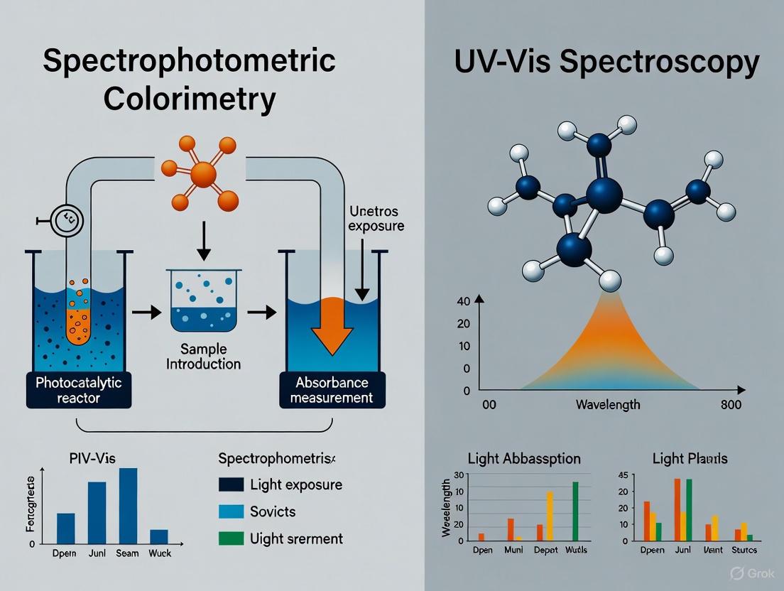
Spectrophotometric Colorimetry vs. UV-Vis Spectrophotometry: A Practical Guide for Photocatalytic Assessment
This article provides a comparative analysis of spectrophotometric colorimetry and UV-Vis spectrophotometry for evaluating photocatalytic efficiency, a critical process in environmental remediation and advanced oxidation technologies.
Spectrophotometric Colorimetry vs. UV-Vis Spectrophotometry: A Practical Guide for Photocatalytic Assessment
Abstract
This article provides a comparative analysis of spectrophotometric colorimetry and UV-Vis spectrophotometry for evaluating photocatalytic efficiency, a critical process in environmental remediation and advanced oxidation technologies. Tailored for researchers and scientists, the content explores the fundamental principles, distinct methodological approaches, and specific applications of each technique. It offers practical guidance for troubleshooting common issues and optimizing protocols for different sample types, including complex substrates. A direct, evidence-based comparison equips professionals with the knowledge to select the most appropriate, reliable, and cost-effective method for their specific photocatalytic research and development projects.
Understanding the Core Principles: How Colorimetry and UV-Vis Spectrophotometry Work
In photocatalytic assessment research, accurately quantifying changes in color or concentration is fundamental to evaluating material performance. This guide objectively compares two principal instrumental techniques for this purpose: spectrophotometric colorimetry and UV-Vis spectrophotometry. Despite the apparent similarity in their names, these techniques are founded on different measurement principles, yielding data of varying complexity and suitability for specific research applications.
Spectrophotometric colorimetry, often referred to simply as colorimetry, is designed for psychophysical sample analysis, meaning its measurements correlate directly with human color perception [1]. It provides straightforward tristimulus values (e.g., L, a, b) that describe the color appearance of a sample under a fixed illuminant. In contrast, UV-Vis spectrophotometry is a tool for physical sample analysis [1]. It performs full-spectrum measurement, capturing a sample's reflectance or transmittance properties across a wide range of wavelengths, typically from ultraviolet (UV) to visible (Vis) light, from 190 nm to 1100 nm depending on the instrument. This generates a rich spectral fingerprint that can be used for precise quantification, identification, and complex color analysis beyond the capabilities of the human eye. For researchers developing new photocatalysts for applications like organic dye degradation or pharmaceutical pollutant removal, the choice between these techniques can significantly impact the depth, accuracy, and reliability of experimental conclusions.
Technical Comparison: Principles, Capabilities, and Data Output
The core difference between these instruments lies in their optical design and the fundamental nature of the data they produce. A colorimeter is a tristimulus color measurement device. It uses a simple light source and a set of three filtered sensors (red, green, and blue) that simulate the response of the human eye [1] [2]. The result is direct readouts of tristimulus values, which are excellent for determining if two colors look the same to an observer under a specific light.
A UV-Vis spectrophotometer, however, uses a more complex system. It employs a monochromator (comprising a prism, grating, or interference filter) to separate a broadband light source into individual wavelengths [1]. This allows the instrument to scan the sample with light of a specific, narrow bandwidth and measure how much is absorbed or transmitted across the entire spectral range. The primary data output is a spectrum—a plot of absorbance (or transmittance) versus wavelength [1] [3]. This full-spectrum data is the source of its superior analytical power.
Table 1: Core Technical Specifications and Capabilities
| Feature | Spectrophotometric Colorimeter | UV-Vis Spectrophotometer |
|---|---|---|
| Measurement Principle | Tristimulus; measures with 3 broad filters (RGB) [1] [2] | Full spectrum; measures with a monochromator [1] |
| Primary Data Output | Tristimulus values (e.g., X, Y, Z or L, a, b*) [1] | Absorbance/Transmittance spectrum (intensity vs. wavelength) [1] |
| Typical Application Scope | Color difference, fastness, and strength [1] | Quantitative concentration analysis, reaction kinetics, identification of compounds [3] |
| Light Source & Observer | Fixed illuminant; typically 2-degree standard observer [1] | Adjustable illuminant settings; typically 10-degree standard observer [1] |
| Analysis of Metamerism | Not possible [1] | Possible due to full spectral data [1] |
| Key Advantage | Speed, portability, cost-effectiveness for routine color QC [1] | High precision, versatility, and comprehensive data for R&D [1] |
Application in Photocatalytic Assessment: Experimental Evidence
The choice of technique is critically illustrated in the field of photocatalysis, where researchers quantify the degradation of organic dyes or pharmaceuticals to benchmark material performance.
Real-Time Monitoring with UV-Vis Spectrophotometry
Advanced photocatalytic research often requires observing reaction kinetics on a sub-second timescale. A 2023 study demonstrated this using a real-time UV-Vis spectroscopic setup to monitor the photocatalytic degradation of Methylene Blue (MB) by TiO₂ nanoparticles [3]. The experimental setup used a broadband Xenon Arc lamp, and a CMOS camera captured all spectral information simultaneously, enabling the acquisition of spectra every 20 milliseconds [3]. This method allowed researchers to plot the evolution of the MB absorption spectrum in real-time, clearly showing the degradation process and even revealing different mechanisms based on the quantity of TiO₂ used [3]. This level of temporal and spectral resolution is impossible for a standard colorimeter, which can only provide tristimulus values at discrete time points.
Quantitative Analysis of Complex Mixtures
UV-Vis spectrophotometry is also indispensable for specific quantitative analyses where colorimetry would be inadequate. A 2024 study on characterizing hemoglobin-based oxygen carriers (HBOCs) compared various UV-Vis spectroscopy-based methods for quantifying hemoglobin [4]. The research highlighted the sodium lauryl sulfate (SLS) Hb method as a preferred choice due to its specificity, ease of use, and safety [4]. The study emphasized that selecting a quantification method based on full-spectrum UV-Vis data is crucial for accurate characterization, as it allows researchers to analyze the entire absorbance spectrum for potential interferences from other carrier components before selecting the quantification algorithm [4]. This specificity and analytical rigor are hallmarks of spectrophotometric analysis.
Performance in Pigment Prediction
A comparative study on predicting natural pigments in agricultural products found that a color spectrophotometer (a type of reflectance spectrophotometer) provided the best accuracy for predicting Total Carotenoid Content (TCC), while Vis/NIR spectroscopy was superior for predicting Total Flavonoid Content (TFC) [5]. This demonstrates that even within the category of spectrophotometric instruments, the optimal choice depends on the specific analyte and its spectral characteristics.
Experimental Protocols: Key Methodologies from Cited Research
Protocol 1: Real-Time Photocatalytic Degradation Monitoring
This protocol is adapted from the study observing MB degradation with TiO₂ nanoparticles [3].
- Objective: To monitor the photocatalytic degradation of an organic dye (Methylene Blue) in real-time using a custom UV-Vis spectroscopic setup.
- Materials:
- Photocatalyst: TiO₂ nanoparticles [3].
- Target Pollutant: Methylene Blue (MB) solution [3].
- Light Source: 300 W Xenon Arc lamp (or other suitable UV-Vis source) [3].
- Spectroscopic Setup: Broadband light source, grating (1200 grooves/mm), and CMOS camera for simultaneous spectral capture [3].
- Magnetic stirrer for continuous mixing.
- Method:
- Prepare an aqueous solution of MB at a known initial concentration (e.g., ~10 µM).
- Disperse a precise quantity of TiO₂ photocatalyst into the MB solution (e.g., 1 g/L).
- Place the mixture in a reaction vessel under continuous stirring.
- In the dark, allow the system to equilibrate for 30 minutes to establish adsorption-desorption equilibrium.
- Initiate irradiation with the Xenon Arc lamp.
- Simultaneously, begin acquiring absorbance spectra at a high frequency (e.g., every 20 ms) using the CMOS camera.
- Continue data acquisition until the reaction is complete (e.g., 1-2 hours).
- Data Analysis: The degradation is tracked by observing the decrease in the characteristic absorption peaks of MB (~664 nm and ~614 nm) over time. The kinetics can be modeled by plotting the normalized absorbance (C/C₀) versus time.
Protocol 2: Evaluating Photocatalytic Activity for Pharmaceutical Degradation
This protocol is based on research investigating novel photocatalysts for degrading ibuprofen [6].
- Objective: To evaluate the full-spectrum-responsive photocatalytic activity of a material (e.g., YMnO₃-based nanoparticles) for the degradation of a pharmaceutical pollutant.
- Materials:
- Photocatalyst: e.g., YMO or YMO-SO powder [6].
- Target Pollutant: Ibuprofen solution [6].
- Light Source: 300 W Xenon lamp with filters to isolate UV, Vis, and NIR regions [6].
- Analytical Instrument: Standard UV-Vis spectrophotometer (e.g., UV1901 UV-Vis spectrophotometer) [6].
- pH meter and chemicals for adjustment (e.g., NaOH, H₂SO₄).
- Method:
- Prepare an ibuprofen solution at a specified concentration (e.g., 75 mg/L) [6].
- Adjust the solution to the desired pH (e.g., pH 7) [6].
- Add a known amount of photocatalyst (e.g., 1 g/L) to the solution [6].
- Stir in the dark for 30 minutes to reach adsorption equilibrium.
- Turn on the light source and begin irradiation. At fixed time intervals (e.g., every 10 minutes), withdraw aliquots of the reaction solution.
- Centrifuge the aliquots to remove the photocatalyst particles.
- Analyze the concentration of the clear supernatant using a UV-Vis spectrophotometer by measuring the absorbance at the characteristic peak of ibuprofen.
- Data Analysis: The degradation percentage (D%) is calculated as (C₀ - Cₜ)/C₀ × 100%, where C₀ and Cₜ are the initial concentration and concentration at time t, respectively.
The Scientist's Toolkit: Essential Research Reagents and Materials
Table 2: Key Reagents and Materials for Photocatalytic Assessment Experiments
| Item | Function in Experiment | Example from Research |
|---|---|---|
| Target Pollutants | Model compounds to assess photocatalytic efficiency. | Methylene Blue (dye) [3], Ibuprofen (pharmaceutical) [6], Crystal Violet (dye) [7]. |
| Semiconductor Photocatalysts | Active materials that generate charge carriers under light to degrade pollutants. | TiO₂ nanoparticles [3], YMnO₃ (YMO) and its derivatives [6], Gd₂Zr₂O₇ pyrochlore nanoparticles [7]. |
| Chemical Scavengers | Used in trapping experiments to identify the primary active species in the photocatalytic mechanism. | Disodium EDTA (for holes, h⁺), 2-Propanol (for hydroxyl radicals, •OH), 1,4-Benzoquinone (for superoxide radicals, •O₂⁻) [6]. |
| Broadband Light Source | Simulates solar radiation or provides specific light regions (UV, Vis, NIR) to excite the photocatalyst. | 300 W Xenon Arc/Xenon lamp [3] [6]. |
| Buffer Solutions & pH Adjusters | Control the pH of the reaction environment, which can critically influence reaction pathways and degradation rates. | Use of acids (e.g., H₂SO₄) or bases (e.g., NaOH) to set specific pH values [6]. |
Selecting between spectrophotometric colorimetry and UV-Vis spectrophotometry hinges on the research question's complexity and the required data granularity.
Use a Spectrophotometric Colorimeter when: The application is strictly limited to quality control of color [1] [2]. The primary need is to quickly measure color difference (ΔE) between a standard and a sample, or to monitor color fastness and strength in a production or inspection environment [1]. Its advantages are speed, portability, and lower cost [1].
Use a UV-Vis Spectrophotometer when: The research demands physical sample analysis and comprehensive data [1]. This is essential for quantifying concentration via Beer-Lambert law, studying reaction kinetics in real-time, identifying unknown compounds from their spectral signature, detecting metamerism, or performing color formulation [1] [3]. It is the instrument of choice for R&D, precise analysis, and whenever the mechanism of a photoreaction is under investigation [1].
In conclusion, for rigorous photocatalytic assessment research that goes beyond simple color comparison, UV-Vis spectrophotometry is the unequivocally superior and necessary tool. Its ability to provide full spectral data enables researchers to not only quantify degradation but also to uncover intermediate species, understand reaction pathways, and reliably benchmark the performance of next-generation photocatalytic materials.
The objective quantification of color is a critical process in numerous scientific fields, including pharmaceutical development and photocatalytic assessment. Instrumental color measurement moves beyond the limits of human perception and vocabulary, allowing researchers to capture color information as objective data. This creates a common language of color essential for scientific communication and quality control across industries [8] [1]. The two principal advanced color measurement instrument types are colorimeters and spectrophotometers, both of which use sophisticated technologies to accurately quantify color, but through fundamentally different operating principles.
Colorimeters perform psychophysical sample analysis by mimicking human eye-brain perception, meaning their measurements correlate directly to how we see color [8] [1]. In contrast, spectrophotometers are designed for physical sample analysis via full spectrum color measurement, providing wavelength-by-wavelength spectral analysis of a sample's reflectance, absorbance, or transmittance properties [8]. This fundamental distinction in approach—tristimulus values versus full-spectrum analysis—defines their respective capabilities, applications, and suitability for different research environments, particularly in sophisticated applications like photocatalytic assessment where understanding material properties under light exposure is paramount.
Core Operating Principles and Methodologies
The Tristimulus Method: Colorimeter Operation
A colorimeter is designed to perform a type of psychophysical sample analysis by mimicking human eye-brain perception, which means its measurements correlate to human perception. In essence, it is engineered to see color the way we do [8] [1]. Its results are direct readings of tristimulus values, which identify a color with characters representing different dimensions of its visual appearance. These values typically use standardized systems like CIE XYZ or CIELab (L, a, b*) developed by the International Commission on Illumination (CIE) [8] [9].
The colorimeter's operation is often based on the Beer-Lambert law, which establishes that the concentration of a solute in a solution is proportional to its absorbance of light [8] [1]. The instrument starts with a simple light source. With the help of a lens and tristimulus absorption filters, the beam of light becomes a single, focused wavelength which then passes through the sample solution. A photocell detector on the opposite side identifies how much of the wavelength was absorbed. This detector connects to a processor and digital display that provides a readable output of the results [8].
The components of a colorimeter are specifically configured for this tristimulus approach:
- Illuminant: Represents a specific light source (e.g., daylight or incandescent) to project consistent brightness onto the object. In a colorimeter, the illuminant is fixed [8].
- Observer: The standard observer offers a specific field of view for analyzing colors, typically a 2-Degree Standard Observer suitable for color evaluation and quality control [8].
- Tristimulus Absorption Filter: This filter isolates specific wavelengths to be applied to the sample, typically corresponding to red, green, and blue color channels [8] [10].
Colorimeter Measurement Workflow: This diagram illustrates the sequential process of tristimulus color measurement, from light source through to tristimulus value output.
Full-Spectrum Analysis: Spectrophotometer Operation
A spectrophotometer operates on fundamentally different principles than a colorimeter, designed for comprehensive physical sample analysis via full spectrum color measurement [8]. By providing wavelength-by-wavelength spectral analysis of a sample's reflectance, absorbance, or transmittance properties, it produces precise data beyond what is observable by the human eye [8]. While spectrophotometers can calculate psychophysical colorimetric information if desired, their primary strength lies in capturing the complete spectral characteristics of a sample [8].
The components of a spectrophotometer reflect its advanced capabilities:
- Illuminant: The illuminant in a spectrophotometer is versatile, allowing researchers to use standard and fluorescent illuminants representing various types of light conditions [8].
- Observer: The observer of a spectrophotometer is typically larger, at about 10 degrees, which CIE recommends as the most appropriate tool for industrial color applications [8].
- Dispersion Element: Instead of simple absorption filters, a spectrophotometer uses a prism, diffraction grating, or interference filter to isolate specific wavelengths, which allows it to systematically scan across the spectral range [8] [10].
The operational process involves an illuminant projecting a light source onto an object and through a prism, grating, or filter. This optical system isolates specific wavelength bands to sequentially hit the sample. A sensor then detects the light that isn't absorbed by the item and passes the data to a processor or computer equipped with specialized software. This system can detect spectral reflectance, transmittance, and absorbance characteristics across the entire measured spectrum [8] [1].
Spectrophotometer Full-Spectrum Analysis: This diagram shows the comprehensive spectral measurement process, highlighting the wavelength scanning capability across the full spectrum.
Comparative Technical Specifications
The differences between these two instrumental approaches become clear when examining their technical specifications and measurement capabilities side by side. The following table summarizes the key distinguishing characteristics:
Table 1: Technical Comparison of Colorimeters and Spectrophotometers
| Feature | Colorimeter | Spectrophotometer |
|---|---|---|
| Measurement Type | Tristimulus (RGB/XYZ) values [10] | Full spectral data (wavelength-by-wavelength) [10] |
| Data Output | Direct tristimulus values (Lab*, XYZ) [8] | Complete spectral curves, plus calculated tristimulus values [8] |
| Light Separation | Tristimulus absorption filters [8] | Prism, grating, or interference filter [8] |
| Observer Angle | Typically 2-degree standard observer [8] | Typically 10-degree standard observer [8] |
| Illuminant Options | Fixed [8] | Multiple, adjustable (D65, A, F, etc.) [8] |
| Metamerism Detection | Not capable [8] [1] | Capable [8] [1] |
| Color Formulation | Not supported [8] | Supported [8] |
Beyond these fundamental technical differences, the instruments vary significantly in their operational characteristics and suitability for different research environments:
Table 2: Performance and Application Comparison
| Characteristic | Colorimeter | Spectrophotometer |
|---|---|---|
| Accuracy | Moderate [10] | High [10] |
| Measurement Speed | Fast [8] | Varies (generally slower) [8] |
| Portability | Typically high [8] [10] | Varies (portable and benchtop options) [8] |
| Complexity | Simple operation [10] | More complex setup and operation [10] |
| Cost | Lower [8] [10] | Higher [8] [10] |
| Primary Applications | Basic QC, color comparison, concentration measurement [10] | R&D, color formulation, full-spectrum analysis [8] [10] |
| Environmental Robustness | Suitable for production environments [8] | More sensitive, better for laboratory settings [8] |
Experimental Applications and Protocols
Methodology for Photocatalytic Assessment
In photocatalytic assessment research, both colorimeters and spectrophotometers provide valuable data, though of different types and depths. For tristimulus measurement of photocatalytic color changes, the experimental protocol typically involves:
Sample Preparation: Prepare photocatalytic material samples (e.g., TiO₂ coatings, ZnO nanoparticles) on standardized substrates. Ensure consistent surface characteristics and thickness across samples.
Initial Measurement: Using a colorimeter, measure the baseline L, a, and b* values of the untreated samples. Calibrate the instrument according to manufacturer specifications using provided white and black calibration tiles.
Contaminant Application: Apply a standardized amount of organic contaminant (e.g., methylene blue, rhodamine B) uniformly across the sample surface.
Photocatalytic Exposure: Expose samples to UV or visible light source under controlled conditions (specific wavelength, intensity, and duration).
Post-Exposure Measurement: Re-measure the L, a, and b* values using the same colorimeter settings and geometry.
Data Analysis: Calculate color difference (ΔE) using the CIELAB color difference equation: ΔE = [(ΔL)² + (Δa)² + (Δb*)²]^0.5. Correlate the degree of color change with photocatalytic efficiency.
For full-spectrum analysis of photocatalytic processes, the methodology is more comprehensive:
Instrument Calibration: Perform full wavelength calibration of the spectrophotometer using certified standards, verifying accuracy across the UV-Vis spectrum (typically 200-800 nm).
Baseline Spectral Capture: Collect full reflectance or absorbance spectra of the pristine photocatalytic material before contaminant application.
Contaminant Characterization: Measure the spectral characteristics of the contaminant solution alone to establish its spectral signature.
Time-Series Measurements: After contaminant application and during light exposure, collect spectral data at predetermined time intervals (e.g., every 5-15 minutes).
Spectral Deconvolution: Analyze spectral changes using multivariate analysis to identify specific chemical transformations and intermediate species.
Kinetic Modeling: Use the full spectral data to calculate degradation rates, quantum yields, and reaction kinetics based on specific wavelength absorbance changes rather than overall color shift.
The Scientist's Toolkit: Essential Research Reagents and Materials
Table 3: Key Research Reagents and Materials for Photocatalytic Color Assessment
| Item | Function | Application Notes |
|---|---|---|
| Photocatalytic Materials (TiO₂, ZnO, WO₃, g-C₃N₄) | Primary catalysts for light-induced reactions | Different materials respond to different light wavelengths (UV vs. visible light active) [11] |
| Organic Dye Contaminants (Methylene Blue, Rhodamine B) | Model compounds for assessing photocatalytic efficiency | Provide measurable color changes and characteristic spectral signatures [11] |
| Standardized Substrates (Glass slides, quartz plates, reflective tiles) | Consistent surfaces for catalyst deposition | Quartz required for UV light transmission in certain experimental setups |
| CIE Standard Illuminants (D65 for daylight, A for incandescent) | Reference light sources for color measurement | Essential for metamerism assessment and standardized color evaluation [8] |
| Calibration Standards (White and black calibration tiles) | Instrument calibration for accurate measurements | Required before each measurement session to ensure data reliability [9] |
| UV-Vis Light Sources (Xenon lamps, LED arrays) | Activation of photocatalytic processes | Wavelength and intensity must be controlled and documented |
| Spectrophotometer Cells (Quartz, glass cuvettes) | Containers for liquid sample analysis | Quartz necessary for UV range measurements |
Data Presentation and Analysis
The fundamental difference in data output between these two methodologies directly influences analytical capabilities in photocatalytic research. The following comparison illustrates how the same photocatalytic process might be represented through these different measurement approaches:
Table 4: Comparative Data Output from Photocatalytic Degradation Analysis
| Analysis Parameter | Tristimulus Colorimeter Data | Full-Spectrum Spectrophotometer Data |
|---|---|---|
| Color Change Quantification | Single ΔE value representing total color difference | Multiple spectral peaks showing specific chromophore degradation |
| Time Resolution | Overall color change at endpoint or limited intervals | Continuous spectral evolution throughout reaction timeline |
| Sensitivity to Intermediates | Limited to visible color changes; may miss intermediates | Can detect transient intermediates with distinct spectral signatures |
| Metamerism Assessment | Not possible | Can identify when color matches under one illuminant but not another |
| Concentration Correlation | Indirect through overall color change | Direct through Beer-Lambert law at specific wavelengths |
| Kinetic Information | Overall degradation rate from color change | Multiple simultaneous reaction rates for different chromophores |
The tristimulus approach provides a simplified, human-vision-correlated assessment of photocatalytic activity, which may be sufficient for quality control applications where the endpoint is a visible color change. However, for mechanistic studies, reaction optimization, and comprehensive material characterization, the full-spectrum approach provides significantly deeper insights into the photocatalytic process, including identification of reaction intermediates, simultaneous monitoring of multiple contaminants, and understanding of degradation pathways.
The choice between tristimulus colorimetry and full-spectrum analysis fundamentally depends on the research objectives, required depth of information, and application context in photocatalytic assessment. Colorimeters, with their direct tristimulus measurement approach, offer speed, portability, and operational simplicity well-suited for rapid screening, quality control, and field applications where the primary interest is correlation with human visual perception [8] [10]. Their limitations in detecting metamerism, providing spectral data, or supporting color formulation must be considered within the research framework.
Spectrophotometers, through their comprehensive full-spectrum analysis, deliver detailed spectral information, superior accuracy, and versatile measurement capabilities essential for research and development, mechanistic studies, and precise color formulation [8] [10]. The ability to detect metamerism, measure colorant strength, and analyze a comprehensive range of sample types makes spectrophotometers particularly valuable for advanced photocatalytic research where understanding the fundamental processes beyond simple color change is required [8].
For photocatalytic assessment research specifically, the decision matrix should consider whether the study requires simple efficiency ranking (where colorimetry may suffice) versus detailed mechanistic understanding (requiring spectrophotometry). As research in this field advances toward more complex material systems and reaction environments, the comprehensive data provided by full-spectrum analysis becomes increasingly valuable, though the simplicity and speed of tristimulus methods retain their utility for specific applied applications.
In photocatalytic assessment research, the selection of appropriate analytical instrumentation is paramount for obtaining accurate and reproducible data. Colorimeters and UV-Visible (UV-Vis) spectrophotometers represent two fundamental tools for material characterization, yet they differ significantly in their technological sophistication and application scope. Colorimetry is a technique designed to mimic human color perception, providing tristimulus values that are directly correlated with how we see color. In contrast, UV-Vis spectroscopy is a comprehensive analytical method that measures the absorption of light across the ultraviolet and visible electromagnetic spectrum, providing detailed information about electronic transitions in molecules [12] [13]. For researchers investigating photocatalytic materials, understanding the distinction between these instruments is crucial for proper experimental design, data interpretation, and ultimately, advancing research in sustainable energy and environmental remediation technologies.
The fundamental operating principle shared by both instruments is the Beer-Lambert Law, which states that the absorbance (A) of a solution is directly proportional to the concentration (c) of the absorbing species and the path length (L) of the light through the sample: A = εcl, where ε is the molar absorptivity coefficient [14] [13] [15]. This relationship forms the quantitative foundation for both techniques, though the manner in which each instrument applies this law differs significantly in complexity and output.
Core Principles and Component-Level Comparison
Fundamental Operating Principles
Colorimeters operate on a relatively straightforward principle of using filtered light to make tristimulus measurements. They typically employ red, green, and blue (RGB) optical filters to separate broad wavelength bands, simulating the response of the human eye's photoreceptors [10] [16] [12]. This approach provides direct colorimetric information but lacks spectral detail. The measurement focuses primarily on quantifying color intensity, which can then be correlated with concentration through the Beer-Lambert law [14]. The simplicity of this approach makes colorimeters robust and easy to operate but limits their analytical capabilities primarily to concentration determination of colored compounds in the visible range.
UV-Vis spectrophotometers employ a significantly more sophisticated approach to spectral analysis. Instead of using fixed filters, these instruments utilize a monochromator (containing a diffraction grating or prism) to disperse polychromatic light from the source into its constituent wavelengths [17] [13]. This monochromator can be precisely adjusted to select narrow wavelength bands, allowing the instrument to scan across the UV and visible spectrum systematically. This capability enables the construction of complete absorption spectra, which provide a "molecular fingerprint" of the sample being analyzed [13] [15]. The detailed spectral information reveals not only concentration but also insights into molecular structure, energy gaps, and reaction kinetics—all critical parameters in photocatalytic research.
Component-by-Component Breakdown
The technological divergence between these instruments becomes evident when examining their core components side-by-side. The table below provides a detailed comparison of the key subsystems in each instrument:
Table 1: Detailed Comparison of Instrument Components
| Component | Colorimeter | UV-Vis Spectrophotometer |
|---|---|---|
| Light Source | Single lamp: Tungsten or halogen lamp for visible range [14] [18] | Multiple lamps: Deuterium (UV) + Tungsten/Halogen (Vis) [17] [13] |
| Wavelength Selection | Fixed tristimulus filters (RGB) [10] [12] | Adjustable monochromator (grating or prism) [17] [13] |
| Spectral Range | Visible range only (~380-780 nm) [14] | UV + Visible ranges (~190-780 nm) [13] [15] |
| Sample Container | Glass or plastic cuvettes [14] [18] | Quartz cuvettes (for UV), glass/plastic (Vis only) [13] |
| Detector | Silicon photodiode or photocell [14] [18] | Photomultiplier tube (PMT) or advanced photodiodes (e.g., InGaAs for NIR) [17] [13] |
| Optical Configuration | Single beam design [14] | Single or double beam design [17] [13] |
| Data Output | Tristimulus values (X, Y, Z or L, a, b*) [10] [12] | Full spectrum (absorbance/transmittance vs. wavelength) [13] [1] |
System Architecture and Light Path
The fundamental differences in component technology directly influence the overall system architecture of each instrument. The following diagrams illustrate the typical optical paths and component arrangements for both systems.
Diagram 1: Colorimeter System Architecture
Diagram 2: UV-Vis Spectrophotometer System Architecture
Performance Comparison for Photocatalytic Assessment
Analytical Capabilities and Limitations
The different technological approaches of colorimeters and UV-Vis spectrophotometers lead to significant variations in their analytical capabilities, which directly impact their suitability for photocatalytic research applications.
Colorimeter Strengths and Limitations: Colorimeters excel in routine quantitative analysis of colored compounds within the visible spectrum. Their simplicity translates to advantages in portability, operational speed, and cost-effectiveness [10] [1]. For photocatalytic assessments, this makes them suitable for quick concentration checks of specific dyes (like methylene blue degradation monitoring) where high spectral resolution is unnecessary. However, colorimeters cannot detect metamerism (where colors match under one light source but not another) [10] [1], lack capability for color formulation [10], and provide no information about absorption events in the ultraviolet region [14]—a critical limitation since many photocatalytic reactions involve UV activation.
UV-Vis Spectrophotometer Strengths and Limitations: UV-Vis instruments provide comprehensive spectral data essential for fundamental research [1]. They can identify and quantify multiple compounds simultaneously based on their unique spectral fingerprints, monitor reaction kinetics at specific wavelengths, and determine crucial optical properties such as band gap energy of semiconductor photocatalysts [13] [15]. The ability to measure into the UV range is particularly important for characterizing wide-bandgap semiconductors like TiO₂. The primary trade-offs include higher cost, greater operational complexity, and typically larger instrument footprint [10] [1].
Quantitative Performance Metrics
The performance differences between these instrument classes can be quantified through several key metrics, as summarized in the table below:
Table 2: Performance Metrics for Photocatalytic Applications
| Performance Parameter | Colorimeter | UV-Vis Spectrophotometer |
|---|---|---|
| Spectral Resolution | Low (broadband filters) [10] | High (adjustable, down to ≤1 nm SBW) [17] [15] |
| Photometric Accuracy | Moderate for color difference [1] | High (with linearity to 2-3 AU typically) [15] |
| Wavelength Accuracy | Fixed to filter characteristics [10] | High (typically ±0.5 nm) [15] |
| Stray Light | Not typically specified [14] | Critical specification (~3 AU for single monochromator) [15] |
| Measurement Speed | Very fast (seconds) [14] [1] | Slower (scanning required) but modern arrays are fast [13] |
| Metamerism Detection | No [10] [1] | Yes [10] [1] |
| Turbidity Measurement | Limited [14] | Yes (specialized models) [1] |
Experimental Protocols for Photocatalytic Assessment
Protocol 1: Dye Degradation Monitoring Using Colorimeter This protocol is optimized for rapid, routine assessment of photocatalytic activity using a colorimeter:
- Calibration: Prepare standard solutions of the target dye (e.g., methylene blue) at known concentrations. Using the colorimeter with the appropriate filter (e.g., blue filter for methylene blue), measure the absorbance of each standard and construct a calibration curve of concentration versus absorbance [14].
- Sample Preparation: Suspend the photocatalyst in the dye solution. Aliquot samples at predetermined time intervals during light irradiation and remove catalyst by centrifugation or filtration [14].
- Measurement: Measure the absorbance of each aliquot using the same colorimeter settings as calibration. Determine concentration from the calibration curve [14].
- Data Analysis: Plot concentration versus time to determine degradation kinetics. This method provides quantitative degradation efficiency but no information about intermediate products.
Protocol 2: Comprehensive Photocatalyst Characterization Using UV-Vis Spectrophotometer This advanced protocol provides complete optical characterization of both the photocatalyst and the reaction process:
- Band Gap Determination:
- Prepare a dilute suspension of the photocatalyst powder in a non-absorbing solvent or use an integrating sphere for solid samples.
- Acquire absorbance spectrum across the UV-Vis range (e.g., 250-800 nm) [13] [15].
- Transform the absorbance data to Tauc plot coordinates: (αhν)^(1/n) vs. hν, where α is absorption coefficient, hν is photon energy, and n depends on transition type (½ for direct, 2 for indirect band gaps) [15].
- Extrapolate the linear region of the Tauc plot to the energy axis to determine the optical band gap.
- Reaction Kinetics and Mechanism:
- Follow Protocol 1 steps but acquire full spectra (e.g., 300-800 nm) at each time point instead of single wavelength measurements [13].
- Use spectral deconvolution techniques to identify and quantify reaction intermediates based on their characteristic absorption peaks.
- Monitor isosbestic points to identify simple versus complex reaction pathways.
Essential Research Reagent Solutions
Successful photocatalytic assessment requires not only proper instrumentation but also appropriate selection of reagents and materials. The following table outlines essential research solutions for conducting these analyses:
Table 3: Essential Research Reagent Solutions for Photocatalytic Assessment
| Reagent/Material | Function/Application | Technical Considerations |
|---|---|---|
| Standard Dye Solutions (e.g., Methylene Blue, Rhodamine B) | Model pollutants for quantifying photocatalytic degradation efficiency [5] | Prepare fresh solutions; establish linear calibration curve within instrument's dynamic range (typically Abs < 1) [13] |
| Reference Catalysts (e.g., Degussa P25 TiO₂) | Benchmark materials for validating experimental protocols and instrument performance | Use consistent specific surface area and crystal phase composition for comparable results |
| Spectrophotometric Cuvettes | Sample containers with defined path length (typically 1 cm) [14] | Quartz for UV measurements, glass/plastic for visible-only; ensure proper cleaning to avoid contamination [13] |
| Solvent-Grade Water | Preparation of aqueous solutions and catalyst suspensions | Use high-purity water (18.2 MΩ·cm) to minimize interference from impurities [13] |
| Band Gap Reference Standards | Validation of UV-Vis instrument performance for Tauc plot analysis | Use materials with known band gaps (e.g., silicon, ZnO) to verify measurement accuracy |
| Neutral Density Filters | Checking photometric linearity of spectrophotometers [15] | Certified filters with known absorbance values at specific wavelengths |
The choice between a colorimeter and a UV-Vis spectrophotometer for photocatalytic assessment research depends fundamentally on the specific research objectives and required information depth.
Select a colorimeter when the application involves:
- High-throughput, routine quality control of photocatalytic materials
- Quantitative measurement of specific colored compounds where full spectral data is unnecessary
- Field measurements or environments where portability and robustness are prioritized
- Situations with budget constraints where basic concentration data is sufficient [10] [1]
Select a UV-Vis spectrophotometer when the research requires:
- Fundamental characterization of new photocatalyst materials (band gap determination, optical properties)
- Detection and quantification of reaction intermediates and complex degradation pathways
- Method development for novel photocatalytic processes
- Research requiring the highest possible accuracy and comprehensive spectral information [13] [1]
For complete photocatalytic characterization, many research laboratories ultimately utilize both instruments: colorimeters for rapid screening and quality control, and UV-Vis spectrophotometers for fundamental material characterization and method development. This dual approach maximizes both efficiency and analytical depth, advancing the development of more efficient photocatalytic systems for environmental and energy applications.
The Role of the Beer-Lambert Law in Quantitative Analysis
Table of Contents
- Introduction to the Beer-Lambert Law
- Fundamental Principles and Mathematical Formulation
- Instrumentation for Quantitative Analysis
- Experimental Protocols in Photocatalytic Assessment
- Comparative Analysis: Colorimetry vs. UV-Vis Spectroscopy
- Advanced Applications and Current Research
- Conclusion
The Beer-Lambert Law is a fundamental principle in analytical chemistry that describes the relationship between the absorption of light and the properties of a material through which the light is traveling [19] [20]. This law forms the cornerstone of quantitative analysis across numerous scientific disciplines, enabling researchers to determine the concentration of solutes in solution by measuring light absorbance [21]. In the specific context of photocatalytic assessment research, where understanding reaction kinetics and degradation rates is paramount, the Beer-Lambert Law provides the theoretical foundation for using spectroscopic techniques to monitor chemical changes [3] [22]. Its reliability and straightforward application make it an indispensable tool for researchers, scientists, and drug development professionals who require precise and accurate concentration measurements.
Fundamental Principles and Mathematical Formulation
The Beer-Lambert Law establishes a linear relationship between the absorbance (A) of a solution and the concentration (c) of the absorbing species, as well as the path length (L) of the light through the solution [19] [20]. The law is mathematically expressed as:
A = εlc
In this equation:
- A is the absorbance, a dimensionless quantity [19] [21].
- ε is the molar absorptivity (also known as the molar extinction coefficient), a constant specific to the substance and the wavelength of light, typically with units of M⁻¹cm⁻¹ [21] [23].
- l is the path length of the light through the solution, usually measured in centimeters (cm) [21].
- c is the concentration of the absorbing species, typically in moles per liter (M) [21].
Absorbance is defined through the concepts of incident light intensity ((I0)) and transmitted light intensity ((I)). Transmittance (T) is the ratio (I / I0), and absorbance is the negative logarithm of transmittance: A = -log₁₀(T) = -log₁₀(I / I₀) [19] [21]. This logarithmic relationship means that absorbance increases linearly with concentration, while transmittance decreases exponentially. The following table shows how absorbance and transmittance values correlate [19]:
| Absorbance | % Transmittance |
|---|---|
| 0 | 100% |
| 1 | 10% |
| 2 | 1% |
| 3 | 0.1% |
| 4 | 0.01% |
| 5 | 0.001% |
The following diagram illustrates the core relationship described by the Beer-Lambert Law and the process of light absorption in a sample:
Instrumentation for Quantitative Analysis
The practical application of the Beer-Lambert Law relies on instruments designed to measure the transmission or absorption of light by a sample. The two primary types of instruments used are colorimeters and spectrophotometers, each with distinct capabilities.
Colorimeters are tristimulus color measurement devices that typically use filters to isolate broad bands of red, green, and blue light, simulating human eye response [2] [1]. They are based on the Beer-Lambert law and are designed for straightforward color comparison and quality control [1]. Their advantages include portability, speed, and relatively low cost, making them suitable for routine inspections on production lines [1]. However, they lack versatility and cannot provide full spectral data, making them unsuitable for complex analysis like identifying metamerism or formulating colors [2] [1].
Spectrophotometers are more sophisticated instruments that perform full-spectrum analysis [2] [1]. They use a monochromator (e.g., a prism or diffraction grating) to isolate individual wavelengths of light across a wide range, typically from ultraviolet (UV) to visible (VIS) and near-infrared [24] [1]. This allows them to measure the spectral reflectance, transmittance, or absorbance of a sample at each wavelength [2]. UV-Vis spectrophotometers, which analyze materials in the ultraviolet and visible wavelengths, are the instrument of choice for research, color formulation, and quantitative analysis where high precision and extensive data are required [3] [1]. Modern advancements include real-time UV/VIS spectroscopy, which uses a broadband light source and a CMOS camera to capture all spectral information simultaneously, enabling the observation of fast dynamic processes like photocatalytic degradation on a sub-second timescale [3].
The table below summarizes the key differences between these two instruments:
| Feature | Colorimeter | UV-Vis Spectrophotometer |
|---|---|---|
| Core Principle | Tristimulus color measurement (psychophysical analysis) [2] [1] | Full-spectrum color measurement (physical sample analysis) [2] [1] |
| Light Separation | Absorption filters (red, green, blue) [1] | Monochromator (prism or diffraction grating) [24] [1] |
| Output Data | Tristimulus values (e.g., L, a, b) [1] | Absorbance/Transmittance spectrum across wavelengths [3] [1] |
| Accuracy & Precision | Accurate for color difference measurements [2] | High precision and accuracy; suitable for research [2] [1] |
| Primary Applications | Quality control, inspection, simple color comparison [2] [1] | Research & development, quantitative analysis, complex color analysis, kinetic studies [3] [1] |
| Cost | Lower cost [2] | More expensive [2] |
Experimental Protocols in Photocatalytic Assessment
Photocatalytic assessment often involves monitoring the degradation of organic dyes, such as methylene blue (MB), using a catalyst like TiO₂ nanoparticles under light irradiation [3]. The following is a generalized experimental protocol for such a study using UV-Vis spectroscopy.
1. Principle and Objective The objective is to evaluate the efficiency of a photocatalyst by tracking the decrease in concentration of a model pollutant (e.g., methylene blue) over time. The Beer-Lambert Law is used to convert measured absorbance values at the dye's characteristic absorption peak (e.g., ~664 nm for MB) into concentration data [3].
2. Reagent and Instrumentation Setup Key research reagents and materials essential for this experiment include:
| Research Reagent Solution | Function in the Experiment |
|---|---|
| Photocatalyst (e.g., TiO₂ nanoparticles) | The active material that degrades the dye under light [3]. |
| Model Pollutant (e.g., Methylene Blue solution) | The compound whose degradation is being monitored [3]. |
| Cuvette | A transparent container (typically with a 1 cm path length) to hold the sample during measurement [19]. |
| UV-Vis Spectrophotometer | The instrument used to measure the absorbance spectrum of the solution at specific time intervals [3]. |
| Calibration Standards | A series of solutions with known concentrations of the dye, used to create a calibration curve [19] [21]. |
3. Step-by-Step Workflow The following diagram outlines the key steps in a photocatalytic degradation experiment:
- Calibration Curve Creation: Prepare a series of standard solutions with known concentrations of the dye (e.g., methylene blue). Measure the absorbance of each standard at the analytical wavelength (λ_max) using the UV-Vis spectrophotometer. Plot absorbance versus concentration to generate a calibration curve; the slope of this line is equal to εl, validating the linear range of the Beer-Lambert Law for the system [19] [21].
- Photocatalytic Reaction: In a reaction vessel, mix the photocatalyst with the dye solution. Begin irradiation with a light source of the appropriate wavelength (e.g., UV or visible light) to initiate the photocatalytic degradation [3].
- Real-Time Monitoring: At predetermined time intervals, withdraw samples from the reaction mixture. For real-time setups, a spectrophotometer with a fast CMOS camera can capture spectra continuously (e.g., every 20 ms) without manual sampling [3].
- Data Analysis: For each sample, measure the absorbance at the analytical wavelength. Use the calibration curve to convert these absorbance values into concentrations. Plot the concentration (or normalized concentration) as a function of time to visualize the degradation kinetics and determine the reaction rate [3].
Comparative Analysis: Colorimetry vs. UV-Vis Spectroscopy
When selecting an analytical technique for photocatalytic research, the choice between colorimetry and UV-Vis spectroscopy is critical and depends on the specific requirements of the study.
- Data Richness and Specificity: The most significant difference lies in the data output. A colorimeter provides a few tristimulus values, which are an average of the sample's interaction with broad red, green, and blue filters [2] [1]. This is sufficient for simple color matching but lacks detail. A UV-Vis spectrophotometer provides a full absorption spectrum, revealing fine details such as shifts in peak position, the appearance of isosbestic points, and the formation or disappearance of intermediate compounds with distinct spectral signatures [3] [1]. This is invaluable for understanding complex reaction mechanisms.
- Application in Photocatalysis Research: Colorimeters are generally inadequate for advanced photocatalytic assessment. As noted in research on methylene blue degradation, a real-time spectroscopic approach is necessary for a "comprehensive understanding of the mechanism" and enables "the detection of peak position shifts of new peaks on a sub-second timescale, which are not easily accessible using conventional UV/VIS spectroscopy" [3]. This level of detail is impossible to achieve with a colorimeter.
- Quantitative Accuracy and Limitations: Both techniques rely on the Beer-Lambert Law, which has known limitations. Deviations from linearity can occur at high concentrations (typically >10 mM) due to electrostatic interactions between molecules or changes in refractive index [20] [25]. Scattering of light by particulate matter (e.g., the photocatalyst itself) can also cause apparent deviations, a factor that must be accounted for in photocatalytic studies [20]. Spectrophotometers are better equipped to identify and correct for some of these issues, for example, by measuring and subtracting baseline scattering.
Advanced Applications and Current Research
The application of the Beer-Lambert Law in quantitative analysis, particularly with advanced spectrophotometry, continues to evolve, pushing the boundaries of photocatalytic research.
- Real-Time Kinetic Analysis: Traditional spectrophotometers require wavelength scanning, which can miss rapid transient events. The development of real-time UV/VIS spectroscopy using simultaneous detection with a CMOS camera allows for the complete spectral evolution of a reaction to be captured within a single camera frame [3]. This technology has been used to show how different quantities of TiO₂ nanoparticles lead to different photocatalytic degradation mechanisms for methylene blue, with all spectral changes plotted every 20 milliseconds [3].
- Addressing Analytical Challenges: Researchers continue to highlight the challenges in spectrophotometric analysis for photocatalysis. A key issue is ensuring a reliable evaluation of photocatalytic activity, especially under visible light irradiation and when dealing with dye decolorization, which may not always represent complete mineralization [22]. The precise, full-spectrum data provided by modern UV-Vis spectrophotometers is essential for differentiating between simple decolorization and the breakdown of chemical structure.
- Broad Utility: Beyond photocatalysis, the Beer-Lambert Law remains a workhorse technique. It is fundamental to analytical chemistry for determining chemical species [21], used in atmospheric science to model the absorption of sunlight by gases and particles [20], and critical in clinical settings, such as measuring bilirubin levels in blood samples [20]. The pursuit of high accuracy in these fields has led to the design of specialized, high-precision spectrophotometers, such as the one developed by the National Institute of Standards and Technology (NIST) for certifying Standard Reference Materials [24].
The Beer-Lambert Law remains an indispensable principle in quantitative chemical analysis. Its simple mathematical formulation, A = εlc, provides a direct link between a measurable physical quantity (absorbance) and the concentration of an analyte. In the demanding field of photocatalytic assessment research, the choice of instrumentation for applying this law is crucial. While colorimeters offer simplicity and speed for basic color measurement, UV-Vis spectrophotometers provide the comprehensive, high-fidelity spectral data necessary to unravel complex reaction mechanisms and kinetics. The advent of real-time spectroscopic techniques further enhances this capability, allowing researchers to observe fast dynamic processes with unprecedented temporal resolution. As photocatalytic research continues to strive for greater efficiency and understanding, the synergy between the robust Beer-Lambert Law and advanced spectrophotometric technology will undoubtedly continue to be a driving force in scientific discovery and innovation.
Advantages and Inherent Limitations of Each Foundational Approach
In the field of photocatalytic assessment research, the selection of an appropriate analytical instrument is paramount for obtaining reliable and meaningful data. The two foundational approaches for colorimetric analysis are spectrophotometric colorimetry, often referred to simply as colorimetry, and ultraviolet-visible (UV-Vis) spectroscopy. While both techniques measure how light interacts with materials, they differ significantly in their principles, capabilities, and applications. This guide provides an objective comparison of these instrumental approaches, focusing on their performance characteristics for evaluating photocatalytic processes. Such processes often involve monitoring the decolorization of dyes or the degradation of colorless compounds under visible light irradiation [22]. Understanding the inherent strengths and limitations of each method enables researchers and drug development professionals to select the most appropriate tool for their specific experimental needs, thereby ensuring accurate evaluation of photocatalytic activity.
Fundamental Principles and Instrumentation
At their core, both colorimeters and UV-Vis spectrophotometers function by passing light through a sample and measuring the resulting interaction. However, their underlying methodologies and technological sophistication differ.
Spectrophotometric Colorimetry: A colorimeter is designed to perform psychophysical sample analysis, meaning its measurements correlate directly to human color perception [1]. It typically uses filters to isolate broad bands of light, often corresponding to red, green, and blue (RGB) regions, and provides results as tristimulus values (e.g., Lab*) [1] [26]. Its operation is fundamentally based on the Beer-Lambert law, which draws a correlation between the concentration of a solute and its absorbance of light [1] [27].
UV-Vis Spectrophotometry: A UV-Vis spectrophotometer is designed for physical sample analysis via full-spectrum measurement [1]. Instead of using simple filters, it employs a monochromator (containing a prism or diffraction grating) to isolate a single, precise wavelength at a time and scans across a specified range, typically 200 to 800 nm, covering both ultraviolet and visible regions [13] [28]. This process generates a complete absorption spectrum, providing data on a sample's reflectance, absorbance, or transmittance properties at each wavelength [1].
The following diagrams illustrate the core operational workflows for each technique, highlighting key differences in components like wavelength selection.
Colorimeter Measurement Workflow
UV-Vis Spectrophotometer Measurement Workflow
Comparative Analysis: Performance and Limitations
The choice between a colorimeter and a UV-Vis spectrophotometer involves trade-offs between speed, cost, data comprehensiveness, and precision. The following table summarizes the key performance characteristics of each foundational approach.
Table 1: Instrument Comparison for Photocatalytic Assessment
| Feature | Spectrophotometric Colorimeter | UV-Vis Spectrophotometer |
|---|---|---|
| Measurement Type | Tristimulus color difference (e.g., ΔE) [26] | Full spectral reflectance/transmittance [26] |
| Primary Output | Lab* values, color difference (ΔE) [1] [26] | Absorbance/Transmittance spectrum, concentration data [13] [29] |
| Wavelength Range | Visible light only (∼400–760 nm) [27] | Ultraviolet & Visible (∼200–760 nm) [13] [27] |
| Data Precision | Moderate, suitable for color difference [26] | High, suitable for quantitative analysis [26] |
| Key Advantage | Speed, portability, cost-effectiveness for routine QC [1] [30] | Comprehensive data, versatility, high accuracy [1] [29] |
| Inherent Limitation | Cannot identify metamerism; limited spectral data [1] | Higher cost and complexity; can be excessive for simple checks [1] |
| Ideal Use Case | Quick pass/fail color checks in quality control [26] | Color formulation, R&D, quantitative concentration analysis [1] [26] |
Advantages of Spectrophotometric Colorimetry
- High Efficiency and Speed: Colorimeters provide rapid measurement results, enabling high-throughput analysis and efficient color control on production lines or during fast-paced experimental screening [1] [30].
- Cost-Effectiveness: Colorimeters are generally more affordable than advanced spectrophotometers, both in terms of initial investment and operational expenses, offering an economical solution for basic color measurement requirements [26] [30].
- Portability and Convenience: Compact and portable designs allow for easy operation on production floors or in the field, making them ideal for on-site inspections and measurements outside a controlled laboratory [1] [30].
- Ease of Operation: Designed for simplicity, colorimeters require minimal training, making them accessible to operators with varying levels of technical experience [30].
Limitations of Spectrophotometric Colorimetry
- Limited Precision and Data: Colorimeters typically measure color using RGB or tristimulus filters, which limits their ability to detect subtle spectral nuances and provide the detailed data needed for color formulation or in-depth analysis [1] [30].
- Inability to Detect Metamerism: Metamerism occurs when colors match under one light source but not another. Colorimeters, with their fixed illuminant, cannot identify this phenomenon, which is a critical drawback for applications where color consistency under different lighting is important [1].
- Difficulty with Complex Colors: These instruments may struggle to accurately measure fluorescent, pearlescent, or metallic colors, as they do not perform a full spectral analysis [30].
- Sensitivity to External Conditions: Measurements can be affected by ambient lighting conditions and surface texture, potentially compromising accuracy if not carefully controlled [30].
Advantages of UV-Vis Spectrophotometry
- Comprehensive Data and Versatility: UV-Vis spectrophotometers provide wavelength-by-wavelength spectral analysis, offering an expansive range of data beyond what is observable by the human eye or a colorimeter. This allows for adjustable illuminant and observer settings [1].
- High Accuracy and Quantitative Capability: These instruments excel in providing precise and accurate measurements, offering high sensitivity and resolution for detecting minute changes in absorbance, which is crucial for quantifying analytes across various concentrations [29] [31].
- Non-Destructive Nature: UV-Vis allows for non-destructive analysis, meaning the sample remains unchanged after measurement. This is particularly advantageous for precious or limited samples and allows for continuous monitoring of reactions over time [29] [28].
- Broad Application Range: The technique is versatile and can be used for quantitative analysis, identifying substances based on their unique absorption spectra, and measuring turbidity in liquids simultaneously with color [1] [29].
Limitations of UV-Vis Spectrophotometry
- Cost and Complexity: UV-Vis spectrophotometers are typically more expensive than colorimeters and have a more complex design. This complexity can make them more sensitive and potentially less suited for harsh factory environments [1] [26].
- Potential for Over-specification: For applications that require only simple color difference measurements, a UV-Vis spectrophotometer may offer more functionality than necessary, resulting in an unjustified investment [1].
- Sensitivity to Stray Light: The accuracy of absorption measurements can be influenced by stray light, which can affect the accuracy of the collected spectra [28].
- Sample Requirements: For UV light analysis, quartz cuvettes are required because glass and plastic absorb UV light, adding to the cost and procedural considerations [13].
Experimental Protocols for Photocatalytic Assessment
The reliable evaluation of photocatalytic activity, particularly under visible light irradiation, requires carefully designed experimental protocols. The choice between colorimetry and UV-Vis spectroscopy will depend on the specific goals of the analysis, such as monitoring dye decolorization or tracking the degradation of colorless compounds [22].
Protocol for Dye Decolorization Monitoring
This protocol is suitable for assessing the degradation of colored organic dyes, a common test for photocatalytic activity.
Objective: To quantify the rate of decolorization of a model dye (e.g., methylene blue) by a photocatalyst under visible light.
Materials and Reagents: Table 2: Key Research Reagent Solutions
| Item | Function in Experiment |
|---|---|
| Photocatalyst Powder (e.g., TiO₂-based material) | The active material that, upon light absorption, generates charge carriers to degrade the dye. |
| Model Dye Solution (e.g., Methylene Blue, Rhodamine B) | The target pollutant whose concentration change is monitored via absorbance. |
| Aqueous Reaction Buffer | Provides a stable pH environment for the photocatalytic reaction. |
| Quartz or UV-Transparent Cuvettes | Holds the sample solution for measurement; quartz is essential for UV-Vis analysis in the UV range. |
Procedure:
- Sample Preparation: Prepare a known concentration of the dye solution in an appropriate aqueous buffer. Disperse a precise mass of the photocatalyst powder into the dye solution to create a suspension.
- Initial Absorbance Measurement: Before light exposure, take an aliquot of the suspension. Separate the catalyst (e.g., via centrifugation or filtration) if it interferes with the measurement. Using a UV-Vis spectrophotometer, measure the absorbance spectrum of the clear supernatant, noting the maximum absorbance value (λ_max) for the dye.
- Light Irradiation: Place the main reaction suspension under visible light irradiation with constant stirring to keep the catalyst suspended. Maintain constant temperature.
- Kinetic Sampling: At predetermined time intervals (e.g., every 15 minutes), withdraw aliquots from the reaction mixture. Separate the catalyst from each aliquot.
- Analysis: Measure the absorbance of each supernatant at the dye's λ_max using either the UV-Vis spectrophotometer or a colorimeter calibrated for that specific wavelength.
- Data Calculation: The decolorization efficiency can be calculated as:
% Decolorization = [(A₀ - Aₜ) / A₀] × 100%, where A₀ is the initial absorbance and Aₜ is the absorbance at time t. For kinetic studies, plotln(A₀/Aₜ)versus time to check for pseudo-first-order behavior.
Protocol for Analysis of Colorless Compound Degradation
Monitoring the degradation of colorless compounds requires a UV-Vis spectrophotometer, as changes occur primarily in the ultraviolet range.
Objective: To track the photocatalytic degradation of a colorless organic pollutant (e.g., phenol) by observing changes in the UV absorption spectrum.
Materials and Reagents: The materials are similar to those in Protocol 4.1, with the model dye replaced by a colorless compound like phenol or salicylic acid.
Procedure:
- Sample Preparation: Prepare a known concentration of the colorless compound in an aqueous buffer and disperse the photocatalyst as before.
- Spectral Scanning: Use a UV-Vis spectrophotometer to perform a full scan (e.g., 200-400 nm) of the solution before irradiation to identify the characteristic absorption peak(s) of the target compound.
- Light Irradiation and Sampling: Expose the suspension to visible light and collect samples at regular intervals, separating the catalyst each time.
- Spectral Analysis: For each sample, perform a full UV spectrum scan. Monitor the decrease in the characteristic UV peak(s) of the parent compound. Additionally, observe the potential appearance and subsequent disappearance of new absorption peaks, which may indicate the formation and further degradation of intermediate products.
- Data Interpretation: Quantify the degradation rate of the parent compound by the decrease in its peak absorbance. The complex spectral changes provide insight into the reaction pathway and the catalyst's efficiency in mineralizing the pollutant, not just breaking it down initially.
Application in Photocatalytic Research: A Case Example
Research into non-destructive measurement methods for agricultural commodities highlights the performance differences between these instruments. A study comparing a color spectrophotometer and Visible/Near-Infrared (Vis/NIR) spectroscopy (381–1065 nm) for predicting natural pigments in cucumber fruit found that the accuracy of each instrument depended on the target analyte [5].
Table 3: Experimental Data from Pigment Prediction in Agricultural Research
| Pigment | Instrument | Calibration Correlation (Rcal) | Prediction Correlation (Rpred) | RPD | Key Finding |
|---|---|---|---|---|---|
| Total Carotenoid Content (TCC) | Color Spectrophotometer | 0.89 | 0.90 | 2.44 | The color spectrophotometer provided the best prediction accuracy for TCC [5]. |
| Total Flavonoid Content (TFC) | Vis/NIR Spectroscopy | 0.86 | 0.83 | 1.78 | The Vis/NIR spectrometer provided the best prediction accuracy for TFC [5]. |
This case demonstrates that the choice of instrument can significantly impact the quality of experimental data. The color spectrophotometer outperformed the more advanced spectroscopic technique for one specific pigment (carotenoids), while the Vis/NIR system was superior for another (flavonoids). This underscores the importance of aligning the instrument's inherent capabilities with the specific analytical goal, a principle that directly translates to photocatalytic assessment research where different reaction products and pathways may need to be monitored.
Practical Protocols: Applying Colorimetry and UV-Vis to Photocatalytic Reactions
Standardized Workflow for Dye Degradation Monitoring via Colorimetry
The quantitative monitoring of dye degradation is a cornerstone in evaluating the efficiency of photocatalytic processes, particularly in environmental remediation and advanced oxidation technologies. Within the broader context of comparing spectrophotometric colorimetry and UV-Vis spectroscopy for photocatalytic assessment, researchers must navigate a landscape of instrumental capabilities, methodological approaches, and data interpretation frameworks. Photocatalytic degradation of organic dyes serves as a key model system for assessing catalyst performance, with colorimetric methods providing accessible, cost-effective analytical pathways [32] [33]. The selection between dedicated colorimeters and versatile UV-Vis spectrophotometers significantly influences the depth of information obtained, experimental design considerations, and ultimately, the scientific conclusions drawn regarding catalytic efficiency [1].
This comparison guide objectively examines both methodologies through the specific application of dye degradation monitoring, providing experimental data, standardized protocols, and analytical frameworks to support informed methodological selection. By establishing a standardized workflow, we aim to enhance reproducibility across photocatalytic studies while clarifying the distinct advantages and limitations inherent to each analytical approach for researchers operating at the intersection of materials science, environmental chemistry, and analytical methodology.
Fundamental Principles: Colorimeters vs. UV-Vis Spectrophotometers
Understanding the core operational differences between colorimeters and UV-Vis spectrophotometers is essential for appropriate method selection in photocatalytic dye degradation studies.
Colorimeters perform psychophysical sample analysis, meaning their measurements correlate directly to human color perception. These instruments are designed with fixed components: an illuminant representing a specific light source, a 2-degree standard observer for color evaluation, and tristimulus absorption filters that isolate specific wavelengths. They provide direct tristimulus color readings (typically as L, a, b or X, Y, Z values) based on the CIE Color System, which characterizes color appearance across different dimensions [1]. The operational principle relies on the Beer-Lambert law, which establishes that solute concentration correlates with light absorbance. A colorimeter projects a single, focused wavelength through a sample solution, with a photocell detector measuring the transmitted light, and a processor converting this measurement into a readable output [1].
UV-Vis Spectrophotometers conduct physical sample analysis through full-spectrum measurement, generating data beyond human visual capability. These instruments offer greater component versatility compared to colorimeters, including adjustable illuminants that can represent various light sources, a larger 10-degree standard observer (recommended by CIE for industrial applications), and wavelength isolation through prisms, gratings, or interference filters. This enables wavelength-by-wavelength analysis of a sample's reflectance, absorbance, or transmittance properties [1]. While they can calculate psychophysical colorimetric data, their primary strength lies in capturing spectral information that facilitates advanced analytical applications.
Table 1: Fundamental Operational Differences Between Colorimeters and UV-Vis Spectrophotometers
| Feature | Colorimeter | UV-Vis Spectrophotometer |
|---|---|---|
| Analysis Type | Psychophysical (correlates to human perception) | Physical (full spectrum analysis) |
| Primary Output | Tristimulus values (Lab*, XYZ) | Spectral reflectance/absorbance/transmittance |
| Illuminant | Fixed | Adjustable (multiple standard illuminants) |
| Observer | Typically 2-degree | Typically 10-degree |
| Wavelength Selection | Tristimulus absorption filters | Prism, grating, or interference filter |
| Data Complexity | Direct color values | Wavelength-by-wavelength spectral data |
Experimental Protocols for Dye Degradation Monitoring
Standardized Photocatalytic Testing Framework
A robust protocol for assessing photocatalytic dye degradation begins with careful experimental design. The following methodology, adapted from research on nitrogen-doped TiO₂ nanoparticles, provides a standardized approach applicable to various catalyst-dye systems [32].
Materials and Reagents:
- Photocatalyst (e.g., N-TiO₂, ZnO, CdS nanostructures)
- Target azo dyes (e.g., Methylene Blue, Rhodamine B, Acid Orange 7)
- Deionized water
- Reaction vessels (batch reactors)
- Light source (visible or UV, depending on catalyst)
- Analytical instrumentation (colorimeter or UV-Vis spectrophotometer)
Procedure:
- Catalyst Preparation: Synthesize or obtain photocatalytic material. Characterize using appropriate techniques (XRD, SEM, TEM) to confirm morphology and composition [32] [33].
- Dye Solution Preparation: Prepare stock solutions of target dyes at known concentrations (typically 10-20 mg/L) using deionized water.
- Adsorption-Desorption Equilibrium: Combine catalyst (0.5-1.5 g/L) with dye solution in photocatalytic reactor. Stir in darkness for 30-60 minutes to establish adsorption-desorption equilibrium before illumination.
- Photocatalytic Degradation: Initiate illumination using appropriate light source (e.g., visible LED for modified catalysts). Maintain constant stirring throughout irradiation.
- Sampling and Analysis: Withdraw aliquots at predetermined time intervals. Separate catalyst from solution via centrifugation or filtration. Analyze supernatant using colorimeter or UV-Vis spectrophotometer.
- Data Recording: Record color values (colorimeter) or full absorbance spectra (UV-Vis) for each time point. Continue measurements until degradation plateaus or completes.
The Taguchi experimental design method, employing orthogonal arrays, can efficiently optimize multiple parameters affecting photocatalytic activity (e.g., pollutant selection, catalyst amount, distance from radiation source, time protocol) with a reduced number of experiments [32].
Instrument-Specific Methodologies
Colorimetric Monitoring Protocol: For dedicated colorimeters, calibrate the instrument according to manufacturer specifications using standard references. Set appropriate measurement parameters for the target dye. For each sample aliquot, measure and record tristimulus values (L, a, b). Calculate color difference (ΔE) between initial and degraded samples using the formula: ΔE = √[(ΔL)² + (Δa)² + (Δb)²] Plot ΔE versus irradiation time to generate degradation kinetics. Alternatively, use specific color channels (particularly those showing maximum change) for quantitative assessment [1] [34].
UV-Vis Spectrophotometric Protocol: Calibrate the spectrophotometer across the relevant wavelength range (typically 200-800 nm for organic dyes). For each sample aliquot, record the full absorbance spectrum. Identify the characteristic absorption peak(s) of the target dye. Monitor the decrease in peak intensity at λmax versus irradiation time. Calculate degradation percentage using the formula: Degradation (%) = [(A₀ - Aₜ)/A₀] × 100 Where A₀ is initial absorbance and Aₜ is absorbance at time t. Plot degradation percentage versus time for kinetic analysis [34] [35].
Performance Comparison: Experimental Data and Analysis
Direct comparison of colorimeters and UV-Vis spectrophotometers for dye degradation monitoring reveals distinct performance characteristics that influence their applicability to different research scenarios.
Table 2: Performance Comparison for Dye Degradation Monitoring
| Performance Metric | Colorimeter | UV-Vis Spectrophotometer |
|---|---|---|
| Measurement Speed | Rapid (seconds per measurement) | Moderate (varies with scan range) |
| Data Comprehensiveness | Limited to color appearance values | Extensive (full spectral information) |
| Detection Sensitivity | Moderate | High |
| Metamerism Detection | No | Yes |
| Suitability for Kinetic Studies | Excellent for rapid sampling | Excellent with defined wavelengths |
| Multi-Component Analysis | Limited | Excellent (spectral deconvolution) |
| Turbidity Compensation | Limited | Advanced capabilities |
| Early Degradation Detection | Moderate | Superior |
Experimental studies demonstrate that UV-Vis spectrophotometry detects color variations earlier and more precisely than visual examination or basic colorimetry. In stress testing of pharmaceutical compounds, color changes were detected significantly earlier by UV-Vis spectrophotometry than by visual assessment [34]. This enhanced sensitivity is particularly valuable for detecting initial degradation stages or subtle catalytic effects.
For photocatalytic applications, the comprehensive data from UV-Vis spectrophotometers enables advanced analysis beyond simple degradation kinetics. The technique can identify intermediate degradation products through spectral deconvolution and chemometric methods, as demonstrated in studies of sulfamethoxazole degradation where UV-Vis coupled with multivariate curve resolution-alternating least squares (MCR-ALS) provided detailed pathway analysis [35].
The morphology of photocatalytic nanomaterials significantly influences degradation efficiency, with different morphologies (nanorods, nanoflowers, nanosheets) exhibiting varying performance even within the same material system. For instance, while TiO₂ nanorods generally demonstrate superior photocatalytic activity, CdS nanoflowers may outperform other morphologies of the same material [33]. These morphological effects necessitate precise monitoring capabilities that UV-Vis spectrophotometry readily provides.
Experimental Workflow and Decision Pathways
The selection between colorimetric and spectrophotometric approaches depends on research objectives, analytical requirements, and available resources. The following workflow diagrams illustrate standardized protocols and decision pathways for implementing these methodologies in dye degradation studies.
Dye Degradation Monitoring Workflow
Instrument Selection Decision Pathway
The Scientist's Toolkit: Essential Research Reagents and Materials
Successful implementation of dye degradation monitoring requires specific reagents and materials optimized for colorimetric and spectrophotometric analysis.
Table 3: Essential Research Reagents and Materials for Dye Degradation Studies
| Reagent/Material | Function/Application | Specification Notes |
|---|---|---|
| Photocatalysts | Materials that drive degradation under light | TiO₂ (anatase/rutile), ZnO, CdS, Ag₃PO₄; Various morphologies (rods, flowers, spheres) [33] |
| Azo Dye Standards | Target pollutants for degradation studies | Methylene Blue, Rhodamine B, Acid Orange 7; High purity for calibration [32] |
| Solvents | Medium for dye dissolution and reaction | Deionized water; Organic solvents (methanol, acetonitrile) for hydrophobic dyes |
| Reference Materials | Instrument calibration and validation | Color reference standards; Spectral calibration standards (holmium oxide) [34] |
| Filtration Materials | Catalyst separation before analysis | Syringe filters (0.22-0.45 μm); Centrifugation equipment |
| Reaction Vessels | Containment for photocatalytic reactions | Quartz cuvettes (UV studies); Batch reactors with reflective walls [32] |
| Light Sources | Photoactivation of catalysts | Visible LED arrays; UV lamps (mercury, xenon); Solar simulators |
| Analytical Instruments | Degradation monitoring and quantification | Colorimeters (tristimulus); UV-Vis spectrophotometers (scanning) [1] |
The comparative analysis of colorimetry and UV-Vis spectrophotometry for dye degradation monitoring reveals distinct application domains for each technique. Colorimeters provide optimal solutions for quality control environments, high-throughput screening, and applications where simple color difference metrics sufficiently address research questions. Their advantages include speed, portability, operational simplicity, and cost-effectiveness for basic degradation assessment [1]. UV-Vis spectrophotometers offer superior capabilities for research-grade applications, mechanistic studies, and scenarios requiring comprehensive spectral information. Their strengths encompass detection of metamerism, identification of degradation intermediates, turbidity measurement, and enhanced sensitivity for early degradation detection [1] [34] [35].
For photocatalytic assessment research, the selection between these methodologies should be guided by specific research objectives. Studies focused solely on degradation efficiency comparison across catalyst formulations may find colorimetric methods sufficient and more efficient. Conversely, research investigating degradation mechanisms, pathway identification, or complex reaction kinetics will benefit from the comprehensive data provided by UV-Vis spectrophotometry. The standardized workflows and decision pathways presented herein offer researchers structured frameworks for implementing these methodologies, potentially enhancing reproducibility and comparability across photocatalytic studies.
As photocatalytic materials evolve toward greater complexity and specificity, the analytical demands for their characterization will similarly intensify. Future methodological developments will likely focus on hyphenated techniques, increased automation, and advanced data processing approaches that extract maximum information from degradation monitoring experiments, regardless of the core analytical platform employed.
UV-Vis Spectrophotometry Protocols for Kinetic Studies and Reaction Monitoring
Kinetic studies are fundamental to understanding chemical processes, providing critical data on reaction mechanisms, rates, and optimal conditions. In both academic research and industrial applications, monitoring these reactions accurately is essential for catalyst development, process optimization, and quality control. Ultraviolet-Visible (UV-Vis) Spectrophotometry has established itself as a cornerstone technique for such analyses, particularly in homogeneous systems where its ability to measure absorbance as a direct function of analyte concentration enables precise kinetic monitoring. The measured absorbance is directly proportional to the concentration of a given reactant, product, or intermediate, making it a productive technique for monitoring reactions [36].
However, the landscape of analytical techniques includes another important instrument: spectrophotometric colorimeters. These devices, which are designed to perform psychophysical sample color analysis, offer a different approach to measurement. This guide provides an objective comparison between these two methodologies, focusing on their application in photocatalytic assessment research. By examining experimental data, protocols, and performance characteristics, we aim to equip researchers and drug development professionals with the information necessary to select the most appropriate technique for their specific kinetic monitoring requirements.
Technical Comparison: UV-Vis Spectrophotometers vs. Colorimeters
Understanding the fundamental differences between UV-Vis spectrophotometers and colorimeters is crucial for selecting the appropriate instrument. The table below summarizes their core characteristics:
| Feature | UV-Vis Spectrophotometer [1] [10] | Colorimeter [1] [10] |
|---|---|---|
| Measurement Type | Spectral analysis (wavelength-by-wavelength) | Tristimulus (RGB values) |
| Principle | Measures light intensity across full spectrum; absorbance, reflectance, or transmittance [10] | Measures absorbed light based on Beer-Lambert law; correlates to human perception [1] |
| Light Source & Range | Adjustable illuminant (often including UV and visible) [1] | Fixed illuminant (e.g., daylight, incandescent) [1] |
| Data Output | Full spectral data (absorbance/transmittance vs. wavelength) | Tristimulus values (e.g., L, a, b or X, Y, Z) [1] |
| Key Components | Prism, grating, or interference filter; 10° observer is common [1] | Tristimulus absorption filters; 2° Standard Observer [1] |
| Metamerism Detection | Yes, due to full-spectrum analysis under different lights [1] [10] | No, due to fixed light source and filters [1] |
| Typical Applications | R&D, color formulation, turbidity measurement, quantitative concentration, reaction kinetics [1] [10] | Basic quality control, color difference checks, routine comparisons [1] [10] |
The workflow for selecting and applying a technique for kinetic monitoring depends on the nature of the chemical system and the analytical requirements. The decision pathway below outlines the core considerations:
Experimental Performance in Photocatalytic Assessment
Quantitative Performance Data
The following table compiles key performance metrics for both techniques as demonstrated in published photocatalytic and kinetic studies:
| Experiment / Application | Instrument Used | Key Performance Metric | Reported Value / Outcome | Reference |
|---|---|---|---|---|
| Hydrolysis of 4-Nitrophenyl Acetate (Homogeneous) | UV-Vis Spectrophotometry | Pseudo-first order rate constant | 0.01848 min⁻¹ | [37] |
| Hydrolysis of 4-Nitrophenyl Acetate (Homogeneous) | Smartphone RGB Analysis (Colorimetric) | Pseudo-first order rate constant | 0.01854 min⁻¹ | [37] |
| Reduction of 4-Nitrophenolate (Heterogeneous, Pd/C) | Smartphone RGB Analysis (Colorimetric) | High reproducibility & analytical frequency | Bypassed sampling needs; suitable for kinetics | [37] |
| Photocatalytic Methane Degradation (Ag-ZnO) | UV-Vis Spectrophotometry | Monitored degradation in flow-bed reactor | Enabled kinetic model establishment | [38] |
| General Applicability | Colorimeter | Color difference, fastness, strength | Accurate for routine comparison of similar colors | [1] |
| General Applicability | Spectrophotometer | Color formulation, metamerism, turbidity | Comprehensive data for R&D and complex QC | [1] [10] |
Detailed Experimental Protocols
Protocol 1: Monitoring Homogeneous Hydrolysis via UV-Vis and RGB Analysis
This protocol is adapted from a study that directly compared both techniques for the base-promoted hydrolysis of 4-nitrophenyl acetate (4-NPA) to 4-nitrophenolate (4-NP) [37].
- Objective: To validate a smartphone-based RGB method against traditional UV-Vis by determining the pseudo-first-order rate constant of 4-NPA hydrolysis.
- Reagents: 4-Nitrophenyl Acetate (4-NPA), carbonate/hydrogen carbonate buffer (pH 9.9).
- UV-Vis Procedure:
- Prepare a 33 µmol L⁻¹ solution of 4-NPA in the carbonate buffer.
- Transfer the solution to a spectrophotometer cuvette.
- Monitor the increase in absorbance at 400 nm (λmax for 4-NP) as a function of time.
- Fit the absorbance-time data to a first-order kinetic model to obtain the rate constant.
- RGB Colorimetric Procedure:
- Prepare the reaction solution identically and place it in a vial on a white background.
- Use a smartphone camera in a fixed geometric arrangement to capture images of the reaction vial at regular time intervals.
- Analyze the images using a program written in Python to extract the intensity of the Blue channel, which was found to be linearly proportional to the 4-NP concentration.
- Fit the Blue channel intensity-time data to a first-order kinetic model.
- Outcome: The rate constants from both methods showed a difference of less than 1%, validating the RGB colorimetric method for homogeneous kinetic studies [37].
Protocol 2: Heterogeneous Photocatalytic Degradation of Methane
This protocol outlines the use of UV-Vis spectrophotometry in the context of a more complex, gas-phase heterogeneous reaction [38].
- Objective: To investigate the photocatalytic performance of Ag-ZnO in degrading low-concentration methane (20-200 ppm) under UV-Vis light.
- Reagents & Catalyst: Methane gas standard, Ag-ZnO photocatalyst (prepared by reducing AgNO₃ with NaBH₄ and loading onto ZnO).
- Apparatus: Flow-bed reactor, Xe lamp (UV-Vis source), UV-Vis spectrophotometer for system characterization and/or product analysis.
- Procedure:
- The Ag-ZnO catalyst is placed inside the flow-bed reactor on slides.
- A gaseous mixture of methane (at the desired concentration) is passed through the reactor.
- The reactor is irradiated by the Xe lamp, initiating the photocatalytic oxidation of methane to CO₂.
- The reaction kinetics are studied by varying light intensity, methane concentration, and flow rate.
- The performance is evaluated, and a comprehensive mathematical model incorporating reaction kinetics is developed and validated.
- Outcome: The study demonstrated the feasibility of removing atmospheric methane via photocatalysis and established a kinetic model for engineering applications [38].
The Scientist's Toolkit: Essential Research Reagents and Materials
The table below lists key reagents and materials used in the featured experiments and the broader field of photocatalytic assessment.
| Item Name | Function / Application | Example from Research |
|---|---|---|
| 4-Nitrophenyl Acetate (4-NPA) | Model substrate for hydrolysis kinetics; produces yellow 4-nitrophenolate for easy tracking. | Used to validate RGB vs. UV-Vis in homogeneous kinetics [37]. |
| Palladium on Carbon (Pd/C) | Heterogeneous catalyst for reduction reactions. | Used in the model reduction of 4-nitrophenolate monitored by RGB analysis [37]. |
| Titanium Dioxide (TiO₂) | Standard semiconductor photocatalyst. | Used in photocatalytic decomposition of methyl orange [37]. |
| Silver-Zinc Oxide (Ag-ZnO) | Modified photocatalyst with enhanced activity. | Used for photocatalytic oxidation of low-concentration methane under UV-Vis light [38]. |
| Methyl Orange | Azo-dye used as a model pollutant for photocatalytic degradation tests. | Decomposed with H₂O₂ and TiO₂ under UV light in a heterogeneous system [37]. |
| Carbonate/Bicarbonate Buffer | Maintains constant pH for reactions, ensuring pseudo-first-order conditions. | Used in the hydrolysis of 4-NPA at pH 9.9 [37]. |
| Montmorillonite | Clay mineral used as an adsorbent in heterogeneous processes. | Studied for the adsorption of rhodamine dye [37]. |
Both UV-Vis spectrophotometry and spectrophotometric colorimetry offer distinct advantages for kinetic studies and reaction monitoring. UV-Vis spectrophotometry remains the gold standard for obtaining comprehensive spectral data, enabling detailed mechanistic insights in both homogeneous and heterogeneous systems, albeit sometimes requiring sample processing for the latter. Its ability to provide full-spectrum information makes it indispensable for research and development. In contrast, colorimeters and emerging RGB analysis techniques provide a streamlined, cost-effective, and often highly portable alternative for applications where tracking a specific color change is sufficient. Their simplicity and ability to bypass sampling in heterogeneous mixtures make them particularly valuable for high-throughput quality control and fieldwork. The choice between them is not a matter of superiority, but of alignment with the specific experimental needs, required data depth, and operational constraints of the research.
In photocatalytic assessment research, the accurate measurement of color and optical properties is fundamental for evaluating material performance and reaction progress. Two principal spectroscopic techniques are employed: spectrophotometric colorimetry, which quantifies color perception in reflected light, and UV-Visible (UV-Vis) spectroscopy, which analyzes light absorption characteristics across ultraviolet and visible wavelengths [2]. The choice between these techniques dictates all subsequent sample preparation, handling, and measurement protocols. This guide objectively compares the experimental workflows for analyzing opaque photocatalytic surfaces versus liquid solutions, providing researchers with detailed methodologies for each approach.
The core distinction lies in the light-matter interaction measured. Spectrophotometric colorimetry primarily measures reflectance—how light reflects from a surface—making it ideal for solid catalysts and coated materials [39] [2]. Conversely, UV-Vis spectroscopy typically measures transmittance—how light passes through a material—making it essential for analyzing solutions and suspensions [13] [40]. This fundamental difference necessitates completely separate sample preparation and handling procedures, which must be meticulously followed to generate reliable, reproducible data for photocatalytic assessment.
Comparative Analysis: Opaque Surfaces vs. Liquid Solutions
The table below summarizes the key experimental attributes for handling opaque surfaces versus liquid solutions in photocatalytic research.
Table 1: Experimental comparison of sample handling for opaque surfaces versus liquid solutions
| Experimental Attribute | Opaque Surfaces | Liquid Solutions |
|---|---|---|
| Primary Measurement | Reflectance [39] [2] | Transmittance/Absorbance [13] [40] |
| Sample Form | Solid films, powders, coated substrates [41] | Dissolved analytes, suspensions, liquid catalysts [42] [40] |
| Key Instrument | Reflectance Spectrophotometer (45°/0° or Integrating Sphere) [39] | UV-Vis Spectrophotometer [13] |
| Sample Container | None (direct placement) [39] | Cuvette (typically quartz or glass) [13] |
| Critical Preparation Step | Surface uniformity and presentation [39] | Concentration adjustment and solvent selection [13] [40] |
| Path Length Consideration | Not applicable | Critical (typically 1 cm) [13] |
| Primary Data Output | Lab* values, Reflectance Spectrum [39] [2] | Absorbance Spectrum, λmax, ε [13] [43] |
| Major Challenge | Surface texture, opacity, metamerism [39] [44] | Solvent absorption, concentration limits, air bubbles [13] [40] |
Experimental Protocols
For Opaque Surfaces
Protocol 1: Reflectance Measurement of Photocatalytic Films
- Principle: This method uses a spectrophotometer with a built-in integrating sphere to measure diffuse reflectance, simulating how the human eye perceives color under standard lighting conditions. It is suitable for evaluating the color of photocatalytic coatings, powders, and solid substrates [39].
- Materials:
- Spectrophotometer with integrating sphere geometry [39]
- Standard white calibration tile
- Zero calibration black trap or tile
- Opaque photocatalytic sample (e.g., coated tile, compressed powder pellet)
- Procedure:
- Instrument Calibration: Turn on the spectrophotometer and allow it to warm up. Calibrate first with the black trap, followed by the white reference tile as per the manufacturer's instructions [39].
- Sample Presentation: Ensure the sample is completely opaque. For powders, compress them into a pellet or place them in a sample cup with a quartz window to present a uniform, flat surface. For coated surfaces, ensure the coating is thick enough to prevent any background interference [42] [39].
- Measurement: Place the sample firmly against the measurement port of the integrating sphere. Acquire the reflectance spectrum over the desired wavelength range (typically 360-780 nm for color). The instrument will output values such as L, a, b*, and the full spectral reflectance curve [39] [2].
- Replication: Take a minimum of three measurements from different areas of the sample to account for potential inhomogeneity.
For Liquid Solutions
Protocol 2: Transmission Measurement of Photocatalytic Solutions or Suspensions
- Principle: This method directs a beam of light through a transparent cuvette containing the liquid sample. The instrument detects the intensity of light transmitted through the sample, which is used to calculate absorbance based on the Beer-Lambert law. This is ideal for monitoring dye degradation in photocatalytic tests [13] [40].
- Materials:
- Procedure:
- Cuvette Selection: Use high-quality quartz cuvettes for measurements involving ultraviolet light (below ~350 nm), as glass and plastic absorb strongly in this region [13].
- Blank Measurement: Fill a cuvette with the pure solvent used to prepare the sample (the "blank"). Place it in the sample holder and record the baseline or blank measurement. This corrects for any light absorption by the solvent or cuvette [13].
- Sample Preparation & Loading: Prepare the sample solution at an appropriate concentration. The target absorbance should ideally be below 1.0 to remain within the instrument's linear dynamic range. If necessary, dilute the sample [13]. Pipette the sample into a clean cuvette, ensuring no air bubbles are trapped in the light path. Wipe the outside of the cuvette with a lint-free tissue to remove fingerprints or droplets.
- Measurement: Place the sample cuvette in the holder and run the spectrum. The instrument will output an absorbance or transmittance spectrum from which the absorption maximum (λmax) and molar absorptivity (ε) can be derived [13] [43].
Workflow Visualization
The following diagrams illustrate the core decision pathways and experimental workflows for sample preparation and analysis in photocatalytic research.
The Scientist's Toolkit: Essential Research Reagents and Materials
The table below details key materials and reagents essential for conducting spectrophotometric analyses in photocatalytic assessment.
Table 2: Essential research reagents and materials for spectrophotometric analysis
| Item | Function & Application |
|---|---|
| Quartz Cuvettes | Holds liquid samples for UV-Vis analysis; transparent to UV and visible light, unlike glass or plastic [13]. |
| Solvents (HPLC Grade) | High-purity water, acetonitrile, methanol, etc.; dissolve analytes without introducing interfering absorbances [13] [40]. |
| Certified Calibration Tiles | Provides known reflectance values for calibrating spectrophotometers before measuring solid surfaces [39]. |
| Syringe Filters (0.45 μm, 0.22 μm) | Clarifies liquid samples by removing suspended particles that could cause light scattering [40]. |
| Compression Pellet Die | Prepares powdered photocatalyst samples into smooth, opaque solid disks for reflectance measurements [39]. |
| Standard Reference Materials | Compounds with known absorption spectra (e.g., potassium dichromate), used for verifying instrument wavelength and photometric accuracy [13] [43]. |
The selection between methodologies for opaque surfaces and liquid solutions is deterministic in photocatalytic research. Spectrophotometric colorimetry provides unmatched quantitative data for solid-state properties, tracking color changes in coatings and surfaces. UV-Vis spectroscopy remains the cornerstone for solution-based analysis, enabling precise concentration quantification and reaction kinetics monitoring through the robust Beer-Lambert law. A meticulous approach to the distinct preparation and handling protocols outlined in this guide is paramount for generating accurate, reproducible, and scientifically valid data, thereby advancing the development and optimization of photocatalytic materials and processes.
Real-Time and In-Situ Monitoring Techniques with UV-Vis Spectroscopy
Ultraviolet-visible (UV-Vis) spectroscopy is an analytical technique that measures the amount of discrete wavelengths of UV or visible light absorbed by or transmitted through a sample in comparison to a reference or blank sample. This property is influenced by the sample composition, providing critical information about what is in the sample and at what concentration. The fundamental principle relies on light energy being inversely proportional to its wavelength, with shorter wavelengths carrying more energy than longer wavelengths. A specific amount of energy is needed to promote electrons in a substance to a higher energy state, which we detect as absorption. Electrons in different bonding environments require different specific energy amounts for promotion, which is why absorption occurs at different wavelengths for different substances. This characteristic makes UV-Vis spectroscopy particularly valuable for identifying substances and determining concentrations across numerous scientific fields [13].
In contemporary research, especially in photocatalytic assessment and drug development, the ability to monitor reactions and processes in real-time and in situ has become increasingly important. Traditional UV-Vis spectroscopy has evolved from offline sample analysis to sophisticated real-time monitoring systems that provide researchers with instantaneous data on reaction kinetics, material properties, and dynamic processes. This evolution addresses the critical need for techniques that can capture instantaneous information without disrupting the system being studied, a limitation common to ex situ methods that require sample extraction and processing. The development of automated in situ platforms capable of precisely revealing heterogeneous processes provides the foundation for data-driven chemical reaction discovery and new scientific discipline revelation [45].
Comparative Analysis of UV-Vis Monitoring Techniques
The table below summarizes the core characteristics, advantages, and limitations of different UV-Vis spectroscopy monitoring approaches used in scientific research:
Table 1: Comparison of UV-Vis Spectroscopy Monitoring Techniques
| Technique | Core Principle | Best For | Key Advantages | Major Limitations |
|---|---|---|---|---|
| Traditional Offline UV-Vis | Sample extraction and cuvette-based measurement | Standard concentration measurements, quality control | Simple operation, established protocols, high precision for clear solutions | No temporal resolution, potential sample disturbance, labor-intensive |
| In Situ Probe-Based Monitoring | Direct immersion of fiber-optic probes into reaction vessels | Homogeneous reactions, simple kinetic studies | Minimal sample volume requirement, real-time data acquisition | Signal interference from stirring and excitation light, limited to less complex mixtures |
| Online Circulatory Systems (OCS) | Continuous pumping of suspension through flow cell | Suspended reactions with moderate particle loads | Separates reaction and detection, reduces interference from excitation light | Particle deposition issues, bubble formation, requires more complex tubing |
| Advanced DAS-OCS-AMS | Debubbling, aeration, and segmented flow incorporated into OCS | Complex heterogeneous systems, photocatalytic studies with solid particles | Addresses bubble and particle deposition issues, provides precise data for both mixture and solid particles | Increased system complexity, requires optimization of multiple parameters |
Each approach offers distinct advantages depending on the research context. Traditional offline methods, while reliable for simple analyses, fail to capture instantaneous information due to lack of temporal resolution. Moreover, solid-liquid separation operations such as centrifugation might destroy weak and loosely adsorbed layers, propagating misinformation in unpredictable and non-systematic ways. In contrast, in situ techniques track processes without disruption, but often face challenges with signal interference. Advanced systems like DAS-OCS-AMS represent the cutting edge, specifically engineered to overcome the persistent problems of bubble formation and particle deposition that plague other monitoring methods in heterogeneous systems [45] [46].
For photocatalytic assessment research specifically, the choice of monitoring technique significantly impacts the reliability and depth of obtainable data. The heterogeneity, polydispersity, and multi-component characteristics of suspended photocatalytic batch reactions (SPBRs) pose particular challenges in acquiring accurate kinetic data. Traditional methods that focus narrowly on the substrate while overlooking the optical characteristics of both the mixture and solid particles provide incomplete information. Advanced circulatory systems with debubbling and aeration capabilities have demonstrated superior performance in these applications by simultaneously monitoring both liquid-phase and solid-phase components while maintaining system integrity throughout extended experimental durations [46].
Experimental Protocols and Methodologies
Advanced In Situ Monitoring with Multivariate Analysis
A sophisticated approach coupling in situ UV-Vis spectroscopy with a two-step algorithm strategy has been developed for quantitatively monitoring heterogeneous reactions in a compact closed-loop incorporation. The methodology involves the following key components and steps:
Equipment Setup: The system utilizes a UV-Vis spectrophotometer equipped with an integrating sphere for simultaneous measurement of total reflectance and transmittance spectra. A closed-loop circulation system maintains sample suspension homogeneity during extended monitoring periods.
Light Scattering Correction: The Inverse Adding-Doubling (IAD) method is applied to extract pure optical absorption properties from the aqueous adsorption system. This step is crucial because the optical properties inherent in a suspension consist of two fractions: absorption fractions reflecting the electronic energy level structure, and scattering fractions related to physical collision between photons and particles/molecules. The IAD method effectively isolates scattering signals to obtain accurate absorption coefficient spectra [45].
Spectral Deconvolution: The Multivariate Curve Resolution-Alternating Least Squares (MCR-ALS) method processes the time-evolving UV-Vis absorption spectroscopic datasets to identify spectral profiles of chemical compositions and their corresponding abundance. This algorithm decomposes spectroscopic datasets into a linear combination of a weighted set of pure spectral profiles, where the weight factor is proportional to their concentrations.
Theoretical Spectral Assignment: Theoretical molecular calculations are employed to connect MCR-ALS solutions with chemical molecular structural evolution without prior information for reference spectra. This innovative approach assists in spectral signal assignment and chemical structure interpretation, particularly valuable for intermediate species that are challenging to isolate experimentally [45].
This integrated protocol was successfully applied to quantify the aqueous adsorption kinetics of bisphenol A onto polyamide microparticles in a one-step UV-Vis spectroscopic measurement. The results demonstrated a decrease in absorption intensity (approximately 60%) along with peak redshift (approximately 6 nm), clearly indicating observable spectral changes of BPA molecules due to adsorption. The practical applicability was confirmed by rapidly screening a superior adsorbent from commercial materials for antibiotic wastewater adsorption treatment [45].
Automated Monitoring System for Photocatalytic Applications
For photocatalytic assessment research, specifically in monitoring suspended photocatalytic batch reactions (SPBRs), an Automated Monitoring System (DAS-OCS-AMS) incorporating online circulatory spectrophotometry with debubbling, aeration, and segmented flow has been developed:
System Configuration: The DAS-OCS-AMS consists of five core units: (1) reaction unit providing necessary conditions and environment for the chemical reaction; (2) debubbling unit for signal noise reduction; (3) aerated and segmented unit to prevent particle deposition; (4) detection unit with a flow-cell spectrophotometer; and (5) data processing unit for real-time analysis [46].
Debubbling Mechanism: A debubbler achieves separation through gravity by utilizing the density disparity between gas and liquid. This addresses the critical issue of signal noise caused by bubbles (SNB), which manifests as sudden increases in absorbance signal intensity during monitoring.
Particle Deposition Prevention: An aerated and segmented device (ASD) generates high-velocity slug flow to eliminate particle deposition on the inner wall of pipelines (PDP) and on the windows of the flow cell (PDW). The segmented flow analysis decreases total circulation time, enhancing the prevention of PDP and PDW [46].
Validation Protocol: The system's performance was validated by monitoring the kinetics of photocatalytic degradation of Acid Orange II by TiO2 (P25), with comparison to traditional OCS-AMS. Results demonstrated that the DAS-OCS-AMS effectively mitigated the influence of SNB, PDP, and PDW, yielding precise results for both the mixture and solid particles [46].
This methodology provides a highly flexible universal framework for online circulatory automated monitoring and establishes a robust hardware foundation for subsequent data processing research in photocatalytic studies and beyond.
Diagram 1: Workflow of Advanced UV-Vis Monitoring System showing integration of hardware components and data processing algorithms for real-time in-situ analysis.
The Scientist's Toolkit: Essential Research Reagents and Materials
Table 2: Essential Research Reagents and Materials for UV-Vis Monitoring Experiments
| Item | Specification/Type | Function/Application | Key Considerations |
|---|---|---|---|
| Photocatalysts | TiO2 (P25), g-C3N4, ZnO | Primary catalytic materials for photocatalytic studies | Particle size, crystal structure, surface area, band gap energy |
| Model Pollutants | Acid Orange II, Bisphenol A, Methylene Blue | Standard compounds for degradation studies and method validation | Stability, absorption characteristics, environmental relevance |
| Adsorbents | Polyamide microparticles, Diatomite, Carclazyte | Materials for adsorption studies and comparative assessments | Surface functionality, porosity, particle size distribution |
| Spectrophotometer | Fiber-optic equipped, Integrating sphere capability | Core measurement instrument for absorbance/transmittance | Wavelength range, resolution, signal-to-noise ratio |
| Flow Cells | Quartz cuvettes, Microvolume flow cells | Sample containment for spectral measurements | Path length, material transparency (quartz for UV), volume requirements |
| Reference Materials | NIST-traceable standards, Solvent blanks | Calibration and baseline establishment | Purity, stability, matrix matching |
| Computational Tools | MCR-ALS software, Theoretical calculation packages | Data deconvolution and structural assignment | Algorithm flexibility, compatibility with experimental data |
The selection of appropriate materials and reagents is critical for successful implementation of real-time UV-Vis monitoring techniques. Photocatalyst choice significantly influences both the reaction kinetics and the optical properties of the system. For instance, TiO2 (P25) is widely used as a benchmark photocatalyst due to its well-characterized properties and reliable performance in degradation studies. Similarly, model pollutants like Acid Orange II provide consistent spectral signatures that facilitate quantitative tracking of reaction progress. The materials used in system construction must also be carefully considered—quartz flow cells and components are essential for UV-transparent measurements, as regular glass and plastic cuvettes absorb UV light and would compromise data quality, particularly in the valuable UV range where many compounds exhibit characteristic absorption [13] [46].
Advanced monitoring approaches additionally require specialized computational tools for data processing and interpretation. The MCR-ALS algorithm has proven particularly valuable for deconvoluting complex spectral datasets obtained from heterogeneous systems, enabling researchers to distinguish between dissolved and adsorbed species without prior spectral information. When combined with theoretical molecular calculations, this approach provides unprecedented insight into molecular structural evolution during reactions, creating a powerful synergy between experimental observation and computational prediction that significantly enhances the information yield from UV-Vis monitoring experiments [45].
Quantitative Performance Data and Comparison
The table below presents experimental data comparing the performance of different UV-Vis monitoring systems in photocatalytic and adsorption studies:
Table 3: Quantitative Performance Comparison of UV-Vis Monitoring Techniques
| Parameter | Traditional Offline Methods | Basic In Situ Probes | Advanced DAS-OCS-AMS |
|---|---|---|---|
| Temporal Resolution | Minutes to hours (sampling limited) | 1-10 seconds | 2-5 seconds |
| Signal Noise from Bubbles | Not applicable (samples clarified) | High interference | Effectively eliminated with debubbler |
| Particle Deposition Issues | Not applicable | Moderate to severe | Minimized with aerated segmented flow |
| Light Scattering Correction | Limited or none | Partial | Comprehensive IAD method implementation |
| Spectral Recovery Accuracy | N/A | 70-85% | >95% with MCR-ALS |
| Monitoring Duration | Limited by manual sampling | Potentially unlimited | Extended operation (24+ hours demonstrated) |
| Data Information Content | Single component focus | Limited multi-component | Full spectral deconvolution capability |
Quantitative assessments demonstrate the superior performance of advanced UV-Vis monitoring systems. In adsorption studies using the integrated IAD and MCR-ALS approach, researchers observed a decrease in absorption intensity of approximately 60% along with peak redshift of about 6 nm, clearly indicating spectral changes of Bisphenol A molecules due to adsorption. The method successfully quantified the transfer of BPA from aqueous solution to suspended polyamide microparticles, revealing that the molar absorption coefficient of aqueous BPA is larger than that of adsorbed BPA on particles, attributed to aggregation effects at the solid-liquid interface [45].
For photocatalytic applications, the DAS-OCS-AMS system has demonstrated exceptional capability in maintaining measurement precision over extended durations. Comparative analysis with traditional OCS-AMS confirmed that the proposed system effectively mitigates the influence of signal noise from bubbles, particle deposition on pipelines, and particle deposition on flow cell windows. This performance is achieved through the debubbler unit that separates gas and liquid phases by gravity, and the aerated segmented device that generates high-velocity slug flow to prevent adhesion of particles to surfaces. The result is precise monitoring of both liquid-phase and solid-phase components throughout photocatalytic reactions, providing comprehensive kinetic data essential for understanding reaction mechanisms and optimizing process parameters [46].
Integrated Workflow for Comprehensive Analysis
The synergy between hardware innovations and advanced data processing algorithms creates a powerful framework for real-time in situ monitoring. The integrated workflow begins with proper system configuration and calibration, followed by continuous monitoring with simultaneous data acquisition from multiple detection modes. The application of light scattering correction methods is essential for accurate interpretation of spectral data from heterogeneous systems, as uncorrected scattering effects can significantly distort absorption measurements and lead to erroneous conclusions about concentration changes and reaction progress [45].
Diagram 2: Integration Framework of UV-Vis Monitoring showing the synergy between experimental hardware, computational algorithms, and analytical interpretation for comprehensive reaction analysis.
The multivariate curve resolution alternating least squares (MCR-ALS) method serves as the computational core of advanced UV-Vis monitoring systems, enabling the decomposition of complex spectroscopic datasets into pure component spectra and concentration profiles without requiring prior information about the system composition. This capability is particularly valuable for studying heterogeneous reactions where intermediate species may form transiently, making them difficult to isolate and characterize by conventional means. When MCR-ALS analysis is coupled with theoretical spectral simulations, researchers can connect the resolved spectral components with specific molecular structures and transformations, creating a powerful link between experimental observation and molecular-level understanding [45].
This integrated approach has demonstrated practical utility across multiple applications, from quantifying adsorption kinetics to screening superior adsorbent materials for wastewater treatment. The method's ability to provide quantitative information about both dissolved and adsorbed species in a single measurement represents a significant advancement over traditional approaches that often require separate experiments or infer adsorption indirectly through solution depletion measurements. As these techniques continue to evolve, they offer promising potential for integration into fully automated experimental platforms that could dramatically accelerate research in photocatalysis, materials development, and pharmaceutical research by providing comprehensive real-time insights into complex chemical processes [45] [46].
The functionalization of building and environmental materials with titanium dioxide (TiO2) nanoparticles provides self-cleaning properties and facilitates the decomposition of pollutants, enhancing durability and functionality [47]. A critical challenge in this field is the lack of an international standard test method for calculating the photocatalytic efficiency (PE) of cementitious substrates, necessitating comparative evaluation of available assessment techniques [47]. This case study objectively compares spectrophotometric colorimetry (SPC) and UV-Vis spectrophotometry (UV-Vis) for assessing TiO2 photocatalytic efficiency using Rhodamine B (RhB) and Methylene Blue (MB) as model pollutants. This comparison is framed within broader research on standardized photocatalytic assessment protocols for heterogeneous catalysts.
Fundamental Principles of Photocatalysis
Photocatalysis involves chemical reactions in semiconductor materials where electrons in the valence band, when excited by light with energy equal to or greater than the band gap, migrate to the conduction band, leaving positive holes that promote redox reactions at the semiconductor surface [47] [48]. The electrons reduce molecular oxygen to form superoxide radicals (•O₂⁻), while the holes oxidize hydroxyl groups or water to form hydroxyl radicals (•OH). These reactive oxygen species subsequently degrade adsorbed organic impurities, mineralizing them into CO₂, H₂O, and other non-toxic inorganic products [47]. TiO2, particularly in its anatase phase with a band gap of 3.2 eV, serves as an excellent photocatalyst and catalyst support due to its high chemical stability, strong metal-support interaction, and effectiveness in various reduction and oxidation reactions [49].
Pollutant Selection Rationale
Rhodamine B and Methylene Blue serve as ideal model pollutants for photocatalytic assessment due to their complex aromatic structures, stability against traditional degradation methods, and well-defined spectroscopic properties [50] [51]. Their distinct degradation pathways provide valuable mechanistic insights: MB degradation typically proceeds through N-demethylation, while RhB degradation occurs through sequential deethylation and fragmentation of the xanthene ring [48]. These characteristics make them excellent benchmark compounds for evaluating photocatalytic performance across different experimental conditions and material systems.
Comparative Experimental Assessment
Methodologies and Experimental Protocols
Spectrophotometric Colorimetry (SPC)
Procedure: SPC tracks visible color changes in pollutant degradation on photocatalytic surfaces. The method involves applying dry dyes (RhB or MB) onto TiO2-functionalized surfaces, followed by irradiation with UV-Vis light. Color changes are measured using a spectrophotometer at specific time intervals, with photocatalytic efficiency calculated based on the degradation rate [47]. This method is particularly effective for opaque substrates like cementitious materials where transmission-based measurements are challenging.
Key Considerations: Sample surface properties significantly influence measurements, requiring careful standardization of surface roughness and porosity. Light sources typically utilize LEDs with sensors and control units to optimize exposure conditions. Software algorithms process sensor data to adjust light intensity and duration for maximum efficiency [52].
UV-Vis Spectrophotometry (UV-Vis)
Procedure: UV-Vis quantifies changes in pollutant concentration by analyzing light absorbance according to the Beer-Lambert law [47]. In real-time UV-Vis setups, a broadband Xenon Arc lamp serves as the light source, focused through a vertical slit (typically 100 µm width). The light passes through the sample, illuminates a diffraction grating (1200 grooves/mm), and the dispersive spectrum is captured using a CMOS camera, enabling simultaneous acquisition of all spectral information without wavelength tuning [3].
Key Considerations: This method requires careful application on cement-based substrates due to their unique porosity and adsorption characteristics. Neutral density filters (optical density = 1.0, transmission = 10%) are often employed to prevent signal saturation in the CMOS camera [3]. The integration time of the camera determines the maximum speed of measurement, enabling observation of degradation processes on sub-second timescales.
Performance Comparison Data
Table 1: Photocatalytic Efficiency Assessment Method Comparison
| Assessment Parameter | Spectrophotometric Colorimetry (SPC) | UV-Vis Spectrophotometry |
|---|---|---|
| Measurement Principle | Surface color change tracking | Light absorbance quantification (Beer-Lambert law) |
| Substrate Compatibility | Excellent for opaque surfaces (cementitious materials) | Requires careful adaptation for porous substrates |
| Spatial Information | Limited surface averaging | Can monitor inhomogeneous spatial variations |
| Temporal Resolution | Minutes to hours | Sub-second capabilities with CMOS detection |
| Equipment Complexity | Moderate | High (requires specialized optics) |
| Quantitative Precision | Reliable for efficiency calculations | High precision for concentration changes |
| Dye Systems Evaluated | RhB, MB on cementitious surfaces | MB degradation by TiO2 nanoparticles |
| Key Advantage | Practical for real-world surfaces | Comprehensive mechanistic insights |
Table 2: Photocatalytic Degradation Performance of Various Catalysts
| Photocatalyst System | Target Pollutant | Degradation Efficiency | Experimental Conditions | Reference |
|---|---|---|---|---|
| TiO2-coated cementitious | Rhodamine B | 29-31% (via SPC) | UV-Vis irradiation | [47] |
| NiO@GLY nanostructures | Methylene Blue | 99% | UVA light, 50 mg catalyst | [50] |
| NiO@GLY nanostructures | Rhodamine B | 96% | UVA light, 50 mg catalyst | [50] |
| NiO@GLY nanostructures | Mixed MB/RhB | 93% | UVA light, 50 mg catalyst | [50] |
| Ag-TiO2-Fuller's earth | Methylene Blue | 98.66% | Visible light, 0.12 g/L catalyst, pH 9 | [51] |
| Ag-TiO2-Fuller's earth | Rhodamine B | 96.34% | Visible light, 0.08 g/L catalyst, pH 8 | [51] |
| Zn₂SnO₄/SnO₂ nanocomposite | Methylene Blue | 99.1% | Natural sunlight, 120 min, 50 mg catalyst | [48] |
| Zn₂SnO₄/SnO₂ nanocomposite | Rhodamine B | 70.6% | Natural sunlight, 120 min, 50 mg catalyst | [48] |
Experimental Workflow
The following diagram illustrates the generalized experimental workflow for assessing photocatalytic efficiency using both SPC and UV-Vis methodologies:
Essential Research Reagent Solutions
Table 3: Key Research Reagents and Materials for Photocatalytic Assessment
| Reagent/Material | Function/Application | Specifications/Alternatives |
|---|---|---|
| Titanium Dioxide (TiO₂) | Primary photocatalyst | Anatase phase (100 nm), band gap 3.2 eV [53] [49] |
| Rhodamine B (RhB) | Model organic pollutant | C₂₈H₃₁ClN₂O₃, MW = 479.02 g/mol [50] |
| Methylene Blue (MB) | Model organic pollutant | C₁₆H₁₈ClN₃S, MW = 319.85 g/mol [50] |
| Nickel Oxide (NiO) | Alternative photocatalyst | Wide band gap, p-type semiconductor [50] |
| Zn₂SnO₄/SnO₂ nanocomposite | Heterojunction photocatalyst | Improved charge separation under sunlight [48] |
| Silver-doped TiO₂ | Enhanced visible light photocatalyst | Fuller's earth supported for improved adsorption [51] |
| Glycine fuel | Combustion synthesis agent | Creates oxygen vacancies in NiO@GLY [50] |
Discussion and Research Implications
Method-Specific Advantages and Limitations
The comparative analysis reveals distinct advantages and limitations for each assessment methodology. SPC proves particularly valuable for evaluating photocatalytic performance on real-world surfaces like cementitious materials, where it demonstrated practical efficiency with measured PE values of 29-31% for RhB degradation on TiO2-coated samples [47]. Its reliability and accessibility make it suitable for rapid screening of photocatalytic materials under conditions mimicking practical applications.
UV-Vis spectrophotometry offers superior mechanistic insights, enabling researchers to observe peak position shifts and intermediate formation on sub-second timescales [3]. This method captured the complete degradation profile of MB by TiO2 nanoparticles, revealing nuanced reaction kinetics that would be missed by conventional endpoint measurements. However, this technique requires careful adaptation for porous substrates like cementitious materials due to their unique adsorption characteristics [47].
Performance Trends Across Catalyst Systems
The data reveal significant performance variations across different catalyst systems. Modified TiO2 catalysts, particularly silver-doped TiO2 supported on Fuller's earth, demonstrated exceptional degradation efficiencies exceeding 96% for both RhB and MB under visible light irradiation [51]. The supporting Fuller's earth increases dye adsorbability on the catalyst surface, promoting degradation while enabling easy catalyst recovery and reuse for multiple cycles.
Alternative catalyst systems including NiO@GLY and Zn₂SnO₄/SnO₂ heterojunctions also showed promising results, with the latter achieving 99.1% MB degradation under natural sunlight [50] [48]. The heterojunction between Zn₂SnO₄ and SnO₂ facilitates better electron-hole separation, reducing recombination rates and enhancing photocatalytic efficiency. The superior performance of most catalysts with MB compared to RhB highlights the significant influence of molecular structure, particularly planarity and electron-donating groups, on diffusion dynamics and degradation efficiency [53].
Standardization Challenges and Future Directions
The absence of standardized international protocols for photocatalytic efficiency assessment remains a significant challenge, particularly for cementitious substrates [47]. Current standards like ISO 10678 primarily target ceramic surfaces, requiring careful adaptation for cement-based applications. Future methodology development should focus on integrated approaches that combine the practical advantages of SPC with the mechanistic precision of UV-Vis spectroscopy, potentially incorporating digital image processing-based colorimetry (DIP) as an emerging, cost-effective alternative [47].
The growing emphasis on visible-light-active and natural sunlight-driven photocatalysis [51] [48] further necessitates methodological adaptations to simulate real-world environmental conditions more accurately. Such developments will facilitate more reliable comparison of photocatalytic materials across different research studies and accelerate the development of efficient environmental remediation technologies.
Overcoming Challenges: Optimization and Troubleshooting for Accurate Results
Managing Sample Turbidity and Light Scattering Effects
In photocatalytic assessment research, such as studying the degradation of organic pollutants like methylene blue (MB), accurately monitoring reaction kinetics is paramount [3]. The choice of analytical instrument can significantly impact data quality and interpretation. Spectrophotometric colorimetry and UV-Vis spectroscopy represent two tiers of analytical approaches with distinct capabilities for handling complex samples, particularly those exhibiting turbidity and light scattering effects caused by photocatalytic nanoparticles [3] [1].
This guide provides an objective comparison of these techniques, focusing on their fundamental principles, performance in turbid environments, and appropriate applications within photocatalytic research. Understanding these distinctions enables researchers to select the optimal methodology, ensuring the collection of reliable and meaningful data.
Fundamental Principles and Instrumentation
Spectrophotometric Colorimeters
- Measurement Principle: Colorimeters perform tristimulus measurement, simulating human eye response by using red, green, and blue filters to measure light absorption [2] [1]. They provide direct data on color intensity but do not capture the full light absorption spectrum.
- Primary Output: Results are typically given as tristimulus values (e.g., L, a, b) or concentration values based on the Beer-Lambert law, which correlates analyte concentration to light absorbance [1].
- Best Suited For: These instruments are ideal for routine quality control and straightforward color difference measurements of clear, non-scattering solutions [2] [1].
UV-Vis Spectrophotometers
- Measurement Principle: Spectrophotometers perform full-spectrum analysis, measuring a sample's reflectance, transmittance, or absorbance across the entire range of visible and ultraviolet wavelengths (typically 200-700 nm) [13] [2] [54].
- Primary Output: They generate a spectral reflectance curve, which acts as a unique "fingerprint" for the color, allowing for sophisticated analysis like metamerism detection and color formulation [1] [54].
- Best Suited For: These are used for high-precision analysis, research and development, and applications requiring detailed spectral data, including the study of turbid samples [3] [2] [1].
Table 1: A direct comparison of spectrophotometric colorimeters and UV-Vis spectrophotometers.
| Feature | Spectrophotometric Colorimeter | UV-Vis Spectrophotometer |
|---|---|---|
| Core Principle | Tristimulus (psychophysical) [1] | Full Spectrum (physical) [2] [54] |
| Data Output | Tristimulus values (e.g., Lab*) [1] | Spectral reflectance/absorbance curve [54] |
| Light Separation | Absorption filters (RGB) [1] | Monochromator (grating or prism) [13] |
| Cost | Generally lower cost [2] | Higher investment [2] [1] |
| Versatility & Complexity | Lower versatility, simpler operation [1] | High versatility and precision, more complex [2] [1] |
Performance in Turbid and Scattering Samples
The presence of suspended particles, such as the TiO₂ nanoparticles used in photocatalytic degradation, introduces significant challenges for optical measurements. These particles cause light scattering, which can be misinterpreted as absorption by the instrument, leading to inaccurate data [55].
Limitations of Colorimeters with Scattering Samples
Colorimeters are particularly susceptible to error in turbid samples. Their tristimulus filters cannot distinguish between light that is truly absorbed by the analyte and light that is merely scattered away from the detector [1]. This results in an overestimation of the analyte concentration and provides no built-in mechanism to correct for this effect.
Capabilities of UV-Vis Spectrophotometers with Scattering Samples
UV-Vis spectrophotometers offer a significant advantage for analyzing scattering samples. Because they capture the entire spectrum, they provide the data necessary to identify and correct for scattering effects.
- Direct Turbidity Measurement: Some advanced spectrophotometers can measure both color and turbidity (haze) simultaneously, providing a direct quantitative assessment of the scattering effect [1].
- Scatter Identification and Subtraction: The spectral data can be processed to model and subtract the scattering component. Rayleigh scatter intensity is proportional to λ⁻⁴, meaning it has a distinct spectral signature that increases sharply at shorter wavelengths (blue end of the spectrum) [55]. Software tools can fit a scatter function to regions of the spectrum where the analyte does not absorb and subtract this baseline from the entire spectrum, revealing the true absorption profile [55].
Table 2: Impact of turbidity and instrument performance in photocatalytic degradation studies.
| Aspect | Impact on Colorimeter | Impact on UV-Vis Spectrophotometer |
|---|---|---|
| Data Accuracy | High error; cannot distinguish absorbance from scatter [1] | Higher accuracy; capable of scatter correction [55] |
| Measured Analyte Concentration | Overestimated | Can be corrected to near-true values |
| Handling of Nanoparticles | Poor; results are compromised by scattering | Managed with scatter-fitting protocols [3] [55] |
| Reaction Kinetics Analysis | Unreliable for quantitative kinetics | Suitable for monitoring real-time kinetics [3] |
Experimental Protocols for Photocatalytic Assessment
Real-Time UV-Vis Spectroscopy for Photocatalytic Degradation
The following protocol, adapted from research on real-time UV/VIS spectroscopy, is designed to monitor fast photocatalytic processes [3].
- Objective: To observe the real-time photocatalytic degradation of Methylene Blue (MB) by TiO₂ nanoparticles.
- Materials:
- Photocatalyst: TiO₂ nanoparticles (e.g., Degussa P25) [3].
- Analyte: Methylene Blue (MB) solution.
- Broadband Light Source: UV-enhanced Xenon Arc lamp [3].
- Spectrometer Setup: A system with a grating (e.g., 1200 grooves/mm) and a CMOS camera for simultaneous capture of all spectral information [3].
- Cuvettes: Quartz cuvettes, transparent to UV light [13].
- Methodology:
- Setup: Focus the Xenon Arc lamp light into a vertical slit (e.g., 100 µm width). The light passes through the sample, is dispersed by the grating, and the spectrum is captured by the CMOS camera [3].
- Calibration: Perform a wavelength calibration using laser line filters (e.g., 488, 532, and 632.8 nm) to establish a pixel-to-wavelength correlation [3].
- Measurement: Mix the MB solution with TiO₂ nanoparticles and expose to UV light to initiate photocatalysis. The CMOS camera captures spectra at a high frequency (e.g., every 20 ms).
- Data Processing: Plot the series of spectra to visualize the degradation of MB in real-time. The decay of the characteristic MB absorption peaks (around 664 nm and 614 nm) is tracked over time [3].
- Key Advantage: This real-time setup captures the entire spectrum simultaneously, enabling the observation of fast dynamic processes, including potential peak shifts or the appearance of intermediate species, which are not easily accessible with conventional spectroscopy [3].
Managing Scattering in Spectral Data
For samples containing scattering particles, follow this workflow to correct absorption spectra, ensuring the baseline accurately reflects zero absorption [55].
Diagram 1: Scatter correction workflow.
Step-by-Step Scatter Subtraction Protocol [55]:
- Identify Non-Absorbing Regions: Using the data cursor tool in the analysis software, select spectral regions where the analyte does not absorb. This typically includes:
- The long-wavelength ("red") part of the spectrum, well beyond the analyte's absorption bands.
- Additional single points at higher-energy wavelengths ("blue" end) where absorption is known to be zero.
- Fit the Scatter Function: Use the software's fitting routine (e.g., Tools -> Background Tools -> Subtract Scatter) to model the scatter over the selected data range. The standard function is based on Rayleigh scatter (proportional to λ⁻⁴). If the fit is poor, alternate functions may provide a better empirical estimate.
- Validate the Fit: Visually inspect the fitted scatter baseline (plotted over the original spectrum). The baseline must lie below the absorption spectrum across the entire wavelength range.
- Subtract the Baseline: Execute the subtraction function. The software will subtract the fitted scatter baseline from the original absorption spectrum, resulting in a corrected spectrum with a proper baseline.
The Scientist's Toolkit: Essential Research Reagents and Materials
Table 3: Key materials and equipment for photocatalytic assessment experiments.
| Item | Function / Relevance |
|---|---|
| TiO₂ Nanoparticles | A standard and highly effective photocatalyst for degrading organic dyes like MB [3]. |
| Methylene Blue (MB) | A model organic pollutant; its degradation is a standard test for photocatalytic activity [3]. |
| Quartz Cuvettes | Sample holders that are transparent to ultraviolet (UV) light, required for UV-Vis analysis [13]. |
| Broadband Light Source | (e.g., Xenon Arc Lamp). Provides the full spectrum of UV and visible light needed for spectroscopy and to activate the photocatalyst [3] [13]. |
| Monochromator / Grating | A critical optical component (e.g., 1200 grooves/mm) that disperses light into its constituent wavelengths for spectral analysis [3] [13]. |
| CMOS Camera Detector | Enables simultaneous capture of the entire dispersed spectrum, facilitating real-time kinetic measurements [3]. |
The choice between a spectrophotometric colorimeter and a UV-Vis spectrophotometer for photocatalytic assessment is clear when sample turbidity is a factor. Colorimeters, while cost-effective and suitable for simple color measurements of clear solutions, are fundamentally limited by their inability to distinguish absorption from scattering [1].
For rigorous research involving photocatalytic nanoparticles, UV-Vis spectrophotometry is the unequivocally superior technique. Its capacity for full-spectrum analysis provides not only more comprehensive data but also the means to correct for the light scattering effects inherent in these systems [55]. The application of real-time spectroscopic methods and scatter-correction protocols ensures accurate interpretation of reaction kinetics and mechanisms, forming a reliable foundation for scientific discovery [3].
Ensuring Optimal Absorbance Range and Avoiding Saturation
In the field of photocatalytic assessment research, the selection of appropriate analytical instrumentation is paramount for obtaining accurate, reproducible, and meaningful data. Two principal techniques employed for such analyses are spectrophotometric colorimetry and UV-Vis spectroscopy. While both methods measure the interaction of light with matter to quantify chemical substances, they differ significantly in their technical capabilities, applications, and suitability for complex research environments. This guide provides an objective comparison of these techniques, focusing on their performance in maintaining optimal absorbance ranges and avoiding detector saturation—critical factors in photocatalytic studies where reaction kinetics and catalyst efficiency are under investigation.
UV-Vis spectroscopy operates on the fundamental principle of the Beer-Lambert law, which states that the absorbance (A) of a solution is directly proportional to the concentration (c) of the absorbing species and the path length (l) of the light through the sample: A = εlc, where ε is the molar attenuation coefficient [56] [57]. This relationship forms the basis for quantitative analysis across both techniques, yet the instruments differ in how they apply this principle. Maintaining absorbance within the optimal linear range of 0.1 to 1.0 AU (corresponding to 90% to 10% light transmission, respectively) is crucial for reliable quantitative measurements [56]. Values outside this range, particularly above 3.0 AU, introduce significant errors and compromise data integrity, necessitating sample dilution to remain within the instrument's dynamic range [56].
Technical Comparison: Colorimetry vs. UV-Vis Spectroscopy
The fundamental differences between colorimeters and UV-Vis spectrophotometers directly impact their performance in photocatalytic research applications. The table below summarizes the key technical distinctions:
Table 1: Technical Comparison of Colorimeters and UV-Vis Spectrophotometers
| Feature | Colorimeter | UV-Vis Spectrophotometer |
|---|---|---|
| Measurement Type | Psychophysical color analysis (trichromatic) [1] | Physical sample analysis (full spectrum measurement) [1] |
| Output Data | Tristimulus values (X, Y, Z or L, a, b) [1] | Full spectral absorbance/transmittance curves [1] |
| Light Separation | Tristimulus absorption filters [1] | Prism, grating, or interference filter [1] |
| Wavelength Range | Limited to specific filter wavelengths [1] | Broad range (e.g., 200-2500 nm for UV-Vis-NIR) [58] |
| Versatility | Low; fixed illuminant and observer [1] | High; adjustable illuminant and observer settings [1] |
| Metamerism Detection | Cannot identify [1] | Capable of identification [1] |
| Ideal Absorbance Range | 0.1 - 1.0 AU [56] | 0.1 - 1.0 AU [56] |
For photocatalytic assessment, UV-Vis spectrophotometers offer significant advantages due to their full-spectrum capability, which enables researchers to detect unexpected intermediates, observe peak shifts, and identify new absorption features that occur during fast dynamic processes [3]. This comprehensive spectral view is particularly valuable for understanding complex degradation mechanisms, such as those involving methylene blue (MB) with TiO2 nanoparticles, where real-time spectral evolution provides insights into reaction pathways [3]. Furthermore, specialized UV-Vis-NIR instruments can probe a wider array of electronic and vibrational transitions, including ligand-to-metal charge transfer (LMCT) and metal-to-ligand charge transfer (MLCT) in the UV region, d-d transitions in the visible range, and overtone vibrations in the NIR region [58]. These capabilities make UV-Vis spectroscopy exceptionally well-suited for characterizing catalytic materials and monitoring reactions in real-time under operational conditions.
Experimental Protocols for Photocatalytic Assessment
Real-Time UV/VIS Spectroscopy for Photocatalytic Degradation
The following protocol, adapted from catalytic research on TiO2 nanoparticles, details how to implement real-time spectroscopic monitoring of photocatalytic reactions [3]:
- Objective: To observe the photocatalytic degradation of methylene blue (MB) by TiO2 nanoparticles in real-time, capturing spectral changes on a sub-second timescale.
- Materials Required: Broadband Xenon Arc lamp (UV-enhanced), complementary metal-oxide-semiconductor (CMOS) camera-based spectrometer, grating (1200 grooves/mm), off-axis parabolic mirrors, neutral density (ND) filters, vertical slit (100 µm width), quartz cuvette, methylene blue solution, TiO2 nanoparticle catalyst [3].
- Setup Configuration:
- Focus the broadband light source into the vertical slit using two plano-convex lenses.
- Direct the collimated light through the sample chamber containing the MB and TiO2 mixture.
- Pass the transmitted light onto the diffraction grating to disperse the spectrum.
- Recollimate the dispersive spectrum using an off-axis parabolic mirror onto the CMOS camera.
- Install ND filters (e.g., OD=1.0) in the beam path if necessary to prevent camera saturation [3].
- Wavelength Calibration:
- Perform pixel-to-wavelength conversion using laser line filters with known center peaks (e.g., 488, 532, and 632.8 nm).
- Validate calibration using a reference material like didymium glass filter, comparing obtained absorbance peaks with certified values or commercial spectrometer data [3].
- Data Acquisition:
- Set camera integration time to capture spectra without saturation (typically 20 ms frames for rapid processes).
- Plot absorbance spectra sequentially to visualize MB degradation in real-time.
- Analyze changes in characteristic MB absorption peaks (around 664 nm and 614 nm) over time to determine degradation kinetics [3].
- Saturation Avoidance: Monitor signal intensity in the reference spectrum (without sample) and adjust integration time or use ND filters to ensure the maximum signal remains below 80-90% of detector saturation [57] [59].
Ensuring Repeatable Absorbance Measurements
Achieving reproducible results in absorbance spectroscopy requires careful attention to experimental technique. The following practices are recommended:
- Cuvette Handling: Use optically clean cuvettes on both interior and exterior surfaces. When moving between measurements, maintain consistent orientation using the marker near the top. Secure the cuvette snugly in its holder to minimize movement, adjusting set screws carefully to avoid cracking, especially with quartz cells [57].
- Sample Preparation: For transmission measurements with liquids, ensure the sample is sufficiently transmissive in the spectral region of interest. Visually clear samples may still strongly absorb in the UV range. For highly absorbing samples, reduce concentration or use a shorter pathlength cuvette to maintain absorbance within the optimal 0.1-1.0 AU range [57].
- Reference Measurements: Always use an appropriate reference sample that accounts for solvent and container effects. When measuring molecules in solution within a cuvette, use a cuvette containing only the solvent as the reference. This corrects for absorption and reflection losses from the cuvette walls and solvent [59].
- Instrument Parameters: Set integration time to approximately 80% of detector saturation for the reference measurement. Apply boxcar smoothing (pixel averaging) of at least 1 to reduce noise while maintaining spectral features. Use spectral averaging to improve signal-to-noise ratio, with the number of averages determined by the desired balance between data quality and acquisition speed [57].
Research Reagent Solutions for Photocatalytic Studies
The table below outlines essential materials and their functions for conducting absorbance-based photocatalytic assessments:
Table 2: Key Research Reagents and Materials for Photocatalytic Absorbance Measurements
| Item | Function | Application Example |
|---|---|---|
| Broadband Light Source | Provides wavelengths across spectral region of interest | Xenon Arc lamp for UV-Vis studies [3] |
| CMOS Camera Spectrometer | Enables simultaneous capture of full spectral information | Real-time monitoring of photocatalytic degradation [3] |
| Neutral Density Filters | Reduces light intensity without spectral distortion | Preventing detector saturation in high-absorbance samples [3] |
| Reference Materials | Verifies wavelength accuracy and system performance | Didymium glass filters, laser line filters [3] |
| Photocatalyst Nanoparticles | Facilitates light-driven degradation reactions | TiO2, ZnO, or rGO-TiO2 composites [3] [60] |
| Organic Dye Targets | Model pollutants for assessing photocatalytic efficiency | Methylene blue, methyl orange [3] [60] |
| Quartz Cuvettes | Holds liquid samples; transparent down to UV wavelengths | Transmission measurements of photocatalytic reactions [57] |
Workflow Diagrams for Absorbance-Based Analysis
The following diagrams illustrate the logical workflows for implementing these techniques in photocatalytic research.
The selection between spectrophotometric colorimetry and UV-Vis spectroscopy for photocatalytic assessment research hinges on the specific experimental requirements. Colorimeters offer simplicity, speed, and cost-effectiveness for routine quality control measurements at specific wavelengths, making them suitable for established protocols where only concentration changes of known compounds need monitoring. However, UV-Vis spectrophotometers provide comprehensive spectral data, greater versatility, and enhanced mechanistic insights through full-spectrum analysis, making them indispensable for research environments investigating novel catalysts or complex reaction pathways. For both techniques, maintaining absorbance within the optimal 0.1-1.0 AU range through appropriate sample preparation and instrument configuration is essential for obtaining reliable, quantitative data. As photocatalytic research advances toward increasingly complex systems and faster timescales, the real-time monitoring capabilities and broad spectral range of modern UV-Vis spectrophotometers make them particularly valuable tools for unlocking deeper catalytic insights.
The photocatalytic assessment of cementitious materials is a critical area of research for developing innovative solutions in environmental remediation, such as self-cleaning surfaces and air-purifying infrastructures. However, the accurate evaluation of photocatalytic activity is significantly challenged by the inherent properties of the cement substrate, primarily its porosity and adsorption capacity. These properties can lead to substantial substrate interference, complicating data interpretation and potentially skewing performance comparisons between different photocatalytic materials. This guide provides a detailed objective comparison between two principal analytical techniques used in this field: spectrophotometric colorimetry and UV-Vis spectroscopy. The focus is on their respective capabilities to manage and mitigate the confounding effects introduced by the porous and adsorbent cement matrix, providing researchers with a clear framework for selecting the appropriate methodological approach.
Technical Comparison: Spectrophotometric Colorimetry vs. UV-Vis Spectroscopy
At their core, both techniques measure the interaction of light with matter. However, their technological approaches and the breadth of data they provide differ substantially, which directly influences their application in complex systems like cementitious materials.
A colorimeter is designed primarily for psychophysical sample analysis, meaning its measurements are tailored to correlate with human color perception [1]. It operates using fixed illuminants and tristimulus absorption filters to measure color values, typically providing direct readouts in tristimulus color spaces such as Lab* [1]. Its operation is often based on the Beer-Lambert law, drawing a correlation between solute concentration and light absorbance [1]. In contrast, a spectrophotometer is engineered for physical sample analysis via full-spectrum color measurement [1]. It uses a prism, grating, or interference filter to isolate wavelengths and captures data across a wide spectral range, providing a complete profile of a sample's reflectance, absorbance, or transmittance properties at each wavelength [1]. This fundamental difference is summarized in the table below.
Table 1: Fundamental Technical Characteristics of Colorimetry and UV-Vis Spectroscopy
| Feature | Spectrophotometric Colorimetry | UV-Vis Spectroscopy |
|---|---|---|
| Core Principle | Tristimulus color measurement; psychophysical analysis [1] | Full-spectrum physical analysis [1] |
| Primary Output | Tristimulus values (e.g., L, a, b*, X, Y, Z) [1] | Spectral reflectance/absorbance/transmittance curves [1] |
| Light Separation | Tristimulus absorption filters [1] | Prism, grating, or interference filter [1] |
| Typical Light Source | Fixed illuminant [1] | Versatile, adjustable illuminants (e.g., Xenon Arc) [3] |
| Data Comprehensiveness | Limited to color-correlated values [1] | High; provides wavelength-by-wavelength data [1] |
Substrate Interference Mechanisms in Cementitious Materials
The porous nature of cementitious materials is not merely a passive background but an active contributor to experimental outcomes. The interference mechanisms can be categorized as follows:
- Physical Entrapment and Surface Area Effects: Cementitious materials contain a complex pore network, with sizes ranging from nanometers (gel pores) to micrometers (capillary pores) [61]. This extensive surface area provides numerous sites for the physical adsorption of probe molecules, such as organic dyes, independent of any photocatalytic reaction. This can lead to an overestimation of degradation efficiency if the initial adsorption is not properly accounted for [62].
- Chemical Interactions: The cement matrix is chemically active. The release of calcium (Ca²⁺) and hydroxide (OH⁻) ions elevates the pH of the environment [62]. This alkaline condition can itself induce the decomposition of certain organic dye pollutants, a phenomenon distinct from photocatalysis. Furthermore, chemical mechanisms like ion exchange, complexation, and precipitation can permanently remove pollutants from solution, confounding photocatalytic degradation measurements [62].
- Optical Interference: The surface roughness, inherent color, and turbidity of cement pastes can scatter light, reducing the effective light penetration and photon availability for activating the photocatalyst [63]. This scattering effect can lower the apparent efficiency of the photocatalytic process.
Table 2: Interference Mechanisms and Their Impact on Photocatalytic Assessment
| Interference Mechanism | Effect on Measurement | Primary Substrate Property |
|---|---|---|
| Physical Adsorption | Non-catalytic removal of probe molecules; overestimation of initial degradation rate [62] | High specific surface area; porosity [61] |
| Chemical Degradation | Degradation via non-photocatalytic pathways (e.g., high pH) [62] | Release of Ca²⁺ and OH⁻ ions [62] |
| Light Scattering | Reduced light penetration and photocatalyst activation [63] | Surface roughness and internal porosity [64] |
Comparative Experimental Performance
When applied to the challenging context of photocatalytic cement, the two techniques demonstrate distinct advantages and limitations.
Handling Porosity and Adsorption Effects
- UV-Vis Spectroscopy: Its comprehensive spectral data is a key advantage. A real-time UV-Vis setup can capture the entire absorption spectrum of a probe molecule like methylene blue (MB) every 20 milliseconds [3]. This allows researchers to distinguish between the rapid, initial adsorption of the dye onto the cement pores (which may cause a uniform decrease in absorbance across the dye's spectrum) and the subsequent photocatalytic degradation (which can alter the shape of the spectrum, cause peak shifts, or generate new peaks from intermediate compounds) [3]. This detailed, time-resolved spectral information is crucial for deconvoluting adsorption from true photocatalysis.
- Spectrophotometric Colorimetry: While a colorimeter can track the overall fading of a colored solution through changes in tristimulus values like Lab*, it lacks the spectral resolution to identify reaction intermediates or subtle changes in the chemical nature of the pollutant [65] [1]. It may interpret a decrease in color intensity solely as degradation, potentially missing the complexity of the underlying processes. Its fixed optical configuration also makes it more susceptible to errors from light scattering caused by the rough cement surface.
Accuracy, Sensitivity, and Versatility
- UV-Vis Spectroscopy: Offers superior accuracy for mechanistic studies. It can detect metastable intermediate compounds formed during the photocatalytic process, providing insights into the degradation pathway [3]. Furthermore, it can be configured to measure not just the catalyst's activity but also to characterize the substrate itself, for instance, by analyzing the turbidity of the solution, which relates to particle leaching from the porous matrix [1].
- Spectrophotometric Colorimetry: Excels in speed and portability for quality control. It is well-suited for rapid, routine comparisons of similar samples, such as determining if a photocatalytic cement batch meets a predefined activity standard [1]. However, it is not versatile enough for in-depth research and development, as it cannot identify metamerism (where colors match under one light source but not another) or provide data for color formulation [1].
Table 3: Experimental Performance Comparison for Assessing Photocatalytic Cement
| Performance Metric | Spectrophotometric Colorimetry | UV-Vis Spectroscopy |
|---|---|---|
| Resolution of Adsorption vs. Degradation | Low; infers degradation from color loss [65] | High; identifies spectral shifts and intermediates [3] |
| Sensitivity to Intermediates | Low [1] | High [3] |
| Robustness to Light Scattering | Low; fixed optical setup [1] | Moderate; can use background correction [3] |
| Measurement Speed | Very high; suitable for production lines [1] | High; real-time capable (ms scale) [3] |
| Portability | High; many portable models available [1] | Variable; benchtop and portable models exist [1] |
Detailed Experimental Protocols
To ensure reproducible and meaningful results, a rigorous experimental protocol is essential. The following outlines standard methodologies for assessing photocatalytic activity using a probe molecule like methylene blue (MB), with specific steps to account for substrate interference.
Protocol for UV-Vis Spectroscopy
1. Substrate Preparation: Fabricate cementitious specimens (e.g., 40x40x160 mm mortar bars) according to relevant standards (e.g., EN 196-1) [63]. Incorporate the photocatalyst (e.g., TiO₂ nanoparticles) into the cement matrix, typically at 1-3% by weight of cement, or apply it as a surface coating [63]. Cure the specimens for 28 days in controlled conditions.
2. Baseline Measurement (Critical for Interference Control):
- Immerse the photocatalytic specimen in the probe molecule solution (e.g., MB solution) in the dark with constant stirring.
- Use a real-time UV-Vis setup [3] or take periodic measurements with a conventional spectrometer to monitor the concentration until it stabilizes. This establishes the adsorption baseline, quantifying the amount of pollutant removed by the substrate's porosity and chemical action alone.
- The remaining concentration after stabilization is the true starting point for the photocatalytic test.
3. Photocatalytic Reaction:
- Irradiate the specimen with a simulated solar light source (e.g., Xenon Arc lamp) while continuing to stir.
- Continuously monitor the solution's UV-Vis spectrum in real-time [3]. Observe for a decrease in the characteristic peak of the target pollutant (e.g., ~664 nm for MB) and the potential appearance of new absorption peaks, indicating intermediate products.
4. Data Analysis: Calculate the photocatalytic degradation efficiency based on the concentration change from the adsorption baseline, not the initial concentration.
Protocol for Digital Colorimetric Analysis (DCC)
This method, a form of spectrophotometric colorimetry, uses digital images and color analysis software.
1. Sample Preparation: Prepare specimens identically to the UV-Vis protocol.
2. Dye Solution and Imaging:
- Apply a droplet of the probe molecule solution onto the surface of the cement specimen.
- Place the specimen in a controlled imaging box with consistent, diffuse lighting.
- Use a digital camera (e.g., a mobile phone camera) to capture images of the droplet at regular time intervals, both in the dark (for adsorption baseline) and under light irradiation (for photocatalysis) [65].
3. Image Analysis:
- Use image processing software (e.g., ImageJ, Adobe Photoshop) or a dedicated app to analyze the color of the droplet in each image [65].
- Extract the sRGB values and convert them into an analytical parameter. Research indicates that the apparent absorbance, ( A(sG) = \log(sGo / sG) ), where ( sGo ) and ( sG ) are the green channel intensities of the reference and sample, often provides a good linear correlation with concentration measured by UV-Vis [65].
4. Data Analysis: Plot the apparent absorbance or other color-based parameter (e.g., ΔE) over time to derive a degradation curve, ensuring to subtract the adsorption effect observed in the dark phase.
Experimental Workflow for Photocatalytic Assessment
Essential Research Reagent Solutions
Successful experimentation in this field relies on a set of key materials and reagents.
Table 4: Essential Research Reagents and Materials
| Reagent/Material | Function/Description | Experimental Role |
|---|---|---|
| Titanium Dioxide (TiO₂) Nanoparticles | Benchmark photocatalyst (e.g., P25) [63] | Active component for pollutant degradation under light. |
| Portland Cement (CEM I 42.5 N) | Primary binder for substrate [63] | Forms the porous cementitious matrix being studied. |
| Methylene Blue (MB) | Model organic pollutant [3] | Probe molecule to quantify photocatalytic efficiency. |
| Polycarboxylate Superplasticizer | Chemical admixture [66] | Disperses cement particles, controls workability, and can influence porosity. |
| Xenon Arc Lamp | Broadband light source [3] | Simulates solar irradiation to activate the photocatalyst. |
| Silica Fume | Pozzolanic additive [64] | Used to control viscosity, reduce segregation, and modify pore structure. |
The choice between spectrophotometric colorimetry and UV-Vis spectroscopy for evaluating photocatalysts on cementitious substrates is not merely a matter of preference but a strategic decision based on the research objectives. UV-Vis spectroscopy is the unequivocal tool for fundamental research, where understanding complex reaction mechanisms, deconvoluting substrate interference, and achieving high accuracy are paramount. Its ability to provide full-spectrum, time-resolved data makes it indispensable for rigorous scientific inquiry. Spectrophotometric colorimetry, particularly digital methods, offers a viable alternative for rapid screening and quality control, where speed, cost, and portability are critical, and the experimental system is well-characterized to minimize interference artifacts. Ultimately, researchers must align their choice of technique with the specific demands of their work, acknowledging that the intricate porosity and adsorption of cementitious materials necessitate a methodologically careful approach for generating valid and reliable data.
Selecting Appropriate Wavelengths and Reference Samples
In the field of photocatalytic research, the accurate assessment of reaction progress and efficiency hinges on the precise selection of both analytical instrumentation and experimental parameters. The choice between spectrophotometric colorimetry and UV-Vis spectroscopy represents a critical methodological crossroad, with each technique offering distinct advantages for specific applications. Photocatalytic assessment fundamentally involves monitoring the degradation or transformation of organic dyes or pharmaceutical compounds through changes in their optical properties. This process requires instruments capable of detecting subtle variations in color and absorption characteristics over time.
The broader thesis of this comparison centers on how these two analytical approaches fulfill different roles within photocatalytic research. While both techniques measure light interaction with matter, their underlying principles, data richness, and application suitability differ substantially. Spectrophotometric colorimetry provides rapid, human-perception-correlated measurements ideal for routine quality control, whereas UV-Vis spectroscopy delivers comprehensive spectral data essential for mechanistic studies and method development. Understanding these distinctions enables researchers to align their instrumental selection with specific research objectives, whether for rapid screening of photocatalytic efficiency or detailed investigation of reaction pathways.
Fundamental Technical Distinctions Between Instruments
The operational divergence between colorimeters and UV-Vis spectrophotometers stems from their distinct optical configurations and measurement philosophies. A colorimeter is designed to emulate human color perception using three specific filters (red, green, and blue) to break down color into tristimulus values, typically reported as X, Y, Z or L, a, b values according to the CIE Color System [1]. This approach focuses on quantifying color in terms that correlate directly with visual assessment, making it ideal for applications where human perception is the ultimate benchmark.
In contrast, a UV-Vis spectrophotometer employs a more sophisticated optical system that measures light intensity across the full spectrum of wavelengths, typically from 200-400 nm (UV) to 400-700 nm (Visible), and often extending into the near-infrared region [58]. Instead of fixed filters, spectrophotometers use a grating or prism to disperse light into its constituent wavelengths, allowing for wavelength-by-wavelength analysis of a sample's absorbance, reflectance, or transmittance properties [1]. This fundamental technical distinction enables spectrophotometers to capture the complete spectral fingerprint of a sample, providing data richness beyond what is possible with colorimetry.
Table 1: Core Technical Specifications and Measurement Capabilities
| Feature | Spectrophotometric Colorimeter | UV-Vis Spectrophotometer |
|---|---|---|
| Measurement Type | Tristimulus (RGB values) | Full spectral analysis |
| Light Separation | Fixed absorption filters | Prism, grating, or interference filter |
| Typical Light Source | Fixed illuminant (e.g., daylight) | Adjustable (various lamps, LEDs) |
| Data Output | X,Y,Z or L,a,b values | Absorbance/Transmittance spectrum |
| Spectral Range | Limited to visible spectrum | UV, Visible, often extending to NIR |
| Observer Angle | 2° Standard Observer | Typically 10° Standard Observer |
Application to Photocatalytic Research
Assessment of Photocatalytic Degradation
In photocatalytic research, both instruments play valuable but distinct roles in monitoring degradation processes. Colorimeters excel in straightforward color difference measurements, providing rapid assessment of dye degradation extent in quality control scenarios [1]. For instance, a colorimeter can quickly determine the concentration of methylene blue (MB) in solution based on the Beer-Lambert law, which states that solute concentration is proportional to light absorbance [1] [10]. This approach works well for routine comparisons of similar colors and determination of color strength and fastness.
UV-Vis spectroscopy offers significantly more comprehensive analysis capabilities for photocatalytic studies. It enables researchers to track not only the disappearance of parent compounds but also the formation and decay of intermediates through full spectral monitoring. A compelling example comes from research on real-time UV/VIS spectroscopy to observe photocatalytic degradation of methylene blue by TiO2 nanoparticles [3]. By capturing complete spectra every 20 milliseconds, researchers could monitor the degradation mechanism in real time, observing subtle peak shifts and the appearance of new absorption features that would be invisible to a colorimeter [3]. This level of detail is crucial for understanding complex reaction pathways and kinetic profiles.
Turbidity and Color Measurement
Beyond simple color measurement, UV-Vis spectrophotometers provide the additional capability of simultaneous turbidity assessment, which is particularly valuable in photocatalytic systems involving nanoparticle catalysts [1]. Turbidity, the haziness in liquids caused by suspended particles, can significantly affect both the visual quality of solutions and the efficiency of photocatalytic processes. Spectrophotometers can quantify this parameter by measuring light scattering, providing insights into catalyst dispersion and settling behavior that complement color data [1]. This dual measurement capability makes spectrophotometers indispensable for comprehensive photocatalytic system characterization where both solution clarity and color changes are relevant parameters.
Experimental Protocols for Photocatalytic Assessment
Real-Time UV-Vis Spectroscopy for Photocatalytic Degradation
The investigation of photocatalytic mechanisms benefits tremendously from real-time spectroscopic monitoring. The following protocol, adapted from research on methylene blue degradation, provides a framework for such analyses [3]:
Instrument Configuration: Employ a broadband light source (e.g., Xenon Arc lamp) and a detection system capable of capturing full spectral information simultaneously, such as a CMOS camera. This configuration eliminates the need for wavelength scanning, enabling true real-time monitoring [3].
Wavelength Calibration: Perform pixel-to-wavelength conversion using laser line filters with known emission peaks (e.g., 488, 532, and 632.8 nm) to establish a correlation between detector position and wavelength. Validate calibration using reference materials like didymium glass filters with characteristic absorption features [3].
Spectral Acquisition: Position the photocatalytic reaction system in the light path and initiate both illumination and data acquisition. Capture spectra at regular intervals (as short as 20 ms) to monitor rapid degradation processes [3].
Data Processing: Calculate absorbance values by comparing sample spectra to reference measurements. Plot the time-dependent spectral changes to visualize degradation kinetics and identify isosbestic points, which indicate clean conversion between species without accumulating intermediates [3].
This methodology enables researchers to observe photocatalytic degradation mechanisms directly, capturing both the disappearance of starting materials and the potential formation of transient intermediates that provide insights into reaction pathways.
Reference Sample Selection and Preparation
Appropriate reference samples are fundamental to obtaining reliable photocatalytic data. The selection strategy varies based on the experimental goals:
Blank References: For catalyst-free systems, use the pure solvent or matrix to establish baseline absorption. For catalyst-containing systems, a reference with catalyst but no degradable compound accounts for catalyst absorption and light scattering [67].
Degradation Monitoring: When tracking specific compound degradation, reference samples with known concentrations of the target compound enable calibration curve establishment according to the Beer-Lambert law [1] [68].
Wavelength Selection: Identify the optimal analytical wavelength by determining the absorption maximum of the target compound through full spectral scanning. For methylene blue, this typically occurs around 664 nm, while methyl orange shows maximum absorption near 464 nm [67].
Table 2: Essential Research Reagent Solutions for Photocatalytic Assessment
| Reagent/Material | Function in Photocatalytic Assessment | Application Example |
|---|---|---|
| TiO₂ Nanoparticles | Semiconductor photocatalyst | Degradation of organic dyes like methylene blue and methyl orange [3] [67] |
| Methylene Blue (MB) | Model organic pollutant | Assessing photocatalytic efficiency under UV/visible light [3] |
| Methyl Orange (MO) | Azo-dye model compound | Evaluating catalyst performance across different pollutant classes [67] |
| Fe-doped TiO₂ | Modified photocatalyst | Studying enhancement of charge separation and visible light activity [67] |
| Bandpass Filters | Wavelength selection | Isolating specific spectral regions for action spectrum determination [3] |
| Neutral Density Filters | Light intensity control | Preventing detector saturation in high-sensitivity measurements [3] |
Wavelength Selection Strategies for Photocatalytic Studies
The strategic selection of excitation wavelengths is paramount in photocatalytic research, as it directly influences both reaction efficiency and mechanistic understanding. The optimal approach involves aligning the emission spectrum of the light source with the absorption characteristics of the photocatalyst [69]. For titanium dioxide (TiO₂), a widely used photocatalyst, this typically means employing UV-A light (315-400 nm) corresponding to its bandgap energy [67]. However, for modified catalysts such as Fe-doped TiO₂, which exhibits enhanced visible light absorption, appropriate wavelength selection shifts to the visible region [67].
Advanced methodological considerations involve correcting experimental action spectra to account for the actual spectrum shape of irradiation sources. As demonstrated in photocatalytic studies, too-wide emission peaks from radiation sources can blur the features of actual action spectra and distort information about photosystem properties at specific wavelength regions [70]. A robust correction method involves recalculating experimental photoresponse data according to actual spectra of exciting radiation, significantly improving correlation between experimental action spectra and actual photosystem properties [70].
Determination of Optimal Photocatalyst Concentration
Beyond wavelength selection, determining the appropriate photocatalyst concentration represents another critical experimental parameter. A method grounded in the limitations of total light dose delivery has proven effective for this purpose [68]. This approach shifts the calculation focus from the concentration of the compound to be oxidized to the system's light absorption characteristics, following these steps:
Measure the molar extinction coefficient (ε) of the photocatalyst at the LED emission maximum under reaction conditions (same solvent, temperature, etc.) [68].
Apply the Bouguer-Lambert-Beer law using the path length of the photoreactor (e.g., 0.04 cm for microreactors) rather than a standard cuvette [68].
Calculate the catalyst concentration required to achieve an absorption >2 (transmission <1%) using the formula: Concentration = 2 / (ε × path length) [68].
This methodology ensures efficient light utilization by matching catalyst concentration to the optical path length and light intensity of the specific experimental setup, maximizing photocatalytic efficiency while minimizing catalyst waste.
Decision Framework and Comparative Analysis
The choice between spectrophotometric colorimetry and UV-Vis spectroscopy for photocatalytic assessment should be guided by research objectives, required data richness, and practical constraints. The following decision framework facilitates appropriate instrument selection:
Select Colorimetry When: The research requires rapid, routine measurements of color change or concentration; the application involves straightforward quality control of similar samples; budget constraints are significant; portability or field use is necessary; and metamerism detection or full spectral data is not required [1] [10].
Choose UV-Vis Spectroscopy When: The research demands comprehensive spectral data for mechanistic interpretation; identification of metamerism (where colors match under one light source but not another) is essential; characterization of reaction intermediates is needed; turbidity measurement alongside color assessment is valuable; or flexibility in light sources and observer angles is required [1] [10].
Table 3: Performance Comparison for Photocatalytic Applications
| Performance Characteristic | Spectrophotometric Colorimeter | UV-Vis Spectrophotometer |
|---|---|---|
| Analysis Speed | Very fast measurements | Moderate (full spectrum takes longer) |
| Data Comprehensiveness | Limited to tristimulus values | Complete spectral fingerprint |
| Metamerism Detection | Unable to identify | Capable of detection under different lights |
| Intermediate Detection | Limited to color changes | Can identify spectral signatures of intermediates |
| Quantitative Precision | Good for simple systems | Excellent across multiple wavelengths |
| Equipment Cost | Lower | Higher |
| Operational Complexity | Simple | More complex, requires training |
Diagram 1: Instrument Selection Workflow for Photocatalytic Assessment
Calibration Procedures and Ensuring Measurement Consistency
In the realm of photocatalytic assessment research, the selection and proper use of color measurement instruments are critical for generating reliable, reproducible data. The comparative analysis between spectrophotometric colorimetry and UV-Vis spectroscopy is foundational, as each technique offers distinct advantages and limitations in quantifying photocatalytic degradation processes. The precision of these instruments is inherently tied to rigorous calibration protocols and consistent measurement practices. Environmental factors, sample preparation techniques, and regular maintenance significantly influence data quality [71]. This guide provides an objective comparison of these methodologies, underpinned by experimental data and detailed protocols, to equip researchers with the knowledge to ensure measurement consistency in their investigations.
Instrument Comparison: Fundamental Principles and Capabilities
Table 1: Fundamental Comparison Between Spectrophotometric Colorimeters and UV-Vis Spectrophotometers
| Feature | Spectrophotometric Colorimeter | UV-Vis Spectrophotometer |
|---|---|---|
| Measurement Principle | Tristimulus (psychophysical); measures based on human eye response [2] [1] | Full spectrum (physical); measures reflectance/transmittance across wavelengths [2] [54] |
| Measured Data | Tristimulus values (e.g., L, a, b*, X, Y, Z) [1] | Spectral reflectance/transmittance curve [54] |
| Typical Light Source | Fixed illuminant (e.g., LED) [1] | Versatile, tunable sources (e.g., Xenon Arc lamp) [3] |
| Data Application | Ideal for routine color difference and quality control [2] [1] | Suitable for color formulation, metamerism detection, and complex analysis [2] [1] |
| Cost & Complexity | Generally lower cost, simpler operation [1] | Higher cost, more complex and sensitive [1] |
Calibration Procedures for Measurement Consistency
Core Calibration Philosophy and Environmental Control
Calibration transforms a precision instrument into a traceable measurement tool. The fundamental process involves standardizing the instrument's photometric scale using certified calibration tiles [71].
- Environmental Factors: A stable environment is paramount for consistent calibration. Key factors include:
- Temperature: Maintain a stable temperature, as fluctuations can cause thermochromic changes in samples and instrument drift [71].
- Humidity: Keep relative humidity stable, non-condensing, and between 20% and 85% [71].
- Air Quality & Light: Protect the instrument from contaminants (chemical vapors, smoke) and direct sunlight, which can heat the device and cause inaccurate readings [71].
- Calibration Frequency: While daily calibration is a common baseline, recalibrating every two to four hours—and immediately before critical measurements—can significantly enhance performance and reduce drift errors [71].
Hands-On Calibration Protocol
Table 2: Step-by-Step Calibration and Maintenance Protocol
| Step | Procedure | Purpose & Rationale |
|---|---|---|
| 1. Preparation | Power on the instrument and allow it to warm up for the manufacturer-recommended time. Ensure a stable environmental conditions [71]. | Delivers continuous power and avoids ambient temperature changes from instrument warm-up, preventing thermal drift [71]. |
| 2. Tile Handling | Inspect the white, black, and green calibration tiles for debris, damage, or contamination [71]. | Ensures calibration standards are flawless; any imperfection directly impacts the accuracy of all subsequent measurements [71]. |
| 3. Tile Cleaning | Gently clean the white tile if needed. Use compressed air to remove dust from the black trap [71]. | Prevents scratches on delicate tile surfaces and removes light-scattering debris that would compromise calibration [71]. |
| 4. Calibration | Follow the instrument's specific calibration routine, typically involving placing each calibration tile on the measurement port. | Establishes a precise photometric scale across the instrument's wavelength range, traceable to standard references [71]. |
| 5. Verification | Measure a known reference standard after calibration to verify accuracy. | Provides immediate feedback on the success of the calibration procedure and confirms instrument performance [71]. |
| 6. Maintenance | Schedule regular professional maintenance checks by a service professional. | Ensures the integrating sphere and optical components meet reflectance guidelines and operate to specification [71]. |
Experimental Protocols for Photocatalytic Assessment
Real-Time UV-VIS Spectroscopy for Photocatalytic Degradation
The following protocol, adapted from research on methylene blue (MB) degradation, demonstrates the application of a real-time UV-Vis spectrometer [3].
- Objective: To observe the photocatalytic degradation of Methylene Blue (MB) by TiO2 nanoparticles in real time.
- Materials:
- Photocatalyst: TiO2 nanoparticles (e.g., undoped or Fe-doped TiO2) [3] [67].
- Pollutant Model: Methylene Blue (MB) solution [3].
- Light Source: UV-A light source to initiate photocatalysis [67].
- Real-Time Spectrometer: Setup with broadband Xenon Arc lamp, grating, and CMOS camera for simultaneous spectral capture [3].
- Methodology:
- Setup Preparation: The optical path is configured with a Xenon Arc lamp focused through a vertical slit (e.g., 100 µm), passing through the sample cell, illuminating a diffraction grating (e.g., 1200 grooves/mm), and the dispersed spectrum is captured by a CMOS camera [3].
- Wavelength Calibration: Perform a pixel-to-wavelength conversion using laser line filters (e.g., 488, 532, and 632.8 nm) to establish a linear relationship between camera pixels and wavelength [3].
- Sample Measurement:
- Place the MB solution with suspended TiO2 nanoparticles in the sample cell.
- Expose the sample to UV-A light to initiate the photocatalytic reaction.
- The CMOS camera captures the entire spectrum every 20 ms, plotting the absorbance in real-time [3].
- Data Analysis: The series of spectra show the decrease in characteristic MB absorbance peaks over time, directly indicating the degradation rate and revealing any intermediate species or peak shifts [3].
Workflow Visualization
Figure 1: Real-Time Photocatalytic Assessment Workflow
Essential Research Reagent Solutions
Table 3: Key Reagents and Materials for Photocatalytic Experiments
| Reagent/Material | Function in Research | Example Application |
|---|---|---|
| TiO2 Nanoparticles | Acts as a photocatalyst; absorbs light energy to generate electron-hole pairs that drive redox reactions [3] [67]. | Primary catalyst for degrading organic dyes like Methylene Blue and Methyl Orange under UV-A light [3] [67]. |
| Fe-doped TiO2 | Enhances charge separation and can modify the band gap, potentially improving photocatalytic efficiency under certain conditions [67]. | Used as a modified catalyst to compare degradation rates and mechanisms against undoped TiO2 [67]. |
| Methylene Blue (MB) | A model organic pollutant (dye) used to quantify photocatalytic degradation efficiency [3]. | The target analyte in real-time degradation studies; its decreasing absorbance is measured over time [3]. |
| Methyl Orange (MO) | An alternative model azo dye pollutant for assessing photocatalytic performance [67]. | Degraded under UV-A and visible light to test catalyst efficacy [67]. |
| Laser Line Filters | Used for precise wavelength calibration of the spectrometer, enabling accurate pixel-to-wavelength conversion [3]. | Establishing a linear correlation between camera pixels and specific wavelengths (e.g., 488, 532, 632.8 nm) during setup [3]. |
The choice between spectrophotometric colorimetry and UV-Vis spectroscopy for photocatalytic assessment hinges on the specific research requirements. Colorimeters offer speed and simplicity for routine quality control and basic color difference measurements, while UV-Vis spectrophotometers provide comprehensive, full-spectrum data essential for understanding complex reaction mechanisms, kinetics, and for color formulation [2] [1]. The critical factor for ensuring data integrity with either instrument is an unwavering commitment to consistent calibration procedures and meticulous control of the measurement environment [71]. The advent of real-time UV-Vis spectroscopy, as demonstrated, offers a powerful tool for capturing rapid photocatalytic dynamics, providing researchers with a deeper, more instantaneous insight into degradation processes that traditional methods might miss [3].
Head-to-Head Comparison: Validating Method Selection for Your Research
Direct Comparison of Sensitivity, Accuracy, and Precision
In photocatalytic assessment research and pharmaceutical development, the selection of an appropriate analytical technique is paramount for generating reliable and actionable data. Spectrophotometric colorimetry and Ultraviolet-Visible (UV-Vis) spectroscopy are two foundational techniques employed for colorimetric analysis, yet they differ significantly in their operational principles and performance characteristics [1]. This guide provides a direct, data-driven comparison of these methods, focusing on the critical parameters of sensitivity, accuracy, and precision. The objective is to equip researchers and scientists with the empirical evidence necessary to select the optimal technique for their specific application, particularly in the context of advanced materials research and drug development.
Fundamental Principles and Instrumentation
Spectrophotometric Colorimetry
A colorimeter is designed for psychophysical sample analysis, meaning its measurements correlate directly to human color perception [1]. It operates primarily on the Beer-Lambert law, which states that the concentration of a solute is proportional to its absorbance [1]. The instrument utilizes a simple light source, a lens, and tristimulus absorption filters to produce a single, focused wavelength of light that passes through the sample solution [1]. A photocell detector then measures the amount of light absorbed. Colorimeters typically employ a fixed illuminant and a 2-Degree Standard Observer, outputting direct tristimulus values such as L, a, b, or X, Y, Z [1]. Their design is inherently simpler, making them robust for quality control in production environments.
UV-Visible Spectroscopy
A UV-Vis spectrophotometer is designed for physical sample analysis via full-spectrum color measurement [1] [13]. It provides wavelength-by-wavelength spectral analysis of a sample’s reflectance, absorbance, or transmittance properties, generating data beyond human perception [1]. Unlike the fixed filters of a colorimeter, a spectrophotometer uses a monochromator (e.g., a prism or diffraction grating) to isolate specific, user-selectable wavelengths [13]. Key components include a versatile light source (often both deuterium and tungsten/halogen lamps), an entrance slit, a collimating mirror, a sample holder, and a highly sensitive detector such as a photomultiplier tube (PMT) or charge-coupled device (CCD) [13]. This allows for adjustable illuminants and observers, most commonly the 10-degree standard observer recommended by CIE for industrial applications [1].
Experimental Comparison of Performance Metrics
Quantitative Performance Data
A recent comparative study of optical sensing methods for colorimetric bio/chemical detection provides robust experimental data, evaluating laboratory-grade spectrophotometry (a form of UV-Vis), portable camera-based imaging, and low-cost LED photometry (PEDD) [72]. The study, conducted using bromocresol green (BCG) solutions across a range of pH values in a controlled laboratory environment, revealed significant performance differences.
Table 1: Direct Performance Comparison of Spectrophotometry and LED Photometry (PEDD)
| Performance Metric | Spectrophotometer (UV-Vis) | LED Photometry (PEDD) | Improvement Factor (PEDD vs. UV-Vis) |
|---|---|---|---|
| Measurement Range | Baseline | Superior | ×16.39 |
| Dynamic Range | Baseline | Superior | ×147.06 |
| Accuracy | Baseline | Superior | ×1.79 |
| Sensitivity | Baseline | Superior | ×107.53 |
| Limit of Detection | Higher | Lower | Not Quantified |
The study concluded that the LED-based PEDD system "outperforms the other two methods in key sensory metrics—such as sensitivity, resolution, and limit of detection" [72]. It is crucial to note that while PEDD is a specific implementation of photometry, the results highlight the potential for photometric systems to exceed the performance of traditional spectrophotometers in certain metrics, challenging the conventional assumption that more complex instrumentation is always superior.
Sensitivity in Pharmaceutical Analysis
Sensitivity is critically important for detecting low analyte concentrations, such as in drug monitoring. A study validating methods to quantify methotrexate (MTX) in plasma and rat skin tissue found that a standard UV-Vis spectrophotometric method could only determine MTX in pharmaceutical samples directly [73]. For the analysis of MTX in complex biological matrices, the researchers had to employ a colorimetric method using the Folin-Ciocalteu (F-C) reagent to shift the wavelength of MTX into the visible range, thereby enabling more sensitive detection and overcoming background interference from the biological matrix [73]. This demonstrates that while UV-Vis is a powerful general tool, specific colorimetric derivatization can enhance sensitivity for particular applications.
The accuracy of both techniques can be compromised by various factors. UV-Vis spectroscopy is susceptible to stray light, which is any light that reaches the detector without passing through the sample [74] [13]. This can lead to inaccurate measurements, especially at high absorbance values (low transmittance), and decrease the linearity of the instrument [74]. Furthermore, the presence of multiple absorbing components in a sample can lead to overlapping absorption bands, making it difficult to discern individual substances accurately without prior separation [74]. The Beer-Lambert law, which forms the basis for quantitation in both techniques, is only strictly obeyed when a single absorbing species is present in the solution [75]. For colorimeters, their lack of versatility and adjustable components can lead to a lack of accuracy, making them less suited for research applications where conditions may need to be optimized [1]. They are also unable to identify metamerism—a phenomenon where colors match under one light source but not another [1].
Precision and Reproducibility
Precision, or the reproducibility of measurements, is a key strength of UV-Vis spectroscopy in controlled environments. The technique is known for its high sensitivity and ability to detect minute changes in absorbance, which is crucial for precise measurements in research and quality control [74]. However, this precision can be highly dependent on sample preparation. Turbidity or particulate matter in the sample can scatter light, significantly affecting the accuracy and precision of the analysis [74] [75]. Proper sample preparation, including dilution and the use of appropriate solvents, is often required to maintain precision [74]. Colorimeters, with their simpler optical paths, can offer excellent precision for routine comparisons of similar colors and are often designed for rapid, repetitive measurements on production lines, providing consistent and precise tristimulus data for quality control [1].
Experimental Protocols for Method Validation
Protocol for UV-Vis Spectrophotometric Quantification
The following methodology was adapted from a validated protocol for quantifying methotrexate and can be generalized for other analytes [73].
- Sample Preparation: Prepare analyte solutions in an appropriate solvent. For biological matrices, a protein precipitation step is often necessary. This can involve mixing the sample (e.g., plasma) with an organic solvent like methanol or acetonitrile in a specific ratio (e.g., 1:1.5), vortex mixing, and then centrifuging to remove precipitated proteins. The supernatant is used for analysis [73].
- Instrument Calibration: Turn on the UV-Vis spectrophotometer and allow the lamps to warm up for the time specified by the manufacturer. Perform a baseline correction (blank measurement) using the pure solvent or buffer.
- Wavelength Selection: If the analyte is known, set the instrument to its wavelength of maximum absorption (λmax). For unknown compounds, first run a full spectrum scan (e.g., from 200 nm to 800 nm) to identify the λmax.
- Quantitative Measurement: Prepare a series of standard solutions of known concentration. Measure the absorbance of each standard at the predetermined λ_max and construct a calibration curve (Absorbance vs. Concentration). The absorbance of the unknown sample is then measured, and its concentration is determined from the calibration curve using the Beer-Lambert law [75].
Protocol for Colorimetric Analysis with Derivatization
This protocol is used when greater specificity or sensitivity in a complex matrix is required.
- Reagent Addition: To the prepared sample, add a specific color-forming reagent. In the case of methotrexate, the Folin-Ciocalteu (F-C) reagent was used to form a colored complex, shifting the absorption into the visible spectrum [73].
- Incubation: Allow the sample-reagent mixture to incubate for a defined period at a controlled temperature to ensure complete color development.
- Measurement: The absorbance of the resulting colored complex is measured using either a colorimeter with a fixed filter corresponding to the complex's absorption or a UV-Vis spectrophotometer at the new λ_max. The use of a dedicated colorimeter can make this a very fast and efficient process [1] [73].
- Validation Parameters: Following International Council for Harmonisation (ICH) guidelines, validate the method by determining its linearity, accuracy, precision, limit of detection (LOD), and limit of quantification (LOQ) [73].
Workflow and Decision Pathway
The following diagrams illustrate the general operational workflow for UV-Vis spectroscopy and a logical framework for selecting between the two techniques.
UV-Vis Instrument Workflow
Technique Selection Decision Pathway
The Scientist's Toolkit: Essential Research Reagents and Materials
The following table details key materials and reagents essential for conducting experiments using these techniques, based on protocols from the cited studies.
Table 2: Essential Research Reagents and Materials
| Item | Function/Application | Example from Literature |
|---|---|---|
| Bromocresol Green (BCG) | A pH indicator dye used in comparative performance studies of optical sensing methods. | Used to prepare solutions across a pH range (2-8) for instrument comparison [72]. |
| Folin-Ciocalteu (F-C) Reagent | A colorimetric reagent used to shift analyte absorption into the visible range for enhanced detection. | Employed to react with methotrexate for sensitive quantification in biological samples [73]. |
| Methanol & Acetonitrile | Organic solvents used for protein precipitation in sample preparation for biological matrices. | Used in 1:1.5 ratio with plasma to precipitate proteins and extract the analyte for analysis [73]. |
| Standard Buffer Solutions | Used for pH meter calibration to ensure accurate pH adjustment of sample solutions. | pH buffers (4 and 7) used to calibrate the pH meter prior to sample measurement [72]. |
| Quartz Cuvettes | Sample holders that are transparent to UV light, required for analysis in the ultraviolet range. | Necessary for UV examination, as glass and plastic cuvettes absorb UV light [13]. |
The direct comparison of sensitivity, accuracy, and precision reveals that the choice between spectrophotometric colorimetry and UV-Vis spectroscopy is highly application-dependent. UV-Vis spectroscopy provides a comprehensive, versatile, and information-rich platform suitable for research, development, and applications requiring full spectral analysis or the detection of metamerism [1] [13]. However, it can be more susceptible to errors from stray light and sample complexity [74]. In contrast, spectrophotometric colorimetry offers a robust, rapid, and cost-effective solution for routine color measurement and quality control, particularly when portability or speed is critical [1]. Emerging technologies like LED-based photometry (PEDD) demonstrate that simplified systems can, in some cases, surpass traditional spectrophotometers in key performance metrics like sensitivity and dynamic range [72]. For researchers in photocatalytic assessment and drug development, this evidence-based guide underscores the need to align analytical technique selection with specific experimental requirements, performance expectations, and operational constraints.
In the field of photocatalytic assessment research, the choice of analytical technique fundamentally shapes the quantity and quality of data available for interpretation. Researchers are often faced with a critical decision: to utilize the straightforward, color-focused data from tristimulus colorimetry or to harness the comprehensive, wavelength-resolved information from UV-Vis spectrophotometry. The former provides simplified colorimetric values that are easy to process, while the latter delivers rich spectral data capable of revealing intricate chemical pathways. This guide provides an objective comparison of these two approaches, detailing their performance, applications, and supporting experimental data to inform scientists and drug development professionals in their methodological selections.
Fundamental Principles and Data Output
The core difference between these methods lies in the nature and richness of the data they generate.
Colorimetry & Tristimulus Values: A colorimeter is designed for psychophysical analysis, measuring how a human observer would perceive color [76]. It operates by passing light through three tristimulus absorption filters (red, green, and blue) that simulate the standardized response of the human eye's cone cells [76] [77]. The output is a set of three values, such as XYZ or Lab, which represent the color of a sample in an abstract color space [78]. This process is lossy, as it projects the complex information of a full light spectrum down to just three numbers [77]. Different spectral distributions that produce the same cone response are perceived as the same color, a phenomenon known as metamerism [79] [77].
UV-Vis Spectrophotometry & Spectral Data: A UV-Vis spectrophotometer performs a physical analysis by measuring the amount of light a sample absorbs or transmits across a wide range of wavelengths, typically from 200 nm to 800 nm [80] [76]. Instead of three values, it produces a continuous absorption/transmission spectrum—a graph showing how the sample interacts with light at each wavelength [80]. This spectral data is a unique fingerprint that contains detailed information about the electronic structure and composition of the sample [3]. A key advantage is that spectral data can be used to derive tristimulus values for color specification, but the reverse is not possible [78].
The relationship between these concepts is summarized below:
Technical and Performance Comparison
The fundamental differences in data output lead to distinct technical performance characteristics, which are critical for selecting the right tool for a given application. The following table summarizes these key differences.
| Feature | Colorimeter (Tristimulus) | UV-Vis Spectrophotometer (Spectral) |
|---|---|---|
| Principle of Operation | Measures intensity at 3 broad wavelengths (RGB filters) [76]. | Measures absorption/transmission at multiple narrow wavelengths [80]. |
| Primary Data Output | Tristimulus values (e.g., XYZ, Lab) [78]. | Full absorption/transmission spectrum [80]. |
| Wavelength Range | Narrow, typically 400-700 nm (visible light only) [80]. | Wide, typically 200-800 nm or greater (UV-Vis-NIR) [80] [81]. |
| Measurement Accuracy | Lower accuracy; suitable for color difference measurement [80]. | Higher accuracy; suitable for precise quantitative analysis [80]. |
| Information Richness | Low (3 data points per sample). | High (100s of data points per sample). |
| Cost & Complexity | Generally less expensive and more portable [80] [76]. | More expensive and complex; often benchtop instruments [80]. |
| Key Advantage | Simplicity, speed, cost-effectiveness for color matching. | Versatility, detailed material characterization, future-proof data. |
Experimental Protocols and Applications in Photocatalysis
The choice between these methods has profound implications for experimental design and data interpretation in photocatalytic research.
Protocol for Monitoring Photocatalytic Degradation
A key application in photocatalysis is monitoring the degradation of organic dyes, such as Methylene Blue (MB). The following workflow contrasts the two approaches using a real-time UV/VIS spectroscopy setup [3]:
Detailed Methodology (Real-Time UV/Vis Spectrophotometry) [3]:
- Setup: A broadband Xenon Arc lamp is used as the light source. The beam is focused through a vertical slit, passes through the reacting sample (e.g., MB with TiO₂ nanoparticles), and is then dispersed by a diffraction grating.
- Detection: Instead of a single sensor, a CMOS camera captures the entire dispersed spectrum simultaneously. This allows for the acquisition of a full wavelength scan with every camera frame (as fast as every 20 ms).
- Data Collection: The series of captured spectra are plotted over time to create a real-time movie of the photocatalytic degradation process.
Comparison of Experimental Outcomes
The data output from the above protocol leads to vastly different analytical insights:
With Tristimulus Colorimetry: The output would be a simple plot of, for example, the b-value (yellowness-blueness) decreasing over time. This confirms that the blue color is fading but provides no insight into the degradation mechanism or the formation of intermediate compounds [78].
With UV-Vis Spectrophotometry: The full spectral data reveals not only the decay of the main MB absorption peak at 664 nm (used for kinetic analysis) but can also show shifts in this peak or the appearance of new absorption peaks at other wavelengths. These features are critical for identifying degradation intermediates and reaction pathways, which are invisible to a colorimeter [3].
Supporting this, a study on photocatalytic degradation highlighted that real-time spectroscopic methods are essential for understanding mechanisms that occur on a sub-second timescale and for detecting subtle spectral features like peak shifts [3].
The Scientist's Toolkit: Essential Research Reagent Solutions
The following table details key materials and reagents commonly used in photocatalytic assessment experiments, as featured in the cited research.
| Item | Function / Relevance |
|---|---|
| TiO₂ Nanoparticles | A standard, high-activity photocatalyst used to drive the degradation of organic dyes like methylene blue under light irradiation [3]. |
| Methylene Blue (MB) | A model organic dye pollutant. Its degradation under visible light is a standard test reaction for evaluating the performance of photocatalysts [3]. |
| Cuvettes | Sample holders that contain the liquid reaction mixture (e.g., dye and catalyst) during analysis in a spectrophotometer or colorimeter [76]. |
| Monochromator / Diffraction Grating | A core component of a spectrophotometer that disperses polychromatic light into its constituent wavelengths, enabling spectral analysis [76] [81]. |
| Standard Observer Functions | A mathematical model (e.g., CIE 1931 2° Standard Observer) that allows spectrophotometric data to be converted into standardized tristimulus color values [79] [77]. |
The choice between tristimulus values and spectral data is not a matter of which is universally better, but which is more appropriate for the research objective.
- Opt for Tristimulus Colorimetry when the goal is rapid, cost-effective color quality control. Its simplicity is ideal for applications like confirming batch-to-batch color consistency or monitoring the endpoint of a decolorization process where detailed mechanistic data is not required [80] [76].
- Choose UV-Vis Spectrophotometry when the research demands a deep understanding of reaction kinetics, mechanisms, and chemical composition. Its rich spectral data is indispensable for identifying intermediates, studying electronic transitions, and uncovering complex photocatalytic pathways [3] [81]. The ability to re-analyze spectral data under different theoretical frameworks later makes it a more future-proof and versatile choice for fundamental research [78].
For rigorous photocatalytic assessment research, where understanding the "how" and "why" behind a reaction is paramount, the richness of spectral data provided by UV-Vis spectrophotometry most often makes it the superior and recommended analytical tool.
Evaluating Cost, Speed, and Throughput for Laboratory and Industrial Settings
The selection of appropriate analytical instrumentation is a critical decision in photocatalytic assessment research, directly influencing data reliability, operational efficiency, and project feasibility. This guide provides an objective comparison between two fundamental techniques—spectrophotometric colorimetry and UV-Vis spectrophotometry—focusing on key performance metrics essential for researchers and drug development professionals. The evaluation examines cost considerations, operational speed, and analytical throughput to inform instrument selection for both routine laboratory analysis and industrial-scale applications. As photocatalytic research increasingly bridges materials science and pharmaceutical development, understanding the capabilities and limitations of these analytical tools becomes paramount for optimizing research workflows and ensuring reproducible, high-quality results across diverse experimental conditions.
Technical Comparison: Operating Principles and Data Output
Table 1: Fundamental Operating Principles and Measurement Capabilities
| Feature | Spectrophotometric Colorimeter | UV-Vis Spectrophotometer |
|---|---|---|
| Measurement Basis | Tristimulus (RGB) values mimicking human eye perception [1] [10] | Full spectral analysis across UV and visible wavelengths [1] [10] |
| Data Output | Tristimulus values (e.g., Lab*, XYZ) for color difference [1] | Full spectral reflectance/absorbance/transmittance data [1] |
| Light Separation | Fixed tristimulus absorption filters [1] | Prism, grating, or interference filter [1] |
| Observer Angle | Typically 2° Standard Observer [1] | Typically 10° Standard Observer (CIE recommended for industry) [1] |
| Metamerism Detection | Cannot detect [1] [10] | Can detect [1] [10] |
| Additional Capabilities | Primarily for color comparison [10] | Color formulation, turbidity measurement, identification of colorant strength [1] |
The core distinction lies in data comprehensiveness. Colorimeters provide simplified, psychophysical color data directly relevant to human perception, whereas spectrophotometers generate complete physical spectral data, enabling deeper material analysis [1] [10]. This fundamental difference dictates their respective applications: colorimeters excel in rapid quality control against a standard, while spectrophotometers are indispensable for research, development, and complex color analysis.
Performance Metrics: Cost, Speed, and Throughput
Table 2: Quantitative Performance Comparison for Assessment
| Performance Metric | Spectrophotometric Colorimeter | UV-Vis Spectrophotometer |
|---|---|---|
| Relative Instrument Cost | Lower cost [10] | Higher cost [1] [10] |
| Operational Speed | Fast, ideal for assembly lines [1] | Modern models offer rapid results (e.g., as little as 4 seconds) [1] |
| Measurement Throughput | High, suited for routine comparisons [1] | Varies; high-throughput models available with automation [82] [83] |
| Data Complexity & Depth | Limited to tristimulus data [1] | Comprehensive, wavelength-by-wavelength spectral data [1] |
| Process Integration | Good for standalone color checks | Excellent, with trends toward cloud connectivity and advanced software integration [82] [83] |
| Typical Application Context | Production and inspection phases [1] | R&D, precise analysis, and sophisticated quality control [1] |
For photocatalytic assessment, this comparison reveals a clear trade-off. Colorimeters offer superior speed and lower cost for high-volume, routine checks where the parameter of interest is a simple color change or difference. Conversely, UV-Vis systems provide unmatched data depth and can be integrated into automated workflows, making them essential for kinetic studies, reaction mechanism elucidation, and quantifying degradation rates from spectral shifts.
Experimental Protocols for Photocatalytic Assessment
Protocol for Colorimetric Analysis of Photocatalytic Dye Degradation
This protocol leverages a colorimeter's speed for high-throughput screening of photocatalytic activity based on dye decolorization.
- Principle: The photocatalytic degradation of a colored dye (e.g., methylene blue) leads to a reduction in color intensity. A colorimeter quantifies this change using tristimulus values, typically the lightness (L*) or a calculated color difference (ΔE) metric [1] [10].
- Materials:
- Photocatalyst sample
- Standardized dye solution (e.g., 10 mg/L methylene blue)
- Spectrophotometric colorimeter (portable or benchtop)
- Quartz or glass reaction vessel
- Light source matching the catalyst's activation wavelength
- Calibration standards for the colorimeter
- Procedure:
- Calibration: Calibrate the colorimeter according to the manufacturer's instructions using a blank solvent and a standard color reference tile [10].
- Baseline Measurement: Add the dye solution to the reaction vessel without catalyst. Measure and record the initial L* (or ab) values.
- Reaction Initiation: Add a known mass of the photocatalyst to the dye solution. Place the vessel under the light source to initiate the reaction.
- Kinetic Sampling: At predetermined time intervals, briefly agitate the solution and immediately measure the L* value with the colorimeter. Ensure minimal light exposure during sampling.
- Data Analysis: Plot L* (or ΔE from initial) versus time. The rate of color loss can be used as a proxy for photocatalytic efficiency.
Protocol for UV-Vis Spectrophotometric Kinetics and Band Gap Analysis
This protocol utilizes a UV-Vis spectrophotometer for a comprehensive analysis of photocatalytic processes, including reaction kinetics and material properties.
- Principle: A UV-Vis spectrophotometer monitors the change in the full absorption spectrum of a reactant or product over time, allowing for specific kinetic analysis. It can also be used to acquire the diffuse reflectance spectrum (DRS) of a solid photocatalyst to determine its band gap energy [84].
- Materials:
- UV-Vis spectrophotometer with kinetic software module and integrating sphere for DRS
- Photocatalyst sample (powder for DRS, in solution for kinetics)
- Target pollutant solution
- Cuvettes (quartz for UV range)
- Light source with appropriate wavelength filter
- Procedure for Kinetic Analysis:
- Spectrum Acquisition: Obtain the full absorption spectrum (e.g., 300-800 nm) of the target pollutant to identify its characteristic absorption peak (λmax).
- Method Programming: Configure the spectrophotometer's kinetic software to measure absorbance at the predetermined λmax at set time intervals (e.g., every 30 seconds for 1 hour).
- Reaction Monitoring: Initiate the photocatalytic reaction and start the kinetic measurement. The instrument automatically tracks the change in absorbance at λ_max over time.
- Data Processing: The software typically exports a data set of absorbance versus time. This can be used to determine the degradation rate constant.
- Procedure for Band Gap Determination via DRS:
- DRS Measurement: Place the solid photocatalyst powder in the sample holder of the integrating sphere. Acquire the diffuse reflectance spectrum relative to a standard (e.g., BaSO4).
- Data Transformation: Convert the reflectance data to the Kubelka-Munk function: F(R) = (1 - R)² / 2R, where R is the reflectance.
- Tauc Plot Construction: Plot [F(R) * hν]^n versus the photon energy (hν). The value of the exponent n depends on the nature of the electronic transition (e.g., 1/2 for direct band gaps).
- Band Gap Extraction: Extrapolate the linear region of the Tauc plot to the x-axis ([F(R) * hν]^n = 0). The intercept gives the band gap energy.
Essential Research Reagent Solutions
Table 3: Key Materials and Reagents for Photocatalytic Assessment
| Item | Function/Application | Notes |
|---|---|---|
| Standardized Dye Solutions (e.g., Methylene Blue, Rhodamine B) | Model pollutants for assessing photocatalytic degradation efficiency [85]. | Concentration must be precisely prepared for reproducible kinetics. |
| Band Gap Reference Materials (e.g., BaSO4, TiO2 standards) | Used as a baseline (white reference) for diffuse reflectance measurements to determine catalyst band gap [84]. | BaSO4 is a common non-absorbing standard. |
| Quartz Cuvettes | Holding liquid samples for UV-Vis analysis; quartz is transparent to UV light. | Essential for measurements in the UV range, unlike some glass or plastic cuvettes. |
| Calibration Standards | For verifying the wavelength and photometric accuracy of the spectrophotometer. | Can include holmium oxide filters for wavelength and neutral density filters for absorbance. |
| Solid Sample Holders/Integrating Sphere | Accessory for measuring the diffuse reflectance of powder photocatalyst samples. | Critical for determining the optical properties and band gap of solid catalysts [84]. |
Experimental Workflow and Data Interpretation
The following diagram illustrates the logical workflow for selecting and applying these techniques in photocatalytic research.
The choice between spectrophotometric colorimetry and UV-Vis spectrophotometry for photocatalytic assessment is not a matter of one instrument being universally superior, but rather of selecting the right tool for the specific research question and context.
For industrial settings or high-throughput laboratory screening where cost and speed are paramount and the key metric is a gross color change, the spectrophotometric colorimeter offers a robust, cost-effective solution [1] [10]. Its simplicity and portability make it ideal for quality control and rapid comparison of similar samples on the production floor.
For fundamental research and development, where understanding reaction mechanisms, precise kinetic profiling, and material characterization (e.g., band gap determination) are required, the UV-Vis spectrophotometer is indispensable [1] [84]. Its ability to provide full spectral data unlocks a deeper level of analysis, despite the higher initial investment and operational complexity.
A practical approach for comprehensive photocatalytic research programs may involve using both technologies in a complementary manner: colorimeters for rapid initial screening of large catalyst libraries, followed by detailed UV-Vis analysis for the most promising candidates.
The selection of an appropriate analytical technique is fundamental to the success of photocatalytic assessment research. Spectrophotometric colorimetry and Ultraviolet-Visible (UV-Vis) spectroscopy are two pivotal techniques employed for detecting and quantifying pollutants in various sample matrices. While both methods leverage the interaction of light with matter, their underlying principles, operational workflows, and suitability for different analytical challenges exhibit significant distinctions. This guide provides an objective, data-driven comparison of these techniques, focusing on their versatility in detecting different pollutant classes and their performance across diverse environmental and industrial sample matrices. The assessment is framed within the context of photocatalytic research, where tracking the degradation of target pollutants is essential for evaluating catalyst efficiency.
Spectrophotometric colorimetry typically involves the use of specific chemical reagents that react with the target analyte to produce a colored complex. The intensity of this color, measured at a specific wavelength, is proportional to the analyte's concentration. A prominent advancement in this field is the use of functionalized gold nanoparticles (AuNPs) as colorimetric probes. AuNPs exhibit a phenomenon known as Localized Surface Plasmon Resonance (LSPR); a change in their interparticle distance or local environment—often induced by analyte binding—causes a visible color change, typically from red to blue, which can be monitored visually or with a spectrophotometer [86].
In contrast, UV-Vis spectroscopy is a technique that measures the absorption of light in the ultraviolet and visible regions by analyte molecules without the necessity for derivatization. The fundamental principle is the Lambert-Beer Law, which states that absorbance is proportional to the concentration of the absorbing species and the path length of the light through the sample [87]. Many organic and inorganic pollutants inherently absorb light at characteristic wavelengths, allowing for their direct quantification.
The table below summarizes the fundamental characteristics of the two techniques.
Table 1: Fundamental Characteristics of the Analytical Techniques
| Feature | Spectrophotometric Colorimetry (AuNP-Based) | UV-Vis Spectroscopy |
|---|---|---|
| Primary Principle | Analyte-induced aggregation or dispersion of AuNPs, causing LSPR shift and color change [86]. | Direct light absorption by analytes based on electronic transitions, governed by Lambert-Beer Law [87]. |
| Typical Signal Output | Visible color change and/or shift in absorption spectrum (e.g., from 520 nm to longer wavelengths) [86]. | Absorption spectrum (absorbance vs. wavelength) used for qualitative and quantitative analysis [87]. |
| Key Requirement | Often requires synthesis and functionalization of nanoparticles with specific recognition elements (e.g., aptamers, ligands) [86]. | Requires the analyte to be a chromophore (absorb light in UV-Vis range) or form a complex with a chromogenic reagent [88]. |
| Key Strength | High sensitivity and potential for naked-eye, reagent-free detection; tunable selectivity via functionalization [89] [86]. | Non-destructive, rapid, and capable of multi-parameter analysis from a single spectrum; suitable for real-time monitoring [90] [87]. |
Experimental Protocols for Key Applications
Protocol 1: "On-Off-On" Fluorescent Sensor for Pollutants
This protocol, adapted from a study detecting benzidine, cyromazine, and streptomycin, exemplifies a sophisticated sensor that uses AuNPs in a fluorescence-based assay, which shares design principles with colorimetric probes [89].
Core Principle: The method is based on Fluorescence Resonance Energy Transfer (FRET), where Gold Nanoparticles (AuNPs) act as efficient quenchers for the fluorophore Rhodamine B.
- "On" State: Rhodamine B solution exhibits strong red fluorescence.
- "Off" State: Addition of AuNPs quenches the Rhodamine B fluorescence due to FRET.
- "On" State Restoration: Introducing the target pollutant causes the competitive displacement of Rhodamine B from the AuNP surface or induces AuNP aggregation, dissociating the fluorophore and restoring fluorescence [89].
Workflow:
- Synthesis of AuNPs: Prepare ~13 nm diameter AuNPs via the citrate reduction method (Turkevich method) by reacting chloroauric acid (HAuCl₄) with sodium citrate [89] [86].
- Sensor Preparation: Mix the Rhodamine B solution with the synthesized AuNP suspension to achieve the fluorescence "off" state.
- Analysis: Introduce the sample containing the target pollutant into the sensor preparation. Monitor the fluorescence recovery ("on" state) at an emission wavelength of 578 nm (excitation at 510 nm).
- Quantification: Construct a calibration curve by plotting the increase in fluorescence intensity against the concentration of the target pollutant [89].
Table 2: Key Reagents for the "On-Off-On" Fluorescent Sensor
| Reagent/Material | Function in the Experiment |
|---|---|
| Chloroauric Acid (HAuCl₄) | Gold precursor for the synthesis of AuNPs [89]. |
| Sodium Citrate | Reducing and stabilizing agent in AuNP synthesis; controls particle size and prevents aggregation [89] [86]. |
| Rhodamine B | Model fluorophore whose fluorescence is quenched by AuNPs via FRET [89]. |
| Target Pollutants | Analyte of interest (e.g., benzidine, cyromazine, streptomycin) that binds to AuNPs and restores fluorescence [89]. |
Figure 1: Workflow of the "On-Off-On" Fluorescence Sensor.
Protocol 2: Direct UV-Vis Spectrophotometry for Copper Ion Detection
This protocol details a green analytical method for determining copper ion (Cu²⁺) concentration in water using a bio-based ligand [88].
Core Principle: The method relies on the formation of a stable, colored complex between Cu²⁺ ions and the ligand Iminodisuccinate (IDS). The resulting Cu-IDS complex absorbs light strongly at 710 nm. The absorbance at this wavelength is directly proportional to the concentration of copper in the sample, in accordance with the Lambert-Beer Law [88].
Workflow:
- Sample Collection: Collect water samples (e.g., drinking water, wastewater, river water) and filter if necessary to remove particulates.
- Complex Formation: To the water sample, add a tetrasodium iminodisuccinate (Na₄IDS) solution. Mix thoroughly and allow the reaction to proceed for 5 minutes to ensure complete complex formation.
- Spectrophotometric Measurement: Transfer the solution to a cuvette and measure its absorbance spectrum using a UV-Vis spectrophotometer across a relevant wavelength range (e.g., 400-800 nm).
- Quantification: Record the absorbance value at the characteristic peak of 710 nm. Determine the Cu²⁺ concentration by interpolating this absorbance value against a calibration curve prepared from standard Cu-IDS solutions of known concentration [88].
Table 3: Key Reagents for Direct UV-Vis Detection of Copper
| Reagent/Material | Function in the Experiment |
|---|---|
| Iminodisuccinate (IDS) | Bio-based, biodegradable chelating ligand that forms a colored complex with Cu²⁺ ions [88]. |
| Copper Sulfate (CuSO₄·5H₂O) | Source of Cu²⁺ ions for preparation of standard solutions and calibration curves [88]. |
| UV-Vis Spectrophotometer | Instrument for measuring the absorption of light by the Cu-IDS complex, enabling quantification [88]. |
Performance and Versatility Comparison
Suitability for Different Pollutant Classes
The choice between colorimetry and UV-Vis spectroscopy is heavily influenced by the nature of the target pollutant.
Table 4: Technique Suitability by Pollutant Class
| Pollutant Class | Spectrophotometric Colorimetry (AuNP-Based) | UV-Vis Spectroscopy |
|---|---|---|
| Heavy Metal Ions | Highly Suitable. Functionalized AuNPs can achieve high selectivity (e.g., for Hg²⁺, Cu²⁺). LODs in nanomolar range possible [86] [91]. | Suitable with ligands. Requires chromogenic ligands (e.g., IDS for Cu²⁺). LODs typically in mg/L range, generally less sensitive than specialized AuNP probes [88]. |
| Pharmaceuticals & Antibiotics | Highly Suitable. DNA aptamer-functionalized AuNPs enable specific detection of streptomycin, tetracycline, etc., via induced aggregation [89] [86]. | Variable. Direct detection possible if the drug has a chromophore (e.g., ofloxacin). Many may require derivatization, limiting direct application [89] [92]. |
| Pesticides | Suitable. Targets like cyromazine can be detected based on molecular interactions causing AuNP aggregation [89]. | Limited for direct detection. Many pesticides are small molecules with weak UV-Vis absorption, often requiring coupled techniques like chromatography [89]. |
| Organic Dyes | Less Suitable. The strong inherent color can interfere with the AuNP colorimetric signal. | Highly Suitable. Ideal for direct, real-time monitoring of dye degradation in photocatalytic studies via absorbance decay [22] [87]. |
| Nitrogen Compounds | Possible with functionalization. | Highly Suitable. Nitrate (NO₃⁻) and nitrite (NO₂⁻) have strong absorption peaks in the UV range (200-250 nm), allowing direct quantification [87]. |
Performance in Different Sample Matrices
The complexity of the sample matrix can significantly impact analytical performance.
Table 5: Performance Across Sample Matrices
| Sample Matrix | Spectrophotometric Colorimetry (AuNP-Based) | UV-Vis Spectroscopy |
|---|---|---|
| Distilled/Drinking Water | Excellent performance. Low interference allows for high sensitivity and accuracy [89] [88]. | Excellent performance. Ideal for clean matrices, providing reliable and direct measurements of chromophoric pollutants [88] [87]. |
| Surface Water/River Water | Good, but may need control. Humic acids and suspended solids can cause non-specific aggregation or scattering [89]. | Good with modeling. Matrix effects exist, but can be mitigated by cheminformatics (e.g., PCA, wavelet transform) to resolve overlapping spectra [90] [87]. |
| Wastewater | Challenging. High ionic strength can cause non-target AuNP aggregation. Fouling can affect functionalized surfaces. Sample pre-treatment is crucial [86]. | Established application. Used with in-situ probes and advanced data modeling for parameters like COD, nitrate, and turbidity, despite fouling challenges [90]. |
| Complex Biological/Food Samples | Good potential with surface chemistry. High specificity from aptamers/antibodies can overcome interference, but requires extensive probe development and sample clean-up [86]. | Limited for direct analysis. Often requires extensive sample preparation (extraction, filtration) to remove interfering components before analysis [88]. |
Quantitative Performance Data
The following table compiles experimental data from the cited research to provide a direct comparison of detection capabilities for specific pollutants.
Table 6: Experimental Performance Data for Select Pollutants
| Technique | Target Pollutant | Linear Range | Limit of Detection (LOD) | Sample Matrix | Source |
|---|---|---|---|---|---|
| Fluor. Sensor (AuNP-based) | Streptomycin | Not Specified | Low detection limits achieved | Real samples (veterinary drugs) | [89] |
| Fluor. Sensor (AuNP-based) | Benzidine | Not Specified | Low detection limits achieved | Real samples | [89] |
| Fluor. Sensor (AuNP-based) | Cyromazine | Not Specified | Low detection limits achieved | Real samples | [89] |
| UV-Vis Spectroscopy | Copper (as Cu-IDS) | 6.3 - 381 mg L⁻¹ | 1.43 mg L⁻¹ | Drinking, Waste, River Water | [88] |
| AuNP Colorimetry (Literature) | Mercury Ions (Hg²⁺) | Nanomolar range | Nanomolar range | Water | [86] |
Both spectrophotometric colorimetry and UV-Vis spectroscopy offer distinct advantages for pollutant detection in photocatalytic research. AuNP-based colorimetry excels in scenarios requiring high sensitivity and specificity for particular pollutants, especially in complex mixtures, and allows for innovative detection strategies like "on-off-on" fluorescent sensors. Its versatility is enhanced by the ability to tailor surface chemistry for different targets. Conversely, UV-Vis spectroscopy provides a robust, often simpler, and more general-purpose tool for direct quantification of chromophoric pollutants, and is highly effective for real-time monitoring and multi-parameter analysis, particularly in aqueous matrices.
The choice is not mutually exclusive. The optimal technique depends on the research question: AuNP-based methods are superior for detecting specific, non-chromophoric pollutants at trace levels, while UV-Vis is unparalleled for directly tracking the degradation of chromophoric compounds like dyes and nitrates. A synergistic approach, leveraging the strengths of both techniques, will provide the most comprehensive assessment of photocatalytic performance and pollutant fate.
Integrated and Complementary Use of Both Techniques in Photocatalytic Research
The accurate assessment of photocatalytic efficiency (PE) is fundamental to advancing materials for environmental remediation and sustainable energy. Among the various analytical techniques available, spectrophotometric colorimetry (SPC) and UV-Vis spectrophotometry (UV-Vis) have emerged as prominent methods for quantifying pollutant degradation and reaction kinetics. While both techniques leverage light-matter interactions, they differ significantly in their operational principles, applications, and the type of information they yield. This guide provides an objective comparison of SPC and UV-Vis spectroscopy, detailing their performance, underlying experimental protocols, and complementary roles in photocatalytic research. By integrating recent experimental data and standardized methodologies, this article aims to equip researchers with the knowledge to select and synergistically apply these techniques for a more comprehensive understanding of photocatalytic processes.
Fundamental Principles and Comparative Analysis
Spectrophotometric Colorimetry (SPC) is a technique that measures the change in surface color of a solid sample to track the degradation of a pollutant. It is particularly suited for opaque or solid substrates, such as functionalized cementitious materials, where the pollutant is applied as a dry dye. SPC quantifies the photocatalytic efficiency by measuring the color coordinates (e.g., Lab*) of the polluted surface before and after light irradiation. The change in these coordinates directly correlates with the breakdown of the chromophores in the pollutant [47].
UV-Vis Spectrophotometry (UV-Vis), in contrast, is a solution-based technique that operates on the Beer-Lambert Law. This law states that the absorbance (A) of light by a solution is directly proportional to the concentration (c) of the absorbing species, the path length (l) of the light through the solution, and the molar absorptivity (ε) of the species (A = εcl) [29] [13]. It measures how much light is absorbed or transmitted by a sample solution at specific wavelengths in the ultraviolet and visible ranges, providing a direct method for quantifying solute concentration [29] [13].
Direct Performance Comparison on Cementitious Substrates
A recent comparative study evaluated these techniques by monitoring the photocatalytic degradation of Rhodamine B (RhB) on TiO₂-coated cementitious materials. The following table summarizes the key performance characteristics observed.
Table 1: Comparative Performance of SPC and UV-Vis in Photocatalytic Assessment of RhB Degradation on Cementitious Materials [47]
| Feature | Spectrophotometric Colorimetry (SPC) | UV-Vis Spectrophotometry (UV-Vis) |
|---|---|---|
| Measured Quantity | Change in surface color coordinates | Absorbance of light by a solution |
| Typical Substrate | Opaque, solid surfaces (e.g., mortar, concrete) | Liquid solutions or suspensions |
| Key Advantage | Practical, efficient for surface analysis; direct measurement on functionalized materials | High precision and versatility in quantifying concentration changes |
| Primary Challenge | Less direct quantification of concentration | Requires careful application on porous substrates; potential for dye adsorption affecting accuracy |
| Noted Performance | Reliable and accurate measurements of surface degradation | Recognized for precision, but substrate porosity demands careful methodology |
The study concluded that SPC is a practical and efficient approach for tracking visible color changes on surfaces, while UV-Vis stands out for its precision in concentration measurement, though it requires careful application on cement-based substrates due to their unique porosity and adsorption characteristics [47]. This highlights the technique's complementarity: SPC excels in analyzing surface-bound reactions on real-world materials, whereas UV-Vis is optimal for quantitative kinetics in liquid-phase studies.
Experimental Protocols for Photocatalytic Assessment
Protocol for Spectrophotometric Colorimetry (SPC)
The following workflow outlines a standardized protocol for assessing photocatalytic efficiency using SPC, as applied to dye degradation on cementitious surfaces [47].
Figure 1: SPC Experimental Workflow
Detailed Methodology:
- Sample Preparation and Pollutant Application: The photocatalytic material (e.g., TiO₂-coated gray or white cement paste) is prepared. A model pollutant, such as Rhodamine B (RhB), is applied uniformly onto the surface in a dry state [47].
- Initial Color Measurement: A spectrophotometer is used to measure the initial color coordinates (L, a, b*) of the polluted surface before any light exposure. This serves as the baseline [47].
- Light Irradiation: The sample is placed under a UV-Vis light source to activate the photocatalyst. The irradiation continues for a set duration or multiple intervals to monitor degradation over time [47].
- Final Color Measurement: After irradiation, the color coordinates of the surface are measured again using the spectrophotometer [47].
- Data Analysis and Efficiency Calculation: The photocatalytic efficiency (PE) is calculated based on the change in color coordinates. For instance, the efficiency can be derived from the recovery of the lightness coordinate (ΔL) or other relevant color difference metrics (ΔE) [47].
Protocol for UV-Vis Spectrophotometry (UV-Vis)
This protocol describes a real-time UV-Vis setup for monitoring photocatalytic degradation in a liquid suspension, offering high temporal resolution [3] [46].
Figure 2: Real-Time UV-Vis Experimental Workflow
Detailed Methodology:
- Reaction Mixture Preparation: A solution of the target pollutant (e.g., Methylene Blue or Acid Orange II) is prepared in water. The photocatalyst (e.g., TiO₂ P25 nanoparticles) is suspended in this solution at a known concentration [3] [46].
- Automated Circulation and Debubbling: The suspension is continuously circulated from the reactor through a flow cell. An online system with a debubbler is often employed to remove air bubbles, which can cause significant signal noise and interfere with accurate absorbance measurements [46].
- Real-Time Spectral Acquisition: A broadband light source (e.g., a Xenon Arc lamp) illuminates the sample in the flow cell. The transmitted light is dispersed using a diffraction grating, and the entire spectrum is captured simultaneously by a CMOS camera or another array detector. This allows for the acquisition of full spectra at very high speeds, enabling real-time monitoring with integration times as low as 20 milliseconds [3].
- Data Processing and Kinetics Analysis: The acquired spectra are processed to plot absorbance versus wavelength at each time point. Using the Beer-Lambert law and pre-established calibration curves, the absorbance at the characteristic peak of the pollutant is converted into concentration. This data is then used to plot concentration versus time, revealing the kinetics of the photocatalytic degradation reaction [3] [13].
The Scientist's Toolkit: Essential Research Reagents and Materials
The following table lists key materials and their functions as commonly used in photocatalytic assessments employing SPC and UV-Vis techniques.
Table 2: Essential Reagents and Materials for Photocatalytic Assessment
| Item Name | Function/Application in Research | Relevant Technique |
|---|---|---|
| Titanium Dioxide (TiO₂) P25 | A benchmark semiconductor photocatalyst used to degrade organic pollutants. | Both SPC & UV-Vis [3] [47] [46] |
| Rhodamine B (RhB) | A model organic dye pollutant used to test and quantify photocatalytic efficiency on surfaces. | Primarily SPC [47] |
| Methylene Blue (MB) | A common model organic pollutant for evaluating photocatalytic activity in solution. | Primarily UV-Vis [3] [93] |
| Acid Orange II | An azo dye used as a model substrate for kinetic studies of photocatalytic degradation. | Primarily UV-Vis [46] |
| Quartz Cuvette | A container for liquid samples transparent to UV and visible light, required for accurate UV-Vis analysis. | UV-Vis [13] |
| Xenon Arc Lamp | A high-intensity broadband light source that simulates sunlight for activating photocatalysts and for spectroscopic measurement. | UV-Vis [3] [13] |
| CMOS Camera Detector | Enables simultaneous capture of the entire dispersed spectrum for real-time spectroscopic monitoring. | UV-Vis [3] |
Spectrophotometric colorimetry (SPC) and UV-Vis spectrophotometry are not competing but largely complementary techniques in the photocatalytic research toolkit. SPC offers a direct, practical, and reliable method for evaluating surface-bound reactions on opaque, real-world materials like self-cleaning coatings. In contrast, UV-Vis spectrophotometry provides high-precision, quantitative data on reaction kinetics in liquid suspensions, with advanced setups enabling unparalleled real-time insight into fast dynamic processes. The choice of technique should be guided by the specific research question—whether the focus is on the performance of a solid photocatalytic surface or the detailed kinetic mechanism in a solution. For a comprehensive understanding, researchers are increasingly advised to leverage the strengths of both methods in an integrated approach.
Conclusion
The choice between spectrophotometric colorimetry and UV-Vis spectrophotometry is not a matter of one being universally superior, but rather hinges on the specific research objectives and constraints. Colorimetry offers a practical, efficient, and cost-effective solution for routine quality control and rapid assessment of color changes, particularly on solid substrates. In contrast, UV-Vis spectrophotometry provides unparalleled precision, rich spectral data, and versatility for in-depth mechanistic studies and quantitative analysis in solution. Future directions point toward the development of standardized protocols for complex substrates, the integration of real-time UV-Vis monitoring for faster photocatalyst screening, and the complementary use of both techniques to leverage their respective strengths. For biomedical and clinical research, these evolving methodologies promise enhanced capabilities in monitoring photocatalytic therapies and developing self-cleaning or antimicrobial surfaces.
References
- 1. Spectrophotometer vs. Colorimeter: What's the Difference? [https://www.hunterlab.com/blog/spectrophotometer-vs-colorimeter-whats-the-difference/]
- 2. Colorimeters vs. Spectrophotometers [https://sensing.konicaminolta.us/us/blog/colorimeters-vs-spectrophotometers/]
- 3. Real-Time UV/VIS Spectroscopy to Observe Photocatalytic ... [https://www.mdpi.com/2073-4344/13/4/683]
- 4. Comparative Evaluation of UV-Vis Spectroscopy-Based ... [https://pmc.ncbi.nlm.nih.gov/articles/PMC11430504/]
- 5. Comparison of color spectrophotometer and Vis/NIR ... [https://www.sciencedirect.com/science/article/pii/S2405844023097724]
- 6. Synthesis and full-spectrum-responsive photocatalytic ... [https://www.frontiersin.org/journals/chemistry/articles/10.3389/fchem.2024.1424548/full]
- 7. Ultraviolet-stimulated photocatalysis of crystal violet using ... [https://link.springer.com/article/10.1007/s13201-025-02492-4]
- 8. Colorimeter vs. Spectrophotometer: What's the Difference? [https://www.hunterlab.com/blog/colorimeter-vs-spectrophotometer-whats-the-difference/]
- 9. Colorimeters – color measurements, tristimulus values, CIE ... [https://www.rp-photonics.com/colorimeters.html]
- 10. Colorimeter vs Spectrophotometer: What's the Difference ... [https://torontech.com/colorimeter-vs-spectrophotometer/]
- 11. Developments in image-based colorimetric analysis ... [https://www.sciencedirect.com/science/article/pii/S2590156725001197]
- 12. What Is a Colorimeter, and How Does It Work? [https://www.hunterlab.com/blog/lab-vs-lch-coordinates/]
- 13. UV-Vis Spectroscopy: Principle, Strengths and Limitations ... [https://www.technologynetworks.com/analysis/articles/uv-vis-spectroscopy-principle-strengths-and-limitations-and-applications-349865]
- 14. Colorimeter- Definition, Principle, Parts, Uses, Examples [https://microbenotes.com/colorimeter-definition-principle-parts-uses-examples/]
- 15. Ultraviolet–visible spectroscopy [https://en.wikipedia.org/wiki/Ultraviolet%E2%80%93visible_spectroscopy]
- 16. The Working Principle of a Colorimeter for Display… [https://www.admesy.com/articles/the-working-principle-of-a-colorimeter-for-display-applications/]
- 17. Instrumentation of a UV-Visible Spectrophotometer - JASCO Inc [https://jascoinc.com/learning-center/theory/spectroscopy/uv-vis-spectroscopy/instrumentation/]
- 18. The 5 Basic Components of a Colorimeter [https://www.colorimeter.org/the-5-basic-components-of-a-colorimeter]
- 19. Beer-Lambert Law | Transmittance & Absorbance [https://www.edinst.com/resource/the-beer-lambert-law/]
- 20. - GeeksforGeeks Beer Lambert Law [https://www.geeksforgeeks.org/physics/beer-lambert-law/]
- 21. Quantitative Spectrophotometry and Beer's Law (Experiment) [https://chem.libretexts.org/Courses/University_of_California_Davis/Chem_4A_Lab%3A_General_Chemistry_for_Majors_I/Chem_4A%3A_Laboratory_Manual/10_7%3A_Quantitative_Spectrophotometry_and_Beer's_Law_(Experiment)]
- 22. Challenge for a reliable evaluation of photocatalytic activity ... [https://www.sciencedirect.com/science/article/pii/S1389556725000139]
- 23. How do you use the Beer-Lambert Law to perform ... [https://www.ssi.shimadzu.com/service-support/faq/uv-vis/light-and-theory/10/index.html]
- 24. An Accurate Spectrophotometer for Measuring the ... [https://pmc.ncbi.nlm.nih.gov/articles/PMC6716049/]
- 25. - BEER 'S LAMBERT (2).pptx LAW [https://www.slideshare.net/slideshow/beerlamberts-law-2pptx/261280733]
- 26. Colorimeter vs Spectrophotometer: Key Differences ... [http://www.3nh.com/en/news/161-1066.html]
- 27. Colorimeter vs spectrophotometer: Which Is a Better Buy? [https://conductscience.com/colorimeter-vs-spectrophotometer/?srsltid=AfmBOoqKg_njx_InABAa_TGOWViy0o0Sipj-vgurZ-FIyQfNTsLsVlO9]
- 28. What is a UV-Vis Spectrophotometer? [https://www.denovix.com/what-is-a-uv-vis-spectrophotometer/]
- 29. Spectrophotometry: Uses, Advantages & Applications [https://lifesciences.danaher.com/us/en/library/spectrophotometry.html]
- 30. Advantages and Disadvantages of Colorimeters [http://www.3nh.com/en/news/161-1064.html]
- 31. Advantages and Applications of UV-Vis Analysis [https://www.richmondscientific.com/advantages-and-applications-of-uv-vis-analysis?srsltid=AfmBOoqktIf7op_UoHxV3Q3JwltNoRgJPJr-BQK_eAazzxDc8wXDVy_4]
- 32. Photocatalytic Testing Protocol for N-Doped TiO 2 ... [https://www.mdpi.com/2076-3417/13/2/774]
- 33. Assessment of Performance of Photocatalytic ... [https://pmc.ncbi.nlm.nih.gov/articles/PMC9696975/]
- 34. Evaluation of color changes during stability studies using... [https://pubmed.ncbi.nlm.nih.gov/35624232/]
- 35. UV-Vis absorption spectrophotometry and LC-DAD-MS-ESI ... [https://www.sciencedirect.com/science/article/pii/S0003267023007845]
- 36. Monitoring Reactions Through UV-Visible Spectroscopy [https://www.spectroscopyonline.com/view/monitoring-reactions-through-uv-visible-spectroscopy]
- 37. Automated monitoring the kinetics of homogeneous and ... [https://www.nature.com/articles/s41598-022-20123-9]
- 38. Experimental and kinetic studies on the photocatalysis of ... [https://www.sciencedirect.com/science/article/abs/pii/S0306261924017719]
- 39. Color Spectrophotometer in Quality Control: Reduce ... [https://torontech.com/color-spectrophotometer-in-quality-control/]
- 40. -Visible UV | Laboratory Analysis Services | Measurlabs Spectroscopy [https://measurlabs.com/methods/uv-visible-spectroscopy/]
- 41. Inline Spectrophotometers & Color Measurement Systems ... [https://www.xrite.com/categories/inline-spectrophotometers]
- 42. Spectrophotometer Setup for Liquids [https://www.hunterlab.com/blog/spectrophotometer-setup-for-liquids/]
- 43. Comparative dataset of experimental and computational ... [https://www.nature.com/articles/s41597-019-0306-0]
- 44. Liquid Colorimeter: Accurate Liquid Color Measurement [https://www.linshangtech.com/tech/liquid-colorimeter-tech1520.html]
- 45. In Situ Quantitative Monitoring of Adsorption from Aqueous ... [https://pmc.ncbi.nlm.nih.gov/articles/PMC11348127/]
- 46. Automated Monitoring System for Suspended ... [https://pmc.ncbi.nlm.nih.gov/articles/PMC10975201/]
- 47. Comparative Evaluation of Photocatalytic Efficiency ... [https://www.mdpi.com/2073-4344/15/3/201]
- 48. Natural Sunlight Driven Photocatalytic Degradation of ... [https://pmc.ncbi.nlm.nih.gov/articles/PMC12299357/]
- 49. Titanium Dioxide as a Catalyst Support in Heterogeneous ... [https://pmc.ncbi.nlm.nih.gov/articles/PMC4213406/]
- 50. Photocatalytic degradation of methylene blue and ... [https://www.sciencedirect.com/science/article/abs/pii/S1010603023000874]
- 51. Visible light photocatalytic degradation of methylene blue ... [https://pubmed.ncbi.nlm.nih.gov/37872312/]
- 52. How Photocatalytic Fibers Works — In One Simple Flow ( 2025 ) [https://www.linkedin.com/pulse/how-photocatalytic-fibers-works-one-simple-flow-2025-hcdzc]
- 53. Unraveling the photocatalytic degradation kinetics and ... [https://link.springer.com/article/10.1007/s11144-024-02712-z]
- 54. Color Spectrophotometers | Instruments for ... [https://www.xrite.com/page/color-spectrophotometer]
- 55. Scatter in UV-Vis absorption spectra [https://www.fluortools.com/software/ae-uv-vis-ir-spectral-software/ae-documentation-pages/aes-spectral-tools/scatter-in-uv-vis-absorption-spectra]
- 56. Optical density and absorbance measurements [https://www.bmglabtech.com/en/blog/optical-density-for-absorbance-assays/]
- 57. Valuable Techniques for Repeatable Absorbance ... [https://www.spectroscopyonline.com/view/valuable-techniques-for-repeatable-absorbance-measurements]
- 58. Unlocking Catalytic Insights with UV–vis–NIR Absorption ... [https://www.spectroscopyonline.com/view/unlocking-catalytic-insights-with-uv-vis-nir-absorption-spectroscopy]
- 59. How to Measure Absorbance [https://www.ossila.com/pages/absorbance-measurements]
- 60. In situ real-time assessment of wavelength dependent ... [https://www.sciencedirect.com/science/article/pii/S2589004225005656]
- 61. Review of Several Experimental Methods for Characterization ... [https://ijcsm.springeropen.com/articles/10.1186/s40069-020-00431-y]
- 62. A critical review of the mechanisms, factors, and ... [https://www.sciencedirect.com/science/article/pii/S0043135424000022]
- 63. A New Preparation Method of Cement with Photocatalytic ... [https://pmc.ncbi.nlm.nih.gov/articles/PMC7731232/]
- 64. Characterization of Porous Cementitious Materials Using ... [https://pmc.ncbi.nlm.nih.gov/articles/PMC7411982/]
- 65. Spectrophotometric and Digital Colour Colourimetric (DCC ... [https://www.sciencedirect.com/science/article/abs/pii/S092540051831236X]
- 66. Study on Dispersion, Adsorption, and Hydration Effects of ... [https://www.mdpi.com/1996-1944/16/11/4168]
- 67. Physico-chemical characterization and photocatalytic ... [https://www.sciencedirect.com/science/article/pii/S1878535223007931]
- 68. A method to determine the correct photocatalyst ... [https://pmc.ncbi.nlm.nih.gov/articles/PMC7214865/]
- 69. How to choose the wavelength for a photochemical reactor? [https://www.3s-tech.net/en/contents/53/1497.html]
- 70. Method for correction of experimental action spectrum ... [https://www.sciencedirect.com/science/article/pii/S2215016121000133]
- 71. A Guide to Color Spectrophotometer Best Practices [https://www.datacolor.com/business-solutions/blog/using-a-spectrophotometer-color-measurement-instrument/]
- 72. A Comparative Study of Optical Sensing Methods for ... [https://www.mdpi.com/1424-8220/25/13/3850]
- 73. Validation of UV–Vis spectrophotometric and colorimetric ... [https://www.sciencedirect.com/science/article/abs/pii/S1386142524004244]
- 74. What Are The Advantages And Disadvantages of An UV - Vis ... [https://www.acttr.com/en/en-faq/en-faq-uv-vis/134-en-faq-uv-vis-advantage-disadvantage.html]
- 75. Visible UV : Principles, Methods & Applications Spectroscopy [https://www.vedantu.com/chemistry/uv-visible-spectroscopy]
- 76. Colorimeter vs spectrophotometer: Which Is a Better Buy? [https://conductscience.com/colorimeter-vs-spectrophotometer/?srsltid=AfmBOopLrsvf1miuswJmSLq7JoMi6khdGZaPx_65ZlRLj0CaC8_AaaYd]
- 77. A Beginner's Guide to (CIE) Colorimetry [https://medium.com/hipster-color-science/a-beginners-guide-to-colorimetry-401f1830b65a]
- 78. Definition of spectral vs. colormetric data. - ColorForums [https://www.colorforums.com/t/definition-of-spectral-vs-colormetric-data/778]
- 79. CIE 1931 color space [https://en.wikipedia.org/wiki/CIE_1931_color_space]
- 80. Comparison Between Colorimeter and Spectrophotometer [https://www.drawellanalytical.com/comparison-between-colorimeter-and-spectrophotometer/]
- 81. Difference & Similarities Spectrometer vs. Spectrophotometer [https://spectrecology.com/blog/spectrometer-vs-spectrophotometer/]
- 82. How UV-Vis is Evolving in 2025 for Better Lab Efficiency [https://www.richmondscientific.com/how-uv-vis-is-evolving-in-2025-for-better-lab-efficiency?srsltid=AfmBOoq0LETFtjLeCUDOOVZsIDqEe2VT9igyHDvum3lBdMnEzGh-nYB8]
- 83. UV-Vis Spectrophotometers 2025 Trends and Forecasts ... [https://www.archivemarketresearch.com/reports/uv-vis-spectrophotometers-494269]
- 84. Analyzing experimental UV–vis spectra of conjugated ... [https://pubs.rsc.org/en/content/articlehtml/2025/nj/d4nj05537c]
- 85. High-throughput robotic colourimetric titrations using ... [https://pubs.rsc.org/en/content/articlelanding/2025/dd/d4dd00334a]
- 86. colorimetric detection of emerging contaminants with AuNPs [https://pubs.rsc.org/en/content/articlehtml/2025/ra/d5ra05615b?page=search]
- 87. Advances on Water Quality Detection by UV-Vis ... [https://www.mdpi.com/2076-3417/10/19/6874]
- 88. Development and validation of an eco-compatible UV–Vis ... [https://pmc.ncbi.nlm.nih.gov/articles/PMC10386959/]
- 89. Fluorescence detection of three types of pollutants based ... [https://pmc.ncbi.nlm.nih.gov/articles/PMC10359850/]
- 90. Dataset on wastewater quality monitoring with adsorption ... [https://www.nature.com/articles/s41597-025-05459-x]
- 91. Advances in environmental pollutant detection techniques [https://www.sciencedirect.com/science/article/pii/S0160412025001163]
- 92. UV-vis spectrophotometry for environmental and industrial ... [https://www.sciencedirect.com/science/article/abs/pii/B9780128218839000047]
- 93. TD-DFT, and spectrophotometric studies of a synthesized ... [https://www.sciencedirect.com/science/article/abs/pii/S0167732224004689]