Selecting Analytical Methods for Inorganic Ions: A Comprehensive Guide for Researchers and Drug Development
This guide provides a systematic framework for researchers and drug development professionals to select, optimize, and validate analytical methods for inorganic ions.
Selecting Analytical Methods for Inorganic Ions: A Comprehensive Guide for Researchers and Drug Development
Abstract
This guide provides a systematic framework for researchers and drug development professionals to select, optimize, and validate analytical methods for inorganic ions. Covering foundational principles to advanced applications, it details key techniques including Ion Chromatography (IC), Capillary Electrophoresis (CE), and Inductively Coupled Plasma (ICP) methods. The article addresses method development for complex matrices, troubleshooting common challenges, and rigorous validation protocols per ISO 17025 standards to ensure data accuracy, regulatory compliance, and reliability in biomedical and pharmaceutical contexts.
Understanding Inorganic Ions and Core Analytical Principles
Inorganic ions are fundamental, non-carbon-based atoms or molecules that carry a net electrical charge, and they are indispensable to all life forms. These ions can be absorbed as nutrients or biosynthesized, shaping a spectrum of fundamental biological processes at the organismal, cellular, and subcellular scales [1]. In an aqueous biological environment, these compounds often dissociate into their constituent ions, which then participate in a vast array of physiological processes. The presence and concentration of these ions are critical for maintaining homeostasis, and their dysregulation is often a marker of disease states, making their accurate detection and measurement a cornerstone of biomedical research and diagnostic methodologies [2].
The separation and analysis of inorganic species, including metal ions, have been a focus of scientific inquiry for decades, receiving significant impetus in the 1960s [2]. Today, the field is experiencing a boom in innovative detection technologies, particularly genetically encoded biosensors, which are illuminating the dynamic roles of anions in living systems [1]. This guide provides an in-depth technical overview of the core classes of inorganic ions—cations, anions, and oxyanions—framed within the critical context of selecting an appropriate analytical method for research and drug development.
Classification and Biomedical Roles of Inorganic Ions
Inorganic ions are typically classified based on their net charge and atomic composition. The primary classes are cations (positively charged ions), anions (negatively charged ions), and a specific subclass of anions known as oxyanions.
Cations
Cations are typically metal ions or the ammonium ion (NH4+). They are often categorized as essential nutrients or toxic heavy metals based on their biological role and required concentration.
Table 1: Biomedically Significant Cations
| Cation | Symbol | Core Biomedical Functions | Toxic Threshold/Notes |
|---|---|---|---|
| Sodium | Na+ |
Key contributor to osmotic pressure, nerve impulse transmission, and muscle contraction [2]. | Regulated within a narrow concentration range. |
| Potassium | K+ |
Critical for maintaining cellular resting membrane potential and hyperpolarization of cells in the nervous system [2]. | Essential for heart and nerve metabolism [2]. |
| Calcium | Ca2+ |
Primary structural component of bones and teeth; acts as a ubiquitous intracellular signaling molecule in processes like muscle contraction and neurotransmitter release [2]. | |
| Magnesium | Mg2+ |
Essential cofactor for hundreds of enzymes, including those involved in ATP metabolism and DNA synthesis [2]. | |
| Zinc | Zn2+ |
Involved in gene transcription, the transmission of neural signals, and functions as a cofactor for many enzymes [2] [1]. | |
| Copper | Cu2+ |
Regulates mitochondrial respiratory function by modifying enzyme activities and modulates the immune system. Excess levels are toxic [2] [1]. | High levels disrupt neural systems, cause kidney/liver failure [2] [1]. |
| Iron | Fe2+/Fe3+ |
Central component of hemoglobin for oxygen transport and various redox enzymes [2]. | |
| Ammonium | NH4+ |
A product of amino acid metabolism; its concentration is frequently determined in biological assays [2]. | Routinely determined by UV-visible spectrophotometry [2]. |
Anions and Oxyanions
Anions are negatively charged ions. Oxyanions are a specific subclass where a central atom is bonded to one or more oxygen atoms (e.g., PO4^3-). The chloride anion (Cl-), in particular, has been recognized as a significant signalling effector in biological systems [1].
Table 2: Biomedically Significant Anions and Oxyanions
| Anion/Oxyanion | Formula | Core Biomedical Functions |
|---|---|---|
| Chloride | Cl- |
Major extracellular anion; crucial for maintaining osmotic pressure, electrolyte balance, and electrical neutrality. Also acts as a signalling effector [1]. |
| Bicarbonate | HCO3- |
Vital for the blood's carbonate buffer system, regulating pH. A substrate in numerous biochemical reactions. |
| Phosphate | PO4^3- |
Key component of bone mineral (hydroxyapatite), phospholipids in cell membranes, and the energy currency of the cell (ATP). |
| Nitrate/Nitrite | NO3- / NO2- |
Considered for their role in the nitrate-nitrite-nitric oxide pathway, which can influence vasodilation and blood pressure. |
| Sulfate | SO4^2- |
Essential for the synthesis of sulfated glycosaminoglycans and for the sulfation of proteins and steroids. |
| Lactate | C3H5O3- |
A product of anaerobic glycolysis; once considered a waste product, it is now recognized as a key metabolic fuel and signaling molecule [1]. |
| Glutamate | C5H8NO4- |
The major excitatory neurotransmitter in the central nervous system [1]. |
Analytical Methods for Inorganic Ion Detection
Selecting the correct analytical method is paramount for obtaining accurate, reproducible, and biologically relevant data on inorganic ion concentrations. The choice depends on factors such as required sensitivity, specificity, whether the analysis is in vivo or in vitro, and the need for spatial resolution.
Established Analytical Protocols
1. Ion Chromatography (IC)
- Principle: Separates ions based on their affinity for a stationary ion-exchange resin under a liquid mobile phase. Different ions elute at different retention times and are detected by conductivity or spectrophotometry.
- Detailed Protocol (for water samples, adaptable to biofluids):
- Sample Preparation: Biological samples (e.g., serum, urine) typically require protein precipitation and dilution with an appropriate eluent to minimize matrix effects. Centrifugation or filtration (0.2 µm) is used to remove particulates.
- System Setup: Equip the IC system with a guard column and a high-capacity anion-exchange or cation-exchange analytical column. Use a suppressor device to reduce background conductivity if using chemical suppression.
- Eluent Preparation: For anion analysis, a common eluent is a carbonate/bicarbonate buffer or a potassium hydroxide gradient. For cation analysis, a methanesulfonic acid eluent is typical.
- Calibration: Prepare a series of standard solutions with known concentrations of the target ions. Inject these to create a calibration curve of peak area vs. concentration.
- Analysis: Inject the prepared sample. Identify ions based on their retention time compared to standards. Quantify by integrating the peak area and referencing the calibration curve [3].
- Applications: Ideal for the simultaneous determination of multiple common anions (e.g.,
F-,Cl-,NO3-,PO4^3-,SO4^2-) or cations in biofluids, cell culture media, and tissue extracts [3].
2. Spectrophotometric Methods
- Principle: Utilizes a reagent that forms a colored complex with the target ion, the concentration of which is determined by measuring absorbance at a specific wavelength.
- Detailed Protocol (for Ammonium Ion,
NH4+):- Reagent Preparation: Prepare a working reagent containing phenol, sodium nitroprusside (catalyst), and sodium hypochlorite (alkaline) in a suitable buffer.
- Sample Preparation: Deproteinize the biological sample if necessary via centrifugation or filtration.
- Reaction: Mix a known volume of the sample or standard with the working reagent.
- Incubation: Heat the mixture at 37°C for 15-30 minutes to allow for the development of indophenol blue.
- Measurement: Measure the absorbance of the solution at a wavelength of 625-630 nm using a UV-visible spectrophotometer.
- Calculation: Determine the ammonium concentration from a standard curve prepared with known amounts of ammonium chloride [2].
- Detailed Protocol (for Phosphate,
PO4^3-):- Reagent Preparation: Prepare an acidic ammonium molybdate solution and an ascorbic acid solution.
- Reaction: Mix the sample with the ammonium molybdate solution, followed by the ascorbic acid solution.
- Incubation: Allow the mixture to stand for 10-15 minutes for the reduction of the 12-molybdophosphate heteropolyacid to phosphomolybdenum blue.
- Measurement: Measure the absorbance at 820-830 nm.
- Calculation: Quantify the phosphate concentration by comparison to a standard curve [2].
- Applications: Routinely used in autoanalyzers for clinical chemistry and for measuring specific metabolites like lactate [2].
3. Genetically Encoded Fluorescent Biosensors (GEFBs)
- Principle: These are recombinant proteins that undergo a change in fluorescence intensity (e.g., FRET-based) or a shift in excitation/emission spectra upon binding a specific target ion. They can be expressed in live cells, allowing for real-time, non-destructive tracking.
- Detailed Protocol (for intracellular
K+orCl-):- Sensor Selection & Delivery: Choose a sensor with appropriate affinity and dynamic range for the expected intracellular concentration (e.g., GEFBs for
K+,Cl-,cAMP). Introduce the DNA plasmid encoding the biosensor into the target cells via transfection (e.g., lipofection, electroporation) or generate stable cell lines. - Cell Culture & Imaging: Culture the transfected cells on glass-bottom imaging dishes under standard conditions.
- Live-Cell Imaging: Use a confocal or epifluorescence microscope equipped with environmental control (37°C, 5% CO2) and appropriate filter sets for the biosensor's fluorophores.
- Data Acquisition: Capture time-lapse images of the cells. For rationetric sensors, collect images at two different wavelengths (e.g., FRET donor and acceptor channels).
- Calibration & Quantification: In situ calibration is often necessary. This may involve perfusing cells with solutions containing ionophores (e.g., valinomycin for
K+) and known concentrations of the target ion to establish a relationship between the fluorescence ratio and the ion concentration [1].
- Sensor Selection & Delivery: Choose a sensor with appropriate affinity and dynamic range for the expected intracellular concentration (e.g., GEFBs for
- Applications: Uniquely suited for monitoring real-time fluctuations of ions like
K+,Cl-,Zn2+,Cu2+, and cyclic nucleotides in specific organelles or cellular compartments, capturing ions in action across time and space [1].
Advanced and Emerging Methods
Other critical methods include Inductively Coupled Plasma-Mass Spectrometry (ICP-MS) for ultra-trace multi-element analysis of metals, Atomic Absorption/Emission Spectroscopy for specific metal quantification, and Capillary Electrophoresis for high-efficiency separations of ions in small sample volumes. Regulatory bodies like the U.S. EPA continuously update and approve methods for environmental and biological monitoring, which can be adapted for biomedical research [4] [5].
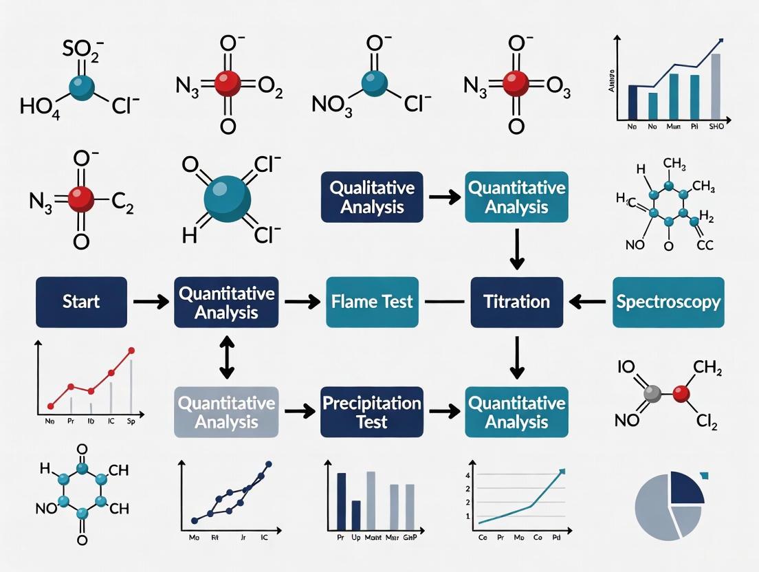
Visualizing Experimental Workflows
The following diagrams outline logical workflows for two primary analytical approaches: a generalized protocol for sample preparation and analysis, and a specific pathway for applying genetically encoded biosensors.
Diagram 1: Generalized Workflow for Inorganic Ion Analysis. This flowchart outlines the core steps from sample collection to data analysis, highlighting two common methodological branches.
Diagram 2: Workflow for Live-Cell Ion Imaging with Biosensors. This chart details the process for real-time, spatially-resolved ion detection in living systems using genetically encoded tools.
The Scientist's Toolkit: Key Research Reagent Solutions
A successful inorganic ion research project relies on a suite of specialized reagents and materials. The following table details essential items for the experiments and methodologies cited in this guide.
Table 3: Essential Research Reagents and Materials for Inorganic Ion Analysis
| Item | Function/Description | Example Application Context |
|---|---|---|
| Ion Chromatography System | Instrumentation for separating and detecting ions. Comprises pump, injector, guard/analytical column, suppressor, and conductivity detector. | Simultaneous quantification of common anions (Cl-, NO3-, PO4^3-) or cations (Na+, K+, Ca2+) in biofluids [3]. |
| Genetically Encoded Biosensor Plasmid | DNA vector encoding a fluorescent protein-based sensor (e.g., for Cl-, K+, cAMP). Allows for expression of the sensor in live cells. |
Real-time imaging of intracellular ion dynamics in response to pharmacological or physiological stimuli [1]. |
| Ionophore | A chemical agent that facilitates the transport of a specific ion across cell membranes. Used for calibrating biosensors or manipulating intracellular ion levels. | Valinomycin (for K+) is used to clamp intracellular potassium at known levels for biosensor calibration [1]. |
| Colorimetric Assay Kits | Pre-formulated reagent kits for spectrophotometric determination of specific ions (e.g., Ammonia, Phosphate). | Quick and routine measurement of ammonium or phosphate levels in cell culture supernatants or tissue homogenates [2]. |
| Certified Reference Materials | Standards with known, certified concentrations of specific ions. Used for calibrating analytical instruments and validating methods. | Creating calibration curves for IC or ICP-MS to ensure quantitative accuracy [4]. |
| Solid Phase Extraction (SPE) Cartridges | Used for sample clean-up to remove interfering substances (e.g., proteins, lipids) from complex biological matrices prior to analysis. | Pre-treatment of serum samples to remove proteins that could foul the IC column. |
The accurate analysis of inorganic ions is a cornerstone of research and development in pharmaceuticals, environmental science, and materials characterization. Selecting the appropriate analytical technique is paramount, as the choice directly impacts the reliability, sensitivity, and efficiency of the results. This guide provides an in-depth overview of four core analytical techniques—Ion Chromatography (IC), Capillary Electrophoresis (CE), Inductively Coupled Plasma Mass Spectrometry (ICP-MS) and Optical Emission Spectrometry (ICP-OES), and Electrospray Ionization Mass Spectrometry (ESI-MS). Framed within the context of selecting a method for inorganic ions research, this document details the principles, applications, and practical methodologies for each technique, serving as a comprehensive resource for researchers and drug development professionals.
Technique Summaries and Comparative Analysis
The following table provides a high-level comparison of the core techniques, highlighting their primary uses, key strengths, and typical limits of detection to guide initial method selection.
Table 1: Core Analytical Techniques at a Glance
| Technique | Primary Function | Key Strengths | Common Ions Analyzed | Typical Detection Limits |
|---|---|---|---|---|
| Ion Chromatography (IC) | Separation and quantification of ionic species [6] [7] | High selectivity for anions/cations; can analyze multiple ions simultaneously; automated operation [7] [8] | Fluoride, Chloride, Nitrate, Sulfate, Phosphate, Bromate, Ammonium, Sodium, Calcium, Magnesium [6] [7] | Low µg/L (ppb) levels [7] |
| Capillary Electrophoresis (CE) | Separation of charged molecules based on mobility in an electric field [9] | High efficiency; very low sample/reagent consumption; fast analysis [10] [9] | Inorganic anions/cations, organic acids, charged biomolecules [10] [9] | Varies widely (µg/L to mg/L) depending on analyte and detection mode [10] |
| ICP-MS & ICP-OES | Elemental analysis and quantification [11] [12] | Exceptionally low detection limits (ICP-MS); wide linear dynamic range; multi-element capability; isotopic information (ICP-MS) [11] [12] | Virtually all metals and metalloids; limited non-metals [11] | ICP-MS: sub-ng/L (ppt) for most elements; ICP-OES: low µg/L (ppb) [11] |
| Electrospray Ionization MS (ESI-MS) | Determination of molecular mass and structure of ionizable compounds [13] | Excellent for thermally labile molecules; can be coupled with separation techniques like LC and CE; enables structural elucidation via MS/MS [13] [10] | Ionizable organics, metals as complexes, biomolecules (proteins, peptides) [13] [10] | Femtomole to picomole levels for biomolecules [13] |
Ion Chromatography (IC)
Principles and Instrumentation
Ion Chromatography is a specific form of high-performance liquid chromatography designed for the separation and quantification of ions [6]. Separation is primarily based on ion-exchange, where analytes in the sample compete with the eluent ions for sites on the stationary phase [6]. The instrumentation includes a pump, injector, separation column, suppressor device, and conductivity detector. Modern systems often feature Reagent-Free IC (RFIC), which uses electrolytically generated eluents from deionized water, minimizing manual preparation and improving reproducibility [6] [8]. The suppressor device is a key component that chemically reduces the background conductivity of the eluent, thereby enhancing the signal-to-noise ratio for the analyte ions [6].
Key Workflow and Protocols
A standard IC protocol for the analysis of common anions (e.g., Cl⁻, NO₃⁻, SO₄²⁻) in a water sample is as follows:
- Sample Preparation: Filter the aqueous sample through a 0.45 µm or 0.2 µm membrane filter to remove particulates. Dilute if necessary to bring analyte concentrations within the calibration range [7].
- Instrument Setup:
- Column: Use a high-efficiency anion-exchange column, e.g., with quaternary ammonium functional groups [6].
- Eluent: For RFIC, a potassium hydroxide (KOH) or sodium carbonate/bicarbonate gradient is commonly generated electrolytically [6] [8].
- Flow Rate: Typically 0.5 - 1.5 mL/min for standard bore columns [8].
- Detection: Suppressed conductivity detection [6].
- Analysis: Inject the recommended sample volume (e.g., 25 µL). The ions are separated based on their affinity for the stationary phase and detected as peaks on a chromatogram.
- Data Analysis: Identify anions by comparing retention times to known standards and quantify them using peak areas from an external calibration curve.
Diagram 1: IC Instrumental Workflow
Capillary Electrophoresis (CE)
Principles and Instrumentation
Capillary Electrophoresis separates ionic and charged species based on their electrophoretic mobility under the influence of an applied electric field within a narrow-bore fused silica capillary [9]. The electrophoretic mobility (μ_ep) is proportional to the ion's charge and inversely proportional to its size and the solution's viscosity [9]. A critical phenomenon in CE is electroosmotic flow (EOF), a pump-like flow of the bulk solution towards the cathode, which is generated by the charged capillary wall. The apparent mobility of an analyte is the sum of its electrophoretic mobility and the electroosmotic mobility [9]. CE instruments consist of a high-voltage power supply, a capillary, two buffer reservoirs, and a detector (e.g., UV-Vis, MS) [10] [9].
Key Modes and Protocols
CE encompasses several separation modes, each suited to different analytical challenges:
- Capillary Zone Electrophoresis (CZE): The most common mode, ideal for simple ions and small molecules in a free solution [10] [9].
- Capillary Gel Electrophoresis (CGE): Uses a gel-filled capillary for size-based separation of large biomolecules like DNA and proteins [9].
- Micellar Electrokinetic Chromatography (MEKC): Employs surfactant micelles to separate both neutral and charged molecules [9].
A basic CZE protocol for inorganic anion analysis:
- Capillary Conditioning: Before first use, rinse a new fused silica capillary (e.g., 50 µm inner diameter, 50-100 cm length) sequentially with 1M sodium hydroxide, deionized water, and run buffer for 10-20 minutes each [9].
- Background Electrolyte (BGE) Preparation: Prepare a buffer, such as 20-50 mM chromate with an osmotic flow modifier (e.g., hexamethonium hydroxide), adjusted to pH 8.0 [10].
- Sample Preparation: Filter and dilute samples in deionized water or a low-ionic-strength buffer.
- Instrument Operation:
- Hydrodynamically inject the sample (e.g., 50 mbar for 5-10 seconds).
- Apply a separation voltage (e.g., -25 kV for anion analysis with reverse polarity).
- Detection: Use indirect UV detection at 254 nm if the BGE contains a UV-absorbing ion like chromate.
ICP-MS and ICP-OES
Principles and Instrumentation
Inductively Coupled Plasma Optical Emission Spectrometry (ICP-OES) and Inductively Coupled Plasma Mass Spectrometry (ICP-MS) are techniques for elemental analysis.
- ICP-OES measures the intensity of light emitted by excited atoms or ions in the plasma, which is characteristic of specific elements [12].
- ICP-MS measures the mass-to-charge ratio (m/z) of ions generated in the plasma, providing isotopic information and much lower detection limits [11] [12].
The sample introduction system converts the liquid sample into an aerosol, which is transported to the argon plasma where temperatures of ~6000-10,000 K cause desolvation, atomization, and ionization [11]. In ICP-MS, the resulting ions are extracted through an interface into a mass spectrometer (typically a quadrupole) for separation and detection. A collision/reaction cell (CRC) is often used before the mass analyzer to remove polyatomic interferences [11].
Key Workflow and Protocols
A standard ICP-MS protocol for trace metal analysis in a water sample:
- Sample Preparation: Acidify the water sample with high-purity nitric acid to a concentration of 1-2% (v/v) to keep metals in solution and prevent adsorption to container walls. For solid samples (e.g., tissue, soil), a microwave-assisted acid digestion is required.
- Instrument Setup & Tuning:
- Nebulizer & Spray Chamber: Select appropriate for sample matrix (e.g., concentric nebulizer, cyclonic spray chamber).
- Plasma Conditions: Optimize RF power, nebulizer gas flow, and torch alignment.
- Tuning: Use a tuning solution containing Li, Y, Ce, Tl to maximize sensitivity and minimize oxides (CeO+/Ce+) and doubly charged ions (Ba++/Ba+).
- CRC Gases: Introduce helium (for kinetic energy discrimination) or hydrogen (for reaction) to mitigate interferences like ArCl⁺ on As⁺ [11].
- Analysis: Introduce samples and standards. Quantification is typically performed using an external calibration curve with internal standardization (e.g., adding Rh, In, or Bi to all samples and standards to correct for instrument drift and matrix suppression).
Diagram 2: ICP-MS Instrumental Pathway
Electrospray Ionization Mass Spectrometry (ESI-MS)
Principles and Instrumentation
Electrospray Ionization (ESI) is a soft ionization technique that transfers ionic species from a solution into the gas phase for mass spectrometric analysis [13]. It is particularly well-suited for the analysis of large, non-volatile, and thermally labile biomolecules. In the ESI process, a solution containing the analyte is pumped through a fine needle held at a high voltage (several kV), creating a fine spray of charged droplets. As the solvent evaporates, the charge density on the droplets increases until Coulombic repulsion causes the ejection of gas-phase ions [13]. ESI is most powerful when coupled with a separation technique like Liquid Chromatography (LC) or Capillary Electrophoresis (CE), and is almost universally paired with tandem mass spectrometry (MS/MS) for structural analysis [13] [10]. Common mass analyzers used with ESI include quadrupoles, time-of-flight (TOF), and Orbitrap instruments [14].
Key Workflow and Protocols
ESI-MS is less directly applied to simple inorganic ions but is powerful for speciated analysis, metal complexes, and biomolecules. A protocol for characterizing a metallodrug using LC-ESI-MS:
- Sample Preparation: Dissolve the metallodrug in a suitable solvent (e.g., methanol/water mixture) at a concentration in the low µM range.
- LC-ESI-MS Setup:
- LC Column: Use a reversed-phase C18 column.
- Mobile Phase: A gradient of water and acetonitrile, both modified with 0.1% formic acid to promote protonation in positive ion mode.
- ESI Source Conditions: Capillary voltage ~3-4 kV; desolvation gas (N₂) flow and temperature optimized for solvent removal.
- MS Analysis:
- Perform an initial full scan (e.g., m/z 100-2000) to determine the molecular ion(s).
- Select the precursor ion of interest and fragment it in the collision cell (using argon gas) via Collision-Induced Dissociation (CID).
- Mass analyze the resulting product ions to obtain a structural fingerprint.
Research Reagent Solutions
Successful implementation of these techniques relies on high-purity reagents and consumables. The following table lists essential materials and their functions.
Table 2: Key Research Reagents and Consumables
| Technique | Essential Reagent/Consumable | Function |
|---|---|---|
| Ion Chromatography (IC) | High-Purity Deionized Water (>18 MΩ·cm) | Base for eluent and standard preparation; minimizes background contamination [6] [8] |
| Anion/Cation Exchange Columns | Stationary phase for separation of ionic analytes [6] | |
| Potassium Hydroxide (KOH) or Carbonate/Bicarbonate Salts | For generating the eluent that displaces analytes from the column [6] | |
| Capillary Electrophoresis (CE) | Fused Silica Capillaries | The separation channel where electrophoresis occurs [10] [9] |
| Background Electrolyte (BGE) Reagents | Creates the pH and ionic environment necessary for separation and EOF control [10] [9] | |
| ICP-MS / ICP-OES | High-Purity Nitric Acid & Hydrogen Peroxide | For sample digestion and stabilization of metal ions in solution |
| Multi-Element Standard Solutions | For instrument calibration and quality control | |
| Argon Gas | Plasma gas and auxiliary/nebulizer gas [11] | |
| Internal Standard Solution (e.g., Rh, In, Sc) | Corrects for instrument drift and matrix effects [11] | |
| ESI-MS | Volatile Buffers (Ammonium Acetate, Formic Acid) | Compatible with ionization process; prevent source contamination [13] |
| High-Purity Organic Solvents (Acetonitrile, Methanol) | Mobile phase components for LC separation and ESI stabilization |
Method Selection Framework
Choosing the right technique depends on the specific analytical question. The following diagram outlines a logical decision process for inorganic ion analysis.
Diagram 3: Analytical Technique Selection Guide
Selection Rationale:
- For total elemental analysis at trace/ultra-trace levels, ICP-MS is the undisputed choice due to its exceptional sensitivity and wide dynamic range [11] [12]. If budgets are constrained and detection limits in the low ppb range are sufficient, ICP-OES is a robust alternative.
- For speciation analysis or molecular structure elucidation (e.g., identifying the oxidation state of a metal or characterizing a metal-organic complex), ESI-MS, particularly when coupled with a separation technique, is required to preserve the molecular information [13].
- For the routine determination of common anions and cations, Ion Chromatography offers robust, automated, and highly selective quantification [6] [7] [8].
- For high-efficiency separation of charged species with minimal sample and solvent consumption, Capillary Electrophoresis is an excellent tool, especially for complex matrices or when rapid method development is desired [10] [9].
The core analytical techniques of IC, CE, ICP-MS/OES, and ESI-MS provide a powerful toolkit for tackling a wide spectrum of challenges in inorganic ion research. IC stands out for its specificity and automation in ionic analysis, CE for its high-efficiency separations, ICP-MS for its unparalleled sensitivity in elemental quantification, and ESI-MS for its ability to provide molecular-level insight. The optimal choice is never universal but is dictated by the specific analytical requirements—be it detection limits, the need for speciation information, sample complexity, or throughput. By applying the structured selection framework and understanding the fundamental principles and protocols outlined in this guide, researchers can make informed decisions to ensure accurate, reliable, and efficient results in their scientific endeavors.
The selection of an appropriate analytical method is a critical step in the research and development of pharmaceuticals and the analysis of environmental samples. For the determination of inorganic ions, this choice directly impacts the reliability, efficiency, and cost-effectiveness of the analysis. The process involves finding a balance between several, often competing, analytical criteria to ensure the method is fit for its intended purpose. Within a structured framework, four key selection criteria emerge as paramount: sensitivity, selectivity, sample matrix, and throughput. These pillars form the foundation of a robust analytical method, guiding researchers to make informed decisions that align with their project's goals, whether for drug development, quality control (QC), or environmental monitoring [15] [16].
This guide provides an in-depth technical examination of these four core criteria. It delves into their precise definitions, their practical implications for method development, and the strategies used to optimize them. Furthermore, it explores the advanced techniques and experimental protocols that are central to modern inorganic ion analysis, such as ion chromatography (IC) and sample preparation methods. By synthesizing these elements, this document serves as a comprehensive resource for researchers, scientists, and drug development professionals tasked with selecting, developing, and validating analytical methods for inorganic ions.
Core Selection Criteria Explained
The evaluation of an analytical method requires a clear understanding of its fundamental performance characteristics. The following criteria are essential for ensuring data quality and methodological robustness.
Sensitivity
Sensitivity is defined as the ability of a method to demonstrate that two samples have statistically different amounts of an analyte. It is quantitatively represented by the proportionality constant, ( kA ), in the analytical calibration function (( S{total} = kA CA + S{mb} )), where ( S{total} ) is the measured signal, ( CA ) is the analyte concentration, and ( S{mb} ) is the signal from the method blank [15]. A method with higher sensitivity will produce a larger change in signal for a given change in analyte concentration.
It is crucial to distinguish sensitivity from the detection limit. The detection limit is the smallest amount of analyte that can be determined with confidence, whereas sensitivity relates to the method's ability to discriminate between different concentrations. Highly sensitive techniques are indispensable for applications like quantifying ultratrace levels of toxic oxyanions (e.g., chromium, arsenic, and selenium) in complex environmental matrices [17].
Selectivity
Selectivity refers to a method's ability to measure the analyte accurately in the presence of interferences, such as other sample components, reagents, or excipients [15]. A selective method isolates and quantifies the target ion without significant bias from these other substances.
In Ion Chromatography (IC), selectivity is often achieved through the careful choice of stationary phase. Columns are available with varying capacities and selectivities optimized for specific analytical needs. For instance, some columns can manage sodium-to-ammonium ratios as high as 10,000:1 isocratically, effectively separating these ions without complex sample preparation [18]. The use of mass spectrometric (MS) detection further enhances selectivity by providing definitive identification based on mass-to-charge ratios [16].
Sample Matrix
The sample matrix encompasses all other components in the sample besides the analyte of interest. The matrix can profoundly affect the analysis by altering the elution behavior, suppressing or enhancing the detector response, or fouling the analytical column [19] [18]. The matrix effect is a well-known phenomenon in ion chromatography, where high concentrations of ions can cause shifts in retention times, peak deformation, or split peaks [19].
Addressing matrix effects is a central challenge. Strategies include:
- Sample Preparation: Techniques like solid-phase extraction (SPE) are used to remove interfering matrix species. For example, a silver-form resin cartridge can remove chloride from a brine sample to allow for the accurate determination of nitrite [18].
- Matrix Utilization: In some cases, the matrix effect can be harnessed to improve separation. A 2024 study demonstrated that adding ammonium hydroxide to a sample at concentrations greater than 1% could enhance the chromatographic resolution of tris and sodium ions, which could not be separated otherwise on a high-capacity column [19].
Throughput
Throughput, or the number of samples that can be analyzed per unit time, is a critical efficiency and cost metric. High-throughput methods are essential for quality control (QC) environments and for screening large numbers of samples [16].
Throughput is influenced by several factors:
- Analysis Time: Faster separations, such as determining multiple anions in less than 3 minutes using capillary electrophoresis, directly increase throughput [20].
- Sample Preparation: Streamlined or automated sample preparation, such as in-line matrix elimination in IC, reduces hands-on time and accelerates overall workflow [18].
- System Equilibration: Techniques like IC with reagent-free eluent generation can reduce system start-up and equilibration times, thereby improving readiness for the next analysis [16].
Table 1: Summary of Key Selection Criteria and Optimization Strategies
| Criterion | Definition | Key Influence on Method | Common Optimization Strategies |
|---|---|---|---|
| Sensitivity | Ability to distinguish between different analyte concentrations. | Affects the lower limits of quantification and the confidence in distinguishing concentrations. | Use of preconcentration (e.g., SPE); selection of detection technique (e.g., MS); method derivatization. |
| Selectivity | Ability to measure analyte accurately in the presence of interferences. | Determines the accuracy of the results in complex samples and avoids false positives/negatives. | Choice of chromatographic column; use of selective detectors (e.g., MS, CAD); sample cleanup. |
| Sample Matrix | All components in the sample other than the analyte. | Can cause interference, signal suppression/enhancement, and column fouling. | Sample preparation (e.g., SPE, LLE); matrix-matched calibration; standard addition method; column selection. |
| Throughput | Number of samples analyzed per unit time. | Impacts operational efficiency, cost, and suitability for high-volume testing. | Faster separations; automation; reduced sample preparation; parallel analysis. |
Advanced Techniques and Methodologies
Ion Chromatography (IC) and Related separation Techniques
Ion Chromatography is a premier technique for inorganic ion analysis, known for its sensitivity, selectivity, and ability to handle complex matrices. Its utility spans from inorganic counterion analysis in pharmaceuticals to the determination of anions in environmental and food samples [20] [16].
- Separation Principles: IC separates ions based on their interaction with a stationary phase, typically an ion-exchange resin. Cations are separated on cation-exchange columns with acidic eluents (e.g., methanesulfonic acid), while anions are separated on anion-exchange columns with basic eluents (e.g., potassium hydroxide) [19] [18].
- Detection Modes: Suppressed conductivity detection is the most common mode, where the background conductivity of the eluent is reduced, enhancing the analyte signal. For non-UV-absorbing ions, Charged Aerosol Detection (CAD) with UHPLC provides universal detection. Mass spectrometry (MS) is hyphenated with both IC and UHPLC for definitive identification and enhanced sensitivity [16].
- Exemplary Application – Counterion Analysis: With over 50% of pharmaceuticals being salts, counterion analysis is vital for ensuring drug efficacy and safety. IC with suppressed conductivity is the standard technique for inorganic counterions like chloride, sulfate, and sodium. For more complex analyses involving both organic counterions and the active pharmaceutical ingredient, UHPLC with CAD and UV detection allows for simultaneous determination in a single run [16].
Sample Preparation Techniques
Sample preparation is often the most critical step for managing complex sample matrices and is a key determinant of success in inorganic ion analysis.
- Solid-Phase Extraction (SPE): SPE is a widely used technique for cleaning up samples and preconcentrating analytes. It involves passing the sample through a cartridge containing a selective sorbent.
- Formats: Conventional SPE uses cartridges or columns, while Dispersive SPE involves adding the sorbent directly to the sample solution [17].
- Sorbent Types: A range of sorbents is available. For example, OnGuard II Ag cartridges remove chloride via precipitation, OnGuard II RP removes hydrophobic organics, and OnGuard II H cartridges are used for pH adjustment [18].
- Novel Sorbents – Layered Double Hydroxides (LDHs): LDHs are advanced sorbents with tunable compositions, making them highly effective for separating and preconcentrating oxyanions like chromate, arsenate, and selenate. Their general formula is ( [(M^{2+}){1−x}(M^{3+})x(OH)2]^{x+}(A^{n−}){x/n} \cdot mH_2O ), where ( M^{2+} ) and ( M^{3+} ) are metal cations and ( A^{n−} ) is an exchangeable anion. Their high sorption capacity and ability to be modified for specific applications make them powerful tools for enhancing sensitivity and selectivity in spectrometric detection [17].
- Electrodialysis (ED): ED is an electrochemical separation process using ion-selective membranes under an electrical potential. It has been successfully applied to selectively remove undesired inorganic ions (e.g., Cl⁻, NO₃⁻) from complex mixtures like reconstituted tobacco extract, thereby modifying the matrix to reduce harmful components in cigarette smoke and improve taste [21].
The Scientist's Toolkit: Key Research Reagent Solutions
The following table details essential materials and reagents used in modern inorganic ion analysis, highlighting their critical functions.
Table 2: Essential Research Reagents and Materials for Inorganic Ion Analysis
| Item | Function/Application | Example Use-Case |
|---|---|---|
| Ion Exchange Columns | Chromatographic separation of ions based on charge and size. | Dionex IonPac CS16 column for separating cations (e.g., Li⁺, Na⁺, NH₄⁺) in high-ammonium matrices [19]. |
| OnGuard II Sample Preparation Cartridges | Off-line matrix elimination to remove specific interferents (e.g., halides, metals, organics). | OnGuard II Ag cartridge to remove chloride from brine for nitrite analysis [18]. |
| Layered Double Hydroxides (LDHs) | Advanced sorbents for selective extraction and preconcentration of oxyanions. | Preconcentration of chromium, arsenic, and selenium oxyanions from water samples prior to spectrometric analysis [17]. |
| Methanesulfonic Acid (MSA) | Eluent for cation-exchange chromatography, can be generated electrolytically. | Used as the eluent (e.g., 8-17 mM) for the separation of Li⁺, Na⁺, and tris ions [19]. |
| Cetyltrimethylammonium bromide (CTAB) | Surfactant and flow modifier in electrophoretic and chromatographic methods. | Used as an electrosmotic flow (EOF) modifier in capillary electrophoresis [20] [22]. |
Detailed Experimental Protocols
Protocol 1: Utilizing Matrix Effect to Enhance Resolution in IC
This protocol is adapted from a 2024 study that improved the resolution of tris and sodium ions on a Dionex CS16 column by leveraging the ammonium matrix effect [19].
1. Materials and Equipment:
- Chromatography System: Dionex ICS 5000 HPIC system with suppressed conductivity detection.
- Column: Dionex IonPac CS16 (3 × 250 mm) analytical column with CG16 (3 × 50 mm) guard column.
- Eluent: Methanesulfonic acid, 8 mM, generated by an Eluent Generator Cartridge.
- Flow Rate: 0.38 mL/min.
- Sample Preparation:
- Stock Solutions: Lithium chloride (0.6 mg/L Li⁺), sodium chloride (0.6 mg/L Na⁺), and tris (40 mg/L).
- Matrix Solution: Prepare ammonium hydroxide solutions at varying concentrations (0.25%, 0.50%, 0.75%, 1.00%, 1.25% m/m) by diluting from a 25% NH₄OH stock.
- Final Sample: Spike the stock ion solutions into each of the ammonium hydroxide matrix solutions.
2. Method:
- Column Temperature: Maintain at 30 °C.
- Injection Volume: 25 µL.
- Data Collection: Process chromatograms using software (e.g., Chromeleon 7).
- Optimization Steps:
- Systematically vary the eluent concentration (6-17 mM), column temperature (25-40 °C), and injection volume (15-40 µL) to observe their interaction with the ammonium matrix concentration.
- Monitor the retention times of lithium, sodium, and tris ions. The optimal condition for baseline separation of tris and sodium is expected at 8 mM MSA, 30 °C, 1% NH₄OH matrix, and 25 µL injection.
3. Data Analysis:
- Plot the retention time of each ion against the ammonium hydroxide concentration for each set of conditions. A linear increase in retention for Na⁺ and Li⁺ with increasing NH₄OH, with minimal change for tris, indicates the matrix effect is functioning to improve resolution.
Protocol 2: Solid-Phase Extraction of Oxyanions Using LDHs
This protocol outlines the use of Layered Double Hydroxides for the dispersive solid-phase extraction of oxyanions prior to spectrometric quantification [17].
1. Materials and Equipment:
- Sorbent: Synthesized or commercial Layered Double Hydroxide (e.g., Mg-Al-CO₃ LDH).
- Target Analytes: Standard solutions of chromate (CrO₄²⁻), arsenate (AsO₄³⁻), and selenate (SeO₄²⁻).
- Equipment: Centrifuge, mechanical shaker, pH meter, analytical instrument for quantification (e.g., ICP-MS).
2. SPE Procedure:
- Sorbent Conditioning: (If required) Suspend the LDH in a weak alkaline solution and centrifuge to prepare it for use.
- Sample Loading:
- Adjust the pH of the aqueous sample (e.g., 100 mL) to an optimal value (typically alkaline for oxyanions) to maximize sorption efficiency.
- Add a known, precise amount of LDH sorbent (e.g., 20 mg) to the sample.
- Agitate the mixture vigorously using a mechanical shaker for a predetermined time (e.g., 30 min) to allow for analyte adsorption.
- Phase Separation: Centrifuge the mixture to separate the sorbent with adsorbed analytes from the solution.
- Analyte Elution / Measurement:
- Option A (Elution): Separate the sorbent, then elute the target oxyanions using a small volume (e.g., 2-5 mL) of a suitable solvent (e.g., concentrated carbonate or phosphate solution). The eluate is then analyzed.
- Option B (Slurry/Solid Analysis): After centrifugation, the sorbent is collected and either re-suspended in a small volume of solvent to create a slurry for techniques like ETAAS, or dried and analyzed directly by techniques like XRF.
3. Data Analysis:
- Calculate the enrichment factor (EF) and percentage recovery. The method's accuracy should be validated using certified reference materials or spike recovery tests.
Workflow and Decision Pathways
The following diagram illustrates the logical decision process for selecting and optimizing an analytical method based on the four key criteria.
The selection of an analytical method for inorganic ions is a multifaceted process that demands a strategic approach. As detailed in this guide, the four key criteria—sensitivity, selectivity, sample matrix, and throughput—are deeply interconnected. A decision that prioritizes one, such as using extensive sample cleanup for a complex matrix, will inevitably impact the others, such as analysis throughput. The modern analytical scientist must therefore navigate these trade-offs with a clear understanding of the available tools and techniques. From the robust separation power of Ion Chromatography and the selective power of novel sorbents like LDHs to the strategic use of matrix effects, the available methodologies are powerful and versatile. By systematically applying the principles and protocols outlined herein, researchers can develop robust, reliable, and efficient methods that are precisely tailored to their specific analytical challenges, thereby ensuring the generation of high-quality data that drives scientific and regulatory decision-making.
The Role of Inorganic Ions in Drug Formulations and Biologics
Inorganic ions are fundamental components in pharmaceutical formulations and biologics, serving critical roles as excipients, stabilizers, and active ingredients. Their precise quantification is essential for ensuring drug efficacy, stability, and safety, making analytical method selection a cornerstone of pharmaceutical development. This guide provides drug development professionals with a comprehensive framework for selecting appropriate analytical techniques to characterize inorganic ions across various pharmaceutical systems, from small molecule drugs to complex biologics like monoclonal antibodies.
The presence and concentration of inorganic ions directly influence critical quality attributes of drug products. In biologics, ions affect protein stability, aggregation, and biological activity [23]. In small molecule formulations, they function as counterions, buffering agents, and osmotic adjusters [24]. Regulatory guidelines emphasize rigorous control and quantification of these components throughout the product lifecycle [25].
The Critical Functions of Inorganic Ions
Roles in Formulation Stability and Efficacy
Inorganic ions perform diverse functional roles that directly impact drug product performance:
- Counterions: Sodium, potassium, calcium, and magnesium ions often serve as counterions for API molecules, influencing crystallinity, solubility, and bioavailability [24].
- pH Regulation: Phosphate, citrate, and carbonate buffers maintain optimal pH for drug stability and compatibility with physiological conditions [23].
- Tonicity Adjustment: Sodium chloride and other ions adjust osmotic pressure to match physiological levels, preventing tissue irritation and hemolysis upon administration [24].
- Catalytic Cofactors: Metal ions including Zn²⁺, Mg²⁺, and Ca²⁺ can act as essential cofactors for enzyme function in biologics [23].
Impact on Biologics and Monoclonal Antibodies
For complex biomolecules like monoclonal antibodies (mAbs), inorganic ions significantly influence higher-order structure, colloidal stability, and binding affinity [23]. The presence of specific ions can either promote or inhibit protein aggregation—a critical quality attribute affecting both safety and efficacy. The immunoglobulin G (IgG) structure, with its constant (Fc) and antigen-binding (Fab) regions, exhibits specific ionic interactions that must be characterized and controlled during development [23].
Table 1: Critical Inorganic Ions in Pharmaceutical Development
| Ion Category | Specific Ions | Primary Functions | Common Formulations |
|---|---|---|---|
| Cations | Na⁺, K⁺ | Osmotic balance, counterions | Injectable suspensions, buffered solutions |
| Divalent Cations | Ca²⁺, Mg²⁺ | Structural cofactors, enzymatic activity | Biologics, diagnostic assays |
| Anions | Cl⁻, PO₄³⁻ | pH regulation, counterions | Buffer systems, reconstitution solutions |
| Trace Metals | Zn²⁺, Cu²⁺, Fe²⁺/³⁺ | Catalytic centers, structural stability | Enzyme therapeutics, metalloprotein drugs |
Analytical Techniques for Inorganic Ion Characterization
Chromatographic Methods
High-Performance Liquid Chromatography (HPLC)
HPLC coupled with specialized detectors addresses diverse analytical needs for inorganic ions:
Mixed-Mode HPLC with ELSD: Trimodal columns combining reversed-phase, cation-exchange, and anion-exchange mechanisms enable simultaneous separation of cations and anions. When paired with Evaporative Light Scattering Detection (ELSD), this approach provides robust quantification of non-chromophoric ions like sodium and phosphate in complex matrices such as aripiprazole injectable suspensions [24]. ELSD detects non-volatile particles after nebulization and evaporation, making it ideal for ions lacking UV chromophores.
Ion Chromatography (IC): High-resolution separation of ionic species is achieved through dedicated ion-exchange columns, typically with conductivity or mass spectrometric detection. IC applications range from counterion analysis in APIs to impurity profiling [25].
Table 2: Chromatographic Methods for Inorganic Ion Analysis
| Technique | Detection | Key Applications | Sensitivity | Limitations |
|---|---|---|---|---|
| Mixed-Mode HPLC | ELSD | Simultaneous cation/anion analysis in complex matrices | Moderate (μg/mL) | Limited sensitivity for trace analysis |
| Ion Chromatography | Conductivity, MS | Counterion quantification, impurity profiling | High (ng/mL) | Matrix interference in biological samples |
| Reversed-Phase HPLC | CAD, UV (derivatized) | Ion analysis after derivatization | Variable | Requires complex sample preparation |
Electrophoretic Techniques
Capillary Electrophoresis (CE)
CE with capacitively coupled contactless conductivity detection (C⁴D) enables rapid, high-efficiency separation of inorganic anions in complex matrices like oils and biological fluids. Key advantages include:
- Minimal sample consumption (nanoliters)
- High separation efficiency with resolution of multiple anions in under 3 minutes
- Simple sample preparation with ultrasound-assisted extraction [20]
This technique has been successfully applied to analyze chloride, nitrate, sulfate, fluoride, and formate in virgin olive oil, demonstrating utility for challenging matrices [20].
Spectroscopic and Mass Spectrometric Methods
Inductively Coupled Plasma-Mass Spectrometry (ICP-MS)
ICP-MS provides exceptional sensitivity and multi-element capability for trace metal analysis:
- Ultra-trace detection (parts-per-trillion level) of elemental impurities
- Wide linear dynamic range covering over 9 orders of magnitude
- Isotopic information for specialized applications [25]
ICP-MS is particularly valuable for compliance with ICH Q3D elemental impurity guidelines, ensuring patient safety by controlling toxic metals like cadmium, lead, and arsenic [25].
Atomic Absorption Spectroscopy (AAS)
AAS remains a robust, cost-effective technique for targeted metal analysis with excellent sensitivity for specific elements and relatively simple operation compared to ICP-MS [25].
Method Selection Framework
Strategic Approach to Analytical Selection
Choosing the optimal analytical technique requires systematic evaluation of multiple factors:
Experimental Design Considerations
Sample Preparation Requirements
Effective sample preparation is foundational to accurate ion analysis:
- Protein Removal: For biological matrices like plasma, acetonitrile or methanol precipitation removes interfering proteins while maintaining ion integrity [26].
- Phospholipid Depletion: Specialized cartridges with scavenger chemistry reduce matrix effects from phospholipids in plasma samples [26].
- Homogenization: Tissue samples require mechanical disruption (bead milling, grinding) followed by aqueous or organic extraction to liberate ions [26].
- Dilution Factors: Optimal dilution balances matrix mitigation with maintaining analytes above detection limits [24].
Validation Parameters
Robust methods require demonstration of:
- Linearity (R² > 0.99) across the analytical range
- Precision (RSD < 10%) for repeatability and intermediate precision
- Accuracy (90-110% recovery) through spiked samples [24]
- Specificity against placebo and matrix components
- Solution stability under processing and storage conditions
Case Study: Simultaneous Analysis of Sodium and Phosphate
Experimental Protocol
A validated method for simultaneous quantification of sodium and phosphate ions in aripiprazole extended-release injectable suspensions demonstrates practical application:
- Column: Trimodal stationary phase (250 × 4.6 mm, 5 μm) with reversed-phase/cation-exchange/anion-exchange functionality
- Mobile Phase: 20 mM ammonium formate (pH 3.2)/acetonitrile (70:30 v/v)
- Flow Rate: 1.0 mL/min
- Detection: ELSD with nebulizer gas (N₂) at 3.2 bar, drift tube temperature 70°C
- Sample Preparation: 10-fold dilution with water, centrifugation at 20,000 rcf for 15 minutes, filtration through 0.45 μm PTFE [24]
Method Performance
The developed method demonstrated excellent analytical performance:
- Linearity: R² > 0.99 across 50-150% of specification range
- Precision: RSD < 10% for both ions
- Accuracy: 95-105% recovery
- Specificity: Base resolution from placebo components (carboxymethyl cellulose, mannitol) [24]
The Scientist's Toolkit: Essential Research Reagents
Table 3: Key Reagents for Inorganic Ion Analysis
| Reagent/Chemical | Function/Application | Technical Notes |
|---|---|---|
| Ultra-Pure Acids (HNO₃, HCl) | Sample digestion, mobile phase preparation | Sub-boiling distilled grade minimizes trace metal contamination [27] |
| High-Purity Inorganic Standards | Calibration, method validation | Certified reference materials (TraceCert) ensure accuracy [24] |
| Mixed-Mode Chromatography Columns | Simultaneous cation/anion separation | Trimodal chemistry (reversed-phase/ion-exchange) enhances selectivity [24] |
| Ion-Exchange Membranes | Electrodialysis, sample cleanup | CEMs/AEMs with tailored selectivity for specific applications [21] |
| Ultrapure Water (>18 MΩ·cm) | Sample preparation, mobile phases | Minimizes background ions in trace analysis [27] |
Regulatory and Compliance Considerations
Pharmaceutical analysis of inorganic ions must align with global regulatory standards:
- ICH Q3D Guidelines: Establish permitted daily exposures for 24 elemental impurities across administration routes [25].
- ICH Q6A Specifications: Define testing criteria for new drug substances and products, including inorganic impurities [25].
- Good Manufacturing Practice (GMP): Require validated methods, equipment qualification, and data integrity throughout analysis [24].
The strategic selection of analytical methods for inorganic ion analysis is paramount in pharmaceutical development. Technique choice must balance sensitivity requirements, matrix complexity, and regulatory expectations. As demonstrated through the case study, advanced approaches like trimodal HPLC-ELSD provide robust solutions for challenging applications in complex formulations.
Future directions point toward increased automation, miniaturization, and integration of artificial intelligence for method optimization and data interpretation [25]. Furthermore, the growing emphasis on green analytical chemistry encourages development of environmentally sustainable methodologies without compromising data quality. Through careful method selection and validation, pharmaceutical scientists can ensure the safety, efficacy, and quality of drug products containing inorganic ions.
Selecting and Applying Methods for Specific Sample Types
Ion Chromatography (IC) for Anions and Cations in Aqueous Solutions
Ion chromatography (IC) is a powerful analytical technique for the separation and determination of ionic species in aqueous solutions. As a form of liquid chromatography, IC measures concentrations of ionic species by separating them based on their interaction with a resinous stationary phase [28]. This technical guide provides a comprehensive overview of IC methodology for analyzing anions and cations, framed within the context of selecting appropriate analytical methods for inorganic ion research. For researchers and drug development professionals, IC offers distinct advantages for monitoring ionic impurities, determining counterions in active pharmaceutical ingredients (APIs), and conducting environmental analyses, all with high sensitivity in the parts-per-billion (ppb) to parts-per-million (ppm) range [29] [28]. The technique's ability to simultaneously determine multiple ionic species in a single injection makes it particularly valuable for comprehensive sample characterization [30].
Core Principles of Ion Chromatography
Fundamental Separation Mechanisms
Ion chromatography separates ions based on their differential affinity for a stationary phase under controlled eluent conditions. The separation process follows predictable rules where small ions typically elute before larger ions, and singly-charged ions elute before multiply-charged ions [31]. The retention mechanism primarily involves ion-exchange processes where analyte ions compete with eluent ions for sites on the stationary phase [32] [33].
Selectivity in IC is governed by multiple factors including electrostatic attraction and enforced ion pairing brought about by hydrophobic attraction and water-enforced ion pairing [32]. The polymeric matrix of the ion exchanger significantly affects selectivity, with different resin materials exhibiting varying affinity for particular ions [33]. For instance, polarizable anions such as nitrate and iodide show significantly larger retention factors on coated polyacrylate resins compared to polystyrene resins [33].
Instrumentation and System Components
A typical IC system consists of several key components: an eluent delivery pump, injection system, chromatographic column, suppressor device (for suppressed conductivity detection), and detector. The fundamental flow path of an IC system can be visualized as follows:
This workflow demonstrates the sequential process where samples are introduced via the injector, separated in the column based on ionic properties, chemically suppressed to reduce background conductivity, and finally detected. Modern IC systems often feature Reagent-Free IC (RFIC) technology with electrolytically generated eluents, simplifying method operation and enhancing reproducibility between laboratories [34].
Method Development and Optimization
Column Selection Criteria
Column choice represents the foundational decision in IC method development. Separation columns contain stationary phases with specific ion-exchange functionalities designed for particular applications. Common stationary phases include polystyrene-divinylbenzene (PS-DVB) copolymers or polyvinyl alcohol (PVA) with quaternary ammonium groups for anion exchange or sulfonate groups for cation exchange [30] [35].
The selection depends on several factors:
- Analyte characteristics: Size, charge, and polarizability of target ions
- Matrix complexity: Sample composition and potential interferents
- Detection requirements: Sensitivity and specificity needs
- Eluent compatibility: pH stability and organic solvent tolerance
For pharmaceutical applications, high-capacity columns are often necessary to handle complex matrices and achieve adequate separation of target analytes from interfering substances [30].
Operational Parameter Optimization
Eluent Composition and Strength
Eluent strength significantly impacts separation efficiency and selectivity. Simply changing the eluent concentration can alter elution order and resolution [31]. For example, changing from 30mM to 48mM methanesulfonic acid (MSA) on a CS16 column causes magnesium and potassium to swap elution positions [31]. Gradient elution methods can sharpen peak shapes and improve resolution of later-eluting peaks while enhancing front-end separation [31].
Temperature Effects
Column temperature plays a crucial role in retention time stability and separation selectivity. Varying temperature can change selectivity, with monovalent ions tending to elute quicker compared to divalent ions as temperature increases [31]. For instance, on a CS16 column at 23°C, magnesium and potassium coelute, but separation occurs at 40°C and improves further at 60°C [31]. Each column has a specified temperature range that should be consulted for optimal operation [31].
Detection Strategies
Detection method selection depends on analyte properties and matrix composition. The most common detection method is suppressed conductivity detection, where a suppressor device reduces background conductivity by converting the eluent to weakly conducting forms while enhancing analyte signal [35]. Alternative detection methods include:
Table 1: Detection Methods in Ion Chromatography
| Detection Method | Principle | Typical Applications | Advantages |
|---|---|---|---|
| Suppressed Conductivity | Measures electrical conductivity after chemical suppression | Common anions (F⁻, Cl⁻, NO₃⁻, SO₄²⁻) and cations (Na⁺, NH₄⁺, K⁺, Ca²⁺, Mg²⁺) | Universal for ionic species, high sensitivity |
| Non-Suppressed Conductivity | Direct conductivity measurement without suppression | Transition metals, when using complexing eluents | Simpler instrumentation, effective for specific applications |
| UV/VIS Detection | Absorption of ultraviolet or visible light | UV-active ions (nitrate, nitrite, bromide), when high chloride present | Selective, avoids chloride interference |
| Amperometry | Current measurement from electrochemical reaction | Carbohydrates, cyanide, sulfide, bromate | Highly specific and sensitive for electroactive species |
| Mass Spectrometry | Mass-to-charge ratio measurement | Unknown identification, speciation studies, trace analysis | Structural information, exceptional selectivity |
For challenging applications such as nitrate and nitrite determination in the presence of high chloride concentrations, switching from conductivity to UV detection provides cleaner results since chloride lacks UV activity [31].
Experimental Protocols
Sample Preparation Techniques
Proper sample preparation is critical for accurate IC analysis. Liquid samples should be filtered through 0.45µm or smaller filters to remove particulate matter [28]. For solid samples, aqueous extraction or acid digestion (for cations) is employed to liberate ions from the sample matrix [28]. Samples should be stored cold to preserve integrity until analysis [28].
For trace analysis, automated inline sample preparation techniques such as preconcentration with matrix elimination can significantly enhance sensitivity and robustness. This approach involves loading the sample onto a preconcentration column while washing away interfering matrix components with ultrapure water before transferring the analytes to the separation column [30].
Method Validation Parameters
For regulated environments such as pharmaceutical development, IC methods must be rigorously validated. Key validation parameters include [35]:
- Specificity: Ability to unequivocally assess the analyte in the presence of potential impurities
- Precision: Degree of agreement between multiple measurements (repeatability and intermediate precision)
- Linearity: Ability to obtain results proportional to analyte concentration within a given range
- Accuracy: Closeness of agreement between accepted reference values and measured values
- Robustness: Capacity to remain unaffected by small, deliberate variations in method parameters
- Solution stability: Evaluation of analyte stability in solution over time
The International Conference on Harmonisation (ICH) guidelines establish qualification thresholds for impurities, typically 0.1% for drugs with maximum daily dose ≤2g/day and 0.05% for higher doses [34].
Applications in Pharmaceutical Analysis
Counterion Determination
Pharmaceutical salts commonly incorporate counterions to promote solubility, stability, and bioavailability. Accurate determination of counterion concentration is essential to establish correct molecular mass, stoichiometric relationships, and completeness of salt formation [34] [35]. Common pharmaceutical counterions include chloride, sulfate, fumarate, oxalate, succinate, and tartrate [34] [35].
A validated IC method for multiple counterions can simultaneously quantify fumarate, oxalate, succinate, and tartrate in active pharmaceutical ingredients using a high-capacity anion-exchange column with suppressed conductivity detection [35]. The method employs an isocratic mobile phase containing 7.5 mM sodium carbonate and 2.0 mM sodium bicarbonate in water mixed with acetonitrile (90:10) at a flow rate of 1.0 mL/min with a 25-minute run time [35].
Trace Impurity Analysis
IC provides exceptional sensitivity for detecting trace ionic impurities in pharmaceuticals. A significant application is nitrite determination at trace levels to prevent formation of N-nitrosamines, which are potent carcinogens [30]. Under acidic conditions, nitrite can react with secondary or tertiary amines to produce nitrosamines, which have been detected in various pharmaceuticals [30].
For nitrite analysis at trace levels, IC with UV/VIS detection following sequential suppression (chemical suppression followed by CO₂ removal) enables low detection limits. This approach, combined with automated preconcentration and matrix elimination, allows detection of nitrite at parts-per-billion levels even in complex matrices [30].
Dialysis Concentrate Quality Control
IC offers an efficient alternative to traditional methods like atomic absorption spectroscopy for quality control of dialysis concentrates, which contain specific concentrations of electrolytes including sodium, potassium, calcium, magnesium, chloride, and buffers such as acetate or bicarbonate [30]. Using a two-channel IC system with high-capacity columns enables simultaneous determination of anions and cations from the same sample without extensive sample preparation [30].
For dialysis concentrate analysis, samples are typically diluted 750-fold before injection. High-capacity columns prevent matrix overload and maintain excellent peak shape even for high-concentration analytes like chloride (~137 g/L in the original concentrate) while resolving critical components like acetate [30].
The Scientist's Toolkit: Essential Research Reagents and Materials
Successful IC analysis requires specific reagents and materials optimized for ionic separations. The following table summarizes essential components for IC laboratories:
Table 2: Essential Research Reagents and Materials for Ion Chromatography
| Item | Function | Application Notes |
|---|---|---|
| IC Grade Water (<18 MΩ·cm resistivity) | Mobile phase preparation, sample dilution | Minimizes background contamination; essential for trace analysis [35] |
| Anion/Cation Standards (Certified reference materials) | System calibration, method validation | Must be NIST-traceable; available as single-element or multi-ion mixtures [29] |
| High-Purity Eluent Chemicals (e.g., sodium carbonate, sodium bicarbonate, MSA) | Mobile phase components | Determine separation selectivity and efficiency; must be free of ionic contaminants [35] |
| Suppressor Regenerants (e.g., sulfuric acid for anion suppression) | Chemical suppression of eluent conductivity | Reduces background signal, enhances analyte response [35] |
| Specialized IC Columns | Analytical separation | Choice depends on target analytes and matrix; multiple chemistries available [31] [30] |
| Syringe Filters (0.45µm or 0.2µm pore size) | Sample preparation | Removes particulates that could damage columns; preferably low-extractable [28] |
| PEEK Tubing and Fittings | Fluidic connections | Chemically inert, prevents metal contamination; essential for trace cation analysis [30] |
Advanced Applications and Future Directions
IC continues to evolve with technological advancements expanding its application range. Coupling IC with mass spectrometry (IC-MS) provides exceptional sensitivity and selectivity for speciation studies and unknown identification [31]. Combustion ion chromatography (CIC) has emerged as a powerful technique for analyzing difficult matrices, particularly for determining total fluorine content in various materials including per- and polyfluoroalkyl substances (PFAS) [36] [31].
The pharmaceutical industry increasingly adopts IC for compendial testing following its incorporation into regulatory monographs. The United States Pharmacopeia-National Formulary (USP-NF) has published general chapters on IC (<345> and <1065>), with additional chapters and monographs incorporating IC-based test methods [30]. This regulatory acceptance solidifies IC's position as a vital analytical tool for pharmaceutical quality control.
Method development efficiency has been enhanced through digital tools such as method simulation software, which predicts separation behavior based on known algorithms, allowing researchers to optimize parameters like temperature, eluent strength, and column selection without extensive laboratory work [31].
Ion chromatography represents a versatile, sensitive, and robust analytical technique for determining anions and cations in aqueous solutions. Its ability to simultaneously quantify multiple ionic species, coupled with advanced sample preparation options and detection capabilities, makes it indispensable for pharmaceutical research, environmental monitoring, and industrial quality control. As IC technology continues to advance with improved column chemistries, detection methods, and regulatory acceptance, its application scope continues to expand, offering researchers powerful solutions for challenging analytical problems in inorganic ion analysis.
Capillary Electrophoresis (CE) for Rapid, High-Resolution Separation
Capillary Electrophoresis (CE) has emerged as a powerful and versatile technique for the rapid, high-resolution separation of inorganic ions, offering a compelling alternative to traditional methods like ion chromatography (IC). The technique is characterized by its simplicity, high separation efficiency, minimal sample and solvent consumption, and short analysis times [37]. For researchers and drug development professionals selecting an analytical method, CE provides a "greener" alternative due to its minimal consumption of organic solvents [38]. The core principle of CE involves the separation of ions based on their differential migration in a conductive buffer under the influence of a high-voltage electric field, driven by their distinct electrophoretic mobilities [39] [40].
The analysis of simple inorganic anions such as chloride, nitrate, and sulfate is particularly well-suited to CE [41]. These analytes are water-soluble and highly mobile, though their determination presents specific challenges, notably their lack of chromophores which necessitates indirect UV detection [41]. While Ion Chromatography (IC) maintains a dominant position in many laboratories, CE has become competitive in areas where it offers distinct advantages, including method simplicity and operational cost [39]. The technique has proven its robustness and ruggedness across a variety of application areas, including pharmaceuticals, forensics, and clinical analysis [41].
Fundamental Separation Mechanisms
In Capillary Zone Electrophoresis (CZE), the most common mode for inorganic ion analysis, separation is primarily achieved based on the electrophoretic mobility of the analytes [40]. This mobility is a function of the ion's charge-to-mass ratio; ions with higher charge and smaller size migrate faster towards the electrode of opposite charge [40] [42]. The effective mobility of an ionic constituent (m̄A) under actual separating conditions can be represented as a function of the absolute mobilities of its ionic forms and their activity coefficients [39].
A critical factor influencing the separation is the electroosmotic flow (EOF), which is the bulk flow of the buffer solution through the capillary induced by the applied electric field [40] [42]. For inorganic anions, whose mobilities are highly negative and naturally oppose the direction of the normal EOF in a fused-silica capillary, this situation would result in impractically long analysis times. To overcome this, the EOF direction is reversed using cationic surfactants. These surfactants, such as hexamethonium or tetradecyltrimethylammonium bromide, form a bilayer on the negatively charged capillary wall, generating a positive surface charge that reverses the EOF direction [41]. Consequently, both the EOF and the anions migrate in the same direction towards the anode when a negative voltage is applied, significantly reducing analysis time and improving peak shape and sensitivity [41].
oob-ecg_0
Critical Methodological Components
Detection Strategies: Indirect UV Detection
Most common inorganic anions (e.g., chloride, sulfate) lack chromophores, making direct UV detection ineffective. Indirect UV detection is therefore the favored approach [41]. This technique involves adding a UV-absorbing species (e.g., chromate, pyromellitate) to the background electrolyte (BGE), which creates a high, stable background signal [41]. When a non-UV-absorbing analyte ion passes through the detector, it displaces the UV-absorbing co-ion, causing a decrease in the background signal. This "dip" in absorbance is recorded as a peak, the area of which is linearly related to the analyte concentration [41]. For optimal peak shape and sensitivity, the electrophoretic mobility of the UV-absorbing ion should closely match that of the analyte ions [41].
Background Electrolyte (BGE) Optimization
The composition of the BGE is arguably the most critical parameter for a successful CE separation, as it directly influences the electric field strength, EOF, and electrophoretic mobility of analytes [43].
- pH: The BGE pH determines the ionization state of both the capillary wall silanols and the analytes, thereby controlling the magnitude of the EOF and the charge of the analytes. For inorganic anions, alkaline pH conditions (e.g., pH 8.4) are commonly used to ensure a strong, stable EOF and full deprotonation of the analytes [41] [43].
- UV Absorber and EOF Modifier: The choice of UV-absorbing co-ion (e.g., pyromellitate) and EOF-reversing cationic surfactant (e.g., hexamethonium hydroxide) is fundamental [41]. Their types and concentrations are optimized to match analyte mobility and achieve complete EOF reversal.
- Additives and Ionic Strength: Modifiers such as organic solvents (e.g., methanol) can be added to alter selectivity [43]. The ionic strength of the BGE affects electrophoretic velocity and efficiency; typical concentrations range from 20 mM to 100 mM [43]. Zwitterionic buffers like TRIS are often preferred as they carry little current, minimizing Joule heating [41].
Sample Preparation and Injection
Sample preparation in CE is often minimal, potentially involving only filtration, dilution, or pH adjustment [39]. However, for complex matrices, cleanup steps like solid-phase extraction may be necessary [43]. Sample injection is typically performed via hydrodynamic (pressure) or electrokinetic means.
- Hydrodynamic Injection: The sample is introduced by applying pressure to the sample vial. This method is generally more representative of the sample composition and is preferred for routine analysis [41].
- Electrokinetic Injection: The capillary tip is placed in the sample vial and a voltage is applied, preferentially loading ions based on their mobility. This can provide a 10-fold improvement in detection limits for small anions but is more susceptible to matrix effects [41].
To enhance sensitivity, preconcentration techniques like field-amplified sample stacking (FASS) can be employed. FASS involves preparing the sample in a low-conductivity matrix, which causes ions to stack into a narrow zone at the boundary with the higher-conductivity BGE when voltage is applied, dramatically improving detection limits [43].
Research Reagent Solutions
The following table details key reagents and materials essential for developing and implementing CE methods for inorganic anion analysis.
| Reagent/Material | Function/Purpose | Examples & Notes |
|---|---|---|
| Background Electrolyte (BGE) | Provides conductive medium; defines separation pH and ionic strength. | 20-100 mM concentration range; often uses zwitterionic buffers like TRIS to minimize current [41] [43]. |
| UV Absorber (for Indirect Detection) | Enables detection of non-chromophoric anions. | Chromate or Pyromellitic acid; mobility should match analyte ions for optimal peak shape [41]. |
| Cationic Surfactant | Reverses Electroosmotic Flow (EOF) direction. | Hexamethonium hydroxide, Tetradecyltrimethylammonium bromide (TTAB); forms bilayer on capillary wall [41]. |
| Capillary | The separation channel. | Fused-silica, typically 50-75 µm internal diameter; effective length 40-60 cm [41] [43]. |
| Sample Solvent | Matrix for dissolving/dispersing the sample. | Should be of low conductivity for stacking (e.g., water or dilute BGE) to enhance sensitivity via FASS [43]. |
Quantitative Performance and Method Validation
CE methods for inorganic anions have been rigorously validated against established techniques like IC, demonstrating excellent agreement and reliability. The table below summarizes performance data from comparative studies and validation reports.
Table 1: Quantitative Performance of CE for Inorganic Anion Analysis
| Application / Analyte | Sample Matrix | Key Performance Metrics | Comparison / Validation |
|---|---|---|---|
| Water Analysis [41] | Mineral Water | Quantification of Cl⁻, SO₄²⁻, NO₃⁻, F⁻ | Results showed good agreement with Ion Chromatography (IC). |
| Pharmaceutical Analysis [41] | Drug Substance (Chloride salt) | Assay of Cl⁻ counter-ion; detection of anionic impurities. | CE data agreed with theoretical content and manual titration; method fully validated. |
| Trace Analysis [41] | Nuclear Power Plant Feed Water | Detection limits at ppb levels. | Achieved using electrokinetic injection with octanesulphonate additive. |
| Forensic Analysis [41] | Post-Blast Residues | LOD ~0.5 ppm for 10 anions; Intermediate precision: 2.11% (area), 0.72% (migration time). | Excellent correlation with routine IC method. |
Method validation data for a chloride assay in a pharmaceutical product further underscores the technique's capability, as shown in the table below.
Table 2: Example Method Validation Data for a Chloride Assay by CE [41]
| Validation Parameter | Result |
|---|---|
| Linearity | R² > 0.999 |
| Accuracy (% Recovery) | 99.4 - 100.6% |
| Precision (% RSD) | < 1.0% |
| Limit of Detection (LOD) | 0.05 µg/mL |
| Limit of Quantification (LOQ) | 0.2 µg/mL |
Applications in Industry and Research
The robustness of CE for inorganic ion analysis is demonstrated by its widespread adoption across diverse fields.
- Pharmaceutical Analysis: CE is extensively used for the quantification of anionic counter-ions (e.g., chloride, sulfate) in drug substances, which can constitute 5-30% of the batch weight [41]. A key advantage is the ability to simultaneously monitor for anionic impurities, providing higher data quality than a simple titration [41].
- Water Quality Monitoring: CE is applied to the analysis of drinking, mineral, surface, and ground waters [41] [37]. Its ability to handle very small sample volumes makes it ideal for analyzing individual raindrops or size-classified cloud and fog drops [37].
- Forensic Science: CE is a valuable tool for profiling inorganic anions in explosive residues, where the specific pattern of ions can indicate the ingredients used in bomb manufacture [41]. Methods have been developed for the simultaneous analysis of ten or more anions relevant to post-blast residues and detonator extracts [41].
- Industrial Process Control: CE is used for monitoring trace anionic impurities in industrial solutions like electroplating baths to ensure product quality and comply with environmental regulations like RoHS and WEEE [41].
oob-ecg_1
Operational Considerations and Instrumentation
Effective implementation of CE requires careful attention to instrumental parameters and method robustness.
- Voltage and Joule Heating: The applied voltage is the primary driver of separation speed and efficiency. However, the current generated leads to Joule heating, which can cause band broadening and reduced resolution if not controlled [43]. A systematic voltage study is recommended to find the maximum voltage that does not generate excessive heat, often indicated by a non-linear current-voltage relationship [43]. Modern instruments use forced-air or liquid-cooling to stabilize capillary temperature [43].
- Capillary Dimensions: Capillary internal diameter (ID) and total length are key considerations. A smaller ID (e.g., 50 µm) dissipates heat more effectively but reduces the detection path length, potentially lowering sensitivity. A longer capillary enhances separation power but requires higher voltage and increases analysis time [43].
- Method Robustness: The final stage of method development involves validating robustness—the method's capacity to remain unaffected by small, deliberate variations in parameters like BGE pH, temperature, or voltage. Techniques like Design of Experiments (DoE) are used for this purpose to ensure the method is reliable and transferable [43].
Capillary Electrophoresis stands as a mature, highly efficient, and environmentally friendly analytical technique perfectly suited for the rapid, high-resolution separation of inorganic ions. Its strengths—including minimal sample requirements, high separation efficiency, simplicity, and low operational cost—make it a compelling choice for researchers and analysts in pharmaceuticals, forensics, environmental science, and industrial quality control. While ion chromatography remains a dominant technique, CE has firmly established its place as a complementary and often superior method, particularly for applications requiring high-speed analysis, high resolution, or where sample volume is limited. The continued development of standardized kits and application protocols ensures that CE will maintain and expand its role as an indispensable tool in the modern analytical laboratory.
ICP-MS and ICP-OES for Ultra-Trace Metal and Multi-Element Analysis
The accurate determination of ultra-trace metals and multi-element concentrations is a critical requirement across numerous scientific fields, including pharmaceutical research, environmental monitoring, and clinical diagnostics. Inductively Coupled Plasma Optical Emission Spectroscopy (ICP-OES) and Inductively Coupled Plasma Mass Spectrometry (ICP-MS) have emerged as two of the most powerful analytical techniques for this purpose. While both techniques utilize an inductively coupled plasma as an excitation or ionization source, their fundamental principles, operational parameters, and performance characteristics differ significantly. This technical guide provides an in-depth comparison of ICP-OES and ICP-MS, focusing on their applicability for ultra-trace metal and multi-element analysis within inorganic ions research, to assist scientists in selecting the appropriate methodology for their specific analytical requirements.
Fundamental Principles and Instrumentation
ICP-OES Operating Principle
ICP-OES operates on the principle of optical emission spectroscopy. The technique utilizes an argon plasma, sustained by a radio frequency (RF) generator, which reaches temperatures of approximately 6000-10000 K. When a sample aerosol is introduced into this plasma, the extreme energy causes the constituent atoms to become excited. As these excited atoms return to their ground state, they emit photons of characteristic wavelengths unique to each element [44]. The optical system then disperses this emitted light using a diffraction grating, and a detector measures the intensity at specific wavelengths. This intensity is directly proportional to the concentration of the element in the sample [45]. The fundamental components of an ICP-OES system include the sample introduction system (nebulizer and spray chamber), the RF generator and plasma torch, the optical spectrometer, and the detector.
ICP-MS Operating Principle
ICP-MS similarly uses a high-temperature argon plasma, but primarily as an ionization source rather than an excitation source. The plasma efficiently ionizes the atoms present in the sample aerosol, converting them primarily into singly-charged positive ions. These ions are then extracted from the plasma at atmospheric pressure into a high-vacuum mass spectrometer through a sophisticated interface consisting of sampling and skimmer cones. The extracted ions are focused by ion optics before being separated according to their mass-to-charge ratio (m/z) by a mass analyzer, most commonly a quadrupole [46]. Finally, a detector, typically an electron multiplier, counts the number of ions at each specific mass, providing both quantitative and isotopic information [45]. The six fundamental compartments of a single quadrupole ICP-MS are: the sample introduction system, inductively coupled plasma, interface, ion optics, mass analyser, and detector [46].
Comparative Workflow Visualization
The following diagram illustrates the core operational differences and similarities between ICP-OES and ICP-MS instrumentation and workflows.
Performance Comparison and Technical Specifications
The fundamental differences in detection principles between ICP-OES and ICP-MS directly translate to significant variations in their analytical performance, particularly regarding sensitivity, detection limits, and elemental coverage.
Table 1: Analytical Performance Comparison of ICP-OES and ICP-MS
| Parameter | ICP-OES | ICP-MS |
|---|---|---|
| Detection Principle | Measurement of emitted light [47] | Measurement of ion mass/charge ratio [47] |
| Typical Detection Limits | Parts per billion (ppb) range [47] [48] | Parts per trillion (ppt) range [47] [48] |
| Linear Dynamic Range | Up to 10^6 orders of magnitude [45] | Up to 10^8 orders of magnitude [45] |
| Elemental Coverage | Suitable for ~73 elements; simultaneous multi-element analysis [45] | Can detect ~82 elements; full periodic table coverage with isotopic information [45] |
| Sample Throughput | High (e.g., 1-60 elements/minute) [45] | High (typically <1 minute per analysis) [45] |
| Tolerance for Total Dissolved Solids (TDS) | High (up to 5-30%) [47] [49] | Low (typically <0.2%); requires dilution for high-matrix samples [47] [46] |
| Isotopic Analysis Capability | No | Yes [48] |
Sensitivity and Detection Limits
The most distinguishing factor between these techniques is sensitivity. ICP-MS typically offers detection limits that are 3 to 4 orders of magnitude lower than ICP-OES, achieving part-per-trillion (ppt) levels compared to parts-per-billion (ppb) for ICP-OES [47] [48]. This makes ICP-MS the unequivocal choice for applications requiring ultra-trace element analysis, such as measuring toxic elements in clinical samples or high-purity materials in the semiconductor industry [49] [46]. However, advanced ICP-OES systems equipped with high-efficiency nebulizers can approach the detection limits needed for some applications that traditionally required ICP-MS, potentially offering a more cost-effective solution [49].
Matrix Tolerance and Interferences
ICP-OES demonstrates significantly higher tolerance for complex sample matrices with high total dissolved solids (TDS), handling up to 5-30% TDS, whereas ICP-MS typically requires samples with less than 0.2% TDS [47] [49] [46]. This robustness makes ICP-OES more suitable for analyzing wastewater, soil digests, and biological fluids with minimal dilution. Regarding interferences, ICP-OES is primarily susceptible to spectral interferences caused by overlapping emission lines from different elements or background shifts [48] [44]. ICP-MS, conversely, faces challenges with isobaric overlaps (ions with the same mass-to-charge ratio, e.g., (^{114})Sn and (^{114})Cd) and polyatomic interferences formed from plasma gases and matrix components (e.g., ArO(^+) interfering with (^{56})Fe(^+)) [46] [48]. Modern ICP-MS instruments employ collision/reaction cells and high-resolution mass spectrometers to effectively mitigate these interferences [50].
Methodologies and Experimental Protocols
Sample Preparation for Ultra-Trace Analysis
Proper sample preparation is paramount for achieving accurate and reproducible results in ultra-trace analysis, regardless of the analytical technique.
- Digestion of Solid Samples: For solid samples such as tissues, plant materials (e.g., cannabis), soils, or alloys, acid digestion is necessary to dissolve the metals of interest. A typical protocol involves weighing 0.1-1.0 g of accurately homogenized sample into a digestion vessel, adding 5-10 mL of concentrated nitric acid (HNO(3)), and heating using a microwave-assisted digestion system [49] [44]. For resistant matrices, a mixture of HNO(3) and hydrochloric acid (HCl) or hydrofluoric acid (HF) may be required. The use of trace metal grade acids and labware cleaned with acid and deionized water is essential to prevent contamination [44].
- Liquid Sample Preparation: Biological fluids (e.g., blood, urine) and water samples typically require simple dilution with a dilute acid or alkaline diluent to reduce viscosity and matrix effects, and to stabilize the analytes [46]. A dilution factor of 10-50 is common for serum, which reduces the total dissolved solids to an acceptable level for ICP-MS (<0.2%) [46]. Surfactants like Triton-X-100 may be added to improve aerosol stability and prevent nebulizer clogging [46].
- Filtration: Following digestion or dilution, samples should be filtered through a 0.45 μm polypropylene filter to remove any undissolved particulates that could clog the nebulizer. Polypropylene is preferred over glass fiber to avoid adsorptive losses [44].
Calibration and Quality Control
- Calibration Standards: Calibration requires a series of multi-element standard solutions prepared in a matrix that closely matches the sample (matrix-matching). For digested samples, this involves preparing standards in the same acid mixture and concentration as the final sample solution [49]. For analyses demanding exceptional accuracy (e.g., <0.2% uncertainty), gravimetric preparation of standards is recommended over volumetric methods [51].
- Internal Standardization: The use of internal standards (e.g., Sc, Y, In, Lu, Rh, Bi) is critical for both techniques to correct for instrument drift, plasma fluctuations, and matrix-induced signal suppression or enhancement [50] [51]. Internal standards are added online or to all samples and standards at a constant concentration.
- Quality Control: A rigorous QC protocol includes the analysis of method blanks, certified reference materials (CRMs), and spike recovery samples. The recovery of spikes (typically 70-125%) and the agreement with CRM certified values validate the analytical method's accuracy.
Advanced Techniques for Complex Matrices
For complex or variable matrices, advanced calibration techniques may be necessary to achieve high accuracy:
- Common Analyte Internal Standard (CAIS) Method: This technique, used with HP-ICP-OES, can reduce matrix-induced errors to below 0.2%. It involves measuring two emission lines (often an atom and an ion line) from the same element that respond differently to matrix changes. The ratio of these lines is used to correct the analyte signal [51].
- Standard Addition: This method involves spiking the sample with known concentrations of the analytes and is particularly useful when the sample matrix is complex, unknown, or difficult to match in the calibration standards.
Method Selection Guide and Application-Specific Considerations
Choosing between ICP-OES and ICP-MS requires a systematic evaluation of analytical needs and practical constraints. The following decision pathway provides a structured approach to this selection process.
Regulatory and Application-Specific Compliance
Adherence to established regulatory methods is often a critical factor in method selection.
- Environmental Analysis: For compliance with the U.S. Environmental Protection Agency (EPA) methods, ICP-OES is governed by EPA 200.5 and 200.7, while ICP-MS is governed by EPA 200.8 [47]. It is important to note that for drinking water analysis (Safe Drinking Water Act), neither technique alone is always sufficient. ICP-OES cannot measure elements like arsenic and mercury at their very low regulatory limits using EPA 200.7, while ICP-MS cannot be used to measure minerals (Na, K, Ca, Mg) using EPA 200.8 [47]. A combination of techniques is often required.
- Specific Applications:
- Clinical/Biological Fluids: ICP-MS is typically preferred due to its low detection limits for toxic elements (Pb, Cd, Hg, As) and nutritional elements (Se, Zn, Cu) at trace levels in blood, urine, and serum [46].
- High-Purity Materials: ICP-MS is essential for certifying high-purity metals (e.g., 99.999% Cu) for the semiconductor industry, where impurities must be quantified at sub-ppm levels [49].
- Food, Agriculture, and Cannabis: While ICP-MS is often used for regulatory testing of toxic elements (As, Cd, Hg, Pb) in cannabis and food products due to its superior sensitivity, ICP-OES with optimized sample introduction can be a viable and cost-effective alternative, especially for state-mandated compliance testing [49] [44].
Operational and Economic Considerations
Table 2: Operational and Economic Comparison
| Aspect | ICP-OES | ICP-MS |
|---|---|---|
| Initial Instrument Cost | Lower [48] [45] | 2-3 times higher than ICP-OES [48] [45] |
| Operating Costs | Moderate (analytical grade reagents) [47] [45] | High (ultra-pure reagents, more argon, cone replacement) [48] [45] |
| Maintenance Complexity | Simpler; fewer consumables [48] | More complex; requires maintenance of cones, detectors, and vacuum systems [48] |
| Operator Expertise | Easier to operate; simpler method development [47] [45] | Requires highly skilled personnel [48] [45] |
| Laboratory Requirements | Standard laboratory environment | May require cleanroom conditions, HEPA filters, and dedicated air-conditioning [46] |
The Scientist's Toolkit: Essential Research Reagent Solutions
Successful implementation of ICP-OES or ICP-MS methods relies on the use of specific, high-purity reagents and consumables.
Table 3: Essential Reagents and Materials for ICP Analysis
| Item | Function | Critical Specifications |
|---|---|---|
| Nitric Acid (HNO₃) | Primary digesting acid for organic and inorganic matrices. Its oxidizing power and the solubility of nitrate salts make it ideal. | "Trace Metal Grade" or "Ultra-Pure Grade" to minimize blank contributions [44]. |
| Hydrochloric Acid (HCl) | Used in combination with HNO₃ for more refractory materials and to stabilize certain elements (e.g., Hg). | "Trace Metal Grade." Note: Can cause spectral interferences in ICP-OES and polyatomic interferences (ArCl⁺) in ICP-MS [49] [46]. |
| Internal Standard Solution | Corrects for instrument drift and matrix effects. Added to all samples, standards, and blanks. | Multi-element mixture (e.g., Sc, Y, In, Lu, Bi) at a consistent concentration. Must contain elements not present in the samples [50] [51]. |
| Multi-Element Calibration Standards | Used to establish the calibration curve for quantitative analysis. | Commercially available certified reference materials (CRMs) or custom-prepared gravimetrically from single-element stocks [51]. |
| High-Purity Deionized Water | Diluent and for rinsing. | Resistivity of 18.2 MΩ·cm to prevent contamination [44]. |
| Polypropylene Syringe Filters | Removal of undissolved particulates from digested samples prior to analysis. | 0.45 μm or 0.2 μm pore size. Polypropylene is preferred to avoid adsorption of analytes [44]. |
| Certified Reference Materials (CRMs) | Validation of method accuracy and precision. | Matrix-matched CRMs (e.g., NIST Standard Reference Materials) with certified values for the analytes of interest. |
ICP-OES and ICP-MS are powerful yet distinct techniques for ultra-trace metal and multi-element analysis. The selection between them is not a matter of one being universally superior, but rather of matching the technique's strengths to the specific analytical problem. ICP-MS provides unparalleled sensitivity, ultra-trace detection capabilities, and isotopic information, making it the preferred choice for the most demanding applications in clinical, pharmaceutical, and high-purity materials analysis. ICP-OES offers robustness, high tolerance for complex matrices, and a more accessible operational and economic profile, making it an excellent workhorse for routine environmental, agricultural, and industrial analysis where extreme sensitivity is not required. By carefully considering the required detection limits, sample matrix, regulatory framework, and operational constraints outlined in this guide, researchers can make an informed and optimal selection to ensure the success of their inorganic ions research.
Mass spectrometry (MS) is a cornerstone analytical technique with applications spanning environmental monitoring, biomedical research, and drug development. For the analysis of inorganic ions and volatile compounds, two specialized techniques offer distinct capabilities: Selected Ion Flow Tube Mass Spectrometry (SIFT-MS) for volatile organic compounds (VOCs) and inorganic gases, and Electrospray Ionization Mass Spectrometry (ESI-MS) for oxyanions and soluble ionic species in liquid samples. SIFT-MS provides real-time, quantitative gas analysis with exceptional sensitivity, achieving detection limits in the parts-per-trillion range without requiring sample preparation or chromatographic separation [52]. This technique utilizes controlled soft chemical ionization to minimize fragmentation, enabling direct analysis of complex gas mixtures. ESI-MS, particularly in its "native" mode, enables the analysis of thermally labile compounds, protein complexes, and ionic species from aqueous solutions by producing gas-phase ions through an electrospray process [53]. When dealing with challenging samples containing non-volatile salts, advanced ESI-MS implementations with specialized emitters and activation methods facilitate the analysis of biological macromolecules and ionic complexes under physiologically relevant conditions.
The selection between these techniques depends fundamentally on the analytical question: SIFT-MS excels at direct, real-time monitoring of gaseous analytes, while ESI-MS provides solutions for non-volatile ionic species and complex molecular assemblies in solution. This guide provides researchers with a comprehensive technical comparison, detailed methodologies, and practical implementation frameworks for applying these powerful techniques to inorganic ion analysis.
SIFT-MS for Gas Analysis
Fundamental Principles and Instrumentation
SIFT-MS is a direct-injection mass spectrometry technique that enables real-time quantitative analysis of volatile compounds present in air samples. The method employs precisely controlled soft chemical ionization using multiple reagent ions, allowing for specific compound identification and quantification without pre-concentration or chromatographic separation [52]. The exceptional sensitivity of SIFT-MS, with detection limits typically in the parts-per-trillion (ppt) range, makes it invaluable for applications requiring trace gas analysis, including environmental monitoring, workplace safety, and breath analysis for clinical diagnostics [54] [52].
The SIFT-MS technique operates through three fundamental stages. First, eight reagent ions (H₃O⁺, NO⁺, O₂⁺, O⁻, O₂⁻, OH⁻, NO₂⁻, and NO₃⁻) are generated from air or moisture-controlled air via microwave discharge [52]. Second, a quadrupole mass filter selects a specific reagent ion which is then injected into a flow tube reactor containing an inert carrier gas (typically helium or nitrogen). The carrier gas thermalizes the reagent ions, ensuring they possess uniform, low kinetic energy critical for reproducible soft ionization. The sample gas is introduced at a calibrated flow rate, and analyte molecules undergo ionization through gas-phase reactions with the reagent ions. Finally, the resulting product ions, along with unreacted reagent ions, are sampled into a second mass analyzer (typically another quadrupole) for separation and detection [52]. The concentration of each analyte is calculated in real-time using the known reaction rate coefficient, sample flow rate, and measured product ion counts, enabling absolute quantification without external calibration.
Key Advantages and Operational Characteristics
SIFT-MS offers several distinct advantages over traditional analytical approaches like GC-MS. The absence of chromatographic separation dramatically reduces analysis time from hours to seconds, enabling true real-time monitoring of dynamic processes [52] [55]. The technique's soft chemical ionization generates significantly less fragmentation compared to electron impact ionization, producing simpler mass spectra that facilitate interpretation and quantification [52]. Furthermore, the use of multiple reagent ions provides orthogonal analytical dimensions, as different reagent ions react with the same compound via distinct mechanisms (proton transfer, charge transfer, association, etc.), creating characteristic reaction "fingerprints" that enhance compound identification specificity [52]. This multi-reagent approach is particularly valuable for distinguishing isomeric compounds that produce identical mass spectra with conventional ionization methods.
SIFT-MS Workflow
Operational advantages include remarkable ease of use, with intuitive software interfaces that enable operation by non-specialist personnel, and minimal maintenance requirements due to the absence of chromatographic columns and consumables [52]. Modern SIFT-MS instruments feature remote operation capabilities and long-term calibration stability, maintaining quantitative performance over extended periods with only routine automated performance verification. The technique also demonstrates an exceptionally wide dynamic range, typically spanning six orders of magnitude from ppt to parts-per-million (ppm) concentrations, allowing simultaneous detection of trace and major components in complex samples [52].
Experimental Protocol for VOC Analysis
Method Setup and Sample Introduction:
- Instrument Preparation: Power on the SIFT-MS instrument and allow sufficient warm-up time (typically 30-60 minutes). Execute automated performance checks using a certified gas standard (e.g., Syft Calibrant Standard) to verify instrument response across the mass range [52].
- Method Selection: Choose between Selected Ion Mode (SIM) for targeted analysis of specific compounds or Full Scan Mode for untargeted analysis. For SIM, create a method specifying target compounds, their characteristic product ions, and appropriate reagent ions for each analyte [56].
- Sample Introduction: Connect the sample source directly to the SIFT-MS sample inlet. For gaseous samples, use inert sampling lines (e.g., SilcoNert-treated stainless steel or heated polytetrafluoroethylene) to minimize analyte adsorption. Introduce the sample at a consistent flow rate (typically 10-50 mL/min) using the instrument's mass flow controller [52].
Data Acquisition and Analysis:
- Acquisition Parameters: For SIM analysis, set acquisition parameters including measurement time per ion (typically 100-500 ms), mass resolution, and total method cycle time. For Full Scan analysis, define the mass range (e.g., m/z 20-350) and scan rate [56] [52].
- Data Collection: Initiate data acquisition. For time-resolved monitoring, set the total analysis duration based on experimental needs. The software automatically records product ion counts for target compounds in SIM mode or complete mass spectra in Full Scan mode [52].
- Quantification: The software calculates absolute concentrations in real-time using the fundamental relationship:
[Analyte] = (Product Ion Counts / Reagent Ion Counts) × (Carrier Gas Flow / Sample Flow) × (1 / Reaction Rate Constant)[52]. Results are displayed quantitatively for target compounds and can be exported for further statistical analysis.
Table 1: SIFT-MS Reagent Ions and Their Primary Reaction Mechanisms
| Reagent Ion | Primary Reaction Mechanisms | Typical Application Areas |
|---|---|---|
| H₃O⁺ | Proton Transfer | Oxygen-containing compounds (aldehydes, ketones, alcohols) |
| NO⁺ | Charge Transfer, Association, Hydride Ion Extraction | Aromatic hydrocarbons, unsaturated compounds |
| O₂⁺ | Charge Transfer, Dissociative Charge Transfer | Alkanes, chlorinated compounds |
| O⁻ | Charge Transfer, Proton Abstraction, Association | Acidic compounds, halogenated organics |
| OH⁻ | Proton Abstraction, Charge Transfer | Strong acids, halogenated compounds |
| NO₂⁻ | Proton Abstraction, Association | Super acids, specialized applications |
ESI-MS for Oxyanions and Ionic Species
Technical Foundations and Native ESI-MS
Electrospray Ionization Mass Spectrometry (ESI-MS) has revolutionized the analysis of non-volatile ionic species, including oxyanions, metal complexes, and biological macromolecules, by facilitating their transfer from solution to the gas phase as intact ions. In conventional ESI-MS, a solution containing analytes is pumped through a charged capillary, creating a fine aerosol of charged droplets. As the solvent evaporates, the charge concentration increases until analyte ions are ejected into the gas phase through either the charged residue mechanism (for large molecules) or the ion evaporation mechanism (for smaller ions) [53]. This process preserves non-covalent interactions and enables the analysis of complex ionic species in their native states.
Native ESI-MS specifically aims to maintain the higher-order structures and specific non-covalent interactions of analytes, which is particularly important for protein complexes, metal-ligand coordination compounds, and ionic assemblies [53]. This approach typically employs aqueous solutions with near-physiological pH and volatile buffers such as ammonium acetate to maintain native structures while remaining compatible with the ESI process. The preservation of solution-phase structures and interactions in the gas phase makes native ESI-MS an powerful tool for studying stoichiometry, stability, and composition of ionic complexes and biological assemblies under conditions relevant to their functional states.
Addressing Analytical Challenges with Salts and Complex Matrices
A significant challenge in ESI-MS analysis of ionic species, particularly oxyanions and biological molecules, is the interference from non-volatile salts commonly present in physiological and environmental samples. These salts can suppress analyte ionization through competitive charge removal, generate chemical noise that obscures signals of interest, and form adducts that complicate mass spectra [53]. Traditional approaches involve desalting steps such as dialysis or buffer exchange, but these risk altering complex equilibria, losing precious sample, or disrupting weak non-covalent interactions.
Advanced ESI-MS strategies have been developed to address these challenges. Theta emitters—specialized glass capillaries with an internal septum creating two parallel channels—enable rapid mixing of sample and additive solutions immediately before electrospray [53]. In this configuration, the sample containing non-volatile salts is loaded in one channel while the other contains a volatile electrolyte with additives designed to mitigate salt adduction. Another effective approach involves the addition of anions with relatively low proton affinities (e.g., bromide or iodide) to the spray solution, which can preferentially remove excess metal cations rather than protons, thereby reducing sodium and potassium adduction to analyte ions [53]. Gas-phase activation methods, including beam-type collision-induced dissociation (BTCID) and dipolar direct current (DDC) offset potentials, provide controlled collisional energy to remove weakly-bound salt adducts without causing significant dissociation of the analytes of interest [53].
Experimental Protocol for Oxyanion Analysis in Saline Solutions
Sample and Emitter Preparation:
- Theta Emitter Setup: Pull borosilicate glass capillaries (1.5 mm outer diameter, 1.17 mm inner diameter) using a micropipette puller to create theta emitters with tip diameters of approximately 1.4 μm [53]. Insert dual platinum wires into the open ends of the theta emitter, ensuring each wire contacts the solution in only one channel.
- Solution Preparation: Prepare the sample solution containing oxyanions or ionic complexes in the desired buffer system. In the opposite channel of the theta emitter, load 200 mM ammonium acetate solution potentially supplemented with low proton affinity anion additives (e.g., 1-5 mM sodium bromide or potassium iodide) [53].
- Instrument Configuration: Mount the theta emitter assembly on the ESI source positioned 1-2 mm from the mass spectrometer inlet orifice. Apply voltages of 0.8-2.0 kV to the platinum wires, progressively increasing until stable electrospray is established [53].
Mass Spectrometry Parameters and Data Acquisition:
- Interface Conditions: Optimize declustering and interface parameters to maintain a balance between salt adduct removal and preservation of non-covalent interactions. Typical settings include curtain gas pressure: 10-25 psi, ion source temperature: 20-50°C, and declustering potential: 50-150 V [53].
- Collisional Activation: Implement two-stage collisional activation for adduct removal. First, apply beam-type collision-induced dissociation (BTCID) in a collision cell with nitrogen bath gas (6-10 mTorr). Second, apply dipolar direct current (DDC) offset potential in a linear ion trap to displace ions to regions of higher radiofrequency field strength, increasing collision energies with bath gas [53].
- Data Collection and Analysis: Acquire mass spectra across appropriate m/z ranges. Process data using specialized software (e.g., PeakView, UniDec) to determine molecular masses, assess adduction levels, and calculate signal-to-noise ratios. Perform replicate analyses (n=3 recommended) to ensure reproducibility [53].
ESI-MS Salt Mitigation
Table 2: Comparison of SIFT-MS and ESI-MS Techniques for Ion Analysis
| Parameter | SIFT-MS | Native ESI-MS |
|---|---|---|
| Sample Type | Gases, volatile compounds | Solutions, ionic species, complexes |
| Sample Preparation | Minimal or none | May require buffer exchange or additives |
| Analysis Time | Seconds (real-time) | Minutes to hours |
| Detection Limits | Parts-per-trillion (ppt) | Nanomolar to picomolar |
| Key Applications | Breath analysis, environmental monitoring, process control | Protein characterization, metalloprotein studies, oxyanion analysis |
| Salt Tolerance | Not applicable (gas phase) | Limited, requires mitigation strategies |
| Quantitation Approach | Absolute (based on reaction kinetics) | Relative (requires calibration standards) |
Research Reagent Solutions
Table 3: Essential Research Reagents and Materials for SIFT-MS and ESI-MS
| Reagent/Material | Function/Application | Technical Specifications |
|---|---|---|
| Certified Gas Standards | SIFT-MS calibration and performance verification | Multi-component mixtures at known concentrations (ppb-ppm range) in air or nitrogen [52] |
| Theta Emitters | ESI-MS sample introduction for saline samples | Borosilicate glass, 1.5 mm OD, 1.17 mm ID, ~1.4 μm tip diameter, dual-channel [53] |
| Ammonium Acetate | ESI-MS volatile buffer component | High purity, 50-200 mM concentration, MS-compatible [53] |
| Low Proton Affinity Anions | ESI-MS adduction reduction additives | Bromide or iodide salts, 1-5 mM in spray solution [53] |
| Ion-Selective Membranes | Electrodialysis sample preparation | Cation exchange (CEM) and anion exchange (AEM) membranes with defined exchange capacity [21] |
| Syft Library Database | SIFT-MS compound identification | Contains reaction rate coefficients, product ions, and branching ratios for quantitative analysis [56] |
SIFT-MS and ESI-MS represent complementary powerful approaches for the analysis of ionic species and volatile compounds across diverse research applications. SIFT-MS provides unparalleled capabilities for real-time, direct analysis of gaseous samples with exceptional sensitivity and minimal sample preparation, making it ideal for monitoring dynamic processes and screening large sample sets [52] [55]. Conversely, ESI-MS offers versatile solutions for characterizing oxyanions, ionic complexes, and biological macromolecules in solution, with advanced implementations addressing the persistent challenge of non-volatile salt interference [53].
The selection between these techniques should be guided by the physical state of the sample (gas vs. liquid), the required detection limits, analysis timeframe, and complexity of the sample matrix. For researchers investigating inorganic ions, understanding the complementary strengths and limitations of each technique enables more informed method selection and experimental design. As both technologies continue to evolve, ongoing improvements in ionization efficiency, mass analyzer performance, and data processing algorithms will further expand their applications in analytical chemistry, biomedical research, and environmental monitoring.
This technical guide details core sample preparation strategies for the analysis of inorganic ions, providing a foundational resource for researchers selecting an analytical method. Proper sample preparation is crucial for achieving accuracy, reproducibility, and sensitivity in analytical results, directly impacting the reliability of data in drug development and environmental research [57].
Sample preparation is the critical first step in the analytical workflow, transforming a raw sample into a form compatible with instrumental analysis. For inorganic ion analysis, this involves isolating target analytes from complex matrices, removing potential interferences, and often pre-concentrating the sample to achieve detectable levels. The core challenge lies in managing the sample matrix—which can include proteins, organic matter, and other salts—without causing loss of contamination of the target ions [46] [57]. A "total workflow" approach that considers every step from sample collection to analysis is essential for optimizing laboratory performance in terms of throughput, data quality, and safety [58].
Core Principles and Objectives
The implementation of any sample preparation strategy is guided by several key principles:
- Accuracy and Representativeness: The prepared sample must accurately reflect the composition of the original substance. This requires careful handling to prevent contamination or the loss of volatile analytes [57].
- Reproducibility: Consistent methods are vital for obtaining reliable results that can be replicated over time and across different laboratories. Standardized protocols are a cornerstone of quality control [57].
- Sensitivity Enhancement: Many sample preparation techniques, particularly pre-concentration, are designed to lower the detection limits of an analytical method, enabling the measurement of trace-level ions [57].
- Matrix Management: The primary goal is often to mitigate matrix effects that can suppress or enhance the analytical signal. This involves separating the analytes from interfering components [46].
Foundational Strategies
Dilution
Dilution is one of the simplest sample preparation techniques, involving the addition of a solvent to reduce the concentration of the sample matrix and analytes.
- Procedure: A measured aliquot of the sample is combined with a diluent, typically a weak acid (e.g., nitric acid), alkali (e.g., tetramethylammonium hydroxide), or a solution containing a chelating agent like EDTA [46].
- Applications and Considerations: Dilution is primarily used to bring the analyte concentration within the calibrated range of the instrument and to reduce matrix effects. For highly proteinaceous samples like blood, acidic diluents can cause precipitation; alkaline diluents with surfactants like Triton-X100 are often preferred to solubilize lipids and membrane proteins [46]. A general guideline for ICP-MS analysis is to maintain total dissolved solids (TDS) below 0.2%, often requiring dilution factors between 10 and 50 for biological fluids [46].
Liquid Extraction
Liquid extraction techniques separate analytes based on their relative solubility in two immiscible liquids.
Liquid-Liquid Extraction (LLE)
Traditional LLE uses an organic solvent to extract analytes from an aqueous sample. It is effective for isolating organic pollutants but can be labor-intensive and require large solvent volumes [57] [59].
Advanced Microextraction Techniques
Modern methods have miniaturized LLE principles to use smaller solvent volumes, improving efficiency and environmental friendliness.
- Vortex-Assisted Liquid-Liquid Microextraction (VALLME): This method uses vortex mixing to thoroughly disperse a microliter-volume extraction solvent into the aqueous sample, creating a large surface area for rapid analyte transfer.
- Typical Protocol: A specialized example is the simultaneous derivatization and extraction of primary aliphatic amines from water. Butyl chloroformate (derivatization agent) is mixed with 1,1,2-trichloroethane (extraction solvent) and added to the water sample. The mixture is vortexed, leading to the formation of carbamate derivatives that are extracted into the organic phase. The phases are separated by centrifugation, and the organic phase is collected for GC analysis [59]. This method achieved enrichment factors of 440-515 and recoveries of 88-103% [59].
- Ionic Liquid-Based Cloud Point Extraction (IL-CPE): This technique utilizes a mixed micellar system comprising a non-ionic surfactant (e.g., Triton X-114) and an ionic liquid (e.g., Aliquat 336). The solution is heated to its cloud point, causing it to separate into surfactant-rich and aqueous phases, with hydrophobic analytes partitioning into the surfactant-rich phase.
- Typical Protocol: For copper(II) determination, the chelating agent 4-nitrocatechol (4NC) is added to the sample. The ionic liquid Aliquat 336 forms a mixed micelle with Triton X-114, extracting the resulting (A336+)₂[Cu(4NC)₂] complex into the surfactant-rich phase at pH 6.0. Centrifugation is not always necessary, simplifying the workflow [60]. The method demonstrated a logarithmic extraction constant of 7.9, indicating high efficiency [60].
Pre-concentration
Pre-concentration increases the concentration of target analytes relative to the solvent, directly improving method sensitivity.
- Solid-Phase Extraction (SPE): This is a widely used pre-concentration technique where samples are passed through a cartridge or disk containing a sorbent material that selectively retains the analytes. After washing, the analytes are eluted with a small volume of a strong solvent [57].
- Evaporation: This simple technique involves heating the sample or applying a stream of inert gas (e.g., nitrogen) to volatilize and remove the solvent, leaving behind concentrated analytes. Automated systems like nitrogen evaporators can significantly enhance throughput and reproducibility [57].
- Electrodialysis (ED): An advanced technique for the selective removal or pre-concentration of inorganic ions from complex liquids. ED uses an electrical potential to drive ions through selective ion-exchange membranes.
- Typical Protocol: As applied to reconstituted tobacco extract, the setup consists of alternating anion and cation-exchange membranes between an anode and cathode. The feed solution is passed through "dilute" chambers, where ions migrate into adjacent "concentrate" chambers under the applied electric field. This allows for the selective removal of ions like Cl⁻, K⁺, Ca²⁺, and Mg²⁺, with reported removal ratios for K⁺ higher than for Ca²⁺ and Mg²⁺ [21].
Table 1: Comparison of Foundational Sample Preparation Techniques
| Technique | Principle | Best For | Advantages | Limitations |
|---|---|---|---|---|
| Dilution | Reducing matrix/analyte concentration with a solvent | Simple liquid samples; bringing analytes into calibration range | Rapid, simple, low cost | Does not remove interferences; can dilute analyte below LOD |
| Liquid-Liquid Extraction (LLE) | Partitioning based on solubility in two immiscible phases | Extracting non-polar or semi-polar analytes from aqueous samples | High capacity, well-established | Uses large solvent volumes; tedious; poor for polar analytes |
| Vortex-Assisted LLME | Solvent microextraction aided by vortex mixing | Trace analysis of water samples; combining with derivatization | Minimal solvent use; high enrichment factors; fast | Requires optimization of many parameters |
| Ionic Liquid-Based CPE | Separation using mixed micelles at cloud point temperature | Pre-concentrating metal ions from aqueous samples | High extraction efficiency; eco-friendly; can be centrifuge-less | Limited by surfactant/IL compatibility; thermal sensitivity |
| Solid-Phase Extraction (SPE) | Selective retention on a sorbent followed by elution | Pre-concentration and clean-up of various sample types | High selectivity; can automate; good for large volumes | Sorbent cost; potential cartridge clogging; method development |
| Electrodialysis (ED) | Selective ion transport using electrical potential and membranes | Selective removal or pre-concentration of ions from complex liquids | High selectivity for ions; scalable; no chemicals added | High equipment cost; membrane fouling; not for non-ionic species |
Method Selection and Workflow Integration
Choosing the appropriate sample preparation strategy depends on the analytical goals, sample matrix, and target ions. The following workflow provides a logical framework for this decision-making process.
Diagram 1: Sample Preparation Strategy Selection Workflow
The Scientist's Toolkit: Essential Reagents and Materials
Successful implementation of the strategies above requires specific high-quality reagents and materials.
Table 2: Key Research Reagent Solutions for Sample Preparation
| Reagent/Material | Function | Application Example |
|---|---|---|
| High-Purity Acids (e.g., HNO₃) | Digestion of solid samples; dilution solvent | Releasing bound metals from tissue or soil samples for elemental analysis [58] [46]. |
| Chelating Agents (e.g., EDTA) | Complexes metal ions to prevent precipitation/stabilize | Added to water samples during amine extraction to chelate cations that could cause precipitation [59]. |
| Ionic Liquids (e.g., Aliquat 336) | Acts as an ion-pairing agent and solvent in microextraction | Forms a mixed micelle with Triton X-114 for the efficient extraction of a copper-4NC complex in CPE [60]. |
| Surfactants (e.g., Triton X-114, Triton X-100) | Forms micelles for CPE; disperses lipids/proteins | Cloud point extraction of metal ions [60]; added to alkaline diluents for biological samples to solubilize components for ICP-MS [46]. |
| Derivatization Agents (e.g., Butyl Chloroformate) | Chemically modifies analytes to improve extraction/analysis | Reacts with primary aliphatic amines in water to form less polar carbamate derivatives for GC analysis [59]. |
| Ion Exchange Membranes | Selectively allows passage of cations or anions | Used in electrodialysis stacks for the selective removal of specific inorganic ions from tobacco extract [21]. |
The selection of a sample preparation strategy is a pivotal step in the analytical method for inorganic ions. Simple dilution may suffice for clean samples with high analyte concentrations, while complex matrices or trace-level analysis demand more sophisticated techniques like microextraction or electrodialysis. The optimal choice is guided by the sample matrix, the chemical nature of the target ions, and the required sensitivity of the overall method. By integrating these sample preparation strategies into a coherent "total workflow," researchers can significantly enhance the quality, efficiency, and reliability of their analytical data in inorganic ion research.
Solving Common Analytical Challenges and Enhancing Performance
Overcoming Interferences and Co-elution in Chromatographic Separations
For researchers selecting an analytical method for inorganic ions, interferences and co-elution represent fundamental challenges that compromise data accuracy and reliability. Co-elution occurs when two or more analytes exit the chromatography column simultaneously, preventing proper identification and quantification [61]. In ion chromatography (IC), which is indispensable for environmental monitoring, pharmaceutical quality control, and water quality assessment, these issues are particularly prevalent due to the complex matrices and similar chemical properties of many target ions [62].
The resolution equation (Rs) serves as the mathematical foundation for understanding and addressing these separation challenges. This equation demonstrates that resolution is governed by three key factors: efficiency (N), selectivity (α), and retention (k') [63]. This technical guide provides researchers with a comprehensive framework for detecting, troubleshooting, and overcoming interference and co-elution challenges specific to inorganic ions analysis, enabling more robust method development and reliable results.
Detection and Diagnosis of Separation Issues
Visual Indicators in Chromatograms
Initial detection of co-elution often begins with visual inspection of chromatographic data. Analysts should scrutinize peaks for these telltale signs:
- Shoulder Peaks: Sudden discontinuities in peak shape often indicate a secondary compound eluting closely with the primary analyte [61]
- Asymmetric Peak Shapes: Ideal chromatographic peaks are symmetrical; tailing or fronting suggests potential interference or column issues [62]
- Unexplained Peak Broadening: Wider than expected peaks may signal co-elution of multiple compounds [63]
- Abnormal Baseline Shifts: Unusual baseline activity during elution can indicate matrix interference [62]
Advanced Detection Techniques
When visual inspection suggests potential issues, confirmatory techniques provide definitive diagnosis:
- Diode Array Detector (DAD) Analysis: Collects approximately 100 UV spectra across a single peak; differing spectra indicate multiple compounds, while identical spectra suggest peak purity [61]
- Mass Spectrometry Detection: Shifting mass spectral profiles across a peak confirm the presence of co-eluting compounds [61]
- Peak Purity Algorithms: Software-based mathematical analysis of peak shape and symmetry can detect hidden co-elution not visible through routine inspection [64]
Table 1: Detection Methods for Co-elution and Interferences
| Detection Method | Principle of Operation | Key Indicators of Co-elution | Advantages |
|---|---|---|---|
| Visual Inspection | Examination of peak shape and symmetry | Shoulders, asymmetry, broadening | Rapid, no additional equipment needed |
| Diode Array Detection | Spectral comparison across peak | Differing UV spectra at different points | High certainty, maintains sample integrity |
| Mass Spectrometry | Mass spectral profile monitoring | Shifting mass fragments across peak | Compound identification capability |
| Peak Purity Algorithms | Mathematical shape analysis | Statistical deviations from ideal shape | Objective, automated detection |
Fundamental Principles of Chromatographic Resolution
The Resolution Equation
Chromatographic resolution (Rs) quantifies the degree of separation between two adjacent peaks and is defined mathematically as:
Rs = 2(t2 - t1) / (w1 + w2)
Where t1 and t2 are retention times of adjacent peaks, and w1 and w2 are their respective baseline widths [63]. For reliable quantification of inorganic ions, Rs ≥ 1.5 generally represents complete baseline separation, though adequate quantification may be possible at lower values depending on analytical requirements [63].
Impact of Resolution on Quantitative Analysis
Inadequate resolution directly impacts analytical accuracy through peak overlap. The relationship between resolution values and quantification error demonstrates why method optimization is critical:
Table 2: Resolution Impact on Quantification Accuracy
| Resolution (Rs) | Peak Overlap | Minimum Quantification Error | Maximum Potential Error | Application Suitability |
|---|---|---|---|---|
| 0.25 | 99.9% | 0.1% | 99.9% | Unacceptable for quantification |
| 0.50 | 93.7% | 6.3% | 93.7% | Qualitative screening only |
| 1.00 | 2.2% | 2.2% | 50.0% | Limited quantitative use |
| 1.50 | 0.1% | 0.1% | 2.3% | Reliable for most quantification |
| 2.00 | <0.1% | <0.1% | <0.1% | Ideal for precise work |
The "maximum potential error" becomes particularly relevant when analyzing inorganic ions with significantly different detector response factors, as similar peak areas may represent substantially different concentrations [63].
Systematic Troubleshooting and Resolution Enhancement
The Three Pillars of Resolution Optimization
Chromatographic resolution depends on three fundamental parameters that provide a systematic framework for troubleshooting:
- Capacity Factor (k'): Measures how long analytes remain in the stationary phase. Ideal range: 1-5 for most applications. Low k' values (<1) indicate insufficient retention, causing peaks to elute with the void volume [61]
- Selectivity (α): Reflects the differential chemical interactions between analytes and the stationary phase. Target α > 1.2 for robust separation. When α = 1.0, separation is impossible regardless of other parameters [61]
- Efficiency (N): Quantifies peak sharpness or "skinniness," expressed as theoretical plate count. Higher efficiency produces narrower peaks, improving resolution and detection limits [61]
Practical Optimization Strategies
Table 3: Systematic Troubleshooting Guide for Resolution Problems
| Observed Symptom | Primary Cause | Immediate Actions | Long-term Solutions |
|---|---|---|---|
| Low retention (k' < 1) | Mobile phase too strong | Weaken mobile phase | Optimize eluent concentration or pH |
| Adequate k' but poor separation | Selectivity issue (α ≈ 1) | Adjust mobile phase composition | Change column chemistry (e.g., C18 to biphenyl) |
| Broad peaks | Low efficiency (N) | Check flow rate, column temperature | Replace with high-efficiency column |
| Variable retention times | Matrix effects | Dilute sample or adjust matrix | Implement sample cleanup procedures |
| High backpressure | Column blockage | Flush column | Improve sample filtration, use guard columns |
For inorganic ions analysis, selectivity optimization often provides the most significant improvements. This can involve switching from conventional C18 columns to specialized phases such as biphenyl, AR columns, or amide columns designed for polar compounds [61]. In ion chromatography, employing gradient elution that changes eluent concentration during analysis can resolve complex mixtures with ions of varying affinities [62].
Advanced Separation Techniques for Complex Samples
Two-Dimensional Chromatography
For highly complex samples containing numerous inorganic ions, comprehensive two-dimensional liquid chromatography (LC×LC) dramatically increases peak capacity and separation power. This technique uses two different separation mechanisms with orthogonal selectivity (e.g., reversed-phase coupled with hydrophilic interaction liquid chromatography) [65]. Recent advancements include:
- Multi-2D LC×LC: Systems that automatically switch between HILIC and RP phases in the second dimension based on elution time in the first dimension, optimizing separation across a wide polarity range [65]
- Active Solvent Modulation (ASM): Technology that reduces elution strength between dimensions by adding modifier solvents, improving focusing at the head of the second dimension column [65]
- Multi-task Bayesian Optimization: Advanced computational approaches that simplify method development for these complex systems, making them more accessible for routine analysis [65]
Computational Peak Deconvolution
When physical separation remains incomplete, computational methods can mathematically resolve co-eluted peaks:
- Exponentially Modified Gaussian (EMG) Functions: The most widely used model for describing chromatographic peak shapes in deconvolution algorithms [64]
- Functional Principal Component Analysis (FPCA): Detects sub-peaks with the greatest variability across multiple chromatograms, particularly valuable for large datasets in metabolomic studies [64]
- Clustering Algorithms: Separates overlapping peaks by grouping similar peak shapes across multiple chromatograms [64]
These computational approaches enable researchers to extract quantitative information from partially resolved peaks, though they require validation against known standards to ensure accuracy.
Experimental Protocols for Method Development
Systematic Method Development Protocol
For researchers developing IC methods for inorganic ions, this structured protocol ensures comprehensive optimization:
Initial Column and Eluent Selection
- Choose a column chemistry appropriate for target ions (e.g., high-capacity anion exchange for halides)
- Select initial eluent composition based on literature values for similar analytes
- Set flow rate to manufacturer recommendations for the column dimensions
Initial Scouting Gradient
- Inject standard mixture containing all target ions
- Run a broad gradient (e.g., 5-100% B over 30 minutes)
- Identify retention characteristics and potential co-elutions
Isocratic Optimization
- Adjust eluent strength to bring earliest eluting peak to k' > 1
- Fine-tune composition to maximize α for critical pairs
- For complex mixtures, develop gradient profile to spread peaks evenly
Selectivity Fine-tuning
- If critical pair co-elution persists, systematically modify selectivity:
- Adjust pH to alter ionization state
- Add organic modifiers (e.g., acetonitrile) to modify interactions
- Change counterions (e.g., carbonate/bicarbonate vs. hydroxide)
- Consider column chemistry change if selectivity remains inadequate
- If critical pair co-elution persists, systematically modify selectivity:
Validation with Real Samples
- Analyze representative sample matrices to identify matrix effects
- Implement sample preparation if needed to reduce interferences
- Verify resolution stability across multiple batches
Sample Preparation Techniques to Minimize Interferences
Proper sample preparation significantly reduces interference challenges:
- Dilution: Simple dilution can reduce ionic strength and minimize matrix effects [62]
- Solid-Phase Extraction (SPE): Selective removal of interferents while retaining analytes of interest
- pH Adjustment: Manipulating sample pH can alter retention and eliminate certain interferences [62]
- Specialty Filtration: Removing particulate matter and specific interferents through selective membranes [62]
Research Reagent Solutions for Ion Chromatography
Table 4: Essential Reagents and Materials for Inorganic Ions Analysis
| Reagent/Material | Function/Purpose | Selection Considerations | Application Notes |
|---|---|---|---|
| High-Purity Eluent Chemicals | Mobile phase generation | Low UV absorbance, minimal contamination | Use eluent generators for consistency |
| Certified Anion Standards | Calibration and quantification | NIST-traceable, ISO 17025 certified | Essential for accurate quantification |
| Specialty Columns | Stationary phase for separation | Selectivity for target ions, pH stability | Guard columns extend lifetime |
| Suppressor Devices | Background conductance reduction | Compatibility with eluent chemistry | Regular maintenance critical |
| Sample Preparation Cartridges | Matrix interference removal | Selectivity for common interferents | pH adjustment may enhance specificity |
| High-Purity Water | Solvent for standards and blanks | ≥18 MΩ·cm resistance | Essential for low detection limits |
Successfully overcoming interferences and co-elution in chromatographic separations of inorganic ions requires a systematic approach that combines theoretical understanding with practical optimization strategies. By methodically addressing capacity factor, selectivity, and efficiency—the three pillars of chromatographic resolution—researchers can develop robust methods that deliver reliable results even in complex matrices.
The increasing availability of advanced instrumentation, including two-dimensional chromatography systems, coupled with sophisticated computational deconvolution tools, provides powerful solutions for the most challenging separation problems. However, fundamental method development principles remain essential for establishing accurate, reproducible analytical methods for inorganic ions analysis across diverse applications from environmental monitoring to pharmaceutical development.
Managing High Backpressure, Baseline Noise, and Column Degradation
In the precise field of analytical method development for inorganic ions, the integrity of chromatographic data is paramount. Researchers and scientists in drug development rely on robust, reproducible results from techniques such as ion chromatography (IC) to accurately quantify anions and cations in complex matrices. However, this pursuit is often challenged by three persistent technical issues: high backpressure, significant baseline noise, and premature column degradation. These problems are not merely operational nuisances; they directly compromise data quality, lead to costly instrument downtime, and can invalidate critical analytical results. This guide provides an in-depth examination of these challenges, offering a structured framework for diagnosis, resolution, and prevention, specifically contextualized within the scope of inorganic ions research. By adopting a proactive approach to system management, professionals can enhance the reliability of their analytical methods and ensure the longevity of their chromatographic instrumentation.
Understanding and Managing High Backpressure
High backpressure is one of the most frequent challenges in liquid chromatography systems, including IC. While some pressure is inherent to the technique, abnormally high levels signal a partial obstruction that can damage the system and degrade performance.
Establishing a Normal Pressure Baseline
The first step in troubleshooting is knowing your system's normal operating pressure. Ideally, you should generate a baseline for the instrument both with and without the column installed [66]. This involves removing the analytical column and replacing it with a zero-dead-volume union. The pressure measured with this union in place is your system's inherent backpressure. Having this reference allows you to quickly determine whether a pressure problem originates from the column itself or from other system components. Furthermore, recording pressure under standard operating conditions, including during gradient analysis where pressure will naturally change with mobile phase viscosity, provides a critical benchmark for identifying deviations [66].
Systematic Diagnosis of Elevated Pressure
A systematic approach is the most efficient way to locate a blockage. The recommended method is to start at the detector and work backward up the flow path, adding or removing one component at a time while monitoring the pressure [66]. This process helps pinpoint the exact location of the obstruction without subjecting the analytical column to unnecessary high-pressure stress.
The following diagnostic workflow outlines this systematic process:
Common Causes and Preventive Strategies
Particulates causing high backpressure typically originate from three main sources: the sample, the mobile phase, or instrument wear and tear [66].
- Sample-Related Particulates: Samples often contain particulates that can clog frits and narrow tubing. Whenever possible, consider filtering your sample prior to analysis using syringe filters or filter vials [66]. Centrifugation is also an effective alternative. Using a guard column or a pre-column filter is a highly effective strategy to protect the more expensive analytical column from particulates and strongly retained compounds [66].
- Mobile Phase Contamination: Bacterial growth in aqueous mobile phases and precipitation of buffer salts are common culprits. Prepare mobile phases with HPLC-grade chemicals and make aqueous phases fresh to prevent microbial growth [66]. For buffered mobile phases, ensure that gradients do not push the composition to a point where salts become insoluble, particularly when moving to highly organic phases [66].
- Instrument Wear and Tear: Pump seals eventually wear down, and the resulting particulates can enter the flow path. Implement a preventative maintenance schedule that includes routine replacement of high-wear components like pump seals, injection needle seats, and rotor valves [66].
Controlling Baseline Noise and Drift
Baseline anomalies, including noise and drift, can obscure peaks of interest and reduce the reliability of quantitative data, especially when detecting trace levels of inorganic ions.
Fundamentals of Baseline Disturbances
Baseline noise refers to the high-frequency, random fluctuation of the signal, while drift is a low-frequency, gradual shift in the baseline position. In techniques like Ion Chromatography with suppressed conductivity detection, a stable baseline is critical for achieving low detection limits. The sample matrix itself can be a significant source of interference; complex matrices containing fats, proteins, surfactants, or high concentrations of heavy metals can contaminate the electrode surfaces or the separation column, leading to poor resolution, irregular baselines, and poor reproducibility [67].
Advanced Signal Processing Techniques
Modern approaches to managing baseline issues involve sophisticated computational algorithms. Recent research has demonstrated the power of neural networks and advanced fitting routines to correct for baseline drift.
- Fitting Neural Networks: One innovative approach involves using a fitting neural network to establish a mapping relationship between the detected signal and the unabsorbed baseline. This method has been shown to effectively correct for baseline drift caused by environmental factors like thermal noise in detectors, thereby improving the signal-to-noise ratio (SNR) and the linearity of the calibration [68]. The network is trained to recognize and filter out the drift component, leaving a clean analytical signal.
- Improved Algorithmic Fitting: For complex spectral data, methods like the Improved Adaptive Gradient-derived Penalized Least Squares (IagPLS) have been developed. This algorithm integrates curvature-driven dynamic regularization and feature protection mechanisms to suppress high-frequency noise while protecting the integrity of key biomarker peaks in the signal [69]. This results in a significant improvement in feature peak prominence and a reduction in negative residual area compared to traditional methods [69].
Table 1: Comparison of Baseline Correction Methods
| Method | Key Mechanism | Best For | Advantages | Limitations |
|---|---|---|---|---|
| Polynomial Fitting | Fits a polynomial curve to the baseline | Simple, smooth baselines | Computationally simple, fast | Can distort peaks if not carefully fitted |
| Asymmetric Least Squares (ALS) | Weighted least squares with asymmetry to favor baseline points | Complex, slowly drifting baselines | More robust than polynomial fitting | Requires optimization of parameters |
| Fitting Neural Network [68] | Learns mapping between noisy signal and true baseline | Complex, non-linear drift due to thermal or environmental noise | High adaptability, powerful denoising | Requires training data and computational power |
| IagPLS [69] | Gradient-driven penalty with feature protection | Spectra with critical biomarker regions needing preservation | Protects key features while denoising | Complex implementation |
Preventing and Diagnosing Column Degradation
The analytical column is the heart of the chromatographic system, and its performance directly dictates the quality of the separation. Protecting it is critical for maintaining method integrity.
Mechanisms of Column Failure
Column degradation manifests as peak tailing, loss of resolution, changes in retention time, and increased backpressure. The primary mechanisms of failure are:
- Chemical Degradation: For silica-based columns, this involves hydrolysis of the silica backbone or loss of bonded phase, especially when operating outside the recommended pH range (typically pH 2-8).
- Physical Degradation: The formation of voids or channels at the column inlet caused by the collapse of the packing material or the accumulation of particulate matter.
- Fouling: The irreversible adsorption of matrix components from samples. In IC, retention of complex matrices such as fats, proteins, and surfactants can lead to the blockage of the column or a reduction in column capacity [67]. High concentrations of metal ions like iron, barium, and calcium can also contaminate the system, leading to poor recovery of analytes like sulfate and phosphate [67].
Proactive Column Protection Strategies
- Sample Cleanup: The most effective strategy is to prevent contaminants from reaching the column. Selecting an appropriate sample pretreatment method is crucial to improve analysis results and protect instruments [67]. As shown in Table 2, numerous purification technologies are available for liquid samples in IC analysis.
- Guard Columns and In-Line Filters: A guard column, which is a short, disposable cartridge containing the same packing as the analytical column, is a low-cost insurance policy. It retains particulate matter and strongly adsorbed compounds, sacrificing itself to protect the analytical column.
- System-Compatible Solvents: Ensure the sample solvent is miscible and has a similar elution strength to the initial mobile phase. A mismatch can cause sample components to crash out of solution and deposit on the column inlet [66].
Table 2: Sample Purification Technologies for Ion Chromatography [67]
| Technology | Principle | Application in IC | Key Benefit |
|---|---|---|---|
| Solid-Phase Extraction (SPE) | Selective adsorption/desorption of analytes or interferences | Removal of specific matrix interferences (organics, ions) | High selectivity and pre-concentration capability |
| Dialysis | Diffusion of small molecules across a membrane | Removal of high-MW interferences (proteins, colloids) | Excellent for complex biological fluids |
| Electrodialysis | Ion transport using an electric field | Desalting or removal of ionic interferences from concentrated samples | Automated pretreatment for highly acidic/alkaline samples |
| Membrane Filtration | Size-exclusion of particles | Removal of particulates using 0.45 µm or 0.2 µm filters | Essential first step for most liquid samples |
The Scientist's Toolkit: Essential Research Reagent Solutions
Successful management of chromatographic systems relies on the consistent use of high-quality reagents and consumables. The following table details key materials and their functions in preventing backpressure, noise, and column degradation.
Table 3: Essential Research Reagents and Consumables
| Item | Function | Technical Notes |
|---|---|---|
| Syringe Filters (0.45 µm or 0.2 µm) | Removes particulates from samples and mobile phases prior to injection/use. | Nylon is a good general-purpose material; check compatibility with your analytes. |
| Guard Column | Captures particulates and strongly retained compounds, protecting the analytical column. | Select a guard column system that is designed specifically for your analytical column [66]. |
| HPLC-Grade Solvents & Salts | Ensures mobile phase purity to minimize baseline noise and prevent system fouling. | Avoids impurities that can accumulate on column frits or detector flow cells. |
| Seal Wash Solvent | Prevents buffer crystallization on pump seals, extending seal life and reducing particulates. | Use a composition recommended by the instrument manufacturer. |
| Pump Seal & Needle Seat Kits | Routine replacement of high-wear parts as part of a preventative maintenance schedule. | Mitigates a key source of particulates that cause high backpressure and system failures [66]. |
| Certified Vials & Caps | Provides inert sample containers with septa that do not leach contaminants or introduce particulates. | Prevents extraneous peaks (ghosting) and sample contamination. |
Integrated Experimental Protocols
Protocol 1: Systematic High Backpressure Diagnosis
This protocol provides a step-by-step methodology for isolating the source of high backpressure.
- Document Baseline Pressure: Record the system pressure at your standard flow rate with the column connected and the system equilibrated.
- Isolate the System: Shut down the flow and carefully disconnect the analytical column. Cap the column ends to prevent it from drying out.
- Connect System Union: Use a low-dead-volume union to connect the injector outlet directly to the detector inlet. Ensure the union is rated for your system's pressure (e.g., PEEK for ≤5000 psi, stainless steel for ≤20,000 psi) [66].
- Measure System Pressure: Restart the flow at the standard rate and measure the pressure. If the pressure is abnormally high compared to your previously recorded system baseline, the problem lies within the LC hardware (pump, injector, tubing, in-line filters, detector).
- Troubleshoot the Hardware (if needed): If the system pressure is high, systematically check and clean/replace components:
- Check and clean the purge valve inline filter.
- Inspect and replace pump seals if necessary.
- Flush the injection valve and inspect the needle and needle seat for blockages.
- Loosen connections one at a time (with the pump at a reduced flow rate) to identify a blocked capillary.
- Reconnect Column (if system pressure is normal): If the system pressure with the union is normal, the problem is with the column. Reconnect the column and restart the flow.
- Flush the Column: Flush the column according to the manufacturer's instructions, typically with a strong solvent that does not damage the stationary phase. Re-equilibrate with mobile phase and re-check pressure.
Protocol 2: Sample Cleanup via Solid-Phase Extraction (SPE) for IC
This protocol outlines a general SPE procedure for purifying samples for ion chromatography analysis.
- Column Selection: Select an appropriate SPE sorbent (e.g., C18 for removal of organic contaminants, a specialized sorbent for ion exchange).
- Conditioning: Pass 3-5 column volumes of a strong solvent (e.g., methanol) through the SPE cartridge, followed by 3-5 volumes of the sample solvent or a weak eluent (e.g., water). Do not allow the sorbent to dry out.
- Sample Loading: Load the sample onto the conditioned SPE cartridge. Use a slow, controlled flow rate to ensure optimal interaction between the analytes/interferences and the sorbent.
- Washing: Pass a wash solution (a solvent strong enough to remove undesired matrix components but weak enough to retain your analytes) through the cartridge. Collect and discard the effluent.
- Elution (for analyte pre-concentration or interference removal): For pre-concentration, elute the retained analytes with a small volume of a strong solvent. For interference removal, the analytes pass through in the load/wash fraction, and the interferences are retained and discarded.
- Analysis: The resulting eluent or load-through fraction can often be injected directly into the IC system, or it may require dilution or pH adjustment to be compatible with the mobile phase.
The relationship between sample preparation, analysis, and data processing is summarized in the following workflow:
Optimizing Sensitivity and Achieving Low Limits of Detection (LOD)
For researchers selecting an analytical method for inorganic ions, understanding and optimizing the Limit of Detection (LOD) and Limit of Quantitation (LOQ) is fundamental to ensuring data reliability. These parameters define the sensitivity and functional range of a method, which is critical for accurately tracing low concentrations of analytes in complex matrices [70].
The LOD is the lowest concentration of an analyte that can be reliably distinguished from a blank sample or background noise, but not necessarily quantified with precision. In practical terms, it is the smallest amount that can be detected, signaling the presence of the analyte. The LOQ, on the other hand, is the lowest concentration at which the analyte can not only be detected but also quantified with acceptable levels of precision and accuracy, typically defined by a predetermined bias and imprecision goal [71]. These concepts are statistically distinct, and their proper determination is a prerequisite for any method to be considered "fit for purpose" [71] [70].
Table 1: Key Definitions and Statistical Formulae for LOD and LOQ
| Parameter | Definition | Common Statistical Formulae |
|---|---|---|
| Limit of Blank (LoB) | The highest apparent analyte concentration expected from a blank sample. | LoB = mean~blank~ + 1.645(SD~blank~) [71] |
| Limit of Detection (LOD) | The lowest concentration reliably distinguished from LoB; detection is feasible. | LOD = LoB + 1.645(SD~low concentration sample~) [71].Alternatively: 3.3σ / S (where σ=standard deviation of response, S=slope of calibration curve) [70]. |
| Limit of Quantitation (LOQ) | The lowest concentration quantified with acceptable precision and accuracy. | Typically 10σ / S [70]. It is ≥ LOD [71]. |
The relationship between these parameters and the analytical signal is visually summarized in the following diagram:
Foundational Principles for Low LOD
Achieving a low LOD ultimately boils down to one core principle: maximizing the signal-to-noise ratio (S/N). This can be accomplished by either increasing the analyte signal, reducing the background noise, or ideally, both simultaneously [72] [73].
A signal that is clearly distinguishable from the noise allows for the confident detection of analytes at increasingly lower concentrations. The generally accepted criteria are an S/N of 3:1 for LOD and 10:1 for LOQ [70] [73]. The following diagram illustrates the core strategy for achieving a low LOD:
Optimization Strategies Across the Analytical Workflow
A holistic approach, optimizing every step from sample preparation to final data processing, is required to significantly improve sensitivity.
Sample Preparation and Pre-Concentration
Effective sample preparation is often the most impactful step for improving LOD. The goals are to concentrate the analyte and remove matrix interferences that contribute to noise.
Sample Clean-Up Techniques:
- Solid-Phase Extraction (SPE): This versatile technique selectively adsorbs analytes and interferences onto a cartridge, allowing for the selective elution and collection of purified analytes. It reduces sample complexity, decreases baseline interferences, and significantly increases detection sensitivity for HPLC, GC, and MS analyses [72].
- Liquid-Liquid Extraction (LLE): One of the oldest techniques, LLE uses immiscible solvents to separate compounds based on their relative solubilities. Modern supported liquid extraction (SLE) offers advantages such as easier automation and lower solvent consumption [72].
- Protein Precipitation: For biological samples, this method removes interfering proteins using agents like ammonium sulfate, trichloroacetic acid, or organic solvents, clearing the matrix for analysis [72].
Pre-Concentration Methods:
- Evaporation and Reconstitution: Techniques like rotary evaporation, nitrogen blowdown, or centrifugal evaporation are used to remove solvent and reconstitute the sample in a significantly smaller volume, thereby concentrating the analyte [72].
- On-Line SPE: This approach integrates the SPE clean-up and concentration step directly with the chromatographic system, automating the process, reducing sample handling, and improving both throughput and reproducibility [72].
Chromatographic Separation Optimization
The separation step is critical for resolving the analyte peak from impurities and ensuring it enters the detector in a narrow, concentrated band.
Column Selection: The choice of column directly influences peak shape and efficiency.
- Particle Technology: Using columns packed with sub-2 μm fully porous or superficially porous (core-shell) particles can dramatically increase column efficiency. These particles reduce band broadening, resulting in narrower, taller peaks and a better signal [72] [73].
- Column Dimensions: A column with a smaller internal diameter (I.D.) reduces the dilution of the sample band. Just halving the column I.D. can increase the analyte concentration reaching the detector by approximately four times. This is a highly effective strategy for sensitivity enhancement in HPLC [73]. In GC, shorter columns (10–15 m) with narrow I.D. (0.18–0.25 mm) are recommended for best peak efficiency [74].
Mobile Phase and Elution Optimization:
- Mobile Phase Additives: Use volatile additives (e.g., formic acid, ammonium acetate) compatible with your detector. Adjusting the pH to promote analyte ionization can enhance signal, particularly in MS detection. For UV detection, ensure solvents like acetonitrile have low UV absorbance at your detection wavelength to minimize baseline noise [72] [75].
- Gradient Elution: A sharp gradient can produce narrower, taller peaks compared to a long isocratic hold, thereby increasing signal intensity [73] [75].
Detector and Instrument-Specific Optimization
Fine-tuning the detector and its interface with the separation system can yield significant sensitivity gains.
Mass Spectrometry (MS) Optimization:
- Ionization Efficiency: Fine-tune source parameters (spray voltage, gas flows, temperatures) for your specific analytes. Consider alternative ionization techniques like APCI for less polar compounds [72].
- Advanced MS Techniques: Leverage high-resolution mass spectrometry (HRMS) for improved selectivity and sensitivity, or ion mobility spectrometry (IMS) to add a separation dimension and reduce chemical noise [72].
- Low-Flow Techniques: Techniques like nano-LC or micro-LC with drastically reduced flow rates (200-500 nL/min) significantly enhance ionization efficiency in ESI-MS, leading to major gains in sensitivity [72].
Flame Ionization Detector (FID) for GC:
- Optimize the fuel-to-oxidizer ratio, typically starting with a 10:1 ratio of hydrogen to air and adjusting in steps of ±5 mL/min [74].
- Ensure the make-up gas flow (often nitrogen) is optimized, as it can have a pronounced effect on sensitivity. Begin with a 1:1 ratio of make-up gas to fuel gas and investigate [74].
UV/Vis Detector for HPLC:
System Maintenance and Data Processing
Contamination Control and Maintenance: A poorly maintained system is a major source of noise.
Data Acquisition and Processing:
- Utilize advanced MS acquisition modes like Parallel Reaction Monitoring (PRM) for improved selectivity and sensitivity in targeted analysis [72].
- Leverage sophisticated software algorithms and machine learning approaches for improved peak detection and integration, which can better extract signals from noisy baselines [72].
The interplay of these strategies across the entire analytical workflow is summarized below:
Experimental Protocols for LOD/LOQ Determination
Protocol 1: Determination via Signal-to-Noise Ratio
This approach is common in chromatographic techniques and is straightforward to implement [73].
- Prepare and Inject a Blank: Inject a sample that does not contain the analyte (e.g., the sample solvent or a processed matrix blank).
- Measure Noise: On the resulting chromatogram, measure the baseline noise (N) over the region where the analyte is expected to elute. This can be the peak-to-peak noise or the standard deviation of the baseline.
- Prepare and Inject a Low-Concentration Standard: Inject a standard containing the analyte at a concentration that produces a small but discernible peak.
- Measure Signal: Measure the height of the analyte peak (S).
- Calculate S/N Ratio: Calculate the Signal-to-Noise ratio (S/N).
- Determine LOD and LOQ: The LOD is the concentration that yields an S/N ≥ 3. The LOQ is the concentration that yields an S/N ≥ 10 [70] [73]. This typically requires analyzing a series of low-level standards to establish the concentration that precisely meets these criteria.
Protocol 2: Determination via Calibration Curve and Standard Deviation
This method, endorsed by guidelines like ICH Q2(R1), uses statistical data from the calibration curve [70].
- Prepare Calibration Standards: Prepare a minimum of five calibration standards, with at least one at a low concentration near the expected limit.
- Analyze and Generate Curve: Analyze each standard multiple times (n ≥ 3 is recommended) and generate a linear calibration curve (signal vs. concentration).
- Calculate Standard Deviation: Calculate the standard deviation (σ) of the response (y-intercept or, preferably, from repeated measurements of a low-concentration standard).
- Apply Formulae:
- LOD = 3.3 × σ / S
- LOQ = 10 × σ / S Where S is the slope of the calibration curve [70].
The Scientist's Toolkit: Essential Reagents and Materials
Table 2: Key Research Reagent Solutions for Optimizing LOD
| Item | Function & Rationale |
|---|---|
| Solid-Phase Extraction (SPE) Cartridges | Selective extraction and concentration of analytes from a complex sample matrix, reducing interferences and improving signal-to-noise [72]. |
| LC-MS Grade Solvents & Additives | High-purity solvents (e.g., acetonitrile, methanol) and volatile additives (e.g., formic acid) minimize baseline noise and prevent source contamination in sensitive detection methods [72] [73]. |
| Narrow-Bore or Micro-Flow HPLC Columns | Columns with small internal diameters (e.g., < 2.1 mm) reduce dilution of the sample band, delivering a more concentrated analyte plug to the detector, thereby enhancing signal intensity [72] [73]. |
| Superficially Porous (Core-Shell) Particle Columns | Provide high chromatographic efficiency (sharp peaks) similar to sub-2μm fully porous particles but with lower backpressure, making them suitable for a wider range of HPLC systems [73]. |
| High-Purity Calibration Standards | Essential for generating accurate calibration curves for LOD/LOQ determination and ensuring precise quantitation of inorganic ions. |
| On-Line SPE Systems | Automate the sample clean-up and concentration process, improving reproducibility, reducing sample handling, and increasing throughput for low LOD analysis [72]. |
The accurate analysis of target analytes within complex matrices represents a significant challenge in fields ranging from clinical diagnostics to environmental monitoring and consumer product safety. Complex samples, such as biological fluids, polymers, and cosmetics, contain numerous interfering components that can compromise analytical accuracy by masking, suppressing, or augmenting the signal of target compounds [76]. These matrix effects can occur chromatographically through co-elution or during ionization processes in mass spectrometric detection, potentially yielding highly variable or unreliable data [76]. The growing demand for precise measurement of trace-level analytes, including inorganic ions, in these challenging environments necessitates robust methodological strategies that encompass sample preparation, analytical technique selection, and data interpretation.
This technical guide provides a comprehensive framework for selecting and optimizing analytical methods for inorganic ions research across three complex matrix categories: biological fluids, polymers, and cosmetics. By integrating theoretical principles with practical protocols, we aim to equip researchers with the systematic approach needed to navigate the intricacies of these matrices, mitigate analytical interferences, and generate reliable, reproducible data for critical decision-making in research and development.
Matrix-Specific Challenges and Characteristics
Biological Fluids
Biological matrices, including blood, urine, and breast milk, present unique analytical challenges due to their inherent complexity and dynamic composition. These fluids contain proteins, lipids, electrolytes, and numerous metabolites that can interfere with analysis [76] [77]. Endocrine-disrupting chemicals (EDCs) and other trace analytes typically exist at very low concentrations (ng/L to μg/L) amidst these potentially interfering compounds, necessitating highly sensitive and selective methods [77]. The complexity of biological samples often requires specific preparation techniques to hydrolyze target analytes or remove matrix components that could compromise analysis [77]. Furthermore, biological samples may exhibit significant variability between individuals and within the same individual over time, adding another layer of complexity to method development and validation.
Cosmetic Formulations
Cosmetics represent highly engineered complex matrices comprising emulsified, liquid, powdered, and wax-based systems with distinctly different physicochemical properties [78]. These products contain intentional combinations of preservatives, emulsifiers, thickeners, humectants, pH adjusters, chelators, fragrances, and active ingredients that create challenging environments for analytical methods [78] [79]. The Chinese National Medical Products Administration's 2021 Cosmetic Classification Rules and Classification Catalogue categorizes cosmetics into 12 types based on formulation, which can be streamlined into four principal categories: emulsified, liquid, powdered, and wax-based cosmetics [78]. Each category demands specific pretreatment approaches to address its unique matrix components and physical properties. Inappropriate selection of pretreatment methods can lead to reduced recovery rates, potentially resulting in undetected illegal additives and regulatory evasion [78].
Polymeric Materials
Polymeric matrices present distinct challenges due to their heterogeneous composition, variable crystallinity, and potential for additive migration. These materials often contain plasticizers, stabilizers, colorants, and fillers that can interfere with analytical signals. The analysis of inorganic ions in polymers requires effective extraction methods to liberate target analytes from the polymer network, followed by precise detection techniques. Matrix complexity is further compounded by the potential for cross-linking, branching, and variations in molecular weight distribution that can affect analyte accessibility and detection.
Analytical Method Selection Framework
Selecting an appropriate analytical method requires matching performance characteristics to specific analytical needs [80]. The fundamental consideration is whether the problem requires knowledge of the absolute amount of analyte or its concentration, as this determines whether the analytical signal is proportional to mass/moles or concentration [80]. Key performance characteristics to consider include accuracy, precision, sensitivity, detection limit, selectivity, dynamic range, robustness, ruggedness, scale of operation, time, and cost [80].
For screening applications where speed is prioritized over extreme accuracy, such as production line quality control, simpler, faster methods may be appropriate. Conversely, for regulatory compliance or research validation, methods with higher accuracy and precision are essential [80]. When analyzing complex mixtures, selectivity becomes a paramount consideration, while for samples with substantial concentration variations, the dynamic range of the method is critical [80].
Table 1: Analytical Method Selection Criteria Based on Performance Characteristics
| Performance Characteristic | Definition | Considerations for Complex Matrices |
|---|---|---|
| Accuracy | Closeness to the true or expected result | Affected by signal source, proportionality constant, and sample handling; assess using standard reference materials |
| Precision | Measure of variability between repeated analyses | Does not imply accuracy; crucial for method reliability in complex backgrounds |
| Sensitivity | Ability to distinguish between small differences in analyte concentration | Especially important for trace analysis in biological and cosmetic matrices |
| Detection Limit | Lowest detectable concentration of analyte | Critical for detecting illegal additives in cosmetics or EDCs in biological fluids |
| Selectivity | Ability to distinguish analyte from interferents | Paramount in complex matrices with multiple potential interferents |
| Dynamic Range | Concentration interval over which method provides accurate results | Important for samples with substantial concentration variations |
Sample Preparation Techniques
Effective sample preparation is crucial for handling complex matrices, as it helps to remove interferences, concentrate analytes, and ensure compatibility with analytical instruments. The choice of preparation technique depends on the nature of the sample matrix and the target analytes.
Solid-Phase Extraction (SPE)
SPE is a versatile sample preparation technique useful for preconcentrating samples, removing interferences, or desalting samples [76]. This approach is particularly valuable for aqueous environmental matrices or liquid cosmetics where analytes are present at low concentrations [76]. The system typically uses a manifold and cartridges to trap and elute analytes, allowing large sample volumes to be loaded and eluted in smaller volumes to preconcentrate the analyte [76]. The selection of an appropriate sorbent chemistry is critical for optimizing recovery and selectivity. For cosmetic matrices, SPE was initially developed as a complement or replacement for liquid-liquid extraction, making it particularly suited for liquid cosmetics [78]. However, for non-liquid cosmetics, additional processing steps must be incorporated to avoid column clogging, extended processing times, excessive solvent use, and low recovery rates [78].
Solid-Phase Microextraction (SPME)
SPME can extract both volatile and non-volatile compounds from liquid or gas matrices using a fiber coated with a stationary phase [76]. This technique can be implemented through direct immersion or headspace sampling and is ideal for offsite sample collection due to its portability [76]. SPME minimizes solvent use and can be easily coupled with chromatographic systems for automated analysis. The fiber chemistry can be selected based on the target analytes' properties, providing flexibility for different application needs.
Liquid-Liquid Extraction (LLE)
LLE separates compounds based on their relative solubility in two immiscible liquids, typically an aqueous and an organic phase. This traditional method remains valuable for certain applications, though it may require large solvent volumes and multiple extraction steps. Modern approaches have miniaturized LLE to reduce solvent consumption, and it can be particularly effective for extracting non-polar analytes from aqueous matrices.
Matrix Solid-Phase Dispersion (MSPD)
MSPD involves mixing the sample with dispersing and solid-phase extraction agents, followed by physical grinding to ensure thorough contact between analytes and the solid-phase extractor [78]. This method is primarily applicable to solid and semi-solid samples, such as certain cosmetic formulations, rather than liquid cosmetics [78]. MSPD integrates sample homogenization, extraction, and purification into a single process, making it efficient for challenging solid matrices.
Derivatization
Derivatization chemically modifies analytes to make them more amenable to analysis, particularly for gas chromatography [76]. This approach can enhance volatility, improve detector response, or enable the analysis of compounds not otherwise detectable. However, unless automated, derivatization can be time-consuming for large sample sets [76]. A specific application includes analyzing formaldehyde, an extremely reactive analyte, where derivatization "traps" the compound to prevent losses during analysis [76].
Analytical Techniques for Inorganic Ions
Ion Chromatography (IC)
Ion chromatography provides high-resolution separation of ionic species, making it particularly suitable for analyzing multiple inorganic ions simultaneously. Modern IC systems employ suppressed conductivity detection for enhanced sensitivity and can be coupled with mass spectrometry for definitive identification and quantification. This technique is especially valuable for analyzing carboxylic acids and inorganic ions in complex matrices like wine, as demonstrated in recent research [81]. The one-pot analysis approach using IC coupled with high-resolution mass spectrometry (IC/CD-HRMS) shows promise for comprehensive ionic composition analysis in challenging matrices [81].
Catalymetry
Catalymetry comprises catalytic methods for determining inorganic ions, representing the inorganic analogue of enzymatic analysis [82]. These methods measure the rate of catalytic chemical reactions in homogeneous solutions or currents on electrodes caused by catalysts dissolved in solutions [82]. The determination is based on the catalyst's effect on an indicator reaction, with the reaction rate proportional to the catalyst concentration [82]. The simplicity of catalytic methods, together with their inexpensive instrumentation and potential for on-site analysis, presents clear advantages for specific applications [82].
The basic principle involves an indicator reaction (A + B → X + Y) catalyzed by the target inorganic ion (C), with the reaction rate (v) expressed as: v = dcx/dt = (k + kcat × cC) × cA × cB where k is the rate constant without catalyst and kcat is the catalytic rate constant [82].
Evaluation can be differential (measuring initial rates) or integral (monitoring reaction progress over time) [82]. For differential evaluation with negligible reaction progress: Δξ/Δt ≈ (k + kcat × cC) × cA,t=0 × cB,t=0 = k' + k'cat × cC where ξ represents the extent of reaction [82].
For integral evaluation with excess reagent B: ln(cA,t=0/(cA,t=0 - ξ)) = (k'' + k''cat × cC) × t [82].
Experimental evaluation involves measuring reaction rates at different catalyst concentrations and constructing calibration curves for unknown samples [82].
Mass Spectrometry-Based Techniques
Mass spectrometry offers exceptional sensitivity and selectivity for trace element analysis. Inductively coupled plasma mass spectrometry (ICP-MS) provides ultra-trace detection capabilities for most elements in the periodic table with minimal interferences. For speciation analysis, coupling liquid chromatography with ICP-MS enables the differentiation of various chemical forms of elements, which is crucial for understanding bioavailability and toxicity. High-resolution mass spectrometry (HRMS) continues to gain prominence for its ability to provide accurate mass measurements and structural information for unknown identification.
Complementary Techniques
Other valuable techniques for inorganic ion analysis include atomic absorption spectroscopy (AAS) for specific element quantification, capillary electrophoresis (CE) for high-efficiency separations of ionic species, and electrochemical methods for selective detection of redox-active ions. Each technique offers distinct advantages and limitations that must be considered in the context of the specific analytical problem, sample matrix, and required performance characteristics.
Experimental Protocols
Protocol 1: Solid-Phase Extraction of Ionic Compounds from Liquid Cosmetics
Principle: This method uses SPE to extract, clean up, and preconcentrate target ionic compounds from complex liquid cosmetic matrices prior to analysis by IC or IC-MS [78] [76].
Materials:
- SPE apparatus (manifold capable of processing multiple samples)
- Reverse-phase or ion-exchange SPE cartridges (select based on target analytes)
- Polar organic solvents (methanol, acetonitrile)
- Aqueous buffers (ammonium acetate, ammonium formate)
- Ultrapure water
- Cosmetic samples
Procedure:
- Cartridge Conditioning: Sequentially pass 5-10 mL of methanol and 5-10 mL of ultrapure water through the SPE cartridge at a flow rate of 1-2 mL/min. Do not allow the cartridge to dry completely.
- Sample Preparation: Accurately weigh 1.0 g of homogeneous liquid cosmetic sample into a clean vial. For water-based cosmetics, dilute with 10 mL of ultrapure water. For alcohol-based products, dilute with 10 mL of a water-alcohol mixture (90:10, v/v). Mix thoroughly until homogeneous.
- Sample Loading: Apply the diluted sample to the conditioned SPE cartridge at a flow rate of 1-2 mL/min. Collect the effluent for disposal.
- Cartridge Washing: Pass 5-10 mL of a weak elution solvent (e.g., 5% methanol in water) through the cartridge to remove weakly retained matrix components. Discard the wash solution.
- Analyte Elution: Elute the target ions with 5-10 mL of a strong elution solvent (e.g., methanol with 2% formic acid or a stepwise gradient of increasing ionic strength buffer). Collect the eluate in a clean vial.
- Concentration: Evaporate the eluate to dryness under a gentle stream of nitrogen at 40°C. Reconstitute the residue in 1.0 mL of mobile phase compatible with the subsequent analytical method.
- Analysis: Analyze the reconstituted extract using IC, IC-MS, or other appropriate analytical techniques.
Critical Notes:
- For cosmetics containing high levels of oils or emulsifiers, an additional pre-extraction with hexane may be necessary to remove lipophilic interferents.
- Cartridge sorbent selection should be optimized for the specific target ions and cosmetic matrix.
- Processing time, solvent volume, and recovery rates should be carefully monitored to ensure method validity [78].
Protocol 2: Catalymetric Determination of Inorganic Ions
Principle: This method quantifies inorganic ions based on their catalytic effect on an indicator reaction, measuring the reaction rate spectrometrically [82].
Materials:
- Spectrophotometer or fluorometer with temperature control
- Reaction substrates (specific to target catalyst)
- Buffer solutions for pH control
- Standard solutions of target inorganic ion
- Stopwatch or automated timer
Procedure:
- Reagent Preparation: Prepare fresh solutions of substrates A and B in appropriate buffer at optimal pH for the indicator reaction. Prepare standard solutions of the target inorganic ion at known concentrations for calibration.
- Initial Rate Measurement (Differential Method): a. Pipette known volumes of substrates A and B into a spectrophotometric cell. b. Initiate the reaction by adding the sample or standard solution containing the target inorganic ion. c. Immediately monitor the change in absorbance or fluorescence over time (至少 5-6 data points in the initial linear region). d. Calculate the initial rate (Δξ/Δt) from the slope of the linear portion of the progress curve.
- Calibration Curve: Repeat step 2 with standard solutions of known concentration covering the expected range of the analyte. Plot initial rate versus catalyst concentration to construct a calibration curve.
- Sample Analysis: Process unknown samples following the same procedure and determine catalyst concentration from the calibration curve.
- Integrated Method (for reactions with substantial progress): a. For reactions where one reactant (B) is in excess, monitor the concentration of the limiting reactant (A) over time. b. Plot ln(cA,t=0/(cA,t=0 - ξ)) versus time. c. The slope of this plot is (k'' + k''cat × cC), from which cC can be determined using appropriate calibration.
Critical Notes:
- Temperature control is crucial as reaction rates are temperature-dependent.
- Substrate concentrations should be optimized to ensure the reaction rate is proportional to catalyst concentration.
- The method should be validated against standard reference materials when available.
Visualization of Analytical Strategies
Diagram 1: Comprehensive Workflow for Analysis of Complex Matrices. This diagram illustrates the systematic approach to analyzing inorganic ions in complex matrices, from sample classification through preparation, analysis, and final data validation.
Research Reagent Solutions
Table 2: Essential Reagents and Materials for Analysis of Inorganic Ions in Complex Matrices
| Reagent/Material | Function/Purpose | Application Notes |
|---|---|---|
| SPE Cartridges | Extract, clean up, and preconcentrate analytes | Select sorbent chemistry (reverse-phase, ion-exchange, mixed-mode) based on target ions and matrix; particularly suited for liquid cosmetics and aqueous samples [76] |
| SPME Fibers | Extract volatiles and non-volatiles without solvents | Ideal for offsite sample collection; available with various coatings for different analyte classes [76] |
| Derivatization Agents | Chemically modify analytes for enhanced detection | Improve volatility for GC analysis or detection properties; essential for reactive analytes like formaldehyde [76] |
| Enzymatic Hydrolysis Reagents | Liberate bound analytes in biological matrices | Specific enzymes (e.g., β-glucuronidase) cleave conjugates for total analyte measurement [77] |
| IS (Internal Standards) | Compensate for variability in sample preparation and ionization | Stable isotopically labeled analogs preferred; correct for matrix effects in MS analysis [76] |
| IC Eluents and Suppressors | Enable high-resolution separation of ionic species | Chemical or electrolytic suppressors reduce background conductivity in IC [81] |
| Catalymetry Substrates | Participate in indicator reactions for catalytic detection | Specific to target catalyst; reaction rate proportional to catalyst concentration [82] |
| Matrix-Matched Calibrants | Provide accurate quantification accounting for matrix effects | Prepared in similar matrix to samples to compensate for extraction efficiency and matrix effects |
The analysis of inorganic ions in complex matrices requires a systematic approach that addresses matrix-specific challenges through appropriate sample preparation and analytical technique selection. By understanding the fundamental principles underlying each method and their compatibility with different matrix types, researchers can develop robust strategies for accurate quantification even at trace levels. The continuing advancement of analytical technologies, including improved separation methods, more sensitive detection systems, and automated sample preparation workflows, promises enhanced capabilities for tackling the analytical challenges presented by biological fluids, polymers, and cosmetic formulations. As regulatory requirements tighten and the need for accurate trace analysis grows, these strategies will become increasingly vital for ensuring product safety, understanding environmental exposure, and advancing scientific knowledge.
Maintenance Schedules and Quality Control for Instrument Longevity
For researchers, scientists, and drug development professionals selecting an analytical method for inorganic ions research, the longevity and reliability of laboratory instrumentation are foundational to data integrity. Equipment failure and inaccurate results can compromise research outcomes and lead to significant financial losses. Companies can lose 5% to 20% of productivity due to machinery malfunctions and repairs [83]. A robust strategy integrating proactive maintenance schedules and rigorous analytical quality control (AQC) is indispensable for ensuring that instruments remain reliable assets throughout their lifecycle, thereby safeguarding your research investments [83] [84].
Establishing Preventive Maintenance Schedules
A preventive maintenance schedule is the cornerstone of instrument longevity, transforming a reactive approach into a strategic, proactive asset management program.
Core Components of a Maintenance Schedule
A comprehensive maintenance schedule should be based primarily on the manufacturer's recommendations, which are founded on extensive testing and real-world performance data [85] [86]. This schedule must be detailed and encompass several key areas:
- Regular Inspections: Daily or weekly visual checks for signs of wear, loose connections, or residue buildup [85].
- Cleaning and Lubrication: Regular cleaning to prevent dirt and debris from compromising moving parts or reducing lubricant effectiveness [83]. A dedicated lubrication management program is critical, as poor lubrication is a leading cause of equipment failure [86].
- Calibration: Frequent calibration against certified reference standards to ensure measurement accuracy [85]. This should be performed by qualified professionals who can provide validation certificates [85].
- Part Replacement: Proactive replacement of components known to have a finite lifespan, based on both manufacturer guidelines and the equipment's documented service history [83] [86].
Implementation and Documentation
Creating the schedule is only the first step; consistent implementation and meticulous documentation are what make it effective. Maintenance activities should be scheduled during planned downtime to minimize disruption to research activities [83]. Every service, repair, and observation must be recorded in a detailed log. This history acts as the equipment's DNA, helping to identify recurring issues, optimize maintenance intervals, and make informed repair-or-replace decisions [86]. Utilizing maintenance management software can greatly facilitate this tracking and provide data-driven insights for future planning [87].
Table: Summary of Key Maintenance Activities and Frequencies
| Activity | Typical Frequency | Key Purpose | Documentation Requirement |
|---|---|---|---|
| Visual Inspection | Daily/Weekly | Identify loose connections, wear, leaks [85] | Log of findings and any actions taken |
| Calibration Verification | According to method/SOP [88] | Ensure measurement accuracy and traceability [85] | Calibration certificate and data |
| Preventive Maintenance | As per manufacturer's schedule [86] | Lubricate, clean, adjust, and replace wear parts [83] | Detailed work order and parts replaced |
| Performance Validation | After major repair or as required by SOP | Confirm system is fit for purpose [84] | Report on accuracy and precision |
Implementing Analytical Quality Control (AQC)
While maintenance preserves the instrument's physical state, analytical quality control ensures the reliability and accuracy of the data it produces. AQC is an all-encompassing system that covers every aspect of the analytical process, from sample collection to result reporting [84].
The Three Stages of Analytical Quality Control
AQC processes should be integrated into three distinct stages of experimentation:
- Pre-Analysis Stage: This phase focuses on everything that happens before the measurement. Key considerations include correct sample identification, consistent sample collection using standard procedures, proper labeling and dating, and the use of suitable containers and storage conditions to prevent sample contamination or deterioration [84].
- Analytical Stage: This involves the actual operation of the instrument, governed by Standard Operating Procedures (SOPs). Critical elements include using trained and competent analysts, following defined and repeatable analytical processes, and ensuring equipment is properly maintained and calibrated [84]. The ongoing reliability of the method is demonstrated through quality control samples like blanks, calibration standards, and matrix spikes [88].
- Post-Analysis Stage: After data collection, robust procedures are needed for how results are calculated, recorded, and communicated. This often involves a statistical approach to determine the acceptability of results and includes timely and accurate reporting to managers or clients [89] [84]. Regular auditing ensures continued compliance with quality standards like ISO/IEC 17025 [84] [90].
Key AQC Experiments and Protocols
The following QC experiments are essential for validating both the analytical method and the ongoing performance of the instrument.
- Initial Method Validation: Before routine use, a new method must be validated. As demonstrated in the ion chromatography method for anions in water, this involves determining key parameters [90]:
- Accuracy and Precision: Assessed through repeatability (within-day) and reproducibility (between-day) experiments.
- Linearity and Range: Evaluation through calibration curves with correlation coefficients (e.g., >0.999) [90].
- Limits of Detection (LOD) and Quantification (LOQ): Calculated from calibration data to establish the method's sensitivity.
- Ongoing Quality Control: During routine operation, the following are analyzed with a frequency defined by the SOPs [88]:
- Continuing Calibration Verification: Standards analyzed periodically to confirm the initial calibration remains valid.
- Method Blanks: Ensure the entire analytical process is free from contamination.
- Control Samples/Laboratory Control Samples (LCS): Verify the measurement system is in control.
- Matrix Spikes/Matrix Spike Duplicates (MS/MSD): Determine the effect of the sample matrix on analytical accuracy and precision.
Table: AQC Parameters for Method Validation
| Validation Parameter | Experimental Protocol | Acceptance Criteria Example |
|---|---|---|
| Accuracy | Analysis of certified reference materials (CRMs) or spiked samples [90] | Recovery of 90-110% for the spike [88] |
| Precision | Repeated analysis of a homogeneous sample (n≥5) [90] | Relative Standard Deviation (RSD) < 5% |
| Linearity | Analysis of calibration standards across the method range [90] | Correlation coefficient (R²) > 0.999 |
| Limit of Detection (LOD) | Based on standard deviation of the response and the slope of the calibration curve [90] | Signal-to-noise ratio ≥ 3 |
| Uncertainty | Estimation of all potential error sources throughout the analytical process [90] | Compliance with fitness-for-purpose requirements |
Integration for Operational Excellence
Maintenance and AQC should not operate in isolation. They form a synergistic feedback loop that drives continuous operational excellence. Data from AQC samples can serve as an early warning for instrument degradation. For example, a consistent drift in calibration verification results or a sudden drop in spike recovery may indicate a need for maintenance before the instrument fails completely [85] [88]. Conversely, a well-maintained instrument provides stable and reliable performance, which is the foundation for generating high-quality analytical data that meets predefined quality standards [84].
The following workflow diagram illustrates the interconnected, cyclical relationship between maintenance and quality control processes and how they collectively contribute to instrument longevity and data integrity.
Detailed Experimental Protocols for Key Experiments
Protocol: Validation of an Ion Chromatography Method for Anions
This protocol outlines the key experiments for validating an IC method for inorganic anions (e.g., chloride, nitrate, sulfate) in water, based on the principles of ISO/IEC 17025 [90].
Instrumentation and Materials:
- Ion Chromatograph with conductivity detector and chemical suppression.
- Anion exchange column.
- Certified anion standards (chloride, nitrate, sulfate).
- Appropriate eluent (e.g., carbonate/bicarbonate).
- High-purity water.
Experimental Procedure:
- Calibration and Linearity:
- Prepare a minimum of five standard solutions across the expected concentration range (e.g., from LOQ to 100 mg/L).
- Inject each standard in triplicate.
- Plot peak area versus concentration and perform linear regression. The correlation coefficient (R²) should be >0.999 [90].
- Determination of LOD and LOQ:
- LOD and LOQ can be determined from the calibration curve as 3.3σ/S and 10σ/S, respectively, where σ is the standard deviation of the response and S is the slope of the calibration curve [90].
- Precision (Repeatability and Reproducibility):
- Repeatability: Inject a mid-level standard or sample 5-7 times within the same day under the same conditions. Calculate the Relative Standard Deviation (RSD).
- Reproducibility: Repeat the analysis of the same mid-level standard or sample over three different days. Calculate the RSD across all results.
- Accuracy:
- Perform a spike recovery study. Analyze a real sample, then spike it with a known concentration of the target anions and re-analyze. Calculate the percentage recovery. Acceptance criteria are typically 90-110% [88].
- Uncertainty Estimation:
- Identify and quantify all significant uncertainty sources (e.g., standard preparation, sample weighing, instrumental precision) using a "bottom-up" or empirical approach like the "black box" model [90]. Combine these components to calculate the combined standard uncertainty for the result.
- Calibration and Linearity:
Protocol: Ongoing Quality Control During Routine Analysis
- Continuing Calibration Verification (CCV):
- After initial calibration, analyze a calibration standard (typically at mid-level) at the beginning of the batch and every 10-20 samples.
- The measured concentration must be within ±10-15% of the true value for the batch to be considered in control [88].
- Method Blank:
- Analyze a volume of high-purity water that has been processed through the entire analytical procedure.
- The blank must not contain any target analytes at a level above the method detection limit, confirming the absence of contamination.
- Laboratory Control Sample (LCS) and Matrix Spike (MS):
- LCS: Analyze a standard of known concentration in a clean matrix. Recovery should be within established control limits (e.g., 90-110%).
- MS/MSD: Spike a duplicate portion of a sample with a known amount of analyte. Analyze both to determine recovery (accuracy) and the relative percent difference (RPD) between duplicates (precision) [88].
The Scientist's Toolkit: Essential Research Reagents and Materials
Table: Key Reagents and Materials for Inorganic Ion Analysis by IC
| Item | Function | Critical Quality Control Consideration |
|---|---|---|
| Certified Anion Standards | Used for instrument calibration and preparation of QC samples. Provides traceability and accuracy [90]. | Must be obtained from a certified/reputable supplier with a valid certificate of analysis (COA) stating concentration and uncertainty. |
| High-Purity Eluents | The mobile phase used to separate ions on the chromatographic column. Its composition is critical for resolution [90]. | Prepared with high-purity water and reagents. Filtered and degassed to prevent system blockages and baseline noise. |
| Quality Control (QC) Check Samples | A sample with a known, certified concentration of analytes, used to verify analytical accuracy and precision [88]. | Should be independent of the calibration standards. Recovery must fall within predefined acceptance limits. |
| Method Blanks | A sample free of the target analytes taken through the entire analytical process to check for contamination [88]. | Must show no detectable levels of target analytes. Any contamination must be investigated and resolved. |
| Matrix Spike Solutions | A concentrated standard solution used to fortify (spike) real samples to assess matrix effects [88]. | Should contain the analytes of interest in a solvent compatible with the sample. The spiking level should be relevant to the study. |
For any research program focused on inorganic ions, a disciplined integration of preventive maintenance and analytical quality control is non-negotiable. This dual approach moves beyond simply fixing broken equipment to creating a culture of proactive care and continuous data verification. By implementing structured maintenance schedules, rigorously validating and monitoring analytical methods, and fostering a feedback loop between the two, laboratories can significantly extend the operational life of their valuable instruments. This commitment ensures the production of reliable, high-quality data that is fit for purpose, ultimately supporting robust scientific decisions, protecting financial investments, and maintaining the integrity of the research itself.
Ensuring Data Reliability: Method Validation and Technique Comparison
The selection of a robust analytical method for inorganic ions research is a critical step in ensuring the generation of reliable, high-quality data. This process transcends the initial method development and is firmly grounded in a rigorous validation procedure. For researchers and drug development professionals, understanding the core validation parameters is essential for assessing a method's performance and fitness for its intended purpose, whether for environmental monitoring, pharmaceutical quality control, or material characterization. Framed within the broader context of selecting an analytical method, this guide provides an in-depth technical examination of four key validation parameters—linearity, limits of quantification and detection (LOQ/LOD), precision, and accuracy—complete with experimental protocols and illustrative data from contemporary research.
Core Validation Parameters: Definitions and Experimental Protocols
The following parameters form the foundation of any method validation, providing quantitative evidence of an analytical procedure's reliability.
Linearity
Definition: Linearity is the ability of an analytical method to produce results that are directly proportional to the concentration of the analyte in a defined range. A linear relationship simplifies quantification and is typically assessed using a calibration curve, with the coefficient of determination (r²) serving as a key indicator.
Experimental Protocol: To establish linearity, a series of standard solutions at a minimum of five concentration levels across the anticipated range are prepared and analyzed. The resulting analyte response (e.g., peak area, fluorescence intensity) is plotted against the nominal concentration. The data is then subjected to linear regression analysis. An r² value >0.999 is often expected for chromatographic methods, indicating an excellent fit. For techniques like Ion Chromatography (IC) with suppressed conductivity detection, the response may not be perfectly linear over broad ranges. In such cases, a risk-based approach is recommended, where linearity is confirmed over a narrower "target working range" centered around the analyte's specification limit [91].
Limits of Detection (LOD) and Quantification (LOQ)
Definition:
- LOD: The lowest concentration of an analyte that can be reliably detected but not necessarily quantified under the stated experimental conditions. It represents a signal-to-noise ratio of approximately 3:1.
- LOQ: The lowest concentration of an analyte that can be quantified with acceptable levels of precision and accuracy. It represents a signal-to-noise ratio of approximately 10:1.
Experimental Protocol: LOD and LOQ can be determined based on the standard deviation of the response (σ) and the slope (S) of the calibration curve. The formulas are:
- ( \text{LOD} = 3.3 \times \sigma / S )
- ( \text{LOQ} = 10 \times \sigma / S ) The standard deviation (σ) can be calculated from the y-intercept of the regression line or from the analysis of a low-concentration sample.
Precision
Definition: Precision is the degree of agreement among individual test results when a method is applied repeatedly to multiple samplings of a homogeneous sample. It is typically expressed as the relative standard deviation (RSD) or coefficient of variation (%CV).
Experimental Protocol: Precision is evaluated at three levels:
- Repeatability (Intra-assay Precision): The precision under the same operating conditions over a short interval of time. It is determined by analyzing a minimum of six replicates of a homogeneous sample at 100% of the test concentration.
- Intermediate Precision: The precision within the same laboratory, incorporating variations such as different days, different analysts, or different equipment.
- Reproducibility: The precision between different laboratories, as in a collaborative study.
An RSD value of <2% is often a target for method precision in pharmaceutical analysis, though values below 3.5% can be acceptable depending on the application [92] [93].
Accuracy
Definition: Accuracy is the closeness of agreement between a test result and an accepted reference value (the true value). It is often reported as a percentage recovery of a known, spiked amount of analyte.
Experimental Protocol: Accuracy is determined by analyzing samples (e.g., a placebo or blank matrix) spiked with known quantities of the analyte, typically at three concentration levels (e.g., 50%, 100%, and 150% of the target concentration) with a minimum of three replicates per level. The recovery (%) is calculated as: [ \text{Recovery (%)} = \frac{\text{(Measured Concentration)}}{\text{(Spiked Concentration)}} \times 100] Acceptance criteria for recovery are often set within 80-120% for trace-level analysis and 95-105% for assay-level methods, depending on the complexity of the matrix [94] [93].
Quantitative Data from Research Studies
The table below summarizes validation data from recent research, illustrating the performance of different analytical techniques for various analytes and matrices.
Table 1: Validation Parameter Data from Analytical Methods Research
| Analytical Technique | Analyte / Matrix | Linearity (r²) | LOD | LOQ | Precision (RSD) | Accuracy (% Recovery) | Citation |
|---|---|---|---|---|---|---|---|
| UPLC-MS | Sulfite Ion / Drinking Water | >0.999 | 0.003 µg/mL | 0.01 µg/mL | <3.5% | 97.55 - 104.49% | [92] |
| Ion Chromatography | Chloride / Vegetable Oils | - | 0.005 µg/g | 0.02 µg/g | - | 94.8% (mean) | [95] |
| Ion Chromatography | Sulfate / Vegetable Oils | - | 0.008 µg/g | 0.03 µg/g | - | 94.8% (mean) | [95] |
| RP-HPLC | Impurities / Fluoxetine HCl API | LOQ to 120% | - | - | Meets ICH criteria | 80 - 120% | [94] |
| Spectrofluorimetry | Mefenamic Acid / Formulations & Plasma | 0.9996 | 29.2 ng/mL | - | <2% | 98.48% | [93] |
Experimental Protocols in Practice
Protocol 1: Determination of Inorganic Anions in Vegetable Oils by Ion Chromatography
This protocol exemplifies sample preparation for a complex, non-aqueous matrix [95].
- Sample Extraction:
- Weigh 4.5 g of the oil sample into a clean tube.
- Add 15 mL of deionized water.
- Cap the tube and shake vigorously for 30 seconds.
- Sonicate the mixture in an ultrasonic bath for 15 minutes.
- Place the tube on a horizontal roller mixer for 15 minutes at 110 rpm.
- Centrifuge the mixture for 15 minutes at 5000 rpm to separate the aqueous and oil phases.
- Sample Clean-up:
- For complex matrices like olive oil, pass the filtered aqueous extract through a C18 solid-phase extraction (SPE) cartridge to remove residual organic compounds.
- Analysis:
- Filter the final aqueous extract through a 0.22 µm PVDF membrane.
- Inject the filtrate into the Ion Chromatography system.
- Chromatographic Conditions: Use an IonPac AS15 analytical column with a potassium hydroxide eluent (33 mM) at a flow rate of 0.33 mL/min and suppressed conductivity detection.
Protocol 2: Validation of an IC Method for Succinate Using a Risk-Based Approach
This protocol highlights the importance of defining a target working range to manage non-linearity in IC [91].
- Define the Target:
- Identify the target concentration of the analyte (e.g., the specification limit for succinate in a drug product).
- Establish the Working Range:
- Determine the concentration range around the target value that must be valid to meet specifications.
- Define a broader range to verify the determination is valid.
- Method Development and Analysis:
- IC Conditions: Use a Dionex IonPac AS11-HC column with a 20 mM sodium hydroxide mobile phase delivered isocratically at 1.0 mL/min.
- Use suppressed conductivity detection with an AERS 500 suppressor.
- Prepare and analyze standards within the defined narrow range to establish linearity and accuracy, thereby minimizing error from non-linear response.
Workflow and Logical Relationships
The following diagram illustrates the logical sequence and interdependence of the key validation parameters in the method selection and qualification process.
The Scientist's Toolkit: Essential Research Reagent Solutions
The table below details key reagents, materials, and instruments commonly used in the development and validation of analytical methods for inorganic ions, along with their critical functions.
Table 2: Key Research Reagent Solutions for Analytical Method Development
| Item | Function / Application | Citation |
|---|---|---|
| C18 Chromatography Column | A reversed-phase stationary phase for separation of organic ions and molecules (e.g., sulfite analysis by UPLC-MS). | [92] |
| Ion Exchange Columns (AS11, AS15) | Specialized stationary phases for the separation of inorganic anions and organic acids in Ion Chromatography. | [95] [91] |
| Suppressed Conductivity Detector | A universal detector for ions in IC that measures electrical conductivity, essential for analytes with no UV chromophore. | [95] [91] |
| Potassium Hydroxide (KOH) Eluent Generator | Produces high-purity, online-generated eluent for IC, crucial for achieving low baseline noise and sensitive detection. | [95] |
| Triethylamine / Buffer Solutions | Mobile phase modifiers in HPLC to control pH and improve peak shape, especially for basic analytes. | [94] |
| Solid-Phase Extraction (SPE) Cartridges (C18) | Used for sample clean-up to remove interfering organic matrix components from aqueous extracts. | [95] |
| Certified Reference Material (CRM) | Provides an accepted reference value with stated uncertainty, essential for verifying method accuracy. | [96] |
The process of selecting an analytical method for inorganic ions research is incomplete without a thorough examination of its validation parameters. As demonstrated, parameters such as linearity, LOD/LOQ, precision, and accuracy are not merely abstract concepts but are quantifiable metrics that define a method's capabilities and limitations. Data from recent studies shows that modern techniques like UPLC-MS and IC can achieve exceptional performance, with r² >0.999, LODs in the sub-ppm range, precision RSDs below 2-3.5%, and accuracy recoveries close to 100%. However, analysts must be aware of technique-specific challenges, such as the non-linear response in IC, and adopt risk-based strategies to mitigate them. By systematically validating these core parameters using established experimental protocols, researchers and drug development professionals can confidently select and implement robust, reliable, and fit-for-purpose analytical methods.
Conducting Recovery Tests and Calculating Measurement Uncertainty
The selection of a robust analytical method for the determination of inorganic ions is a critical step in chemical research and drug development. This process requires not only an understanding of the method's principle but also a rigorous validation of its performance characteristics to ensure the generation of reliable data. Among these characteristics, the recovery test and the calculation of measurement uncertainty are paramount. Recovery tests evaluate the accuracy of a method by measuring its ability to quantify a known amount of analyte added to a sample, while measurement uncertainty provides a quantitative estimate of the doubt associated with a measurement result. This guide provides an in-depth technical framework for conducting these essential procedures, forming a cornerstone for any thesis on selecting analytical methods for inorganic ions.
Theoretical Foundations
The Role of Recovery in Method Validation
A recovery test fundamentally assesses the bias of an analytical procedure. It is performed by analyzing a sample both with and without the addition of a known quantity of the target analyte (the "spike"). The percentage recovery is then calculated, indicating what proportion of the added analyte was measured by the method. According to the ICH Q2(R2) guideline on validation of analytical procedures, assessing accuracy is a fundamental requirement, and recovery experiments are a standard way to demonstrate it [97]. For methods analyzing inorganic ions in complex matrices like geological samples or industrial products, recovery data is indispensable as it accounts for losses during sample preparation (e.g., digestion, extraction) or matrix-induced interferences during analysis [98].
Understanding Measurement Uncertainty
Measurement uncertainty (MU) is a parameter associated with the result of a measurement that characterizes the dispersion of values that could reasonably be attributed to the measurand. It is not a simple "error" but a comprehensive estimate of all potential sources of doubt. The international standard (GUM, ISO/IEC Guide 98-3) provides a framework for quantifying MU, typically expressed as a combined standard uncertainty or an expanded uncertainty using a coverage factor (k), often 2, to give a 95% confidence interval. For analytical chemists, a well-defined MU is crucial as it allows other scientists to assess the reliability of the data and make informed decisions.
Experimental Protocols for Recovery Tests
Sample Preparation and Spiking Techniques
The integrity of a recovery test hinges on proper sample preparation. For inorganic ions in challenging matrices, this often involves digestion or extraction to liberate the analytes into a solution compatible with the analytical instrument.
- Digestion Techniques: The choice of digestion method is matrix-dependent. For solid inorganic samples like ores, cements, or ceramics, microwave-induced combustion in closed vessels has been demonstrated as effective for the subsequent determination of halogens, minimizing volatilization losses [98]. Pyrohydrolysis is another robust method, particularly for fluorine and chlorine determination in refractory materials such as nuclear-grade alumina and coal, where it facilitates the separation of halogens from the complex matrix [98].
- Spiking Procedure: To perform the test, a representative sample is divided into at least three portions. One portion remains unspiked to determine the native amount of the analyte (
C_original). A second portion is spiked with a known, moderate concentration of the analyte standard (C_added) before the sample preparation begins. This is crucial as it assesses the entire methodological workflow. The third portion can be spiked after preparation (post-extraction spike) to check for instrument-specific effects. The spike level should be relevant to the expected concentration in the sample, typically between 50% and 150% of the native level.
Analysis and Calculation
The spiked and unspiked samples are then analyzed using the chosen analytical procedure, such as Ion Chromatography (IC) or ICP-MS, which are widely used for multi-ion determination [98] [21]. The recovery (%) is calculated using the formula:
Recovery (%) = [(C_spiked - C_original) / C_added] * 100
where:
C_spikedis the concentration found in the spiked sample.C_originalis the concentration found in the unspiked sample.C_addedis the known concentration of the spike added.
A recovery of 100% indicates perfect accuracy. Acceptable recovery ranges depend on the analysis, but for many trace inorganic ions, recoveries between 80-110% are often considered satisfactory, provided the range is justified and the uncertainty is accounted for.
A Framework for Calculating Measurement Uncertainty
The "bottom-up" approach to MU involves identifying, quantifying, and combining all significant uncertainty sources. The following workflow outlines this process.
The first step is to create a cause-and-effect diagram to visualize all significant sources. Key contributors for inorganic ion analysis typically include:
- Sample Preparation (
u_prep): Uncertainty from weighing, dilution, digestion efficiency, and extraction recovery. The standard uncertainty from recovery (u_rec) can be quantified asu_rec = (Recovery Bias / 2), whereRecovery Bias = |100 - Mean Recovery%|[98]. - Calibration (
u_cal): Uncertainty from the preparation of calibration standards and the regression fit of the calibration curve. This can be quantified using the standard error of the regression. - Method Precision (
u_prec): The random uncertainty associated with the analysis. This is best estimated as the relative standard deviation (RSD) of repeated measurements of a homogeneous sample, typically over multiple days (intermediate precision). - Instrumental Measurement (
u_inst): This is often encompassed within the method precision but can be separately evaluated from the signal noise of a standard.
Calculation of Combined and Expanded Uncertainty
The individual relative standard uncertainties (ui) are combined geometrically to calculate the combined relative standard uncertainty (ucomb):
u_comb = √(u_prep² + u_cal² + u_prec² + ...)
To provide a confidence interval of approximately 95%, the combined standard uncertainty is multiplied by a coverage factor (k), usually 2, to obtain the expanded relative uncertainty (U_rel).
U_rel = k * u_comb
The final result is reported as: Result ± (U_rel * Result)
For example, if a chloride concentration is measured as 105 mg/L and U_rel is 8%, the result would be reported as 105 mg/L ± 8.4 mg/L.
Table 1: Quantification of Key Uncertainty Components for Inorganic Ion Analysis
| Uncertainty Source | How to Quantify | Example Data | Relative Standard Uncertainty (u_i) |
|---|---|---|---|
| Recovery | From recovery test on certified reference material (CRM) or spiked sample | Mean Recovery = 95% (Bias = 5%) | u_rec = 5/2 = 2.5% |
| Method Precision | Intermediate Precision (RSD of repeated measurements) | RSD = 3.0% | u_prec = 3.0% |
| Calibration Curve | Standard error of the slope and intercept | Standard error of regression = 2.2% | u_cal = 2.2% |
| Sample Weighing | From balance calibration certificate | Balance tolerance = 0.0001g for 0.1g sample | u_w = 0.06% |
Case Study: Chloride in Cement by Ion Chromatography
Context: The analysis of chloride in cement-bound materials is critical for assessing corrosion risk [98]. Ion Chromatography (IC) is a preferred method, but requires rigorous validation.
Sample Preparation: The sample is digested using microwave-induced combustion or pyrohydrolysis to liberate chloride completely without loss [98]. An aliquot of the cement sample is spiked with a known amount of chloride standard before digestion.
Recovery Test: Analysis of the unspiked and spiked digestates by IC yields the following:
- Native
C_original: 0.45% Cl C_spiked: 0.88% ClC_added: 0.40% Cl- Recovery (%):
[(0.88 - 0.45) / 0.40] * 100 = 107.5%
Uncertainty Budget:
u_rec: Bias is 7.5%, sou_rec = 7.5 / 2 = 3.75%u_prec: Intermediate precision RSD = 2.5%u_cal: From IC calibration curve = 1.8%
Combined Uncertainty:
u_comb = √(3.75² + 2.5² + 1.8²) = √(14.06 + 6.25 + 3.24) = √23.55 ≈ 4.85%
Expanded Uncertainty:
U_rel = 2 * 4.85% = 9.7%
Reported Result: For a measured chloride value of 0.45%, the final result is 0.45% w/w ± 0.044% w/w, stating the uncertainty is the expanded uncertainty with a coverage factor k=2.
The Scientist's Toolkit: Essential Research Reagents and Materials
The following table details key materials and reagents essential for conducting recovery and uncertainty studies in the analysis of inorganic ions.
Table 2: Essential Research Reagents and Materials for Inorganic Ion Analysis
| Item | Function / Explanation | Example Application |
|---|---|---|
| Certified Reference Materials (CRMs) | Provides a matrix-matched sample with a certified value for the target ion(s). Crucial for independent accuracy and recovery assessment. | Validating a method for halogens in coal [98]. |
| High-Purity Inorganic Salt Standards | Used to prepare primary calibration standards and for spiking experiments. Purity and accurate weighing are critical. | Preparing a 1000 mg/L chloride stock solution from KCl. |
| Ion Chromatography (IC) System | An instrumental workhorse for the simultaneous separation and determination of multiple anions or cations. | Determining Cl-, NO3-, and SO42- in tobacco extract [21]. |
| Specialized Digestion Systems | Microwave-assisted digestion or pyrohydrolysis systems are needed to decompose resistant inorganic matrices. | Digesting copper concentrates for halogen analysis [98]. |
| Anion/Cation Exchange Membranes | Key components in electrodialysis (ED) and related separation techniques for selective ion removal. | Selective removal of Cl- and NO3- from reconstituted tobacco extract [21]. |
| Stable Isotope-Labeled Standards | Used in isotope dilution mass spectrometry (ID-MS), the gold standard for accuracy and minimizing uncertainty. | Quantifying trace iodine in biological samples via ICP-MS. |
Conducting rigorous recovery tests and thoroughly calculating measurement uncertainty are not merely academic exercises; they are foundational to producing chemically defensible data. This guide has outlined a systematic, practical approach to these procedures, tailored to the challenges of analyzing inorganic ions. By integrating these practices into the analytical method selection and validation process, researchers and drug development professionals can make informed decisions, ensure regulatory compliance, such as that outlined in ICH Q2(R2) [97], and ultimately build confidence in the data that drives scientific conclusions and product development forward.
The accurate characterization of inorganic ions and trace metals in environmental and biological samples is a cornerstone of analytical chemistry, with significant implications for public health, industrial regulation, and scientific research. Researchers and drug development professionals face critical decisions in selecting appropriate analytical methodologies, each with distinct advantages and limitations. This technical guide provides a comprehensive comparative analysis of two predominant methodological approaches: online techniques (including Aerosol Mass Spectrometry - AMS and the Xact series ambient metal monitors) and offline techniques (such as Ion Chromatography - IC and Inductively Coupled Plasma Mass Spectrometry - ICP-MS). Framed within the context of selecting optimal analytical methods for inorganic ions research, this whitepaper examines instrumental principles, performance characteristics, experimental protocols, and practical considerations for method implementation. The selection between these methodologies hinges on multiple factors including required detection limits, temporal resolution, sample matrix complexity, and analytical throughput requirements, all of which will be explored in detail to inform methodological decision-making for scientific applications.
Methodological Fundamentals and Principles
Online Analytical Techniques
Online analytical techniques provide real-time or near-real-time measurement capabilities, enabling rapid characterization of sample composition without extensive sample preparation.
Aerosol Mass Spectrometry (AMS) operates by directly introducing aerosol samples into the instrument where they are vaporized upon impact with a heated surface (typically ~600°C) and subsequently ionized by electron impact before mass analysis. This technique specifically measures the non-refractory components of fine particulate matter, including key secondary inorganic ions such as sulfate (SO₄²⁻), nitrate (NO₃⁻), ammonium (NH₄⁺), and chloride (Cl⁻) with a typical time resolution of 2-5 minutes [99]. The fundamental strength of AMS lies in its high temporal resolution, which enables the capture of dynamic pollution episodes and diurnal variation patterns that would be missed by offline methods.
The Xact series ambient metal monitors utilize X-ray fluorescence (XRF) spectroscopy to directly measure trace metal content collected on a filter tape with a typical time resolution of 30 minutes to 4 hours [99]. This technology is capable of simultaneously quantifying up to 45 elements, ranging from aluminum (Al) to bismuth (Bi), by detecting the characteristic X-rays emitted when inner-shell electrons are displaced from target atoms. This capability makes it particularly valuable for continuous monitoring of heavy and trace metals in ambient air quality studies and industrial hygiene applications.
Offline Analytical Techniques
Offline techniques involve sample collection followed by laboratory-based analysis, typically offering higher sensitivity and broader analyte coverage but with reduced temporal resolution.
Ion Chromatography (IC) separates and quantifies water-soluble inorganic ions based on their interaction with a stationary phase under a liquid mobile phase. The methodology outlined in EPA Method 300.1 governs the determination of inorganic anions in drinking water, providing a standardized framework for regulatory compliance [3]. IC excels in quantifying major anions (Cl⁻, NO₃⁻, SO₄²⁻) and cations (NH₄⁺, Na⁺, K⁺, Ca²⁺, Mg²⁺) with excellent sensitivity and precision, though it requires collection of integrated samples (typically 24-hour for atmospheric studies) followed by laboratory extraction and analysis [99].
Inductively Coupled Plasma Mass Spectrometry (ICP-MS) represents the gold standard for trace metal analysis across biological and environmental matrices. The technique introduces liquid samples into high-temperature argon plasma (~6000-10000 K) where atoms are atomized and ionized, followed by mass separation and detection. Modern ICP-MS instruments offer two primary approaches for interference management: High-Resolution Sector Field ICP-MS (ICP-SFMS) which physically separates interferences through high mass resolution (up to 10,000), and Collision-Reaction Cell Quadrupole ICP-MS (ICP-QMS) which uses gas-phase reactions to eliminate polyatomic interferences [100]. ICP-MS achieves exceptional detection limits extending to parts-per-trillion (ppt) levels and a dynamic range spanning up to 9-12 orders of magnitude, making it suitable for measuring both essential and toxic elements in complex biological samples [46] [100].
Figure 1: Analytical Techniques Workflow and Characteristics. This diagram illustrates the fundamental principles, target analytes, and temporal resolution of major online and offline analytical methods used for inorganic ion and trace metal analysis.
Comparative Performance Analysis
Quantitative Method Comparison
Table 1: Performance Characteristics of Online vs. Offline Analytical Methods
| Parameter | Online AMS | Online Xact | Offline IC | Offline ICP-MS |
|---|---|---|---|---|
| Target Analytes | NO₃⁻, SO₄²⁻, NH₄⁺, Cl⁻ (non-refractory) | ~45 elements (Al, K, Ca, Ti, Zn, Mn, Fe, Ba, Pb, etc.) | Water-soluble inorganic ions (NO₃⁻, SO₄²⁻, NH₄⁺, Cl⁻, etc.) | Virtually all elements (Cr, As, Se, Cd, Pb, etc.) |
| Time Resolution | 2 minutes [99] | 30 minutes [99] | 24 hours (filter sampling) [99] | 24 hours (filter sampling) [99] |
| Detection Limits | Moderate | Moderate (element-dependent) | Low (ppb) | Very low (ppt) [47] |
| Precision | Good (R² > 0.8 for comparable elements) [99] | Good (R² > 0.8 for comparable elements) [99] | Excellent | Excellent |
| Key Advantages | High temporal resolution, real-time monitoring | Multi-element capability, moderate temporal resolution | High sensitivity, standardized methods (EPA 300.1) [3] | Ultra-trace detection, multi-element capability, wide dynamic range [46] |
| Key Limitations | Limited to non-refractory species, higher cost | Limited sensitivity for some trace elements, spectral overlaps | Low temporal resolution, sampling artifacts | High instrumentation cost, complex operation, spectral interferences [100] |
| Sample Throughput | Continuous monitoring | Continuous monitoring | Moderate (batch processing) | High (multi-element) but requires preparation |
| Regulatory Methods | - | - | EPA 300.1 [3] | EPA 200.8, 6020 [47] |
Table 2: Inter-Comparison Results Between Online and Offline Methods from Delhi Study [99]
| Analyte Category | Correlation (R²) | Observed Bias | Primary Reasons for Discrepancy |
|---|---|---|---|
| SO₄²⁻ | Season-dependent | Slopes closer to 1:1 in winter | Formation of particulate (NH₄)₂SO₄ on filters |
| NH₄⁺ | Season-dependent | Slopes closer to 1:1 in winter | Formation of particulate (NH₄)₂SO₄ on filters |
| NO₃⁻ | Variable | Filter-based measurements lower in summer | Volatile nature of NO₃⁻ from filter substrate |
| Cl⁻ | Consistent | Filter-based consistently higher | Not specified in study |
| Elements (Al, K, Ca, Ti, Zn, Mn, Fe, Ba, Pb) | >0.8 [99] | Xact 10-40% higher than ICP-MS | Distance between inlets, spectral interference, digestion protocol variations |
Inter-Method Comparison Studies
Substantial research has directly compared online and offline methodologies to quantify measurement biases and correlations. A comprehensive inter-comparison study conducted in the heavily polluted megacity of Delhi revealed significant seasonal variations in method agreement for secondary inorganic ions [99]. The slopes for SO₄²⁻ and NH₄⁺ were closer to the 1:1 line during winter compared to summer at both sampling sites, attributed to the formation of particulate (NH₄)₂SO₄ on filter substrates [99]. Filter-based NO₃⁻ measurements demonstrated substantial negative artifacts compared to online AMS measurements during summer at IITD and winter at IITMD, reflecting the volatile nature of NO₃⁻ from filter substrates [99].
For trace metals, the Delhi study established high correlation coefficients (R² > 0.8) between Xact and ICP-MS measurements for elements including Al, K, Ca, Ti, Zn, Mn, Fe, Ba, and Pb during summer at IITD and winter at both sites [99]. Despite these strong correlations, the Xact instrument typically reported elemental concentrations 10-40% higher than ICP-MS measurements, with variations dependent on season and sampling site. Researchers identified multiple factors contributing to these discrepancies, including physical distance between instrument inlets, spectral interferences in Xact measurements, different sampling strategies, variable blank filter concentrations, and the specific digestion protocols employed for ICP-MS analysis [99].
Inter-laboratory comparison studies for particulate matter analysis have further validated the comparability between different analytical techniques. Research examining EDXRF, PIXE, and ICP-MS demonstrated good agreement for elements including S, K, Ti, Mn, Fe, Cu, Br, Sr, and Pb in PM₁₀ samples, though variations occurred for lower concentration elements [101]. These findings underscore the importance of understanding methodological biases when comparing datasets derived from different analytical platforms.
Experimental Protocols and Workflows
Online Method Implementation
AMS Operational Protocol:
- Sample Introduction: Ambient aerosol is directly introduced through a critical orifice with aerodynamic lens focusing.
- Particle Sizing: Time-of-flight measurement determines particle size distribution.
- Vaporization & Ionization: Non-refractory components are vaporized at ~600°C and ionized via electron impact (70 eV).
- Mass Analysis: Quadrupole or time-of-flight mass spectrometry separates ions by mass-to-charge ratio.
- Data Processing: Mass spectra are deconvoluted using standard fragmentation patterns for quantification.
Xact Operational Protocol:
- Sample Collection: Ambient air is drawn through a filter tape with controlled flow rate.
- Direct Analysis: The collection spot is positioned in the X-ray beam path without sample preparation.
- XRF Measurement: The instrument irradiates the sample with X-rays and detects characteristic fluorescent X-rays.
- Spectral Deconvolution: Proprietary software resolves overlapping elemental peaks and quantifies concentrations based on calibration curves.
- Tape Advancement: The system automatically advances the filter tape for subsequent measurements.
Offline Method Implementation
IC Analysis Protocol for Water-Soluble Ions:
- Sample Collection: Particulate matter is collected on filters (Teflon, quartz, or PTFE) over 24-hour periods [99].
- Extraction: Filters are extracted in ultrapure water (typically 10-20 mL) via ultrasonic agitation or mechanical shaking for 30-60 minutes.
- Filtration: The extract is filtered through 0.45 μm or 0.22 μm syringe filters to remove insoluble particles.
- Chromatographic Analysis: Processed according to EPA Method 300.1 using anion-exchange column (e.g., AS14, AS23) with carbonate/bicarbonate eluent [3].
- Detection: Suppressed conductivity detection provides quantification against multi-level calibration standards.
ICP-MS Analysis Protocol for Trace Metals:
- Sample Preparation: Filter samples are subjected to acid digestion (typically HNO₃/H₂O₂ mixture) using hot blocks, microwave systems, or room temperature digestion [46].
- Dilution: Digested samples are diluted to total dissolved solids <0.2% to minimize matrix effects [46].
- Instrument Calibration: Multi-element calibration standards covering expected concentration range, with internal standards (Sc, Y, In, Lu, Rh) to correct for matrix effects and instrument drift.
- Analysis: Samples introduced via pneumatic nebulizer, with collision/reaction cell gases (He, H₂, O₂) employed to eliminate polyatomic interferences when necessary [100].
- Quality Control: Continuous verification with blanks, duplicates, and certified reference materials (NIST SRM 2783 for particulate matter) [101].
Figure 2: Essential Research Reagent Solutions for Analytical Methods. This diagram outlines the critical consumables, reagents, and components required for implementing online and offline analytical techniques for inorganic ion and trace metal analysis.
Method Selection Guidance
Application-Based Decision Framework
The selection between online and offline analytical methods should be driven by specific research objectives, data requirements, and operational constraints.
Choose Online Methods (AMS, Xact) When:
- High temporal resolution is critical for capturing dynamic processes (e.g., pollution plume transport, diurnal variations, rapid emission events) [99]
- Real-time data is required for process control or immediate decision-making
- Long-term continuous monitoring is needed without extensive manual intervention
- Sample preservation is challenging due to volatile components
- Research budget permits higher instrumentation costs for enhanced temporal data
Choose Offline Methods (IC, ICP-MS) When:
- Ultra-trace detection limits are required for low-abundance analytes [47]
- Comprehensive speciation analysis is needed beyond bulk elemental composition
- Regulatory compliance must be demonstrated using approved methods (e.g., EPA 300.1, 200.8) [3] [47]
- Sample archiving for future re-analysis is important
- Method development flexibility is required for novel matrices
- Budget constraints favor lower instrumentation costs despite higher labor requirements
Emerging Techniques and Future Directions
The field of inorganic analytical chemistry continues to evolve with several promising developments:
Hyphenated Techniques: The integration of chromatography with elemental detection (LC-ICP-MS, IC-ICP-MS) enables powerful speciation capabilities for determining the molecular form of elements, which is critical for accurate toxicity and bioavailability assessment [100].
Advanced Interference Management: Triple-quadrupole ICP-MS (ICP-QQQ) with mass-shift capabilities provides unprecedented control over spectral interferences, particularly for challenging elements like sulfur, arsenic, and selenium in complex matrices [46].
Novel Sampling Approaches: The development of the AERosol and TRACe gas Collector (AERTRACC) represents an online-measurement-controlled sampler for source-resolved emission analysis, bridging the gap between online and offline methodologies [102].
Automated Sample Preparation: Robotic systems for filter extraction, dilution, and digestion are increasingly being integrated with analytical instruments to improve reproducibility and throughput while reducing labor requirements and contamination risks.
The comparative analysis of online (AMS, Xact) and offline (IC, ICP-MS) methods for inorganic ion research reveals a complementary relationship rather than a competitive one between these analytical approaches. Online techniques provide unparalleled temporal resolution for capturing dynamic environmental and industrial processes, while offline methods deliver superior sensitivity, specificity, and regulatory compliance capabilities. The optimal methodological selection depends fundamentally on the specific research questions, required detection limits, temporal data needs, and available resources. Future methodological developments will likely focus on bridging the gap between these approaches through automated sampling systems, integrated analytical platforms, and advanced data fusion techniques that leverage the respective strengths of both online and offline methodologies. For researchers and drug development professionals, understanding these complementary analytical capabilities enables more informed methodological selections that align with specific research objectives and data quality requirements.
In the development and validation of an analytical method for inorganic ions, the reliability and acceptance of research data are paramount. Adherence to internationally recognized regulatory standards provides the foundational framework that ensures data integrity, technical competence, and ultimately, regulatory approval. Within this context, two complementary yet distinct frameworks emerge as critical: ISO/IEC 17025 for laboratory competence and the GxP family of regulations for product life cycle quality. ISO/IEC 17025 serves as the international benchmark for the competence of testing and calibration laboratories, establishing stringent requirements for the impartiality, technical capability, and consistent operation of laboratories [103] [104]. The standard enables laboratories to "demonstrate that they operate competently and generate valid results," which is crucial for building confidence in laboratory outputs and facilitating international acceptance of test reports and calibration certificates [104].
The GxP guidelines – encompassing Good Laboratory Practice (GLP), Good Manufacturing Practice (GMP), and Good Clinical Practice (GCP) – represent a collection of quality guidelines and regulations created to ensure product safety and efficacy throughout their life cycle [105] [106]. For researchers and drug development professionals, understanding the intersection and application of these frameworks is essential for ensuring that analytical methods for inorganic ions produce defensible data that meets both scientific and regulatory expectations. This is particularly crucial in pharmaceutical development, where regulatory bodies worldwide require documented evidence that systems and processes meet established requirements [106].
Understanding ISO/IEC 17025: The International Standard for Laboratory Competence
Core Structure and Requirements
ISO/IEC 17025:2017 organizes its requirements into a structured framework of five main clauses (4-8), each addressing critical aspects of laboratory operations and management systems [103] [107]. The standard has evolved significantly from its predecessor (ISO/IEC 17025:2005), moving away from a procedure-heavy approach to a more risk-based, outcome-focused framework [103]. The current version introduces a completely restructured format aligned with recent CASCO standards, transitioning from the previous Management/Technical requirements split to five comprehensive sections: General, Structural, Resource, Process, and Management requirements [103].
Table: Core Requirements of ISO/IEC 17025:2017
| Clause | Focus Area | Key Requirements |
|---|---|---|
| Clause 4 | General Requirements | Impartiality, confidentiality, independence [103] [107] |
| Clause 5 | Structural Requirements | Legal entity, management structure, organizational roles [103] [107] |
| Clause 6 | Resource Requirements | Personnel competence, facilities, equipment, metrological traceability [103] [107] |
| Clause 7 | Process Requirements | Method validation, sampling, uncertainty, reporting, data management [103] [107] |
| Clause 8 | Management System | Documentation control, internal audits, corrective actions, management reviews [103] [107] |
Key Technical Requirements for Analytical Methods
For researchers developing analytical methods for inorganic ions, Clause 7 (Process Requirements) contains particularly critical elements. This clause addresses the technical aspects of laboratory operations, including method selection, verification, and validation with supporting records, measurement uncertainty evaluation, and result validity assurance [103]. The standard requires laboratories to validate methods for their intended use, demonstrating that the method meets specified performance criteria such as accuracy, precision, detection limits, and robustness [103]. This is especially relevant when developing new analytical techniques for inorganic ion detection, where demonstrating method validity is essential for regulatory acceptance.
Measurement uncertainty evaluation represents another fundamental technical requirement under ISO/IEC 17025 [103] [107]. For quantitative analysis of inorganic ions, laboratories must identify, quantify, and document all significant sources of uncertainty in their measurements, ensuring results are reported with appropriate confidence intervals. The standard also emphasizes result validity assurance through proficiency testing and inter-laboratory comparisons, providing essential external quality assurance that the laboratory's analytical methods produce accurate and comparable results [103].
Figure 1: ISO/IEC 17025 Compliance Workflow for Method Development. This diagram illustrates the sequential and interconnected requirements for achieving accreditation when developing an analytical method.
Demystifying GxP Requirements: Ensuring Product Quality and Safety
The GxP Framework and Its Components
GxP represents a collection of quality guidelines and regulations designed to ensure product safety and efficacy throughout development, manufacturing, and distribution [105] [106]. The "G" stands for "Good," the "P" for "Practice," and the "x" is a variable representing different areas of focus [105]. For researchers in pharmaceutical development, understanding the specific GxP domains that impact analytical method development is crucial for regulatory compliance.
Table: Key GxP Domains Relevant to Analytical Research
| GxP Domain | Full Name | Primary Focus | Relevance to Analytical Methods |
|---|---|---|---|
| GLP | Good Laboratory Practice | Non-clinical laboratory studies [105] | Directly applies to safety testing methods for inorganic ions |
| GMP | Good Manufacturing Practice | Pharmaceutical manufacturing [105] | Applies to quality control methods for drug substances |
| GCP | Good Clinical Practice | Clinical trials with human subjects [105] | Impacts bioanalytical methods for clinical trial samples |
| GDP | Good Documentation Practices | Data recording and documentation [105] | Foundational to all analytical method documentation |
GxP Validation Fundamentals
GxP validation is a systematic, risk-based approach to providing documented evidence that systems, equipment, and processes consistently meet predetermined specifications and quality attributes throughout their entire lifecycle [106]. This comprehensive validation framework goes beyond simple testing – it establishes a documented trail of evidence that demonstrates systems and processes are designed, monitored, and controlled according to quality standards and regulatory requirements [106].
The core principles of GxP compliance include [105]:
- Data Integrity: Ensuring data remains accurate, consistent, and reliable throughout its entire lifecycle, adhering to ALCOA+ principles (Attributable, Legible, Contemporaneous, Original, Accurate)
- Comprehensive Documentation: Maintaining complete and accurate records of all processes and procedures
- Robust Quality Management Systems: Implementing systems that ensure continuous compliance and quality improvement
- Personnel Training: Ensuring all staff are properly trained and qualified for their responsibilities
- Risk-Based Approach: Focusing resources on areas with highest impact on product quality and patient safety
For analytical method development, GxP compliance requires strict adherence to validated methods, complete and accurate record-keeping, and comprehensive documentation of all analytical procedures [105]. This ensures that the methods used to analyze inorganic ions produce reliable, reproducible results that can withstand regulatory scrutiny.
Implementing Compliance: Practical Methodologies and Protocols
The Gap Analysis Process: Assessing Laboratory Readiness
A gap analysis is a systematic process that helps laboratories identify areas where they fall short of the requirements outlined in ISO/IEC 17025 [108]. It provides a roadmap for bridging the quality gap and achieving accreditation by giving laboratories a clear understanding of the steps they need to take to comply with ISO/IEC 17025 and enhance their overall quality management system [108]. For researchers implementing a new analytical method for inorganic ions, beginning with a thorough gap analysis ensures all requirements are addressed before validation studies commence.
The gap analysis process involves several key steps [109]:
- Team Assembly: Forming a cross-functional team including Quality Manager, Technical Manager, Section Heads, and Document Controller
- Evidence Collection: Gathering all relevant documents including Quality Manual, SOPs, equipment records, training competencies, and previous audit reports
- Clause-by-Clause Assessment: Systematically reviewing each requirement of the ISO/IEC 17025 standard against current laboratory practices
- Gap Identification and Prioritization: Documenting discrepancies and prioritizing based on impact and resource requirements
- Action Plan Development: Creating a detailed implementation plan with assigned responsibilities and timelines
A properly conducted gap analysis should cover all main clauses of the standard, with particular attention to technical requirements relevant to analytical method development [109]. For inorganic ion analysis, this includes method validation protocols, equipment calibration and qualification, measurement uncertainty budgets, and personnel competency requirements for the specific analytical techniques employed.
Validation Protocols for GxP Compliance
For GxP-regulated environments, computer systems and software used in analytical methods require rigorous validation following a structured approach [110]. The validation process for systems supporting inorganic ion analysis typically follows these key phases [110] [106]:
Validation Master Plan (VMP): This foundational document provides a high-level overview of the company's validation approach, policies, and scope, identifying all GxP-relevant systems and defining validation methodology [110]
User Requirement Specifications (URS): Clearly and unambiguously stating what the software must accomplish from a business process perspective, with every requirement being testable [110]
Risk Assessment: Analyzing the system to identify which functions have a direct impact on GxP requirements—namely product quality, patient safety, and data integrity—using a risk-based approach [110]
Vendor Qualification: Auditing the software provider to ensure they have a robust Quality Management System (QMS) in place [110]
Validation Protocols (IQ, OQ, PQ):
- Installation Qualification (IQ): Verifying that software installation meets manufacturer's specifications and internal requirements [110]
- Operational Qualification (OQ): Ensuring the system operates as specified across its full operational range, verifying security features, audit trails, and data handling capabilities [110]
- Performance Qualification (PQ): Confirming that the software consistently performs as intended within the context of the specific business process using real-world data [110]
Traceability Matrix and Validation Summary Report: Providing a clear, auditable trail showing that every single requirement has been adequately tested and verified, followed by a definitive statement on the validation status [110]
Figure 2: GxP Software Validation Lifecycle. This workflow outlines the structured process for validating computerized systems used in regulated analytical environments, from planning through maintenance.
Essential Research Reagent Solutions for Compliant Inorganic Ion Analysis
The selection and qualification of research reagents represent a critical aspect of both ISO/IEC 17025 and GxP compliance for inorganic ion analysis. Proper documentation and quality assurance of all materials used in analytical methods are essential for generating defensible data.
Table: Essential Research Reagent Solutions for Inorganic Ion Analysis
| Reagent/Material | Function in Analysis | Compliance Considerations | Documentation Requirements |
|---|---|---|---|
| Certified Reference Materials (CRMs) | Calibration and method validation | Traceability to national/international standards [103] | Certificate of Analysis, uncertainty values, expiry dating |
| High-Purity Solvents and Reagents | Sample preparation and mobile phases | Purity verification, contamination control | Manufacturer COA, testing records, storage conditions |
| Ion Chromatography Columns | Separation of inorganic ions | Performance qualification, lifetime validation | Installation records, performance logs, change control |
| Standard Solutions | Calibration and quality control | Stability studies, proper preparation protocols | Preparation records, expiration dating, storage conditions |
| Quality Control Materials | Ongoing method performance verification | Commutability with patient samples, stability | Source documentation, assigned values, uncertainty |
| Sample Preparation Kits/Reagents | Sample processing and derivatization | Lot-to-lot consistency, interference testing | Manufacturer validation data, in-house verification |
Integration and Compliance Strategy: A Unified Approach
Synergies Between ISO/IEC 17025 and GxP Requirements
For laboratories operating in regulated environments, both ISO/IEC 17025 accreditation and GxP compliance are often necessary. Fortunately, significant synergies exist between these frameworks that laboratories can leverage for more efficient quality management. Both emphasize a risk-based approach to quality management, requiring systematic identification and control of risks to data quality and product safety [103] [110]. Both require comprehensive document control systems, including version control, approval processes, and change management [103] [105]. Both mandate thorough personnel training and competency assessment programs with complete documentation [103] [105]. Both require robust equipment management programs including qualification, calibration, and preventive maintenance [103] [106]. Both implement internal audit programs and management review processes to drive continuous improvement [103] [107].
For laboratories developing analytical methods for inorganic ions, implementing an integrated management system that addresses both frameworks simultaneously can significantly reduce duplication of effort while ensuring comprehensive quality coverage. This integrated approach is particularly valuable for methods that may be used for both research purposes (where ISO 17025 applies) and regulatory submissions (where GxP compliance is required).
Maintaining a State of Control: Ongoing Compliance Management
Achieving compliance is not a one-time event but rather requires ongoing maintenance and monitoring. Both ISO/IEC 17025 and GxP frameworks emphasize the importance of maintaining a state of control through continuous quality improvement [110] [111]. For ISO/IEC 17025, accreditation requires ongoing surveillance and must be renewed at least every two years, with full reassessment every five years in most regions [111]. This ensures that labs not only meet the standard once but continue to evolve with emerging technologies and methodologies [111].
Key elements for maintaining compliance include [110]:
- Change Control Management: Any change to validated systems or processes must be evaluated for impact on the validated state and managed through formal change control procedures
- Periodic Review: Systems should be reviewed regularly (typically annually) to confirm they remain in a validated state and continue to meet user requirements
- Continuous Training: Ongoing training programs ensure personnel remain current with procedures, technologies, and regulatory requirements
- Trend Analysis and Monitoring: Statistical review of quality control data, audit findings, and method performance indicators to identify potential issues before they impact data quality
- Management Review: Regular formal reviews of the quality system by laboratory management to ensure continued suitability, adequacy, and effectiveness
For analytical methods monitoring inorganic ions, this ongoing compliance approach includes regular participation in proficiency testing programs, periodic method revalidation, continuous monitoring of quality control data, and documentation of all activities to demonstrate sustained control during regulatory inspections.
Successfully developing and implementing an analytical method for inorganic ions within the framework of regulatory standards requires more than just following procedures – it demands building a culture of quality throughout the organization. ISO/IEC 17025 and GxP requirements, while sometimes viewed as bureaucratic hurdles, actually provide a robust framework for ensuring scientific excellence and data integrity. For researchers and drug development professionals, understanding and implementing these standards is not merely about regulatory compliance but about generating data that is reliable, reproducible, and scientifically defensible.
The integration of these frameworks into daily laboratory operations ensures that analytical methods for inorganic ions produce results that can be trusted for critical decisions in drug development and manufacturing. By viewing compliance as an integral part of scientific quality rather than a separate administrative burden, laboratories can position themselves for success in today's highly regulated and competitive research environment.
The accurate determination of inorganic anions is a critical requirement in various scientific and industrial fields, including pharmaceutical development, environmental monitoring, and food safety. Selecting the most appropriate analytical method is fundamental to generating reliable data. Ion Chromatography (IC) and Capillary Electrophoresis (CE) represent two of the most powerful techniques for ion analysis. This case study provides an in-depth technical guide on cross-validating these methods, offering a framework for researchers and drug development professionals to critically assess their performance and determine the optimal application for each technique.
Cross-validation in analytical chemistry is the process of critically assessing scientific data generated by two or more methods [112]. It serves as a vital quality assurance step, confirming a method's reliability across different contexts and ensuring data integrity, which is especially crucial for regulatory compliance in pharmaceuticals and environmental testing [113]. This process moves beyond a simple comparison, establishing the robustness and reproducibility of analytical results under varying conditions.
Theoretical Foundations of IC and CE
Ion Chromatography (IC)
IC is a well-established form of liquid chromatography that separates ions based on their interaction with a stationary phase and an electrolyte solution as the mobile phase. Separation occurs as anions exchange with functional groups on the resin, typically ammonium or alkyl ammonium groups. The separated ions are then detected, most commonly via chemically suppressed conductivity detection, which provides high sensitivity by reducing the background conductivity of the eluent [114]. IC is renowned for its robustness, high efficiency, and ability to simultaneously determine multiple anions in a single run, making it a benchmark technique for routine ion analysis.
Capillary Electrophoresis (CE)
CE separates ions based on their differential migration in an applied electric field within a fused-silica capillary. The separation mechanism is governed by the ions' electrophoretic mobility, which is a function of their charge-to-size ratio. For anion analysis, Indirect Photometric Detection is frequently employed, where a UV-absorbing electrolyte provides a background signal that decreases when a non-UV-absorbing anion passes the detector [114]. More recently, Capacitively Coupled Contactless Conductivity Detection (C4D) has been successfully implemented for CE, enabling direct detection of ions like chloride, nitrate, sulfate, and fluoride with high sensitivity without the need for derivatization [20]. CE offers advantages of high separation efficiency, rapid analysis, and minimal reagent consumption.
Experimental Design and Protocols
Methodology for IC Analysis
Apparatus and Conditions: A standard IC system equipped with a chemically suppressed conductivity detector is used. The separation is performed using an anion-exchange column (e.g., Dionex AS4A-SC). The mobile phase (eluent) consists of a mixture of sodium carbonate and sodium bicarbonate. A post-column suppressor system is employed to enhance sensitivity by reducing the background conductivity.
- Eluent: 1.7 mM NaHCO₃ / 1.8 mM Na₂CO₃
- Flow Rate: 2.0 mL/min
- Injection Volume: 100 µL
- Analysis Time: Approximately 12 minutes for a standard anion mix (chloride, nitrite, nitrate, phosphate, sulfate) [114].
Methodology for CE Analysis
Apparatus and Conditions: A CE system with either indirect UV or contactless conductivity detection (C4D) can be used. For C4D, a fused-silica capillary and a background electrolyte optimized for anion separation are required.
- Capillary: Fused-silica, 47 cm total length (40 cm effective length) × 50 µm I.D. [114]
- Background Electrolyte (for C4D): 15 mmol L⁻¹ histidine, pH 4.0 (adjusted with lactic acid), with 0.6 mmol L⁻¹ cetyltrimethylammonium hydroxide (CTAH) as an electroosmotic flow modifier [20]
- Separation Voltage: 5 kV (for indirect UV) or -10 kV (for C4D with reversed polarity)
- Injection: Hydrodynamic (e.g., 0.5 psi for 35 s) or electrokinetic (5 kV for 5 s)
- Analysis Time: Less than 3 minutes for chloride, nitrate, sulfate, fluoride, and formate [20]
Sample Preparation Protocol
Sample preparation is a critical step, especially for complex matrices like food or biological fluids. For analyzing anions in virgin olive oil, a simple ultrasound-assisted liquid-liquid extraction is effective [20]:
- Weigh approximately 2.0 g of oil sample into a centrifuge tube.
- Add 2.0 mL of an aqueous internal standard solution (e.g., tartrate).
- Mix the solution vigorously for 1 minute.
- Sonicate the mixture for 10 minutes.
- Centrifuge at 5000 rpm for 10 minutes to separate the phases.
- Carefully collect the aqueous (lower) layer for analysis by IC or CE.
- Filter the extract through a 0.45 µm membrane filter before injection.
Table 1: Key Research Reagent Solutions for IC and CE Anion Analysis
| Reagent/Material | Function in Analysis | Example Usage |
|---|---|---|
| Anion-Exchange Column | Stationary phase for separating anions based on affinity. | Dionex AS4A-SC column [114] |
| Carbonate/Bicarbonate Eluent | Mobile phase that carries samples through the IC system. | 1.8 mM Na₂CO₃ / 1.7 mM NaHCO₃ solution [114] |
| Chemical Suppressor | Reduces background conductivity of the eluent, enhancing signal-to-noise. | Post-column anion suppressor [114] |
| Fused-Silica Capillary | The conduit within which electrophoretic separation occurs. | 50 µm I.D., 40-47 cm length capillary [114] [20] |
| Background Electrolyte (BGE) | Provides the medium for current conduction and defines separation pH. | 15 mmol L⁻¹ Histidine (pH 4.0) [20] |
| Electroosmotic Flow Modifier | Reverses or controls the electroosmotic flow for anion analysis. | Cetyltrimethylammonium hydroxide (CTAH) [20] |
Cross-Validation: Parameters and Comparative Data
A rigorous cross-validation assesses key performance parameters to establish the reliability and comparative merits of each method. The following table summarizes typical data from a comparative study of IC and CE for analyzing common anions [114].
Table 2: Cross-Validation Data: Comparative Performance of IC and CE for Anion Analysis
| Performance Parameter | Ion Chromatography (IC) | Capillary Electrophoresis (CE) |
|---|---|---|
| Analysis Time | ~12 minutes for 5 anions (Cl⁻, NO₂⁻, NO₃⁻, PO₄³⁻, SO₄²⁻) [114] | <3 minutes for 5 anions (Cl⁻, NO₃⁻, SO₄²⁻, F⁻, HCOO⁻) [20] |
| Limit of Detection (LOD) | ~0.01-0.05 mg/L [114] | ~0.001-0.3 mg/L (Varies with injection mode & detection) [114] [20] |
| Repeatability (RSD, n=6) | < 1.5% (migration time), < 3.5% (peak area) [114] | < 2.5% (migration time), 3-7% (peak area with electrokinetic injection) [114] |
| Linearity (R²) | >0.999 [114] | 0.9984 - 0.9999 [20] |
| Key Strengths | High reliability, excellent precision, well-established, robust for routine analysis [114] | Very high speed, high separation efficiency, minimal reagent use, small sample volume [114] [20] |
| Limitations / Challenges | Longer analysis time, higher reagent consumption, potential for column degradation | Lower concentration sensitivity vs. IC, precision can be dependent on injection technique [114] |
Workflow and Decision Pathway
The following diagram illustrates a logical workflow for the cross-validation process and method selection, based on the experimental findings and characteristics of IC and CE.
Advanced Applications and Hyphenated Techniques
The combination of IC and CE can be leveraged beyond simple comparison to create powerful multidimensional separation systems. Such systems significantly enhance peak capacity for resolving complex sample mixtures. Recent research has demonstrated the comprehensive hyphenation of capillary anion chromatography with CE, coupled to mass spectrometry (MS), for the simultaneous determination of anions and cations. In one configuration, a switching valve acts as a modulator, periodically injecting the effluent from the IC column into the CE capillary for a second dimension of separation before MS detection. This IC×CE-MS approach has been successfully applied to challenging separations like the speciation of organic and inorganic arsenic compounds, showcasing the potential of these techniques to work in concert rather than in isolation [115].
This cross-validation case study demonstrates that both IC and CE are capable techniques for anion analysis, but with distinct performance profiles that dictate their ideal applications.
- Choose Ion Chromatography (IC) when the application demands high reliability, superior precision, and robust performance for routine analysis of complex matrices. It is the preferred choice for regulatory compliance and when the highest possible sensitivity is required, particularly with conductivity detection [114].
- Choose Capillary Electrophoresis (CE) when analytical speed, high separation efficiency, and minimal consumption of samples and reagents are the primary concerns. It is excellent for high-throughput screening and for situations where sample volume is limited [20].
The cross-validation process itself is not merely an academic exercise; it is a critical scientific and regulatory requirement to ensure data integrity. By systematically comparing the two methods against defined validation criteria, researchers can make an informed, defensible choice for their specific analytical needs, ensuring the generation of accurate and reliable data for inorganic ion research.
Conclusion
Selecting the optimal analytical method for inorganic ions is a critical, multi-faceted decision that hinges on a clear understanding of the sample matrix, required sensitivity, and regulatory goals. A method that excels in one context, such as Ion Chromatography for water-soluble ions, may be less suitable for trace metal analysis than ICP-MS. Success relies not only on initial selection but also on rigorous method validation, proactive troubleshooting, and an awareness of technological advancements like the growing use of mass spectrometry-based techniques. For biomedical and clinical research, these robust analytical strategies are foundational for ensuring drug safety, understanding elemental impacts in biological systems, and meeting the stringent data quality demands of global regulatory bodies.
References
- 1. Illuminating anions in biology with genetically encoded ... [https://pubmed.ncbi.nlm.nih.gov/39657518/]
- 2. Inorganic Ion - an overview [https://www.sciencedirect.com/topics/pharmacology-toxicology-and-pharmaceutical-science/inorganic-ion]
- 3. Determination of Inorganic Anions in Drinking Water by Ion ... [https://www.epa.gov/esam/epa-method-3001-revision-10-determination-inorganic-anions-drinking-water-ion-chromatography]
- 4. Clean Water Act Methods Update Rule 22 for the Analysis ... [https://www.federalregister.gov/documents/2025/01/21/2024-29239/clean-water-act-methods-update-rule-22-for-the-analysis-of-contaminants-in-effluent]
- 5. Superfund Analytical Methods (SFAM) [https://www.epa.gov/clp/superfund-analytical-methods-sfam]
- 6. Ion Chromatography: An Overview and Recent ... [https://www.chromatographyonline.com/view/ion-chromatography-overview-and-recent-developments]
- 7. Ion Chromatography | IC | Analysis of Ions [https://www.eag.com/techniques/chromatography/ion-chromatography-ic/]
- 8. Ion Chromatography Through the Ages with Alexandra ... [https://www.bioanalysis-zone.com/ion-chromatography-through-the-ages-with-alexandra-zatirakha/]
- 9. An Introduction to Capillary Electrophoresis: Theory, ... [https://www.technologynetworks.com/analysis/articles/an-introduction-to-capillary-electrophoresis-theory-practice-and-applications-378737]
- 10. A Comprehensive Review on Capillary Electrophoresis– ... [https://pmc.ncbi.nlm.nih.gov/articles/PMC12461452/]
- 11. ICP-MS: How Inductively Coupled Plasma Mass ... [https://www.spectro.com/inductively-coupled-plasma-mass-spectrometry-icp-ms-principle]
- 12. ICP-MS Highlights: A Spectroscopy Year in Review [https://www.spectroscopyonline.com/view/icp-ms-highlights-a-spectroscopy-year-in-review]
- 13. Electrospray Ionisation Mass Spectrometry: Principles and ... [https://pmc.ncbi.nlm.nih.gov/articles/PMC1853331/]
- 14. Advancements and Emerging Techniques in Mass ... [https://www.chromatographyonline.com/view/advancements-and-emerging-techniques-in-mass-spectrometry-a-comprehensive-review]
- 15. 3.4: Selecting an Analytical Method [https://chem.libretexts.org/Bookshelves/Analytical_Chemistry/Analytical_Chemistry_2.1_(Harvey)/03%3A__The_Vocabulary_of_Analytical_Chemistry/3.04%3A_Selecting_an_Analytical_Method]
- 16. Counter Ion Analysis for Pharmaceuticals [https://www.thermofisher.com/us/en/home/industrial/pharma-biopharma/pharma-biopharma-learning-center/pharmaceutical-qa-qc-information/counter-ion-analysis-information.html]
- 17. Enhancing Analytical Potential for Ultratrace Analysis of ... [https://pmc.ncbi.nlm.nih.gov/articles/PMC11598274/]
- 18. Practical and Popular Sample Preparation Techniques for ... [https://www.chromatographyonline.com/view/practical-and-popular-sample-preparation-techniques-ion-chromatography]
- 19. Utilization of Matrix Effect for Enhancing Resolution in ... [https://pmc.ncbi.nlm.nih.gov/articles/PMC11314505/]
- 20. A simple analytical method for determining inorganic ... [https://www.sciencedirect.com/science/article/pii/S0956713515002534]
- 21. A Comprehensive Analysis of Inorganic Ions and Their ... [https://www.mdpi.com/2077-0375/12/6/597]
- 22. Organic Analytical Reagents for the Determination ... [https://www.nist.gov/publications/organic-analytical-reagents-determination-inorganic-ions]
- 23. Analytical Techniques for the Characterization and ... [https://pmc.ncbi.nlm.nih.gov/articles/PMC9967501/]
- 24. Efficient HPLC-ELSD analysis of sodium and phosphate in ... [https://pubs.rsc.org/en/content/articlehtml/2025/ra/d5ra07182h]
- 25. Review on the modern analytical advancements in impurities ... [https://aber.apacsci.com/index.php/aas/article/view/3159]
- 26. Bioanalytical Analysis of Small-Molecule Drugs and ... [https://www.chromatographyonline.com/view/bioanalytical-analysis-of-small-molecule-drugs-and-metabolites-in-physiological-samples-by-lc-ms-part-2-sample-preparation]
- 27. 3 Research Breakthroughs Enabled by High-Purity ... [https://www.msesupplies.com/blogs/access-by-email/3-research-breakthroughs-enabled-by-high-purity-inorganic-chemicals-in-2024-2025?srsltid=AfmBOoopSI3ecFUQwOVq5yaU7sxmGtUEcAzfM5gNTggfGsGnEpDPHflD]
- 28. Ion Chromatography [https://serc.carleton.edu/microbelife/research_methods/biogeochemical/ic.html]
- 29. IC Standard Solutions - Anion & Cation Standards [https://www.inorganicventures.com/blog/news/ic-standard-solutions-anion-cation]
- 30. Advancing Pharmaceutical Analysis with Ion ... [https://www.chromatographyonline.com/view/advancing-pharmaceutical-analysis-with-ion-chromatography]
- 31. 5 Quick Tips to Develop and Improve Your IC Method [https://www.thermofisher.com/blog/analyteguru/5-quick-tips-to-develop-and-improve-your-ic-method/]
- 32. Factors affecting selectivity in ion chromatography [https://pubmed.ncbi.nlm.nih.gov/16106841/]
- 33. Review Factors affecting selectivity in ion chromatography [https://www.sciencedirect.com/science/article/abs/pii/S0021967305000397]
- 34. Analyzing Pharmaceuticals by Ion Chromatography [https://www.thermofisher.com/us/en/home/industrial/chromatography/chromatography-learning-center/ion-chromatography-information/ion-chromatography-analysis-analyte/analyzing-pharmaceuticals-ion-chromatography.html]
- 35. Development of an Ion Chromatography Method for ... [https://pmc.ncbi.nlm.nih.gov/articles/PMC4727764/]
- 36. Using Ion Chromatography to Determine Anion Cation Levels [https://www.thermofisher.com/blog/analyteguru/using-ion-chromatography-to-determine-anion-cation-levels-in/]
- 37. Application of capillary electrophoresis to the analysis ... [https://www.sciencedirect.com/science/article/abs/pii/S0021967398010073]
- 38. Performance of capillary electrophoresis instruments [https://www.sciencedirect.com/science/article/abs/pii/S0165993623001437]
- 39. Capillary electrophoresis of inorganic anions [https://www.sciencedirect.com/science/article/abs/pii/S0021967398007894]
- 40. Capillary Electrophoresis Systems for High-Resolution ... [https://www.labcompare.com/10-Featured-Articles/133264-Capillary-Electrophoresis-Systems-for-High-Resolution-Separations/]
- 41. Analysis of Inorganic Anions by Capillary Electrophoresis [https://www.chromatographyonline.com/view/analysis-inorganic-anions-capillary-electrophoresis]
- 42. Capillary Electrophoresis in Focus: A Solution for ... [https://www.aurorabiomed.com/capillary-electrophoresis-in-focus-a-solution-for-characterization-and-quality-control/?srsltid=AfmBOorOzAYkasD5bK0FHZJvoKXJJ94hg2XxPQNfRau5xWB5Y0-7S5Gh]
- 43. Method Development for Capillary Electrophoresis [https://www.labx.com/resources/method-development-for-capillary-electrophoresis-optimization-strategies/5638]
- 44. How ICP-OES changed the face of trace element analysis [https://www.sciencedirect.com/science/article/abs/pii/S0048969723058692]
- 45. ICP-MS or ICP-OES: What's the difference? - GenTech Scientific [https://gentechscientific.com/icp-ms-vs-icp-oes/]
- 46. Inductively Coupled Plasma Mass Spectrometry: Introduction ... [https://pmc.ncbi.nlm.nih.gov/articles/PMC6719745/]
- 47. Comparison of ICP-OES and ICP-MS for Trace Element Analysis | Thermo Fisher Scientific - US [https://www.thermofisher.com/us/en/home/industrial/environmental/environmental-learning-center/contaminant-analysis-information/metal-analysis/comparison-icp-oes-icp-ms-trace-element-analysis.html]
- 48. ICP-OES vs. ICP-MS: Selecting the Right Method [https://aelabgroup.com/icp-oes-vs-icp-ms-selecting-the-right-method/]
- 49. ICP-OES as a Viable Alternative to ICP-MS for Trace Analysis: Meeting the Detection Limits Challenge | Spectroscopy Online [https://www.spectroscopyonline.com/view/icp-oes-as-a-viable-alternative-to-icp-ms-for-trace-analysis-meeting-the-detection-limits-challenge]
- 50. Parameters to Consider When Evaluating ICP-MS ... [https://www.spectroscopyonline.com/view/parameters-consider-when-evaluating-icp-ms-configurations-and-performance]
- 51. Assessment of high precision, high accuracy Inductively ... [https://www.sciencedirect.com/science/article/abs/pii/S0584854707004442]
- 52. Real-Time Trace Gas Detection by SIFT-MS [https://www.azom.com/article.aspx?ArticleID=22190]
- 53. Improved Mass Analysis of Proteins and ... [https://pmc.ncbi.nlm.nih.gov/articles/PMC12453605/]
- 54. Hyphenated Mass Spectrometry versus Real-Time ... [https://pmc.ncbi.nlm.nih.gov/articles/PMC8659178/]
- 55. Introducing SIFT-MS: The Final Piece of Your Analytical ... [https://www.chromatographyonline.com/view/introducing-sift-ms-the-final-piece-of-your-analytical-toolkit]
- 56. SIFT-MS Knowledge Base | Real-Time Gas Analyzer ... [https://syft.com/knowledge-base?s=144]
- 57. Sample Preparation: A Comprehensive Guide [https://www.organomation.com/sample-preparation-a-comprehensive-guide]
- 58. How to Use a “Total Workflow” Sample Prep Approach ... [https://www.spectroscopyonline.com/view/how-to-use-a-total-workflow-sample-prep-approach-to-optimize-elemental-analysis]
- 59. Vortex-assisted liquid–liquid microextraction for the ... [https://www.nature.com/articles/s41598-025-04155-5]
- 60. Ionic Liquid-Based Centrifuge-Less Cloud Point Extraction of ... [https://pmc.ncbi.nlm.nih.gov/articles/PMC12348477/]
- 61. Co-Elution: How to Detect and Fix Overlapping Peaks. [https://axionlabs.com/chromatography-training/co-elution-in-chromatography/]
- 62. Overcoming Common Challenges in Ion Chromatography ... [https://www.inorganicventures.com/blog/news/overcoming-common-challenges-ion-chromatography-analysis]
- 63. Chromatography Fundamentals, Part VIII: The Meaning ... [https://www.chromatographyonline.com/view/chromatography-fundamentals-part-viii-meaning-and-significance-chromatographic-resolution]
- 64. Separation of Chromatographic Co-Eluted Compounds by ... [https://pmc.ncbi.nlm.nih.gov/articles/PMC8065729/]
- 65. HPLC 2025 Preview: Boosting the Separation Power of LC ... [https://www.chromatographyonline.com/view/boosting-the-separation-power-of-lc-lc]
- 66. Diagnosing and Preventing High Back Pressure in LC ... [https://www.restek.com/articles/diagnosing-and-preventing-high-back-pressure-in-lc-systems?srsltid=AfmBOorZ0Wrvtvi1wemy1HgBfMuDn6fKQOsT7hlMmRGVC6Qjsm5Ux1N2]
- 67. Pretreatment methods in ion chromatography: A review [https://www.sciencedirect.com/science/article/abs/pii/S0021967324005363]
- 68. A baseline correction and noise suppression method ... [https://www.sciencedirect.com/science/article/abs/pii/S1350449524001087]
- 69. A novel IagPLS baseline correction method for glioma ... [https://pubs.rsc.org/en/content/articlelanding/2025/ay/d5ay00792e]
- 70. What Is LOD and LOQ Determination in Analytical ... [https://altabrisagroup.com/lod-and-loq-determination/]
- 71. Limit of Blank, Limit of Detection and Limit of Quantitation [https://pmc.ncbi.nlm.nih.gov/articles/PMC2556583/]
- 72. Advanced Strategies to Improve Detection Limits in ... [https://www.organomation.com/advanced-strategies-to-improve-detection-limits-in-chromatography-mass-spectrometry]
- 73. HPLC Tips & Tricks: Increase Your HPLC/UHPLC Method ... [https://www.sigmaaldrich.com/US/en/technical-documents/technical-article/analytical-chemistry/small-molecule-hplc/increase-your-hplc-uhplc-method-sensitivity?srsltid=AfmBOoqhpBQl1vBCCign6C0rxiBe-eork49DJSNKCegTgVDWyH5akxTj]
- 74. The Essential Guide to Optimizing Sensitivity in GC–FID ... [https://www.chromatographyonline.com/view/essential-guide-optimizing-sensitivity-gc-fid-analysis]
- 75. How to improve LOD or detection limits in HPLC [https://www.mtc-usa.com/kb-article/aa-01024]
- 76. Overview of Methods and Considerations for Handling ... [https://www.chromatographyonline.com/view/overview-methods-and-considerations-handling-complex-samples]
- 77. Recent advances of mass spectrometry-based analytical ... [https://www.sciencedirect.com/science/article/abs/pii/S0165993624000050]
- 78. Decoding Cosmetic Complexities: A Comprehensive Guide to ... [https://pmc.ncbi.nlm.nih.gov/articles/PMC10818968/]
- 79. Meta-Analysis and Analytical Methods in Cosmetics ... [https://www.mdpi.com/2079-9284/11/1/1]
- 80. 1.4: Selecting an Analytical Method [https://chem.libretexts.org/Bookshelves/Analytical_Chemistry/Instrumental_Analysis_(LibreTexts)/01%3A_Introduction/1.04%3A_Selecting_an_Analytical_Method]
- 81. Quantification of carboxylic acids and inorganic ions in ... [https://www.sciencedirect.com/science/article/abs/pii/S0026265X25024701]
- 82. catalymetry – the inorganic sister of enzymatic analysis [https://link.springer.com/article/10.1007/s40828-025-00210-w]
- 83. Equipment Longevity Best Practices [https://www.bradyid.com/resources/equipment-longevity-best-practices]
- 84. Understanding Quality Control in Analytical Chemistry [https://www.azolifesciences.com/article/Understanding-Quality-Control-in-Analytical-Chemistry.aspx]
- 85. Maximizing the Lifespan of Your Laboratory Instrumentation - [https://www.lighthouselabservices.com/maximizing-the-lifespan-of-your-laboratory-instrumentation/]
- 86. Maintenance Best Practices That Extend Equipment Life by ... [https://packagingsystems.com/maintenance-best-practices-that-extend-equipment-life-by-years-not-months/]
- 87. Effective Equipment Maintenance Management: Ensuring ... [https://semeq.com/en/blog/effective-equipment-maintenance-management-ensuring-longevity-and-efficiency/]
- 88. Quality Control Guidelines for SAM Chemical Methods [https://www.epa.gov/esam/quality-control-guidelines-sam-chemical-methods]
- 89. Analytical quality control [https://en.wikipedia.org/wiki/Analytical_quality_control]
- 90. Validation of an ion chromatographic method for the ... [https://www.sciencedirect.com/science/article/abs/pii/S0011916410003371]
- 91. A risk-based approach to validation of ion chromatography ... [https://aapsopen.springeropen.com/articles/10.1186/s41120-021-00044-z]
- 92. Assessment of inorganic ion in drinking water using new ... [https://www.sciencedirect.com/science/article/pii/S1018364720301038]
- 93. Development of a green spectrofluorimetric method for ... [https://pubs.rsc.org/en/content/articlehtml/2025/ra/d5ra05667e]
- 94. Characterization and Analytical Method Validation for ... [https://pmc.ncbi.nlm.nih.gov/articles/PMC11695798/]
- 95. Development of an extraction method for the determination ... [https://pmc.ncbi.nlm.nih.gov/articles/PMC6133859/]
- 96. Validation of ion chromatographic methods for the trace ... [https://pubmed.ncbi.nlm.nih.gov/9203360/]
- 97. Q2(R2) Validation of Analytical Procedures March 2024 [https://www.fda.gov/regulatory-information/search-fda-guidance-documents/q2r2-validation-analytical-procedures]
- 98. Ion chromatography: A comprehensive review of sample ... [https://www.sciencedirect.com/science/article/abs/pii/S002196732400685X]
- 99. Inter-comparison of online and offline methods for measuring ... [https://amt.copernicus.org/articles/15/2667/2022/]
- 100. Magnet or Cell? A Comparison of High-Resolution Sector ... [https://www.spectroscopyonline.com/view/magnet-or-cell-comparison-high-resolution-sector-field-icp-ms-and-collision-reaction-cell-quadrupole]
- 101. An interlaboratory comparison study on the measurement ... [https://www.sciencedirect.com/science/article/abs/pii/S1352231015305008]
- 102. [PDF] Inter-comparison of online and offline methods for ... [https://www.semanticscholar.org/paper/Inter-comparison-of-online-and-offline-methods-for-Bhowmik-Shukla/fcd9a011cdf53d455db7092fcbe686c8bd67309e]
- 103. ISO 17025 Compliance Guide 2026: Requirements, ... [https://www.scispot.com/blog/iso-17025-compliance-guide-requirements-software-best-practices]
- 104. ISO/IEC 17025: A Complete Guide to Lab Accreditation [https://intuitionlabs.ai/articles/iso-17025-laboratory-accreditation]
- 105. GxP Compliance: A Comprehensive Guide to 2025 [https://www.compliancequest.com/what-is-gxp-compliance-with-fda-regulations/]
- 106. GxP Validation Process: What It Is And How to Implement It [https://www.sware.com/blog/gxp-validation-process]
- 107. ISO 17025: Its Importance and Requirements [https://safetyculture.com/topics/iso-17025]
- 108. ISO 17025 Gap Analysis: Bridging the Quality ... [https://msgweb.com/iso-iec-17025/iso-17025-gap-analysis-bridging-the-quality-gap-for-laboratories]
- 109. ISO/IEC 17025 Gap Analysis Checklist [https://www.qse-academy.com/iso-iec-17025-gap%E2%80%91analysis-checklist/]
- 110. GxP Software Validation: A Roadmap for 2025 Compliance [https://fdainspections.com/gxp-software-validation-roadmap-2025/]
- 111. Why ISO 17025 Accreditation Matters in Regulated Industries [https://reesscientific.com/blog/why-iso-17025-accreditation-matters1]
- 112. Cross-validation (analytical chemistry) [https://en.wikipedia.org/wiki/Cross-validation_(analytical_chemistry)]
- 113. Cross-Validation of Analytical Method: What It Is, How & ... [https://pharmaguru.co/cross-validation-in-analyticalytical-methods/]
- 114. Analysis of anions in aqueous samples by ion ... [https://www.sciencedirect.com/science/article/pii/S0021967399006500]
- 115. Methodical studies of the simultaneous determination of ... [https://pubmed.ncbi.nlm.nih.gov/30027317/]