Metalloproteins and Metallodrugs: Mechanisms, Methods, and Clinical Translation in Bioinorganic Chemistry
This article provides a comprehensive exploration of bioinorganic chemistry, focusing on the intricate mechanisms of metalloproteins and the therapeutic action of metallodrugs.
Metalloproteins and Metallodrugs: Mechanisms, Methods, and Clinical Translation in Bioinorganic Chemistry
Abstract
This article provides a comprehensive exploration of bioinorganic chemistry, focusing on the intricate mechanisms of metalloproteins and the therapeutic action of metallodrugs. Tailored for researchers and drug development professionals, it covers foundational principles, advanced methodological approaches for studying metal-biomolecule interactions, strategies to overcome drug resistance and analytical challenges, and comparative analysis of drug efficacy and validation techniques. The review synthesizes key insights from current literature to highlight the growing potential of metal-based approaches in addressing complex biomedical problems, from cancer therapy to neurodegenerative diseases, and outlines future directions for the field.
The Essential Roles of Metals in Biology: From Fundamental Cofactors to Therapeutic Agents
The metalloproteome encompasses the complete set of metalloproteins within an organism—proteins that require metal ions as cofactors to perform their physiological functions [1] [2]. This domain of study sits at the intersection of proteomics and metallomics, forming a crucial component of systems biology that aims to provide a comprehensive understanding of the role of metal ions in living systems [2]. Metal ions are indispensable to life, involved in approximately half of all enzymatic reactions and critical for processes ranging from cellular signaling to structural stabilization [3]. The field of metalloproteomics has emerged to specifically address the identification, characterization, and quantification of these metal-protein complexes, integrating advanced analytical techniques with fundamental coordination chemistry principles to elucidate their structural and functional roles in biological systems [3] [2].
The study of metalloproteomes presents unique challenges distinct from general proteomics. Unlike covalent post-translational modifications, metal-protein interactions are often non-covalent and can be highly labile, necessitating specialized analytical approaches that preserve these interactions during analysis [3] [2]. Furthermore, the metalloproteome is dynamic, with metal availability and incorporation influenced by cellular conditions, and misincorporation can lead to dysfunctional proteins with pathological consequences [2]. Understanding the metalloproteome is therefore essential not only for fundamental biology but also for elucidating disease mechanisms and developing metallodrugs for therapeutic applications [3] [4].
Prevalence and Distribution of Metalloproteins
Bioinformatic analyses based on protein sequence predictions provide valuable insights into the scope and distribution of metalloproteins across different domains of life. These studies estimate that a significant proportion of all proteins encoded by an organism are metalloproteins, though the exact percentages vary by organism and by specific metal.
Table 1: Estimated Distribution of Metalloproteins Across Domains of Life [1]
| Organism Domain | Zinc Proteome | Nonheme Iron Proteome | Copper Proteome |
|---|---|---|---|
| Archaea | 5-6% | ~7% | <1% |
| Bacteria | 5-6% | ~4% | <1% |
| Eukaryotes | ~9% | ~1% | <1% |
The data reveal several important trends in metalloprotein evolution. Zinc proteins show a substantial increase in higher organisms, representing about 9% of the entire proteome in eukaryotes compared to 5-6% in prokaryotes [1]. This expansion suggests an increased reliance on zinc-dependent processes in more complex organisms. In contrast, nonheme iron proteins remain relatively constant in absolute number across evolution, resulting in a diminished relative share in eukaryotes compared to prokaryotes [1]. Copper proteins consistently represent less than 1% of proteomes across all domains of life [1].
Overall, it is estimated that approximately one-third of all proteins in the human body require a metal cofactor for functionality, though a significant portion of these relationships remain uncharacterized [2]. In the human proteome, consisting of approximately 20,000 protein-encoding genes, this translates to an estimated 6,600 metalloproteins [2]. Current knowledge gaps are substantial, with bioinformatic predictions suggesting that nearly 4,125 human metalloprotein-encoding genes have either incorrect metal association predictions or no recognized metal interactions [2].
Functional Classification of Metalloproteins
Metalloproteins perform diverse biological functions that can be categorized based on the role of the metal ion:
- Catalytic Functions: Metal ions serve as catalytic centers in numerous enzymes, facilitating reactions including redox processes, hydrolysis, and group transfer [3]. For example, superoxide dismutase utilizes copper and zinc to catalyze the disproportionation of superoxide radicals [2].
- Structural Functions: Metal ions stabilize protein conformations and structural motifs, with zinc fingers representing a prominent example of structural metalloproteins involved in nucleic acid recognition [5].
- Regulatory Functions: Metals act as secondary messengers and regulate various cellular processes, including signaling pathways and gene expression [3] [5].
- Transport Functions: Specialized metalloproteins facilitate the storage and transport of essential metal ions (e.g., ferritin for iron) and other molecules (e.g., hemoglobin for oxygen) [5].
Table 2: Essential Metal Ions and Their Key Functions in Biological Systems
| Metal Ion | Key Biological Functions | Example Metalloproteins |
|---|---|---|
| Zinc (Zn) | Structural stabilization, hydrolytic catalysis, transcriptional regulation | Zinc fingers, carbonic anhydrase, alcohol dehydrogenase |
| Iron (Fe) | Oxygen transport, electron transfer, redox catalysis | Hemoglobin, cytochromes, ferritin, NO synthase |
| Copper (Cu) | Electron transfer, oxidative catalysis, oxidant protection | Cytochrome c oxidase, Cu/Zn-SOD, ceruloplasmin |
| Manganese (Mn) | Redox catalysis, oxidative protection, hydrolytic enzymes | Arginase, Mn-SOD, photosystem II |
| Cobalt (Co) | Enzyme cofactor, radical-based reactions | Vitamin B(_{12})-dependent enzymes |
The functional importance of metal cofactors distinguishes them from other protein modifications; while phosphorylation and glycosylation do not always have a one-to-one relationship with protein function, the presence of a metal cofactor is typically intimately linked with enzymatic activity [2]. For instance, Cu,Zn-SOD possesses no superoxide scavenging ability without its copper cofactor and may even produce superoxide rather than scavenge it in the absence of zinc [2].
Analytical Challenges in Metalloproteomics
Preservation of Native Metal-Protein Complexes
The central challenge in metalloproteomics is maintaining the integrity of metal-protein interactions throughout analysis. These complexes are often labile, with coordination bonds that can readily dissociate under non-physiological conditions [3]. Traditional bottom-up proteomic approaches, which rely on protein denaturation and enzymatic digestion, disrupt the non-covalent interactions between metals and proteins, thereby losing crucial information about metal association [5] [2].
Several factors can compromise metalloprotein integrity during analysis:
- Sample Preparation: Reagents and buffers used in collection or preparation may contain chelators or promote metal loss [2]. Detergents, salts, and pH variations can disrupt metal binding sites.
- Separation Conditions: Chromatographic separations requiring non-physiological pH, organic solvents, or high salt concentrations can denature proteins and release metal ions [3] [2].
- Storage Conditions: Repeated freeze-thaw cycles have uncharacterized effects on metal-protein interactions, potentially leading to metal redistribution or loss [2].
The lability of metal-protein complexes varies significantly based on the metal ion's coordination chemistry. Metal ions from the s-, p-, and f-blocks and the 3d row typically form complexes dominated by ionic contributions and are generally labile [3]. In contrast, 4d and 5d metal ions from groups 8-10 form complexes with significant covalent character and tend to be more inert [3]. This fundamental coordination chemistry dictates which analytical approaches are suitable for different classes of metalloproteins.
Separation and Detection Considerations
Successful metalloproteomic analysis requires separation techniques that preserve native conditions while providing sufficient resolution to resolve complex protein mixtures. Size exclusion chromatography (SEC) is particularly valuable as it employs mild, aqueous buffers that help maintain protein structure and metal binding [5]. Other separation modes including ion exchange and hydrophobic interaction chromatography can also be adapted for native separations.
Detection strategies in metalloproteomics typically combine molecular information about proteins with elemental information about metals:
- UV-Vis Detection: Monitors protein absorbance but provides no metal-specific information
- ICP-MS Detection: Provides elemental specificity, high sensitivity, and quantitative capabilities for metal detection [5]
- ESI-/MALDI-MS: Offers molecular mass information and protein identification capabilities
The hyphenation of separation techniques with ICP-MS (e.g., SEC-ICP-MS) creates a powerful platform for metalloproteomics, enabling simultaneous determination of protein size and metal content [5]. This approach allows researchers to correlate metal peaks with specific proteins or protein complexes in biological samples.
Methodologies for Metalloproteome Analysis
Bioinformatic Prediction Tools
Bioinformatics approaches provide valuable tools for initial metalloproteome prediction based on protein sequence analysis. These methods identify metalloproteins through the presence of specific metal-binding motifs, domains, or sequence patterns known to coordinate metal ions [1]. While these predictions are invaluable for estimating metalloproteome size and composition, they have limitations, particularly for novel metal-binding sites not previously characterized.
Current challenges in bioinformatic prediction include:
- Limited Annotation: Many putative metalloproteins lack experimental validation of metal binding
- Metal Misassignment: Incorrect metal association is common in database annotations
- Context-Dependent Binding: Metal binding may be conditional on cellular metal availability, developmental stage, or environmental factors
Despite these limitations, bioinformatic analyses provide essential frameworks for guiding experimental investigations and estimating the scope of metalloproteomes across different organisms.
Experimental Workflows for Metalloprotein Characterization
A robust metalloproteomics workflow requires careful attention to each step from sample preparation to data analysis, with specific measures to preserve metal-protein interactions.
Diagram 1: Metalloproteomics workflow using SEC-ICP-MS
Sample Preparation Protocol
Proper sample preparation is critical for preserving native metal-protein interactions:
- Homogenization: Use Tris-buffered saline (50 mM Tris pH 8.0, 150 mM NaCl) without EDTA or other metal-chelating protease inhibitors [5]. Homogenize tissues or cells by manual Dounce homogenization or sonication for 5 minutes.
- Clarification: Centrifuge homogenates at 16,000 × g for 5 minutes to remove insoluble debris [5]. Collect the supernatant for analysis.
- Protein Quantification: Determine protein concentration at 280 nm using a microvolume UV spectrophotometer [5]. Load between 20-150 μg of protein onto the separation column.
- Buffer Considerations: Avoid phosphate buffers as they may negatively affect the metalloproteome over time or with freeze-thaw cycles [5].
Size Exclusion Chromatography with ICP-MS Detection
SEC-ICP-MS represents a cornerstone technique for quantitative metalloproteomics:
Chromatographic Conditions:
ICP-MS Parameters:
- Tune instrument according to manufacturer protocols
- Monitor specific metal isotopes (e.g., (^{56})Fe, (^{63})Cu, (^{66})Zn, (^{59})Co, (^{127})I)
- Employ collision/reaction cell technology to minimize polyatomic interferences
Quantification:
Table 3: Essential Research Reagents for Metalloproteomics
| Reagent/Category | Specific Examples | Function in Metalloproteomics |
|---|---|---|
| Buffers | Tris-buffered saline, Ammonium nitrate pH 7.6-7.8 | Maintain physiological pH and ionic strength to preserve metal-protein interactions |
| Internal Standards | Cesium (Cs), Antimony (Sb) at 10 ppb | Monitor and correct for instrumental drift in ICP-MS analysis |
| Metalloprotein Standards | Thyroglobulin, Ferritin, Ceruloplasmin, Cu/Zn-SOD, Vitamin B(_{12}) | Calibrate size exclusion columns and generate quantitative calibration curves |
| Chromatography Media | Size exclusion resins with appropriate molecular weight ranges | Separate native protein complexes based on hydrodynamic volume |
| Reference Materials | Certified metal-protein complexes, SRM/CRM materials | Validate analytical methods and ensure measurement accuracy |
Complementary Analytical Techniques
Several additional methodologies provide valuable insights for metalloproteome characterization:
- Electron Paramagnetic Resonance (EPR) Spectroscopy: Provides detailed information about metal coordination environment, redox states, and substrate binding in paramagnetic metalloproteins and metallodrugs [4].
- X-ray Absorption Spectroscopy: Offers element-specific information about metal coordination geometry and oxidation state, even in complex samples [3].
- Native Mass Spectrometry: Enables direct measurement of intact metal-protein complexes, providing information about stoichiometry and metal binding [3].
These techniques can be integrated with separation-based approaches to provide comprehensive characterization of metalloproteins, combining information about metal identity, protein structure, and biological function.
Applications in Biomedical Research
Metalloproteins in Neurodegenerative Diseases
Metalloproteomics has yielded significant insights into the role of metal homeostasis in neurodegenerative disorders:
- Alzheimer's Disease: Metalloproteomic techniques have revealed decreased metal occupancy of transferrin in plasma, suggesting disrupted iron metabolism in AD patients [5].
- Amyotrophic Lateral Sclerosis (ALS): The metalation status of superoxide dismutase (SOD1) directly affects disease progression and lifespan in transgenic mouse models of familial ALS [5] [2].
- General Mechanisms: Disruption of metalloprotein function contributes to oxidative stress, protein misfolding, and aberrant aggregation—hallmarks of many neurodegenerative conditions [2].
The brain presents particular challenges and opportunities for metalloproteomics, as it contains high concentrations of essential metals and exhibits unique metabolic demands, consuming approximately 25% of the body's energy output while representing only 2% of body mass [2]. This high metabolic activity is reflected in metal-dependent processes, with nitric oxide production in the central nervous system approximately 20 times greater than in the vasculature [2].
Metallodrug-Protein Interactions
The study of xenobiotic metal complexes and their interactions with biological systems represents another important application of metalloproteomics. Metallodrugs, including platinum-based chemotherapeutics (e.g., cisplatin), ruthenium complexes, and gold-based antiarthritic agents, exert their biological effects through interactions with proteins and nucleic acids [3]. These complexes often form more stable or inert coordination bonds compared to those of essential metals, requiring adapted analytical approaches [3].
Metalloproteomic studies of metallodrugs aim to:
- Identify specific protein targets responsible for therapeutic and toxic effects
- Characterize metallodrug metabolism and biotransformation products
- Understand mechanisms of resistance and sensitivity
- Guide the rational design of improved therapeutic agents
The integration of metalloproteomic approaches into drug development pipelines promises to enhance our understanding of metallodrug mechanisms and accelerate the development of more effective and selective metal-based therapeutics.
The discovery of cisplatin represents a paradigm shift in cancer chemotherapy, marking a transition from serendipitous discovery to rational drug design in medicinal inorganic chemistry. This whitepaper delineates the historical trajectory of platinum-based drugs, from the initial accidental discovery of cisplatin's biological activity to the contemporary development of targeted metallodrugs with sophisticated mechanisms of action. We examine the fundamental bioinorganic principles governing metallodrug behavior, including hydrolysis activation, DNA adduct formation, and cellular processing. Furthermore, we explore how modern analytical techniques and computational approaches are enabling researchers to overcome the limitations of early platinum agents—specifically, drug resistance and dose-limiting toxicities—through deliberate molecular engineering. The integration of advanced omics technologies with traditional bioinorganic chemistry has unveiled complex metallodrug-protein interactions and provided novel insights for targeted therapy, establishing a robust foundation for the next generation of metallopharmaceuticals.
The genesis of cisplatin as a chemotherapeutic agent remains one of the most compelling examples of serendipity in modern science. In 1965, while investigating the effects of electric fields on bacterial growth, Rosenberg and colleagues made a critical observation: Escherichia coli bacteria ceased to divide but exhibited a 300-fold increase in cell size when placed in an electric field with platinum electrodes [6]. This profound biological effect was traced not to the electric field itself, but to a platinum coordination complex—cis-diamminedichloroplatinum(II), or cisplatin—formed electrolytically from the electrodes [6] [7]. Rosenberg's subsequent hypothesis, that a compound inhibiting bacterial division might also halt uncontrolled tumor cell proliferation, launched a new era in cancer therapeutics [6].
The clinical approval of cisplatin by the U.S. Food and Drug Administration in 1978 established platinum-based chemotherapy as a cornerstone for treating solid tumors [6] [8]. Cisplatin demonstrated remarkable efficacy against testicular, ovarian, and other cancers, earning its place on the World Health Organization's list of essential medicines [9]. Despite this success, significant challenges emerged, including dose-limiting toxicities (notably nephrotoxicity, neurotoxicity, and ototoxicity) and the development of intrinsic and acquired drug resistance [8] [7]. These limitations spurred the development of subsequent generations of platinum and other metal-based drugs, transitioning from fortuitous discovery to rational, mechanism-driven design [6] [10].
Table 1: Clinically Approved Platinum-Based Anticancer Drugs
| Drug Name | Approval Year | Key Structural Features | Primary Cancer Indications | Major Toxicity Concerns |
|---|---|---|---|---|
| Cisplatin (Platinol) | 1978 | Two ammine ligands, two chloride leaving groups | Testicular, ovarian, bladder, head and neck | Nephrotoxicity, neurotoxicity, ototoxicity |
| Carboplatin (Paraplatin) | 1989 | Cyclobutanedicarboxylate ligand in place of chlorides | Ovarian, lung | Myelosuppression |
| Oxaliplatin (Eloxatin) | 1994 | 1,2-diaminocyclohexane (DACH) ligand | Colorectal | Peripheral sensory neuropathy |
| Nedaplatin (Aqupla) | 1994 (Japan) | Glycolate ligand | Lung, esophageal, bladder, testicular, ovarian | Myelosuppression, nephrotoxicity |
| Lobaplatin | 1994 (China) | Lactate ligand, cyclobutane-1,1-dicarboxylate | Breast, lung, chronic myelogenous leukemia | Myelosuppression, thrombocytopenia |
Fundamental Mechanisms of Action: A Bioinorganic Perspective
Activation and Cellular Processing
Cisplatin functions as a prodrug that requires chemical activation within the cellular environment. Its square planar geometry features two relatively inert amine ligands and two labile chloride ligands that govern its reactivity [8] [11]. The activation mechanism is critically dependent on chloride concentration:
- In the extracellular space (chloride concentration ~100-103 mM), cisplatin remains largely intact due to chloride competition [7] [11].
- Upon cellular entry, the intracellular chloride concentration drops significantly (~4-23 mM), facilitating aquation (hydrolysis) where water molecules displace chloride ligands, generating highly reactive electrophilic species [7] [11].
Cellular uptake occurs through multiple mechanisms, including passive diffusion and active transport via copper transporters (CTR1) and organic cation transporters [7]. The aquated platinum species form covalent adducts with biological nucleophiles, with DNA representing the primary pharmacological target [8] [7].
DNA Damage and Repair Mechanisms
Cisplatin exerts its cytotoxic effects primarily through the formation of covalent DNA adducts that disrupt DNA replication and transcription. The platinum atom coordinates preferentially to the N7 position of purine bases, forming several distinct lesions [12] [8]:
- Intrastrand crosslinks (65% GpG, 25% ApG, 5-10% GpNpG)
- Intrastrand crosslinks (1,2-intra-strand and 1,3-intra-strand)
- A lower percentage of interstrand crosslinks
The 1,2-intrastrand d(GpG) crosslink constitutes the major cytotoxic lesion, inducing significant DNA bending and unwinding that disrupts normal DNA processing [12]. Cells respond to these lesions primarily through the nucleotide excision repair (NER) pathway, with contributions from transcription-coupled repair (TCR) and global genomic repair systems [12] [7]. Enhanced DNA repair capacity represents a significant mechanism of cisplatin resistance in many cancers [7].
Figure 1: Cisplatin Mechanism of Action from Cellular Uptake to Apoptosis
Evolution from Serendipity to Rational Design
Second and Third Generation Platinum Drugs
The limitations of cisplatin prompted systematic efforts to develop analogues with improved therapeutic profiles. Carboplatin, approved in 1989, features a bidentate cyclobutanedicarboxylate ligand replacing the two chloride leaving groups [6] [8]. This structural modification confers:
- Reduced reactivity and slower DNA binding kinetics
- Different toxicity profile with diminished nephrotoxicity but increased myelosuppression
- Formation of identical ultimate DNA lesions as cisplatin
Oxaliplatin, approved in 1994, incorporates a bulkier 1,2-diaminocyclohexane (DACH) carrier ligand [6]. This structural alteration enables:
- Activity against colorectal cancers, typically resistant to cisplatin
- Formation of bulkier DNA adducts that are more difficult to repair
- Distinct protein recognition of DNA lesions
Table 2: Structural Modifications and Their Biological Consequences in Platinum Drugs
| Structural Element | Cisplatin | Carboplatin | Oxaliplatin | Impact on Biological Activity |
|---|---|---|---|---|
| Carrier Ligands | Two ammine groups | Two ammine groups | DACH ligand | Alters drug uptake, adduct recognition, and repair |
| Leaving Groups | Two chloride ions | Cyclobutanedicarboxylate | Oxalate group | Modulates hydrolysis rate, reactivity, and toxicity |
| Cross-resistance Profile | Reference drug | Cross-resistant with cisplatin | Often active in cisplatin-resistant models | Dictates spectrum of clinical activity |
| Major Dose-Limiting Toxicity | Nephrotoxicity | Myelosuppression | Peripheral neuropathy | Determines clinical utility and patient management |
Innovative Strategies in Metallodrug Design
Contemporary approaches to metallodrug development extend beyond traditional platinum chemistry to encompass diverse metal centers and innovative targeting strategies:
Octahedral Ruthenium Complexes Ruthenium-based agents such as KP1019 and NAMI-A progressed to clinical trials, leveraging their octahedral geometry for enhanced target discrimination [13] [10]. These complexes exhibit:
- Alternative mechanisms of action including protein targeting and redox modulation
- Reduced general toxicity compared to platinum agents
- Activity against cisplatin-resistant models
Gold-Based Therapeutics The gold(I) complex auranofin, initially developed for rheumatoid arthritis, has been repurposed for oncology applications [13] [10]. Its mechanism involves:
- Inhibition of thioredoxin reductase, a selenium-containing enzyme
- Preference for soft Lewis bases (sulfur and selenium)
- Distinct cytotoxicity profile from platinum drugs
Luminescent Metal Complexes Iridium, ruthenium, and lanthanide complexes offer intrinsic luminescence enabling:
- Real-time tracking of cellular uptake and distribution
- Photodynamic therapy applications
- Theranostic approaches combining diagnosis and treatment
The Scientist's Toolkit: Advanced Methodologies in Metallodrug Research
Analytical Techniques for Metallodrug Characterization
Contemporary metallodrug development employs sophisticated analytical techniques to decipher complex metal-biological system interactions:
- Damage-seq: Maps cisplatin-induced DNA damage with single-nucleotide resolution genome-wide by capturing sites of platinum adduct formation [12].
- XR-seq (Excision Repair-seq): Identifies and sequences oligonucleotides excised during nucleotide excision repair, providing repair maps across the genome [12].
- Metalloproteomics: Combines separation techniques with inductively coupled plasma mass spectrometry (ICP-MS) to identify metallodrug-protein interactions across the proteome [14].
- Synchrotron X-ray spectro(micro)scopy: Enables element-specific mapping of metal distribution in cells and tissues with high sensitivity and spatial resolution [11].
- Molecular Dynamics Simulations: Provide atomic-level insights into metal complex behavior in biological environments, including ligand exchange kinetics and binding modes [13].
Table 3: Essential Research Reagents and Methodologies for Metallodrug Studies
| Research Tool | Composition/Type | Primary Function | Key Applications in Metallodrug Research |
|---|---|---|---|
| Damage-seq | High-throughput sequencing platform | Genome-wide mapping of DNA damage | Identification of cisplatin Pt-d(GpG) di-adduct formation patterns across tissues |
| XR-seq | Antibody-based capture with sequencing | Excision repair product mapping | Comparative analysis of DNA repair efficiency across different organs |
| Metalloproteomics | 2D GE coupled to ICP-MS | Protein target identification | System-wide discovery of metallodrug-protein interactions |
| Ctr1 -/- MEFs | Mouse embryonic fibroblasts lacking CTR1 | Copper transporter studies | Validation of copper transporters in platinum drug uptake mechanisms |
| ICP-MS | Inductively coupled plasma mass spectrometry | Ultra-trace metal quantification | Determination of metal biodistribution and accumulation in tissues and cells |
Experimental Workflows for Mechanism Elucidation
Figure 2: Multi-omics Workflow for Cisplatin Mechanism Studies
A representative experimental workflow for comprehensive mechanism studies integrates multiple omics approaches [12]:
- In Vivo Treatment: Administer cisplatin to model organisms (e.g., mice) via intraperitoneal injection.
- Tissue Collection: Harvest organs of interest (kidney, liver, lung, spleen) after 4 hours of exposure.
- Parallel Multi-omics Analysis:
- Damage-seq: Isolate genomic DNA and map cisplatin adducts using anti-platinum antibodies.
- XR-seq: Capture and sequence excision repair products to generate nucleotide-resolution repair maps.
- RNA-seq: Profile transcriptomic changes in response to cisplatin-induced damage.
- Epigenomic Integration: Incorporate publicly available epigenomic data (ChIP-seq, DNase-seq) from repositories like ENCODE.
- Integrated Data Analysis: Identify correlations between damage formation, repair efficiency, gene expression, and chromatin features across different tissues.
This integrated approach revealed tissue-specific differences in damage formation (highest in kidney, lowest in spleen) and repair, providing insights into the organ-specific toxicity of cisplatin [12].
Contemporary Frontiers in Metallodrug Design
Overcoming Drug Resistance and Toxicity
Modern metallodrug development addresses the fundamental limitations of platinum chemotherapy through innovative chemical and biological strategies:
Targeted Drug Delivery
- Lipoplatin: Liposomal formulation of cisplatin that enhances tumor accumulation while reducing systemic exposure, currently in Phase III clinical trials for lung cancer [6].
- Polymer-based carriers: Platforms such as Prolindac utilize polymer conjugation to modulate drug release kinetics and biodistribution [6].
Mechanism-Based Chemical Design
- Picoplatin: Incorporates a methyl-substituted pyridine ligand that sterically hinders inactivation by thiol-containing biomolecules, overcoming glutathione-mediated resistance [6].
- Satraplatin: Orally bioavailable platinum(IV) complex that undergoes intracellular activation, circumventing intravenous administration requirements [6].
Multi-Targeting Approaches Contemporary designs increasingly incorporate hybrid structures that combine metal-based pharmacophores with organic targeting motifs, enabling:
- Dual targeting of DNA and specific proteins or enzymes
- Organelle-specific accumulation (e.g., mitochondria, nucleus)
- Synergistic mechanisms of action that overcome resistance
Computational and Structural Biology Approaches
Rational metallodrug design increasingly leverages computational methods to predict and optimize drug-target interactions:
- Quantum Mechanical/Molecular Mechanical (QM/MM) Simulations: Model electronic structure changes during metal-ligand binding, providing insights into reaction mechanisms [13].
- Molecular Dynamics with Metal-Capable Force Fields: Simulate metallodrug behavior in biologically relevant environments, accounting for ligand exchange kinetics [13].
- Structure-Activity Relationship (SAR) Analysis: Systematically correlate structural features (ligand identity, coordination geometry, oxidation state) with biological activity [10] [11].
These approaches enable researchers to move beyond traditional trial-and-error screening toward predictive metallodrug design based on fundamental bioinorganic principles.
The journey from Rosenberg's serendipitous observation to contemporary rational design exemplifies the evolving sophistication of medicinal inorganic chemistry. Cisplatin not only established metallodrugs as viable therapeutic agents but also provided fundamental insights into metal-biological system interactions that continue to guide drug development. The future of metallodrug design lies in embracing several key paradigms:
Personalized Metallochemotherapy Integration of genomic, transcriptomic, and proteomic profiling will enable matching of specific metallodrug mechanisms to individual patient tumor characteristics, particularly DNA repair capacity and transporter expression profiles.
Advanced Delivery Platforms Nanotechnology-based delivery systems, including targeted nanoparticles and stimuli-responsive materials, will enhance tumor-specific accumulation while minimizing off-target toxicity.
Multifunctional Agents Next-generation metallodrugs will increasingly incorporate diagnostic (e.g., luminescent, MRI-active) and therapeutic capabilities within single molecules, enabling real-time treatment monitoring and adaptation.
Expanded Medicinal Applications While cancer therapy remains the primary focus, metallodrug principles are expanding to address antimicrobial resistance (e.g., bismuth-based antibiotics), neurodegenerative disorders, and metabolic diseases [14] [10].
The transition from serendipity to rational design in metallodrug development represents a maturation of bioinorganic chemistry into a predictive science capable of addressing complex medical challenges through deliberate molecular engineering. As analytical techniques continue to advance and our understanding of metal-biological interactions deepens, the next decade promises unprecedented opportunities for innovative metallopharmaceuticals with enhanced efficacy and selectivity.
1. Introduction
Metalloproteins, defined as proteins containing metal ion cofactors, constitute a broad and essential class of molecules, estimated to make up approximately one-third to one-half of all proteins in nature [15] [16]. These proteins leverage the unique properties of metal ions to perform functions that are inaccessible to purely organic compounds. The metal cofactors, which include iron, zinc, copper, manganese, and others, expand the functional repertoire of proteins, enabling them to participate in a diverse array of biological processes. This guide provides an in-depth examination of the four primary roles of metalloproteins—structural, catalytic, electron-transfer, and signaling functions—framed within the context of modern bioinorganic chemistry and metallodrug research. We summarize key quantitative data, detail experimental methodologies for probing these systems, and visualize critical concepts to serve researchers and drug development professionals in advancing this interdisciplinary field.
2. Core Functions of Metalloproteins
Metalloproteins achieve their functional diversity through the interplay between the metal ion's electronic properties and its specific protein environment. The metal's reactivity is finely tuned by its primary coordination sphere (the ligands directly bonded to it) and the secondary coordination sphere (the surrounding network of amino acid residues that influence the active site through hydrogen bonding, electrostatic, and hydrophobic interactions) [16]. The following sections break down the core functional categories.
Table 1: Core Functions and Representative Examples of Metalloproteins
| Function | Role of Metal Ions | Key Metalloprotein Examples | Metal Cofactor(s) |
|---|---|---|---|
| Structural | Stabilizes protein folding, tertiary and quaternary structure; facilitates assembly of complexes. | Zinc finger proteins [15] | Zn(II) |
| Catalytic | Serves as active site for enzymatic catalysis; activates substrates, facilitates redox chemistry. | Heme enzymes (e.g., Cytochrome P450), Iron-sulfur cluster enzymes, Zinc metalloproteases [15] | Fe (Heme, Fe-S), Zn(II), Cu, Mn |
| Electron-Transfer | Acts as a reversible redox center, shuttling electrons between biological molecules. | Cytochromes, Iron-sulfur proteins (e.g., in Shewanella oneidensis) [17], Blue copper proteins | Fe (Heme, Fe-S), Cu |
| Signaling & Regulation | Mediates cellular signaling pathways; senses reactive species or small molecules. | Calmodulin (Ca²⁺ signaling), Heme-based gas sensors (e.g., soluble guanylate cyclase) | Ca(II), Fe (Heme) |
2.1. Structural Functions Metal ions play a critical role in maintaining and stabilizing the three-dimensional structures of proteins and larger macromolecular complexes. Zinc, for example, is often found in structural motifs like "zinc fingers," where it coordinates with specific amino acid side chains (typically cysteine and histidine) to create a stable fold that can interact with DNA, RNA, or other proteins [15]. The loss of the metal ion can lead to protein misfolding and loss of function, underscoring its essential structural role.
2.2. Catalytic Functions Metalloenzymes catalyze some of the most challenging reactions in biology. The metal center can activate substrates by binding them directly, participate in redox cycles by changing oxidation states, or stabilize transition states and reaction intermediates [15] [18]. For instance:
- Heme proteins like cytochrome P450 enzymes catalyze the insertion of oxygen atoms into inert C-H bonds.
- Zinc metalloproteases use the Zn²⁺ ion to polarize a water molecule, generating a nucleophile that attacks peptide bonds [15].
- Manganese- and copper-containing enzymes are key players in redox reactions and antioxidant defense, such as in superoxide dismutase [15].
2.3. Electron-Transfer Functions Multicenter redox proteins are essential for biological energy conversion processes, including respiration and photosynthesis [17]. These proteins contain multiple metal centers, such as heme groups or iron-sulfur clusters, that act as "stepping stones" for electrons. The directionality and specificity of electron flow are controlled by the precise thermodynamic and kinetic properties of each individual metal center, which are in turn tuned by their protein environment [17].
2.4. Signaling Functions Metal ions are integral to cellular signaling and communication. Calcium (Ca²⁺) is a classic secondary messenger, with proteins like calmodulin undergoing conformational changes upon Ca²⁺ binding to regulate target enzymes. Heme-based sensors, such as soluble guanylate cyclase, detect gaseous signaling molecules like nitric oxide (NO) or carbon monoxide (CO), triggering signaling cascades that regulate processes like vasodilation and neuronal communication [16].
3. The Scientist's Toolkit: Research Reagent Solutions
Studying metalloproteins requires a specialized set of reagents and tools to handle metal incorporation, stability, and analysis.
Table 2: Essential Research Reagents for Metalloprotein Studies
| Reagent / Material | Function and Application |
|---|---|
| Deuterated Buffers & Media | Used for H/D exchange to enable neutron protein crystallography, reducing background noise and allowing visualization of H/D atoms [18]. |
| Perdeuterated Growth Media | For expressing fully deuterated proteins, which is critical for high-quality neutron scattering experiments and nuclear magnetic resonance (NMR) studies [18]. |
| Metal Chelators (e.g., EDTA) | Used to create metal-free (apo-) protein samples for metal-reconstitution studies or to remove contaminating metals. |
| Stable Isotope-Labeled Nutrients (¹⁵N, ¹³C) | For producing isotopically labeled proteins for advanced NMR spectroscopy and mass spectrometry analyses. |
| Artificial Cofactor Analogues | Synthetic versions of native metal cofactors (e.g., modified porphyrins) used to probe enzyme mechanism or engineer new functions [16]. |
| Redox Buffers | Chemical systems (e.g., dithionite/ferricyanide) to poise and maintain specific electrochemical potentials for studying redox-active metalloproteins. |
4. Key Experimental Methodologies
A combination of biophysical and computational techniques is required to elucidate the structure and mechanism of metalloproteins.
4.1. Neutron Protein Crystallography
- Purpose: To determine the positions of hydrogen (H) and deuterium (D) atoms in a protein structure without causing radiation damage, which is crucial for elucidating protonation states, water molecule orientations, and hydrogen-bonding networks involved in catalysis [18].
- Protocol:
- Protein Deuterization: Crystals are soaked in deuterated buffer or the protein is expressed in deuterated media to exchange H for D. This step is critical because deuterium has a more favorable neutron scattering cross-section [18].
- Data Collection: Large crystals (typically >0.5 mm³) are exposed to a neutron beam, and diffraction data are collected. Data collection can be performed at room temperature without significant radiation damage [18].
- Refinement: The resulting neutron scattering-length density map is used to build an atomic model that includes H/D atoms, revealing protonation states of key residues (e.g., determining if a histidine is δ- or ε-protonated) [18].
4.2. Electron Paramagnetic Resonance (EPR) Spectroscopy
- Purpose: To study paramagnetic metal centers (e.g., Fe, Cu, Mn) in metalloproteins and metallodrugs, providing information on metal coordination geometry, oxidation state, and electronic structure [4].
- Protocol:
- Sample Preparation: Protein or drug sample is prepared in a quartz EPR tube, often frozen to cryogenic temperatures (e.g., 10-50 K) to reduce relaxation broadening.
- Data Acquisition: The sample is placed in a magnetic field and irradiated with microwave frequency. The spectrum is recorded as the first derivative of the microwave absorption as a function of the magnetic field.
- Analysis: Parameters from the spectrum (g-values, hyperfine coupling constants A) are interpreted to infer the metal's ligand field and spin state. Pulse EPR techniques can measure distances between paramagnetic centers [4].
4.3. Integrated Approach: X-ray Crystallography with Mass Spectrometry
- Purpose: To characterize the adducts formed between metallodrugs and proteins ("protein metalation") at an atomic level and determine binding stoichiometry [19].
- Protocol:
- Reaction: A metallodrug (e.g., cisplatin) is incubated with a model protein (e.g., Hen Egg-White Lysozyme, HEWL) in solution.
- X-ray Crystallography: Crystals of the protein-metallodrug adduct are grown. X-ray diffraction locates the metal atom and identifies the protein residues to which it is bound (e.g., His15 in HEWL) [19].
- Electrospray Ionization Mass Spectrometry (ESI-MS): The same reaction mixture is analyzed by ESI-MS. This "soft" ionization technique confirms the binding stoichiometry (e.g., 1:1 or 2:1 drug-to-protein ratio) and can identify the specific metal-containing fragment bound to the protein [19].
5. Visualization of Concepts and Workflows
Diagram 1: Hierarchy of a Metalloprotein Active Site. The metal's inherent properties are defined by its primary coordination sphere, which is fine-tuned by the surrounding secondary coordination sphere to achieve specific biological function [16].
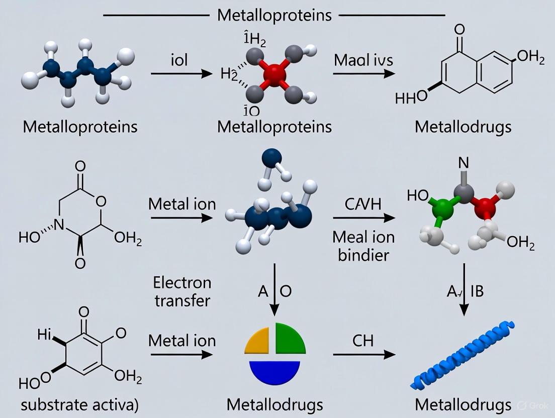
Diagram 2: Generalized Mechanism of a Metallodrug. Many metallodrugs are prodrugs that require activation, often via hydrolysis or reduction, before interacting with their biological targets to exert a therapeutic effect [20].
6. Conclusion
The diverse biological roles of metalloproteins—from providing structural integrity to catalyzing complex reactions, transferring electrons, and mediating signals—are fundamental to life. A deep understanding of these functions, driven by advanced structural and spectroscopic techniques, is paramount. This knowledge not only deciphers basic biological mechanisms but also fuels the rational design of artificial metalloenzymes for biotechnology and the development of novel metallodrugs with unique mechanisms of action for medicine. The continued integration of experimental and computational approaches will undoubtedly uncover new metalloprotein functions and accelerate their application in addressing global health and industrial challenges.
The cellular interactomes of zinc and copper represent a sophisticated regulatory network essential for life. These transition metals, while indispensable as cofactors for up to 10% of proteins in living organisms, exhibit significant functional interplay and potential for cytotoxicity when misregulated [21] [22]. This whitepaper delineates the molecular mechanisms governing zinc and copper homeostasis, trafficking pathways, and their intricate crosstalk, drawing upon recent advances in metalloprotein research. We synthesize findings from structural biology, genetic models, and biochemical studies to present a coherent framework for understanding how cells allocate these essential metals. Furthermore, we explore the biomedical implications of metal homeostasis in therapeutic development and disease pathogenesis, providing methodologies for experimental investigation and quantitative data on metal interactions. This knowledge provides a foundation for developing novel metallodrugs and therapeutic strategies targeting metal-related pathologies.
Zinc and copper are essential transition metals that play critical roles in numerous biological processes through their incorporation into metalloproteins. Zinc (Zn), typically found in the +2 oxidation state, serves as a structural component in zinc finger proteins and a catalytic cofactor in enzymes like metalloproteases [15] [23]. Its filled d-orbital (d¹⁰) provides geometric flexibility without participating in redox chemistry, making it ideal for structural roles. In contrast, copper (Cu) cycles between +1 and +2 oxidation states, participating in electron transfer reactions and oxygen activation in enzymes such as superoxide dismutase and cytochrome c oxidase [23] [24].
The Irving-Williams series (Mn²⁺ < Fe²⁺ < Co²⁺ < Ni²⁺ < Cu²⁺ > Zn²⁺) predicts the relative stability of metal complexes, with Cu²+ forming the most stable complexes among biologically relevant metals [24]. This thermodynamic preference creates a fundamental challenge for cells: how to ensure correct metallation of proteins despite copper's strong binding affinity. Cells have evolved sophisticated metal trafficking pathways involving chaperones, transporters, and storage proteins to overcome this challenge and maintain metal specificity [21] [24].
Recent research has revealed extensive functional interplay between zinc and copper homeostasis, with each metal influencing the distribution and utilization of the other [25] [26]. Understanding this crosstalk is crucial for elucidating normal cellular function and developing treatments for metal-related diseases, including neurodegenerative disorders and cancer.
Molecular Mechanisms of Zinc and Copper Homeostasis
Zinc Homeostasis Pathways
Cellular zinc homeostasis is maintained through coordinated regulation of import, export, and storage mechanisms. The Zap1 transcription factor regulates zinc uptake in yeast by activating expression of ZRT1 and ZRT2 zinc transporters under zinc-deficient conditions [22]. In vertebrates, zinc transporters of the ZIP (SLC39A) and ZnT (SLC30A) families facilitate zinc import into the cytoplasm and export from the cytoplasm, respectively [25].
A breakthrough discovery identified ZNG1 (Zn-regulated GTPase metalloprotein activator 1) as a vertebrate metallochaperone that coordinates zinc trafficking to client metalloproteins [21]. ZNG1 belongs to the COG0523 protein family of G3E GTPases and specifically activates the zinc metalloprotease METAP1 (methionine aminopeptidase 1) by facilitating its proper metallation [21]. This represents the first experimentally verified zinc metallochaperone in any organism, revealing a new layer in zinc homeostasis.
Copper Homeostasis Pathways
Copper homeostasis involves specialized chaperones that deliver copper to specific cellular compartments and enzymes. The Atx1-like chaperones transfer copper to transporters in the secretory pathway [22], while CCS (copper chaperone for superoxide dismutase) specifically delivers copper to Cu/Zn-SOD1 [24] [22]. A third class of chaperones, including Cox17, supplies copper to mitochondria for cytochrome c oxidase assembly [22].
Cellular copper levels are tightly regulated by transcription factors such as CueR in bacteria and Mac1 in yeast, which activate copper efflux systems under high copper conditions [24]. These sensors exhibit remarkable sensitivity, with CueR responding to Cu(I) at zeptomolar (10⁻²¹ M) concentrations [24], ensuring minimal levels of free intracellular copper that could cause oxidative damage.
Interplay Between Zinc and Copper Homeostasis
Zinc and copper homeostasis are interconnected through multiple mechanisms. Studies in Enterococcus faecalis demonstrated that the Zur (zinc uptake regulator) protein mediates responses to both zinc and copper stress [26]. Transcriptomic analyses revealed that bacterial exposure to copper activates zinc homeostasis modules, repressing zinc uptake systems while inducing efflux mechanisms [26].
In marine oysters (Crassostrea gigas), zinc supplementation mitigates copper toxicity by modulating metal accumulation and oxidative stress responses [25]. The molecular basis for this protection involves zinc's ability to competitively inhibit copper uptake through shared transporters and induce metallothionein expression for metal sequestration [25]. This antagonistic interaction highlights the functional interplay between these essential elements.
Table 1: Key Proteins in Zinc and Copper Homeostasis
| Protein | Metal Specificity | Function | Mechanism |
|---|---|---|---|
| ZNG1 | Zinc | Metallochaperone | GTPase that activates METAP1 via zinc transfer [21] |
| Zur | Zinc (and Copper) | Transcriptional regulator | Represses zinc uptake genes; responds to copper stress [26] |
| CueR | Copper | Transcriptional activator | Binds Cu(I) with zeptomolar sensitivity; activates efflux systems [24] |
| CTR1 | Copper | Membrane transporter | High-affinity copper uptake across plasma membrane [22] |
| ZIP/ZnT | Zinc | Transporters | ZIP imports zinc; ZnT exports zinc from cytoplasm [25] |
| METAP1 | Zinc | Metalloenyzme | Removes N-terminal methionine; client of ZNG1 [21] |
Quantitative Analysis of Zinc-Copper Interactions
Recent investigations have provided quantitative data on the physiological and molecular consequences of zinc-copper interactions. In oyster models, zinc supplementation significantly reduced copper accumulation in gill tissues from 121.45 ± 1.89 mg/kg to 65.32 ± 1.21 mg/kg dry weight, demonstrating zinc's protective effect against copper overload [25].
Transcriptomic analyses revealed that zinc co-exposure modulated expression of 72.5% of the immune-related genes dysregulated by copper alone, with particular impact on the Toll-like signaling pathway and apoptosis-related genes [25]. This molecular reprogramming underpins zinc's ability to alleviate copper-induced immunotoxicity.
In antibacterial applications, copper-zinc nanocomposites (ZnFe₂O₄@ZnS/Cu₂S) exhibited potent efficacy with minimum inhibitory concentrations (MIC) of 50 μg/mL against E. coli, 60 μg/mL against S. aureus, and 80 μg/mL against drug-resistant Salmonella [27]. At 200 μg/mL with 80 minutes exposure, these nanocomposites achieved a bacteriostatic rate of 99.99% against all tested bacterial strains [27].
Table 2: Quantitative Effects of Zinc-Copper Interactions in Biological Systems
| System/Parameter | Condition 1 | Condition 2 | Effect | Reference |
|---|---|---|---|---|
| Oyster copper accumulation | Cu only: 121.45 ± 1.89 mg/kg | Cu + Zn: 65.32 ± 1.21 mg/kg | 46.2% reduction in Cu accumulation [25] | |
| Antibacterial activity (MIC) | E. coli: 50 μg/mL | S. aureus: 60 μg/mL | T-Salmonella: 80 μg/mL | Concentration-dependent efficacy [27] |
| Cellular viability (cancer cells) | ZnO NPs: ~60% at 100 μg/mL | Cu-doped ZnO NPs: ~30% at 100 μg/mL | Enhanced cytotoxicity with Cu doping [28] | |
| Bacteriostatic rate | ZZC nanocomposite at 200 μg/mL, 80 min | 99.99% against multiple bacteria | High efficacy against drug-resistant strains [27] | |
| Gene expression modulation | Cu-altered immune genes | Zn co-exposure normalized 72.5% | Transcriptomic reprogramming [25] |
Experimental Approaches and Methodologies
Investigating Metal Homeostasis: Key Protocols
Genetic and Pharmacological Validation of ZNG1 Function The identification of ZNG1 as a zinc metallochaperone employed a multi-faceted approach across model systems [21]. Researchers utilized CRISPR/Cas9-mediated knockout in zebrafish and mouse models to establish ZNG1's essential role in vertebrate zinc homeostasis. Biochemical validation included GTPase activity assays measuring phosphate release under varying zinc conditions. METAP1 interaction studies employed co-immunoprecipitation and surface plasmon resonance to determine binding kinetics. Functional rescue experiments demonstrated that ZNG1-deficient phenotypes could be reversed by wild-type ZNG1 but not GTPase-deficient mutants, establishing the essential role of GTP hydrolysis [21].
Transcriptomic Analysis of Metal Stress Responses The study of zinc and copper interplay in Enterococcus faecalis and oyster models utilized comprehensive transcriptomic approaches [25] [26]. For bacterial systems, microarray analysis was performed on cells exposed to sublethal copper (0.5 mM) and zinc (0.3 mM) concentrations, identifying differentially expressed genes. In oyster hemocytes, RNA-Seq profiled responses to copper exposure (50 μg/L) with and without zinc supplementation (100 μg/L). Bioinformatic analysis included weighted gene co-expression network analysis (WGCNA) to identify metal-responsive modules and Gene Ontology enrichment to determine affected biological processes [25].
Assessment of Metal Toxicity and Protection Physiological metal interactions were quantified using integrated approaches [25]. Inductively coupled plasma mass spectrometry (ICP-MS) measured metal accumulation in tissues under various exposure conditions. Biochemical assays quantified oxidative stress markers including lipid peroxidation (malondialdehyde content), antioxidant enzyme activities (SOD, CAT, GPX), and protein carbonylation. Immunological parameters included flow cytometric analysis of hemocyte viability, phagocytosis activity, and apoptosis rates using Annexin V/propidium iodide staining [25].
The Scientist's Toolkit: Essential Research Reagents
Table 3: Essential Research Reagents for Zinc-Copper Homeostasis Studies
| Reagent/Category | Specific Examples | Function/Application | Experimental Context |
|---|---|---|---|
| Metal Salts | CuSO₄, ZnCl₂, Cu(NO₃)₂, ZnSO₄ | Prepare metal exposure solutions; control concentration and bioavailability [25] [27] | In vivo and in vitro metal treatment studies |
| Molecular Biology Kits | RNA extraction kits, cDNA synthesis kits, qPCR reagents | Gene expression analysis of metal-responsive genes [25] | Transcriptomic studies of metal homeostasis |
| Antibodies | Anti-ZNG1, Anti-METAP1, Anti-MT (metallothionein) | Protein detection, localization, and quantification via Western blot, IHC [21] | Validation of protein expression and localization |
| Cell Culture Media | DMEM, RPMI-1640 with metal-defined FBS | Control metal availability in cell-based assays [28] | In vitro metal studies with cell lines |
| Spectroscopy Standards | ICP-MS metal standards, AAS calibration standards | Quantify metal concentrations in biological samples [25] [28] | Metal quantification in tissues/cells |
| Nanoparticles | ZnO NPs, Cu-doped ZnO NPs, ZZC nanocomposites | Study metal interactions at nano-bio interface [28] [27] | Antimicrobial and anticancer applications |
Biomedical Implications and Metallodrug Development
Dysregulation of zinc and copper homeostasis is implicated in numerous human diseases. Neurodegenerative disorders including Alzheimer's and Parkinson's disease involve metal-protein interactions where copper and zinc promote amyloid-beta aggregation and oxidative stress [22]. Wilson's disease, characterized by copper accumulation in liver and brain, results from mutations in the ATP7B copper transporter [22].
The interplay between zinc and copper informs the development of novel metal-based therapeutics. Copper-doped zinc oxide nanoparticles exhibit enhanced cytotoxicity against cancer cells compared to undoped nanoparticles, showing particular efficacy against bone cancer fibroblasts (G-292) with significant inhibition of cell proliferation at 17.5 μg/mL [28]. This selective toxicity toward cancer cells with minimal effects on normal lung fibroblasts (MRC-5) highlights their therapeutic potential [28].
Antimicrobial applications of copper-zinc nanocomposites (ZZC) demonstrate effectiveness against drug-resistant pathogens, achieving 99.99% bacteriostatic rates against multi-drug-resistant Salmonella through membrane disruption mechanisms [27]. These nanocomposites also promote wound healing in mixed bacterial infection models, indicating promise for clinical application [27].
The cellular interactomes of zinc and copper represent a dynamic regulatory network maintained through specialized trafficking pathways, metallochaperones, and transcriptional regulators. The recent identification of ZNG1 as the first verified zinc metallochaperone provides a new paradigm for understanding intracellular zinc distribution [21]. The antagonistic relationship between zinc and copper, observed across evolutionary lineages from bacteria to vertebrates, reveals fundamental principles of metal biology with significant implications for human health and disease.
Future research directions should focus on structural characterization of metal transfer complexes, particularly the ZNG1-METAP1 interface, to elucidate mechanisms of metal transfer specificity. The development of advanced imaging probes for visualizing metal dynamics in live cells will provide spatial and temporal resolution of metal trafficking events. Exploration of metal-based combination therapies that exploit zinc-copper interactions may yield novel approaches for treating antibiotic-resistant infections and cancers with greater specificity and reduced side effects.
As the field of bioinorganic chemistry advances, integration of metalloprotein data with systems biology approaches will enable comprehensive modeling of metal interactomes, accelerating the rational design of metallodrugs that target specific nodes in metal homeostasis networks. These efforts will deepen our understanding of cellular inorganic chemistry while providing new therapeutic strategies for metal-related pathologies.
The serendipitous discovery of cisplatin's anticancer activity fundamentally transformed cancer chemotherapy, establishing platinumbased drugs as cornerstone treatments for various malignancies [29]. These drugs primarily function as classical chemotherapeutics, inducing apoptosis through DNA crosslinking and disruption of replication processes [30]. Despite their clinical success, platinum-based chemotherapeutics face significant limitations including severe systemic toxicity, inherent and acquired drug resistance, and limited efficacy against certain cancer types [31]. These challenges have motivated the systematic exploration of alternative transition metal-based therapeutics with different mechanistic profiles.
The field of medicinal bioinorganic chemistry has responded by developing non-platinum metallodrugs that offer unique mechanisms of action (MoA), diminished side-effect profiles, and potential activity against cisplatin-resistant cancer cells [32]. Among these, complexes of ruthenium, iridium, and gold have emerged as particularly promising candidates, with some advancing to clinical trials [30]. These metals provide distinctive advantages, including variable oxidation states, favorable ligand exchange kinetics, and the ability to target specific cellular compartments and biomolecules beyond DNA, such as proteins and enzymes involved in critical signaling pathways [33].
This technical guide comprehensively reviews the current state of non-platinum metallodrug development, emphasizing their unique chemical properties, established mechanisms of action, and the experimental methodologies essential for their characterization and evaluation. By moving beyond the platinum paradigm, researchers are developing a new generation of metallopharmaceuticals with enhanced selectivity and novel anticancer mechanisms.
Ruthenium Complexes: Front-Runners in Clinical Translation
Chemical Properties and Classification
Ruthenium complexes have advanced furthest among non-platinum metallodrug candidates, with several compounds having undergone clinical evaluation [34]. Their therapeutic potential stems from several innate characteristics: low systemic toxicity, iron-mimicking behavior that facilitates binding to biomolecules like transferrin, and accessible Ru(II)/Ru(III) oxidation states that allow participation in biological redox chemistry [35]. These properties enable ruthenium complexes to accumulate more effectively in tumor tissues compared to platinum drugs [35].
Structurally, investigational ruthenium compounds fall into several categories:
- Ru(III) coordination complexes including KP1019, KP1339, and NAMI-A
- Ru(II) arene "piano-stool" complexes such as RAPTA-C and RM175
- Polynuclear ruthenium complexes with bridging ligands
- Ru(II) polypyridyl complexes for photodynamic therapy applications
Table 1: Clinically Evaluated Ruthenium-Based Anticancer Complexes
| Complex | Oxidation State | Clinical Status | Primary Indication/Target | Key Characteristics |
|---|---|---|---|---|
| KP1019 | Ru(III) | Completed Phase I | Colorectal cancer | Indazole ligands; apoptosis induction |
| NKP-1339 (IT-139) | Ru(III) | Clinical trials | Solid tumors | Sodium salt of KP1019; improved solubility |
| NAMI-A | Ru(III) | Clinical trials | Metastases | Imidazole ligands; anti-metastatic |
| RAPTA-C | Ru(II) | Preclinical | Primary tumors & metastases | Arene complex; antiangiogenic |
Established Mechanisms of Action
Unlike platinum drugs that primarily target nuclear DNA, ruthenium complexes exhibit diverse mechanisms of action that contribute to their distinct biological profiles:
DNA-Targeting Mechanisms: While not their primary target, some Ru(II) arene complexes can bind DNA, forming monofunctional adducts at the N7 position of guanine bases. This binding is often complemented by intercalative interactions of extended arene ligands and specific hydrogen-bonding interactions between chelating ligands and DNA bases, resulting in structural distortions distinct from cisplatin-induced lesions [30].
Protein-Targeting Mechanisms: Ruthenium complexes frequently interact with protein targets. The Functional Identification of Target by Expression Proteomics (FITExP) methodology has identified multiple protein targets for RAPTA complexes [36]. RAPTA-T causes upregulation of proteins involved in metastasis and tumorigenesis suppression, while RAPTA-EA, which incorporates an ethacrynic acid moiety, primarily upregulates oxidative stress-related proteins and inhibits glutathione S-transferase (GST) activity [36].
Metastasis Inhibition: NAMI-A exhibits a unique profile, showing pronounced anti-metastatic activity with limited effects on primary tumor growth [30]. This activity may involve inhibition of extracellular matrix metalloproteases and interference with cell adhesion and migration processes.
Reactive Oxygen Species (ROS) Generation: Multiple ruthenium complexes, including polypyridyl and cyclopentadienyl derivatives, induce apoptosis through ROS-mediated mitochondrial dysfunction [35]. This oxidative stress triggers downstream effects including cell cycle arrest and DNA damage.
Activation by Reduction: The "activation by reduction" hypothesis proposes that Ru(III) complexes (e.g., KP1019, NAMI-A) serve as prodrugs that are activated to more reactive Ru(II) species in the hypoxic tumor microenvironment, potentially enhancing tumor selectivity [30].
Experimental Protocols for Ruthenium Complex Evaluation
Cytotoxicity Assessment (MTT Assay): The standard 3-(4,5-dimethylthiazol-2-yl)-2,5-diphenyltetrazolium bromide (MTT) assay evaluates in vitro anticancer activity. Cells (e.g., A549, HepG2, MCF-7) are seeded in 96-well plates, treated with compound gradients for 24-72 hours, followed by MTT solution addition. After formazan crystal formation, solubilization solution is added, and absorbance is measured at 570 nm to determine IC₅₀ values [33].
Proteomic Target Identification (FITExP): The FITExP method identifies protein targets using the following workflow [36]:
- Treat appropriate cell lines (e.g., MDA-MB-231 and MCF-7 for breast cancer) with ruthenium complexes
- Perform quantitative proteomic analysis via mass spectrometry
- Reference data against positive controls (e.g., paclitaxel, cisplatin)
- Apply statistical analysis (P < 0.05 cutoff) to identify significantly upregulated proteins
- Validate potential targets through functional assays
DNA Binding Studies: To characterize DNA interactions:
- Incubate ruthenium complexes with calf thymus DNA or oligonucleotides
- Analyze binding via UV-Vis spectroscopy, circular dichroism, or fluorescence quenching
- Employ gel electrophoresis to assess DNA migration changes
- Use X-ray crystallography or NMR spectroscopy for structural characterization of adducts
Iridium Complexes: Emerging Heavyweight Contenders
Structural Diversity and Anticancer Potential
Iridium complexes represent a newer area of investigation in bioinorganic medicinal chemistry, with significant progress achieved over the past 15 years [37]. While no iridium complexes have yet entered clinical trials, their structural diversity and potent cytotoxicity warrant serious attention. Major structural classes include:
Half-Sandwich Iridium(III) Cyclopentadienyl (Ir-Cpx) Complexes: These "piano-stool" complexes have shown particularly promising anticancer activity since their initial report in 2007 [37]. Their stability, intracellular localization, target organelles, and molecular targets have been extensively characterized.
Iridium(III) Polypyridyl Complexes: These octahedral complexes benefit from attractive photophysical properties, making them suitable for photodynamic therapy (PDT) applications and cellular imaging [33].
Iridium N-Heterocyclic Carbene (NHC) Complexes: These complexes demonstrate promising anticancer activity and have been explored as living cell imaging reagents [33].
Unique Mechanisms and Intracellular Targets
Iridium complexes frequently operate through mechanisms distinct from both platinum and ruthenium-based drugs:
Mitochondrial Targeting: A defining characteristic of many iridium complexes is their pronounced mitochondrial localization. A seminal study employing correlative 3D cryo X-ray imaging unambiguously demonstrated exclusive accumulation of a potent half-sandwich iridium complex within mitochondria, without chemical fixation, labeling, or mechanical manipulation [38]. This mitochondrial targeting enables concentrations 15-250 times more potent than cisplatin across multiple cancer cell lines.
Reactive Oxygen Species (ROS) Generation: Iridium complexes effectively generate ROS through multiple pathways. As photosensitizers, they produce cytotoxic singlet oxygen (¹O₂) upon light irradiation for photodynamic therapy [33]. Even without photoactivation, many iridium complexes induce oxidative stress that overwhelms cellular antioxidant defenses.
Protein Inhibition and Enzyme Targeting: Iridium complexes can inhibit specific enzyme systems. Some Ir(III)-NHC complexes strongly and selectively inhibit thioredoxin reductase (TrxR) activity, disrupting redox homeostasis and triggering apoptosis [31]. Other complexes target protein kinases and modulate MAPK signaling pathways.
DNA Binding with Novel Mechanisms: While generally not considered primary DNA binders, some iridium complexes can interact with DNA through non-covalent mechanisms, including groove binding and intercalation, causing distinct conformational changes compared to platinum-induced lesions.
Experimental Protocols for Iridium Complex Analysis
Intracellular Localization via Correlative 3D Cryo X-Ray Imaging: This powerful method precisely localizes and quantifies iridium within hydrated cells at nanometer resolution [38]:
- Grow cancer cells on specialized silicon nitride supports
- Treat with iridium complexes at relevant concentrations and timepoints
- Rapidly vitrify cells by plunge-freezing in liquid ethane
- Acquire cellular ultrastructure via cryo soft X-ray tomography (cryo-SXT) at 50 nm resolution
- Map elemental distribution via cryo hard X-ray fluorescence tomography (cryo-XRF) with 70 nm step size
- Correlate datasets to assign iridium localization to specific organelles
Photodynamic Therapy Evaluation: For photoactive iridium complexes:
- Treat cells with complexes in dark conditions
- Expose to light at specific wavelengths and energies
- Assess dark toxicity vs. photoenhanced toxicity
- Measure singlet oxygen quantum yields using chemical traps or reference compounds
- Evaluate intracellular ROS generation with fluorescent probes (e.g., DCFH-DA)
Thioredoxin Reductase Inhibition Assay:
- Prepare cell lysates or purified TrxR enzyme
- Incubate with iridium complexes at varying concentrations
- Measure TrxR activity using DTNB [5,5'-dithiobis(2-nitrobenzoic acid)] assay
- Determine IC₅₀ values for inhibition potency
- Validate selectivity against related enzymes like glutathione reductase
Gold Complexes: From Arthritis to Oncology
Chemical Classes and Development History
Gold complexes have a longer history in medicine, initially developed for rheumatoid arthritis treatment before their anticancer potential was recognized [30]. Major classes of anticancer gold complexes include:
Gold(I) Phosphine Complexes: Tetrahedral Au(I) complexes, particularly those containing phosphine ligands, display broad-spectrum anticancer activity, including against cisplatin-resistant cell lines [30]. Auranofin, an oral anti-arthritis drug, has demonstrated promising anticancer activity in preclinical models [31].
Gold(III) Porphyrin Complexes: These square-planar complexes exploit the structural similarity between Au(III) and Pt(II) while offering enhanced stability through the robust porphyrin ligand system [30]. They exhibit significant in vitro and in vivo activity against hepatocellular and nasopharyngeal carcinoma.
Gold(III) Bipyridyl Complexes: These complexes further expand the structural diversity of gold-based anticancer agents, though stability under physiological conditions remains a developmental challenge.
Primary Mechanisms of Action
Gold complexes typically operate through mechanisms fundamentally different from platinum drugs, with minimal DNA interaction:
Thioredoxin Reductase (TrxR) Inhibition: A primary mechanism for many gold complexes involves potent and often irreversible inhibition of thioredoxin reductase [30]. This selenocysteine-containing enzyme plays crucial roles in maintaining cellular redox homeostasis, antioxidant defense, and regulating apoptosis. Its inhibition disrupts multiple cellular processes and triggers oxidative stress-mediated cell death.
Mitochondrial Dysfunction: Related to TrxR inhibition, gold complexes frequently induce mitochondrial membrane depolarization, disrupting electron transport chain function and promoting cytochrome c release, which activates the apoptotic cascade [31].
Glutathione Reductase Inhibition: Some gold(I) phosphine complexes additionally inhibit glutathione reductase, further compromising the cellular antioxidant defense system and enhancing oxidative stress [30].
Protein Binding via Thiolate Coordination: Gold complexes exhibit high affinity for cysteine residues in proteins, leading to widespread protein binding that can modulate various enzymatic activities and signaling pathways.
Experimental Protocols for Gold Complex Evaluation
Thioredoxin Reductase Inhibition Assay:
- Incubate purified TrxR with gold complexes at varying concentrations
- Measure enzyme activity using NADPH-dependent DTNB reduction assay
- Monitor absorbance at 412 nm to quantify inhibition
- Determine IC₅₀ values and compare to reference compounds
- Assess inhibition reversibility through dialysis experiments
Mitochondrial Function Assessment:
- Stain treated cells with JC-1 or TMRE fluorescent dyes
- Analyze mitochondrial membrane potential via flow cytometry or fluorescence microscopy
- Measure ATP production levels using luciferase-based assays
- Assess oxygen consumption rates using Seahorse extracellular flux analyzer
Cellular Uptake and Distribution Studies:
- Treat cells with gold complexes for various durations
- Wash, trypsinize, and collect cells
- Lyse cells and fractionate into cytosolic, mitochondrial, and nuclear components
- Quantify gold content using atomic absorption spectroscopy or ICP-MS
- Correlate uptake levels with cytotoxicity measures
Comparative Analysis: Quantitative Assessment of Non-Platinum Metallodrugs
Table 2: Comparative Cytotoxicity and Properties of Non-Platinum Metallodrugs
| Metal Complex | Example Compounds | IC₅₀ Range (μM) | Primary Molecular Targets | Resistance Profile | Key Advantages |
|---|---|---|---|---|---|
| Ruthenium | KP1019, NAMI-A, RAPTA-C | 10-200 | DNA, Proteins, GST | No cross-resistance with cisplatin | Low toxicity, anti-metastatic, iron-mimicking |
| Iridium | Ir-Cpx complexes, Polypyridyl | 0.1-50 | Mitochondria, TrxR, DNA | Effective against cisplatin-resistant cells | High potency, mitochondrial targeting, PDT applications |
| Gold | Auranofin, Au(I) phosphines, Au(III) porphyrins | 0.5-20 | TrxR, Mitochondria | No cross-resistance with cisplatin | Unique enzyme inhibition, nanomolar potency for some complexes |
The Scientist's Toolkit: Essential Research Reagents and Methodologies
Table 3: Key Research Reagent Solutions for Metallodrug Development
| Reagent/Assay | Function/Application | Experimental Utility |
|---|---|---|
| MTT Assay Kit | Cytotoxicity determination | Standardized measurement of cell viability and IC₅₀ values |
| JC-1 Dye | Mitochondrial membrane potential assessment | Fluorescent detection of mitochondrial depolarization |
| DCFH-DA Probe | Intracellular ROS detection | Quantification of reactive oxygen species generation |
| cryo-SXT Setup | Cellular ultrastructure imaging | Nanoscale cellular architecture without chemical fixation |
| cryo-XRF Setup | Elemental mapping | Precise intracellular metal localization and quantification |
| Proteomics Kits | Protein target identification | FITExP analysis for target deconvolution |
| TrxR Activity Assay | Enzyme inhibition studies | Evaluation of thioredoxin reductase inhibition potency |
| DNA Binding Kits | DNA interaction studies | Assessment of metallodrug-DNA interactions and adduct formation |
Visualization of Key Mechanisms and Workflows
Mechanism of Mitochondrial Targeting by Iridium Complexes
Diagram 1: Mitochondrial targeting mechanism of iridium complexes
Proteomic Workflow for Metallodrug Target Identification
Diagram 2: FITExP workflow for metallodrug target identification
The development of non-platinum metallodrugs represents a paradigm shift in cancer chemotherapy, moving beyond the limitations of traditional platinum-based approaches. Ruthenium, iridium, and gold complexes offer distinct mechanistic profiles, targeting diverse cellular components including proteins, enzymes, and organelles beyond nuclear DNA. Their unique properties—including variable oxidation states, ligand exchange kinetics, and the ability to generate reactive oxygen species—enable novel mechanisms of action that can overcome cisplatin resistance and reduce systemic toxicity.
The ongoing clinical evaluation of ruthenium complexes and promising preclinical data for iridium and gold compounds underscore the translational potential of these agents. Future development will likely focus on enhancing tumor selectivity through targeted delivery systems, optimizing combination therapies, and exploiting unique activation mechanisms such as photoactivation for spatiotemporal control of drug activity. As our understanding of the intricate relationships between metal complex structures and their biological activities deepens, the rational design of next-generation metallodrugs with improved efficacy and safety profiles will continue to advance, solidifying the role of non-platinum metallodrugs in the future landscape of cancer therapy.
Advanced Analytical Techniques and Therapeutic Applications in Metallodrug Development
In bioinorganic chemistry, the function of metalloproteins and metallodrugs is dictated by the precise coordination environment of their metal centers. The metal's identity, oxidation state, geometric arrangement, and ligand identity collectively determine reactivity, specificity, and mechanism [15]. Deciphering this structural information at the atomic level is fundamental to understanding diverse biological processes, from oxygen transport and electron transfer to enzymatic catalysis and gene regulation [15]. Furthermore, abnormalities in metal homeostasis and metalloprotein structure are implicated in serious human diseases, including neurodegenerative disorders and cancer, making the characterization of metal sites crucial for biomedical advances [15]. This technical guide details how three powerful spectroscopic techniques—Electron Paramagnetic Resonance (EPR), X-ray Absorption Spectroscopy (XAS), and Nuclear Magnetic Resonance (NMR) spectroscopy—serve as indispensable tools for resolving these metal coordination environments, thereby driving innovation in metalloprotein biochemistry and metallodrug design.
Electron Paramagnetic Resonance (EPR) Spectroscopy
Fundamental Principles and Applications
EPR spectroscopy is a selective and powerful technique for characterizing paramagnetic metal centers, which contain unpaired electrons. It provides detailed information on oxidation states, metal coordination geometry, and the electronic structure of metalloproteins and metallodrugs [4]. The technique is particularly valuable for studying metal ion coordination and redox chemistry, which are central to the function of many bioinorganic systems [4]. EPR can probe the local environment of the metal ion, making it a major tool for understanding metal-based structures and their function.
Advanced pulse EPR methods, such as Electron-Nuclear Double Resonance (ENDOR) and Hyperfine Sublevel Correlation (HYSCORE) spectroscopy, further extend its capabilities by resolving hyperfine interactions with nearby magnetic nuclei, providing insights into the ligand identity and protonation states [4].
Experimental Protocol for Metalloprotein EPR
Sample Preparation:
- Protein Purity: Ensure the metalloprotein is highly purified (>95% homogeneity) to avoid spurious signals.
- Buffer Considerations: Use deuterated buffers or buffers with low dielectric loss to minimize non-resonant absorption of microwaves. Avoid buffers containing high concentrations of paramagnetic ions.
- Sample Volume: Standard quartz EPR tubes typically require 150-200 µL of sample. Optimal protein concentration depends on the metal center's spin state and EPR sensitivity but often ranges from 50-500 µM.
- Cryogenic Requirement: Most biological EPR experiments are performed at cryogenic temperatures (e.g., 10-50 K) to slow relaxation rates and enhance signal resolution. This requires rapid freezing of samples in liquid nitrogen.
Data Acquisition Parameters:
- Microwave Frequency: Most commonly performed at X-band (~9-10 GHz), which offers a good balance of sensitivity and resolution for biological samples.
- Microwave Power: Power saturation studies can provide information on relaxation properties and are used to optimize signal intensity without saturation.
- Magnetic Field Range: The field range is adjusted based on the expected g-values of the metal center. A typical scan for a Fe(III) center (g ~4.3, 2) might be from 0 to 7000 G.
- Modulation Amplitude: Optimized to maximize signal-to-noise without distorting line shapes; typically 5-10 G.
Data Analysis Workflow:
- Signal Averaging: Improve signal-to-noise by accumulating multiple scans.
- Baseline Correction: Subtract a polynomial baseline to correct for instrumental drift.
- Spectral Simulation: Use spin Hamiltonian parameters (g-tensor, A-tensor, zero-field splitting parameters) to simulate the experimental spectrum. This iterative process allows for the extraction of precise metal coordination parameters.
Table: Key EPR Parameters for Common Metal Ions in Bioinorganic Chemistry
| Metal Ion | Common Oxidation State | Typical g-values | Key Information Obtained |
|---|---|---|---|
| Cu(II) | +2 | g∥ ~ 2.2-2.4, g⟂ ~ 2.04-2.08 | Type of ligand donor set (N/O/S), tetragonal distortion |
| Fe(III) (Heme) | +3 | g ~ 2, 4.3, 6 | Spin state (high-spin vs. low-spin), axial vs. rhombic symmetry |
| Mn(II) | +2 | g ~ 2.0, multi-line hyperfine | Coordination number, ligand identity via hyperfine coupling |
| Mo(V) | +5 | g ~ 1.96-2.02 | Ligand environment in enzyme active sites (e.g., sulfite oxidase) |
Diagram 1: Experimental workflow for EPR spectroscopy of metalloproteins and metallodrugs, covering sample preparation, data acquisition, and analysis to extract structural parameters.
X-ray Absorption Spectroscopy (XAS)
Fundamental Principles and Applications
XAS is a premier technique for probing the local structural environment of metal ions, regardless of the physical state of the sample, making it ideal for non-crystalline biological materials [39]. The technique is divided into two main regions: X-ray Absorption Near Edge Structure (XANES) and Extended X-ray Absorption Fine Structure (EXAFS). XANES provides information about the metal's oxidation state and geometry, while EXAFS yields quantitative data on the number, type, and distance of coordinating atoms [39]. This makes XAS uniquely powerful for studying metal sites in metalloproteins whose structures cannot be solved by crystallography, and for characterizing metallodrugs bound to biomolecules.
Experimental Protocol for Metalloprotein XAS
Sample Preparation:
- Concentration: High metal concentration is critical. For transition metals, optimal concentrations are typically 1-10 mM for EXAFS and can be lower for XANES.
- Sample Homogeneity: The sample must be homogeneous to avoid artifacts. For metalloproteins, ensure the metal is fully incorporated into the protein and that the sample is monodisperse.
- Sample Form: Aqueous samples are loaded into specialized cells with X-ray transparent windows (e.g., polycarbonate). Samples can be frozen as glasses in cryoprotectants like glycerol for data collection at cryogenic temperatures to reduce radiation damage.
- Volume: Typical sample volume is 50-100 µL.
Data Collection at Synchrotron Beamline:
- Beamline Setup: Experiments require a synchrotron radiation source. The beamline should be equipped with a double-crystal monochromator for energy selection and ionization chambers for measuring X-ray absorption.
- Energy Range: Data collection spans from ~200 eV below to ~1000 eV above the absorption edge of the element of interest.
- Detection Mode: Data can be collected in transmission mode for concentrated samples or fluorescence mode for dilute samples (e.g., metalloproteins in biological buffers).
- Temperature: Data is often collected at ~10-20 K using a liquid helium cryostat to minimize radiation damage and to reduce the Debye-Waller factor, sharpening the EXAFS oscillations.
Data Processing and Analysis:
- Energy Calibration: Calibrate the energy scale using a metal foil standard measured simultaneously with the sample.
- Background Subtraction: Pre-edge and post-edge backgrounds are subtracted to isolate the absorption edge.
- Normalization: The edge jump is normalized to unity.
- EXAFS Extraction: The normalized absorption is converted from energy to photoelectron wave number (k-space). A spline function is used to remove the low-frequency background to isolate the EXAFS oscillations, χ(k).
- Fourier Transform: χ(k) is weighted and Fourier transformed to produce a radial distribution function (pseudo-radial distribution).
- Curve Fitting: Theoretical scattering paths, generated from a putative structural model (e.g., using FEFF code), are used to fit the experimental EXAFS data to extract quantitative structural parameters (coordination number, bond distance, and Debye-Waller factor).
Table: XAS Signatures for Common Metal Coordination Environments
| Metal & Oxidation State | XANES Edge Energy (eV) | EXAFS Coordination Shell | Typical Bond Lengths (Å) |
|---|---|---|---|
| Fe(II) (High-Spin) | ~7118 | 4-6 N/O @ ~2.1-2.2 Å | Fe-N: 2.10-2.20 |
| Fe(III) (High-Spin) | ~7123 | 4-6 N/O @ ~2.0-2.1 Å | Fe-N: 2.00-2.10 |
| Cu(I) | ~8979 | 2-4 S/N @ ~2.2-2.3 Å | Cu-S: 2.20-2.30 |
| Cu(II) | ~8984 | 4-5 N/O @ ~1.95-2.00 Å | Cu-N: 1.95-2.00 |
| Zn(II) | ~9664 | 4 N/O @ ~2.00-2.05 Å | Zn-N: 2.00-2.05 |
| Mo(IV) | ~20000 | 4-6 S/O @ ~2.3-2.4 Å | Mo-S: 2.30-2.40 |
Nuclear Magnetic Resonance (NMR) Spectroscopy
Fundamental Principles and Applications
While often associated with organic molecules, NMR spectroscopy is a powerful and element-selective tool for characterizing metal coordination environments, particularly for nuclei like 195Pt, 113Cd, and 67Zn [40] [41]. The chemical shift range, especially for nuclei like 195Pt, is extremely large and highly sensitive to the oxidation state, coordination geometry, and ligand identity [40]. This makes NMR ideal for studying metallodrugs and, with advanced methodologies, even heterogeneous systems like single-atom catalysts (SACs) which mimic supported enzymatic sites [40]. Recent advances in ultra-wideline NMR methodology have enabled the acquisition of high-quality 195Pt NMR spectra for atomically dispersed Pt sites, allowing for the resolution of coordination environments with molecular precision [40] [41].
Paramagnetic NMR is another powerful branch of the technique used to study paramagnetic metalloproteins. It exploits hyperfine shifts (contact and dipolar) that contain rich structural information about the metal center and its surroundings, providing long-range distance restraints up to 10 Å from the metal [42].
Experimental Protocol for 195Pt NMR of Single-Atom Catalysts
Sample Preparation:
- Sample Type: Solid powder of the Pt single-atom catalyst on a support (e.g., N-doped carbon).
- Pt Loading: The methodology has been demonstrated for Pt contents down to about 1 wt% [40]. Higher loadings (e.g., 5-15 wt%) improve the signal-to-noise ratio.
- Rotor Packing: For Magic-Angle Spinning (MAS) experiments, the powder is tightly packed into a MAS rotor (e.g., 3.2 mm outer diameter).
Data Acquisition Parameters (Ultra-Wideline NMR):
- Nucleus: 195Pt (spin-1/2, 33.8% natural abundance).
- Spectrometer Frequency: ~107 MHz for 195Pt at a magnetic field of 9.4 T.
- Probe Type: A static or MAS probe capable of generating very short, high-power pulses to excite the broad spectral width.
- Acquisition Mode: Due to the extreme breadth of the spectrum (can exceed 10,000 ppm), the powder pattern is often recorded stepwise in several experiments using frequency-stepped techniques [40].
- MAS Frequency: 10-50 kHz. However, for highly heterogeneous sites with a broad distribution of chemical shifts, MAS may not resolve individual signals but still provides valuable information [40].
- Temperature: Data is often acquired at low temperatures to enhance signal intensity [40].
- Repetition Rate: Fast repetition rates enabled by modern instrumentation make these experiments feasible in hours to a few days [40].
Data Analysis Workflow:
- Spectral Summation: For frequency-stepped data, combine the individual spectral segments to produce the complete ultra-wideline pattern.
- Spectral Simulation: The observed pattern is not from a single site but a distribution of sites. This is modeled using Monte Carlo simulations that account for a Gaussian distribution of the chemical shift tensor components [40].
- Parameter Extraction: The simulation yields average chemical shift tensor parameters: the isotropic chemical shift (〈δiso〉), the span (〈Ω〉 = δ11 - δ33), and the skew (〈κ〉). These parameters are precise reporters of the average Pt local environment, including oxidation state and ligand field [40].
Table: NMR-Active Metal Nuclei and Their Properties in Bioinorganic Chemistry
| Nucleus | Spin | Natural Abundance (%) | Chemical Shift Range (ppm) | Primary Applications |
|---|---|---|---|---|
| 195Pt | 1/2 | 33.8 | ~+5000 to -5000 | Pt anticancer drugs, Pt single-atom catalysts [40] |
| 113Cd | 1/2 | 12.2 | ~1000 | Spectroscopic probe for Zn²⁺ and Ca²⁺ sites |
| 67Zn | 5/2 | 4.1 | ~400 | Direct study of Zn²⁺ sites in metalloenzymes |
| 1H (Paramagnetic) | 1/2 | 99.9 | ~100s | Hyperfine shifts in paramagnetic metalloproteins |
Diagram 2: A synergistic multi-technique approach for determining metal site structure, showing the complementary information provided by EPR, XAS, and NMR spectroscopy.
Research Reagent Solutions for Spectroscopic Studies
Table: Essential Reagents and Materials for Metalloprotein Spectroscopy
| Reagent/Material | Function/Application | Technical Specifications |
|---|---|---|
| Deuterated Solvents (D₂O, etc.) | Minimizes interfering proton signals in NMR; reduces dielectric loss in EPR. | >99.9% D atom purity |
| Cryoprotectants (Glycerol, Sucrose) | Prevents ice crystal formation in frozen EPR and XAS samples; forms a transparent glass. | 15-30% (v/v) final concentration |
| Metal-Free Buffers (CHELEX-treated) | Ensures no spurious metal contamination that could compete for binding sites. | e.g., HEPES, Tris, MOPS |
| Synchrotron Sample Cells | Holds liquid or frozen samples for XAS data collection with X-ray transparent windows. | Polycarbonate or Kapton windows |
| Quartz EPR Tubes | Holds samples for EPR analysis; transparent to microwave radiation. | High-purity quartz, 4 mm outer diameter |
| MAS NMR Rotors | Holds solid samples for Magic-Angle Spinning NMR experiments. | Zirconia or silicon nitride, 3.2 mm diameter |
| Metal Foil Standards (Fe, Cu, Pt) | Energy calibration for XAS experiments. | High-purity (99.99+%), 5-10 µm thickness |
| Stable Isotope Labels (15N, 13C) | Provides enhanced NMR signals and enables structural assignment in proteins. | >98% atom enrichment |
EPR, XAS, and NMR spectroscopy form a powerful, complementary toolkit for deciphering the intricate details of metal coordination environments in bioinorganic systems. EPR provides unparalleled insight into paramagnetic centers, XAS delivers element-specific local structural data regardless of sample state, and NMR offers atomic-resolution information on both diamagnetic and paramagnetic systems, including metal centers themselves. The integration of data from these techniques, often augmented by computational modeling and other biophysical methods, allows researchers to build accurate, three-dimensional models of metalloprotein active sites and metallodrug-target adducts. As these spectroscopic technologies continue to advance—with improvements in sensitivity, resolution, and data analysis methods—they will undoubtedly unlock deeper understanding of bioinorganic mechanisms and accelerate the rational design of new metallodrugs and biomimetic catalysts.
Metalloproteomics represents a critical frontier in bioinorganic chemistry, integrating metallomics and proteomics to comprehensively analyze metal-protein interactions within biological systems. This technical guide details the foundational principles and advanced methodologies for employing liquid chromatography (LC) coupled with inductively coupled plasma mass spectrometry (ICP-MS) in metalloproteomic workflows. This powerful combination enables the precise speciation of metal-binding proteins, providing invaluable insights into metal utilization in physiological processes, disease mechanisms, and the action of metallodrugs. We present standardized protocols, data analysis frameworks, and essential reagent solutions to equip researchers with the tools necessary for rigorous metal-protein speciation studies, thereby contributing to a deeper understanding of the dynamic metalloproteome.
Metalloproteomics is an interdisciplinary field that amalgamates proteomic and metallomic approaches to characterize the entirety of metal and metalloid species within a cell or tissue type [2]. Its primary focus is elucidating the precise interactions between metal ions and proteins, which are fundamental to countless biological processes. It is estimated that approximately one-third to one-half of all proteins require a metal cofactor for functionality [2] [3] [43]. These metalloproteins are indispensable for catalytic activity, structural integrity, electron transfer, oxygen transport, and gene regulation [15]. The functional output of a metalloprotein is intimately linked to its metal cofactor; the absence of the correct metal can lead to a complete loss of, or even aberrant, activity [2].
The study of metalloproteomes faces significant analytical challenges. The metalloproteome is dynamic and its accurate characterization is complicated by the non-covalent, often labile, character of metal-protein complexes [3]. Traditional bottom-up proteomic workflows, which rely on denaturing conditions and enzymatic digestion, invariably disrupt these weak ionic interactions, leading to loss of metal cofactors and misidentification of metalloproteins [2]. Consequently, a targeted metalloproteomics approach that preserves the native state of metal-protein complexes during analysis is essential. This requires careful consideration of coordination chemistry throughout the experimental process, from sample preparation to separation and detection [3].
Fundamental Principles of Metal-Protein Interactions
Coordination Chemistry in Biological Systems
The interaction between a metal ion and a protein is governed by coordination chemistry principles. Metal ions form dative bonds with protein ligands, typically via amino acid side chains such as cysteine thiolates, histidine imidazoles, and aspartate/glutamate carboxylates [3]. The thermodynamic stability and kinetic lability of these complexes are paramount. Lability refers to the speed at which metal-ligand bonds break and form. Most biologically relevant complexes involving s-, p-, and 3d-block metal ions (e.g., Na+, K+, Mg2+, Ca2+, Zn2+, Fe2+/3+, Cu+/2+) are labile, meaning their bonds break and form rapidly [3]. This lability is crucial for metal ion trafficking and signaling but poses a great challenge for analysis, as complexes can dissociate during separation.
In contrast, metal ions from the 4d and 5d blocks (e.g., Pt2+, Ru3+) and some 3d ions in specific configurations (e.g., Cr3+) tend to form more inert complexes with slower ligand exchange kinetics [3]. This fundamental distinction dictates the design of metalloproteomic workflows, especially the choice of separation conditions.
The Challenge of Correct Metal Assignment
A significant issue in metalloprotein annotation is the misassignment of metals. Biological and chemical selectivities can be less stringent than expected, leading to the incorporation of non-cognate metals into protein binding sites, particularly under conditions of metal imbalance [44]. Furthermore, bioinformatic predictions are imperfect; studies suggest that for any organism, up to 50% of metalloproteins may have an incorrectly predicted or unknown metal association [2]. This highlights the critical need for experimental verification of metal-protein partnerships through techniques like LC-ICP-MS, which directly links a specific metal to a specific protein or biomolecular complex.
The LC-ICP-MS Platform: Core Components and Principles
The coupling of liquid chromatography (LC) with inductively coupled plasma mass spectrometry (ICP-MS) creates a powerful hyphenated technique for metal-protein speciation. LC separates complex biological mixtures based on physicochemical properties under native conditions, while ICP-MS acts as an element-specific, highly sensitive detector.
Liquid Chromatography (LC) Separation Modes
The choice of LC separation mode is critical for preserving metal-protein complexes and providing resolution.
- Size Exclusion Chromatography (SEC): Separates proteins based on their hydrodynamic radius. It is performed under gentle, non-denaturing conditions (e.g., physiological pH and ionic strength), making it ideal for preserving labile metal-protein interactions [2] [45]. Its primary drawback is relatively low resolution.
- Anion Exchange Chromatography (AEC) & Cation Exchange Chromatography (CEC): Separate proteins based on their surface charge. These methods can provide high resolution but require careful optimization of pH and buffer composition to avoid stripping metals from proteins.
- Reversed-Phase Chromatography (RPC): Offers high resolution based on hydrophobicity. However, it typically uses organic solvents and acidic conditions that denature proteins and disrupt metal binding, rendering it unsuitable for studying labile complexes but potentially useful for inert metallodrug-protein adducts [3].
Inductively Coupled Plasma Mass Spectrometry (ICP-MS) Detection
ICP-MS is the detector of choice for metalloproteomics due to its exceptional characteristics:
- Element-specificity: It can detect and quantify virtually any metal or metalloid simultaneously.
- High sensitivity: It offers extremely low detection limits, often in the parts-per-trillion range, enabling the study of metalloproteins at physiological concentrations [3].
- Wide linear dynamic range: It can quantify metals across several orders of magnitude, from trace to major components.
- Isotopic information: It provides natural isotopic abundance data, which can be used in isotope-assisted quantification studies.
The interface between the LC and the ICP-MS is a critical component. The liquid effluent from the LC is directly nebulized into the high-temperature argon plasma (~6000-10000 K), where all molecules are atomized and ionized. These ions are then passed into the mass spectrometer for separation and detection based on their mass-to-charge ratio (m/z).
Workflow Visualization
The following diagram illustrates the comprehensive workflow for a typical LC-ICP-MS analysis in metalloproteomics, covering sample preparation, separation, detection, and data analysis.
Detailed Experimental Protocols
Sample Preparation under Native Conditions
Preserving the native metalloproteome is the most critical step. Any deviation can lead to metal loss, exchange, or redistribution.
Protocol: Tissue Sample Preparation for Metalloprotein Analysis
- Homogenization: Rapidly homogenize the fresh or flash-frozen tissue using a pre-cooled Potter-Elvehjem homogenizer in a 3-5 volume of ice-cold buffer (e.g., 20 mM HEPES, 150 mM NaCl, pH 7.4).
- Buffer Selection: The buffer must be non-denaturing and should not contain strong chelators (e.g., EDTA) or high concentrations of denaturing salts. A gentle detergent (e.g., 0.1% CHAPS) may be added to solubilize membrane proteins.
- Clarification: Centrifuge the homogenate at 20,000 × g for 30 minutes at 4°C to remove insoluble debris.
- Protein Stabilization: Add protease inhibitors immediately after homogenization to prevent protein degradation. Avoid repeated freeze-thaw cycles, as they can disrupt metal-protein interactions [2].
- Concentration Determination: Determine the protein concentration of the supernatant using a compatible assay (e.g., Bradford assay). The sample is now ready for LC-ICP-MS analysis.
LC-ICP-MS Operational Methodology
Protocol: SEC-ICP-MS Analysis of Cadmium-Binding Proteins (based on [45])
- Chromatography Conditions:
- Column: High-performance gel permeation column (e.g., TSK-GEL G3000SWx₁, 7.8 mm ID × 30 cm).
- Mobile Phase: 50 mM Tris-HCl buffer, 150 mM NaCl, pH 7.2. Filter (0.22 µm) and degas before use.
- Flow Rate: 0.8 mL/min.
- Temperature: Maintain column temperature at 4-10°C using a column oven to enhance stability.
- Injection Volume: 50-100 µL of clarified tissue supernatant.
- ICP-MS Conditions:
- ICP RF Power: 1550 W.
- Nebulizer Gas Flow: Optimize for maximum signal intensity and stability (typically ~0.9-1.1 L/min Argon).
- Isotopes Monitored: ¹¹¹Cd, ⁶⁶Zn, ⁶³Cu, ⁵⁶Fe, ³⁴S (as a potential proxy for cysteine-rich proteins).
- Dwell Time: 100 ms per isotope.
- Data Collection: Acquire data in time-resolved analysis (TRA) mode. The total run time will depend on the column separation range (typically 20-40 minutes).
Data Integration and Analysis
The raw data consists of separate, simultaneous chromatograms for each monitored isotope.
- Co-elution Identification: A key principle is that a true metalloprotein will show co-elution of the protein (detected indirectly by UV absorption at 280 nm) and its cognate metal (detected by ICP-MS).
- Quantification: Quantify the metal content by integrating the peak area in the metal-specific chromatogram and comparing it to a calibration curve generated from standard solutions of the metal.
- Molecular Weight Estimation: In SEC, the retention time of the metalloprotein peak is compared to a calibration curve of standard proteins of known molecular weight to estimate the apparent molecular mass of the metalloprotein complex.
Key Research Reagent Solutions
The following table details essential reagents and materials required for establishing a robust LC-ICP-MS workflow for metalloproteomics.
Table 1: Essential Research Reagents for LC-ICP-MS Metalloproteomics
| Reagent/Material | Function/Purpose | Key Considerations |
|---|---|---|
| HEPES or Tris-HCl Buffer | Native homogenization and LC mobile phase. | Maintains physiological pH; non-chelating. Avoid phosphate buffers with ICP-MS. |
| Size Exclusion Column | Separates native protein complexes by hydrodynamic radius. | Select pore size appropriate for target protein MW range (e.g., 10-500 kDa). |
| Ion Exchange Column | High-resolution separation based on protein surface charge. | Requires volatile buffers (e.g., ammonium acetate) for downstream ICP-MS. |
| Protein Standards | Calibration of SEC column for molecular weight estimation. | Must be run under identical native conditions as samples. |
| Elemental Standards | Quantification of metal content via ICP-MS calibration. | High-purity, multi-element standard solutions for calibration. |
| Protease Inhibitor Cocktail | Prevents proteolytic degradation during sample prep. | Essential for preserving full-length metalloproteins. |
| CHAPS Detergent | Mild detergent for solubilizing membrane metalloproteins. | Compatible with native structures and ICP-MS detection. |
Applications in Bioinorganic Chemistry and Metallodrug Research
The LC-ICP-MS platform has broad applicability in deciphering metal-related biological mechanisms.
- Characterizing Metallothioneins (MTs): MTs are cysteine-rich metal-binding proteins central to metal detoxification and homeostasis. LC-ICP-MS has been extensively used to characterize the metal composition (Cd, Zn, Cu) of MT isoforms in various tissues and organisms, even in complex matrices like fish bile [46]. The Brdicka reaction, an electrochemical method, can also be used for MT quantification and, when combined with mathematical modeling, can generate tissue-specific electrochemical fingerprints [47].
- Metallodrug-Protein Interactions: The fate and mechanism of metal-based drugs (e.g., Pt-based chemotherapeutics like cisplatin, Ru-based complexes) are prime targets for LC-ICP-MS analysis. Researchers can track the drug distribution in serum, identify specific protein carriers (e.g., albumin, transferrin), and monitor the formation of drug-biomolecule adducts over time [3]. This information is crucial for understanding drug efficacy and toxicity.
- Discovery of Novel Metalloproteins: By analyzing the elution profiles of metals from biological samples, researchers can identify previously uncharacterized metal-binding biomolecules. The discovery of a cadmium-binding metallothionein-like protein in the cyanobacterium Anacystis nidulans R-2 is a classic example of this application [45].
Advanced Considerations and Recent Advances
Handling Metal Lability
The kinetic lability of metal complexes is the single greatest challenge in metalloproteomics. The following diagram outlines the decision-making process for selecting an analytical strategy based on the metal's kinetic properties.
Strategies to mitigate metal loss include:
- Low Temperature: Performing all separation steps at 4°C.
- Short Analysis Times: Minimizing the time from sample preparation to analysis.
- On-line Isotope Dilution: A sophisticated method using enriched stable isotopes to correct for analyte loss and quantify species absolutely.
Complementary Techniques
While LC-ICP-MS is powerful for speciation and quantification, it provides limited structural information about the protein. Therefore, it is often coupled with complementary techniques:
- ESI-MS/MS: Fractions collected from the LC can be analyzed by electrospray ionization tandem mass spectrometry to identify the protein and characterize its primary structure and post-translational modifications.
- EPR Spectroscopy: For paramagnetic metal centers (e.g., Cu²⁺, Mn²⁺, Fe³⁺), Electron Paramagnetic Resonance spectroscopy provides detailed information on the coordination geometry and electronic structure of the metal site, which is vital for understanding enzyme mechanisms and metallodrug coordination [43].
- X-ray Crystallography and Cryo-EM: Provide atomic-resolution and high-resolution structures of metalloproteins, respectively, revealing the precise architecture of the metal-binding site [15].
The integration of liquid chromatography with ICP-MS has established itself as a cornerstone technique in metalloproteomics. Its unparalleled sensitivity, element specificity, and quantitative capabilities make it indispensable for identifying and characterizing metal-protein complexes in complex biological matrices. By adhering to workflows that respect the coordination chemistry of labile metal ions, researchers can accurately map metalloproteomes, elucidate the mechanisms of metalloenzymes, and decipher the bio-transformations of metallodrugs. As this field progresses, further integration with molecular MS and advanced spectroscopic techniques will undoubtedly provide a more holistic and functional understanding of the vital roles metals play in biology and medicine.
Targeted drug delivery systems represent a paradigm shift in therapeutic intervention, designed to maximize efficacy while minimizing off-target effects. Within the broader context of bioinorganic chemistry, these systems provide sophisticated platforms for delivering metallodrugs and studying metalloprotein interactions. The strategic incorporation of metal ions and coordination complexes into biological systems has opened new avenues for diagnostic and therapeutic applications, with targeted delivery systems enhancing their precision. Antibody-Drug Conjugates (ADCs) and nano-drug delivery systems (NDDS) have emerged as leading technologies in this space, leveraging the principles of molecular recognition and supramolecular assembly to achieve selective drug action [48] [49].
Bioinorganic chemistry investigates the roles of metal ions in biological processes, with metalloproteins—proteins containing metal ion cofactors—being essential to numerous physiological functions including oxygen transport, electron transfer, and enzymatic catalysis [15]. The design of metallodrugs often draws inspiration from these natural systems, creating coordination complexes that can interact with specific biological targets. When integrated with advanced delivery platforms, these metal-based therapeutics gain enhanced targeting capabilities, allowing for more precise manipulation of biological pathways and more effective treatment of complex diseases [15] [4].
Platform Architectures and Components
Antibody-Drug Conjugates (ADCs)
ADCs are complex biopharmaceuticals comprising three key components: a monoclonal antibody, a cytotoxic payload, and a chemical linker that connects them [48] [49]. This architecture enables targeted delivery of potent cytotoxic agents to cancer cells expressing specific surface antigens, minimizing damage to healthy tissues—a concept initially envisioned by Paul Ehrlich as the "magic bullet" [49]. The development of ADCs has progressed through multiple generations, each refining these components to enhance therapeutic efficacy and safety profiles.
Antibodies in ADCs serve as targeting moieties, typically immunoglobulin G (IgG) molecules engineered for specific antigen recognition. IgG1 is the dominant subclass (85% of clinical-stage ADCs) due to its superior Fcγ receptor binding capacity and extended serum half-life (14-21 days) [48]. Ideal target antigens demonstrate high expression on cancer cells with minimal presence on healthy tissues, with common examples including HER2, CD30, Trop-2, and Nectin-4 [48] [49]. Antibody engineering has progressed from murine to humanized and fully human antibodies to reduce immunogenicity, with emerging technologies enabling site-specific conjugation for more homogeneous ADC products [49].
Linkers play a critical role in ADC stability and payload release. They must remain stable during systemic circulation to prevent premature payload release while efficiently releasing the cytotoxic agent upon internalization into target cells. Linkers are categorized as cleavable (e.g., hydrazone, dipeptide, protease-sensitive) or non-cleavable (e.g., thioether), with each class offering distinct advantages for specific applications [48] [49]. Recent innovations include hydrophilic linkers to counteract payload hydrophobicity and specialized linkers that enable controlled release based on specific intracellular conditions [49].
Payloads are potent cytotoxic agents responsible for killing target cells. With IC50 values typically in the picomolar to nanomolar range, these molecules are too toxic for systemic administration alone but can be effectively delivered via ADC targeting [48]. Payloads are primarily categorized into three classes: DNA-damaging agents (e.g., calicheamicin, pyrrolobenzodiazepines), microtubule inhibitors (e.g., auristatins, maytansinoids), and topoisomerase inhibitors (e.g., SN-38, deruxtecan) [48] [49]. The selection of payload considers not only potency but also conjugation properties, solubility, and stability to ensure optimal ADC performance.
Table 1: Evolution of ADC Generations
| Generation | Key Characteristics | Representative Examples | Advancements | Limitations |
|---|---|---|---|---|
| First | Murine mAbs, conventional cytotoxics, non-cleavable linkers | Gemtuzumab ozogamicin | First targeted approach | Immunogenicity, linker instability, market withdrawal |
| Second | Humanized mAbs, more potent payloads, improved linkers | Trastuzumab emtansine | Reduced immunogenicity, enhanced stability | Off-target toxicity, heterogeneous DAR |
| Third | Site-specific conjugation, fully human mAbs, hydrophilic linkers | Enfortumab vedotin | Improved homogeneity, reduced off-target effects | - |
| Fourth | High DAR (7-8), Fab fragments, novel conjugation | Trastuzumab deruxtecan, Sacituzumab govitecan | Enhanced tumor drug concentration, superior efficacy | - |
Nano-Drug Delivery Systems (NDDS)
Nano-drug delivery systems encompass a diverse range of nanoscale carriers designed to improve drug pharmacokinetics and biodistribution. These systems leverage the unique physicochemical properties of nanomaterials to overcome biological barriers and achieve targeted delivery. In the context of bioinorganic chemistry, NDDS provide versatile platforms for delivering metallodrugs and studying metal-protein interactions at the subcellular level [50] [51].
Liposomes are among the most extensively used drug delivery systems due to their biocompatibility and ability to encapsulate both hydrophilic and hydrophobic agents [51]. Their amphiphilic structure allows for simultaneous loading of hydrophilic drugs in the aqueous compartment and hydrophobic molecules in the lipid bilayer. Clinically approved liposomal formulations like Doxil demonstrate reduced systemic toxicity while enhancing tumor accumulation via the enhanced permeability and retention (EPR) effect [51]. Recent advancements include stimuli-responsive designs that release their payload in response to specific triggers such as pH changes or light exposure [51].
Polymeric nanoparticles offer superior stability and controlled release profiles compared to liposomes. Composed of synthetic or natural polymers such as poly(lactic-co-glycolic acid) (PLGA) or chitosan, these systems can be engineered with precise degradation rates and release kinetics [51]. Their covalent bond structure provides greater stability than the ester bonds in phospholipids, enabling cargo retention for extended periods—some polymer systems remain stable for up to six months at room temperature [51]. Polymeric carriers also play important roles in gene delivery, forming stable complexes with genetic material through electrostatic interactions.
Metal-based nanoparticles including gold, silver, and iron oxide nanoparticles offer unique properties for both therapeutic and diagnostic applications. Their tunable surface chemistry, distinctive optical characteristics, and potential for multifunctionality make them valuable platforms for targeted delivery [51]. In bioinorganic contexts, metal nanoparticles can serve as both delivery vehicles and active therapeutic agents, particularly when functionalized with targeting ligands for specific metalloproteins or cellular receptors.
Antibody-nanogel conjugates (ANCs) represent an emerging platform that combines the favorable features of polymeric nanocarriers with antibodies. These systems can stably encapsulate various chemotherapeutic agents with diverse mechanisms of action and solubility profiles [52]. ANC platforms have demonstrated selective killing of cancer cells overexpressing disease-relevant antigens including human epidermal growth factor receptor 2, epidermal growth factor receptor, and tumor-specific mucin 1, while maintaining low toxicity to non-targeted cells [52].
Table 2: Nanocarrier Platforms for Targeted Drug Delivery
| Platform | Composition | Size Range | Key Advantages | Therapeutic Applications |
|---|---|---|---|---|
| Liposomes | Phospholipids, cholesterol | 50-200 nm | High biocompatibility, dual drug loading (hydrophilic/hydrophobic) | Cancer therapy (Doxil), gene delivery |
| Polymeric Nanoparticles | PLGA, chitosan, other natural/synthetic polymers | 20-200 nm | Controlled release, high stability, tunable degradation | Sustained release formulations, gene therapy |
| Metal Nanoparticles | Gold, silver, iron oxide | 5-100 nm | Multifunctionality, optical properties, surface tunability | Photothermal therapy, diagnostic imaging |
| Antibody-Nanogel Conjugates | Polymer nanogels with conjugated antibodies | 50-300 nm | High drug loading, multiple mechanisms of action | Broad-spectrum cancer targeting |
Mechanisms of Action and Signaling Pathways
ADC Mechanisms and Intracellular Processing
ADCs employ multiple mechanisms to achieve targeted cell killing. The primary pathway involves antigen binding, internalization, and intracellular payload release, while secondary mechanisms include immune-mediated effects and bystander killing [48] [49].
The canonical ADC mechanism begins with specific binding to target antigens on the cell surface, followed by internalization via receptor-mediated endocytosis. The ADC-antigen complex progresses through early endosomes to late endosomes, eventually fusing with lysosomes. Within the acidic and enzyme-rich lysosomal environment, the linker is cleaved—either through pH-sensitive hydrolysis or enzymatic degradation—releasing the active cytotoxic payload [49]. The freed payload then interacts with its intracellular target: DNA-damaging agents cause double-strand breaks, microtubule inhibitors disrupt cytoskeleton function, and topoisomerase inhibitors prevent DNA replication, ultimately triggering apoptosis [48].
Beyond direct cytotoxicity, ADCs can engage immune effector mechanisms through their Fc domains. Antibody-dependent cellular cytotoxicity (ADCC) involves natural killer cells recognizing bound antibodies and releasing perforins and granzymes to induce target cell apoptosis. Antibody-dependent cellular phagocytosis (ADCP) occurs when macrophages engulf antibody-coated cells, while complement-dependent cytotoxicity (CDC) activates the complement cascade against targeted cells [48] [49].
The bystander effect represents another important mechanism, particularly for tumors with heterogeneous antigen expression. This phenomenon occurs when membrane-permeable payloads released from antigen-positive cells diffuse into neighboring antigen-negative cells, extending the cytotoxic effect beyond directly targeted cells [49]. Payloads with high membrane permeability such as deruxtecan (DXd) exhibit pronounced bystander activity, while charged molecules like monomethyl auristatin F (MMAF) typically do not [49].
Nanocarrier Targeting Strategies
NDDS employ multiple targeting strategies to achieve selective drug delivery, broadly categorized as passive and active targeting approaches.
Passive targeting leverages the anatomical and physiological differences between normal and diseased tissues. In cancer, the enhanced permeability and retention (EPR) effect enables nanocarrier accumulation in tumor tissues due to leaky vasculature and impaired lymphatic drainage [51]. Particle size, shape, and surface properties significantly influence EPR-mediated accumulation, with optimal sizes typically ranging from 50-200 nm [51]. Additionally, specific administration routes can exploit physiological barriers for passive targeting, such as oral delivery systems that utilize pH gradients in the gastrointestinal tract for colon-specific drug release [50].
Active targeting involves surface functionalization of nanocarriers with targeting ligands that recognize specific molecules on target cells. These ligands include antibodies, peptides, aptamers, small molecules, and carbohydrates that bind to receptors overexpressed on target cells [51]. Active targeting enhances cellular uptake through receptor-mediated endocytosis and can improve specificity for particular cell types. In nuclear targeting, nanocarriers functionalized with nuclear localization signals (NLS) can facilitate drug delivery to the nucleus, enabling direct interaction with genetic material or nuclear proteins [51].
Stimuli-responsive systems represent an advanced targeting approach where drug release is triggered by specific physiological or external stimuli. These "smart" nanocarriers respond to cues such as pH changes, enzyme activity, redox potential, or temperature variations in the target environment [50]. For example, pH-sensitive systems remain stable at physiological pH (7.4) but degrade or change conformation in acidic tumor microenvironments (pH 6.5-7.2) or endolysosomal compartments (pH 4.5-6.0) [50]. Enzyme-responsive systems utilize overexpressed enzymes in disease sites (e.g., matrix metalloproteinases in tumors) to trigger drug release through enzymatic cleavage of specific linkers [51].
Experimental Protocols and Methodologies
ADC Characterization and Quality Assessment
Comprehensive characterization of ADCs is essential for ensuring their safety and efficacy. Key analytical methods provide critical quality attributes that must be monitored throughout development and manufacturing.
Drug-to-Antibody Ratio (DAR) Determination: DAR represents the average number of small molecule drugs conjugated per antibody and significantly influences ADC pharmacokinetics, efficacy, and toxicity [48]. Lower DAR values (2-4) typically provide more stable drug distribution and prolonged therapeutic effects, while higher DAR values may lead to excessive drug accumulation in healthy tissues [48]. DAR analysis employs hydrophobic interaction chromatography (HIC) to separate ADC species based on hydrophobicity differences resulting from drug loading. The distribution profile reveals both the average DAR and the heterogeneity of drug loading, with ideal ADCs demonstrating a narrow distribution around the target DAR value [48] [49]. Mass spectrometry techniques, particularly LC-MS, provide complementary DAR assessment by measuring the mass shift associated with drug conjugation.
Stability and Aggregation Assessment: ADC stability must be evaluated under various storage conditions and in biological matrices. Size exclusion chromatography (SEC) is the primary method for quantifying high molecular weight aggregates, which can accelerate clearance and increase immunogenicity [49]. Stability studies typically include thermal stress testing, freeze-thaw cycling, and extended storage at recommended conditions. Plasma stability assessments monitor linker integrity and payload release when incubated in human or animal plasma, providing critical data on potential premature release during circulation [49].
Binding and Potency Assays: Surface plasmon resonance (SPR) quantifies antigen-binding affinity and kinetics, ensuring that conjugation does not compromise target recognition [48]. Cell-based potency assays measure the half-maximal inhibitory concentration (IC50) of ADCs against antigen-positive cell lines, validating the biological activity of the conjugate. These assays typically employ viability indicators such as Alamar Blue or CellTiter-Glo to quantify cell killing after 72-96 hours of ADC exposure [48].
Nanocarrier Formulation and Evaluation
The development of effective NDDS requires careful optimization of formulation parameters and comprehensive physicochemical characterization.
Formulation Methods: Lipid-based nanocarriers are typically prepared using thin-film hydration, ethanol injection, or microfluidic methods [51]. The selection of phospholipids, cholesterol content, and surface modifiers significantly influences carrier stability, drug loading, and biodistribution. Polymeric nanoparticles are commonly formulated using emulsion-solvent evaporation, nanoprecipitation, or interfacial polymerization techniques [51]. Critical parameters include polymer molecular weight, copolymer composition, and drug-polymer ratio, which collectively determine drug loading capacity and release kinetics.
Physicochemical Characterization: Dynamic light scattering (DLS) measures particle size, size distribution (polydispersity index), and zeta potential (surface charge) [51]. Electron microscopy (SEM and TEM) provides visual confirmation of particle morphology and size. Drug loading and encapsulation efficiency are quantified using HPLC or UV-Vis spectroscopy after separating the nanocarriers from unencapsulated drug [51]. X-ray photoelectron spectroscopy (XPS) and Fourier-transform infrared spectroscopy (FTIR) confirm surface modification and successful ligand conjugation.
In Vitro Release Kinetics: Drug release profiles are evaluated using dialysis methods under sink conditions. The release medium is selected to simulate physiological conditions (pH 7.4) or specific target environments (e.g., pH 5.5-6.0 for tumor microenvironments, pH 4.5-5.5 for endolysosomal compartments) [50] [51]. For stimuli-responsive systems, the triggering stimulus (pH change, enzyme addition, redox potential alteration) is applied during the release study to demonstrate controlled release functionality.
Bioinorganic Analysis Techniques
The integration of metallodrugs into targeted delivery systems requires specialized analytical approaches to characterize metal coordination environments and oxidation states.
Electron Paramagnetic Resonance (EPR) Spectroscopy: EPR provides detailed information about the electronic structure and coordination environment of paramagnetic metal centers in metalloproteins and metallodrugs [4]. This technique is particularly valuable for studying metals with unpaired electrons such as Cu(II), Mn(II), Fe(III), and Gd(III). Continuous-wave EPR measurements reveal information about metal oxidation states, coordination geometry, and spin states, while pulse EPR techniques (e.g., HYSCORE, ENDOR) can probe interactions between metal centers and nearby nuclear spins [4]. For metallodrug delivery systems, EPR can monitor metal complex stability, assess interactions with biological molecules, and track changes in metal coordination during drug release.
X-ray Crystallography and Absorption Spectroscopy: Advanced crystallographic methods, including serial synchrotron and XFEL crystallography, provide atomic-resolution structures of metalloproteins and their complexes with metallodrugs [15]. These techniques reveal metal coordination geometry, ligand binding modes, and structural changes induced by metal binding. X-ray absorption spectroscopy (XAS), including extended X-ray absorption fine structure (EXAFS) and X-ray absorption near edge structure (XANES), offers complementary information about metal oxidation states and local coordination environments without requiring crystalline samples [15].
Cryo-Electron Microscopy and Tomography: Cryo-EM enables structural characterization of complex metalloprotein assemblies and nanocarrier systems at near-atomic resolution [15]. Cryo-electron tomography extends this capability to visualize nanocarriers within cellular environments, providing insights into subcellular trafficking and localization [15]. These techniques are particularly valuable for studying the interactions between functionalized nanocarriers and cellular structures, including receptor binding, internalization pathways, and intracellular fate.
The Scientist's Toolkit: Essential Research Reagents
Table 3: Key Research Reagents for Targeted Drug Delivery Studies
| Reagent Category | Specific Examples | Function/Application | Technical Considerations |
|---|---|---|---|
| ADC Components | Trastuzumab (anti-HER2), Brentuximab (anti-CD30) | Targeting antibodies for ADC construction | Humanized IgG1 preferred for Fc-mediated functions; affinity optimization critical |
| Cytotoxic Payloads | Monomethyl auristatin E (MMAE), DM1, SN-38, Calicheamicin | Potent agents for targeted cell killing | IC50 values: pM range for DNA agents, nM for microtubule inhibitors; solubility crucial for conjugation |
| Linker Chemistry | MC-VC-PABC, SMCC, Val-Cit dipeptide, Hydrazone | Connect antibodies to payloads with controlled release | Cleavable vs. non-cleavable; enzyme-sensitive vs. pH-sensitive; plasma stability essential |
| Nanocarrier Materials | PLGA, DSPC, Cholesterol, PEG-lipids, Chitosan | Formulation of lipid and polymeric nanoparticles | Biocompatibility, degradation profile, drug loading capacity, surface functionalization |
| Targeting Ligands | Folic acid, RGD peptides, Transferrin, Aptamers | Active targeting to specific cell types | Binding affinity, receptor density, internalization efficiency, conjugation chemistry |
| Characterization Standards | NIST monoclonal antibody reference material, Size standards | Quality control and method validation | Essential for regulatory compliance; ensures analytical reproducibility |
| Cell Line Models | SK-BR-3 (HER2+), MCF-7 (ER+), MDA-MB-231 (TNBC) | In vitro evaluation of targeting efficiency | Antigen expression level confirmation; isogenic pairs valuable for specificity assessment |
Clinical Translation and Current Challenges
The translation of targeted drug delivery systems from laboratory research to clinical application faces several significant challenges. For ADCs, key limitations include target antigen heterogeneity, drug resistance mechanisms, and on-target/off-tumor toxicity [53] [48] [49]. Tumor cells may downregulate target antigen expression or alter intracellular processing pathways to evade ADC-mediated killing [48]. Additionally, the bystander effect—while beneficial for heterogeneous tumors—can sometimes increase toxicity to adjacent normal tissues [49].
NDDS confront barriers related to biological interactions, including opsonization and clearance by the mononuclear phagocyte system, limited penetration through biological barriers, and potential immune reactions to nanocarrier components [51]. Studies indicate that only 0.7% of administered nanomaterial drugs successfully accumulate near tumors, with most being sequestered in normal tissues [51]. This inefficient targeting remains a major obstacle for clinical translation.
Combination therapies represent a promising strategy to overcome these limitations. ADCs are increasingly being paired with immune checkpoint inhibitors, chemotherapy agents, small-molecule targeted therapies, and other modalities to enhance efficacy and counter resistance mechanisms [54]. For example, the combination of Enfortumab vedotin (anti-Nectin-4 ADC) with Pembrolizumab (anti-PD-1) has shown improved outcomes in advanced urothelial carcinoma [54]. Similarly, nanocarriers are being integrated with imaging modalities for theranostic applications, enabling simultaneous drug delivery and treatment monitoring.
From a bioinorganic perspective, targeted delivery systems offer exciting opportunities for advancing metallodrug applications. The integration of metalloprotein inhibitors, metal-based imaging agents, and catalytic metallodrugs into precision delivery platforms could significantly expand their therapeutic potential while minimizing metal-related toxicity [15] [4]. As these fields continue to converge, we anticipate increasingly sophisticated systems that leverage the unique properties of metal ions for diagnostic and therapeutic applications.
Targeted drug delivery systems represent a convergence of bioinorganic chemistry, materials science, and molecular biology, creating platforms of unprecedented specificity for therapeutic intervention. ADCs and NDDS have transformed cancer treatment paradigms and hold promise for addressing other challenging diseases including autoimmune disorders, persistent infections, and neurodegenerative conditions. The continued integration of metalloprotein insights and metallodrug design principles with advanced delivery technologies will likely yield next-generation systems with enhanced targeting capabilities and therapeutic efficacy. As characterization methods improve and our understanding of biological barriers deepens, these sophisticated delivery platforms will increasingly fulfill the vision of precision medicine—delivering the right therapeutic agent to the right location at the right time, while minimizing collateral damage to healthy tissues.
Metallodrugs represent a unique class of therapeutic agents whose mechanisms of action stem from the versatile chemical properties of metal ions. These agents exploit metal-specific characteristics—including diverse coordination geometries, variable oxidation states, and rich redox chemistry—to achieve therapeutic effects through multiple biological pathways. Unlike purely organic compounds, metallodrugs often function as prodrugs that undergo activation within biological systems through ligand substitution or redox reactions, enabling multifaceted interactions with cellular targets [55].
The bioinorganic chemistry of metallodrugs encompasses a complex interplay between metal centers and biological macromolecules. This review systematically examines three primary mechanistic strategies: direct DNA targeting, reactive oxygen species (ROS) generation, and strategic protein interactions. Understanding these interconnected pathways provides a foundation for rational drug design and the development of targeted therapeutic agents with enhanced efficacy and reduced side effects [56] [55].
DNA Targeting Mechanisms
DNA represents a primary cellular target for many metallodrugs, particularly in anticancer therapy. These compounds employ diverse strategies to interact with and modify DNA structure and function, often resulting in cell cycle arrest and apoptosis.
Covalent DNA Binding
Covalent binding to DNA nucleobases represents one of the most established mechanisms for metallodrugs:
- Cisplatin and its analogs undergo hydrolysis inside cells where chloride concentration is low, generating aquated species that covalently bind to N7 positions of guanine and adenine bases, creating primarily 1,2-intrastrand cross-links that distort DNA structure and inhibit replication [55].
- Activation kinetics are controlled by ligand lability; carboplatin features a chelated dicarboxylate ligand that slows hydrolysis, reducing nephrotoxicity while maintaining DNA-binding capability [55].
Table 1: Metallodrugs Employing Covalent DNA Binding
| Metallodrug | Metal Center | Binding Mode | Cellular Consequence |
|---|---|---|---|
| Cisplatin | Pt(II) | 1,2-intrastrand crosslinks | DNA distortion, replication inhibition |
| Carboplatin | Pt(II) | Same as cisplatin | Reduced toxicity profile |
| Oxaliplatin | Pt(II) | Bulkier adducts than cisplatin | Differentiated activity spectrum |
Non-Covalent DNA Interactions
Beyond covalent binding, metallodrugs utilize various non-covalent interactions:
- Intercalation and groove binding: Planar aromatic ligand systems can insert between DNA base pairs or fit into DNA grooves, often stabilized by electrostatic interactions with the negatively charged DNA backbone [57].
- G-quadruplex targeting: Specific metallodrug structures can stabilize non-canonical DNA secondary structures like telomeric G-quadruplexes, potentially disrupting telomere maintenance in cancer cells [58] [59].
Sequence-Specific DNA Recognition
Engineering sequence specificity represents an advancing frontier in metallodrug design:
- Presenter protein strategy: A ruthenium piano-stool complex was conjugated to biotin and complexed with streptavidin, creating a ternary system that bound more strongly to telomere G-quadruplexes than double-stranded DNA. Both chemo-genetic modifications (varying the complex) and protein mutations modulated binding specificity [58] [59].
- Engineered metalloprotein dimers: Constructing covalent dimers of metallohomeodomain proteins significantly enhanced DNA binding avidity and specificity for 5'-TAATTA-3' sequences, while activating calcium-dependent DNA cleavage activity not observed in monomeric constructs [60].
Reactive Oxygen Species (ROS) Generation
Metal-mediated redox chemistry provides a powerful tool for inducing oxidative stress in pathological cells:
Metal-Catalyzed ROS Production
- Fenton and Fenton-like reactions: Transition metals like iron and copper can catalyze the conversion of cellular hydrogen peroxide to highly reactive hydroxyl radicals through redox cycling between different oxidation states [56].
- Electron transfer processes: Metallodrugs with accessible redox potentials can participate in cellular electron transfer chains, generating superoxide and other reactive oxygen species that overwhelm cellular antioxidant defenses [61].
Photocatalytic ROS Generation
- Light-activated systems: Selected metallodrugs can be activated by specific light wavelengths to generate singlet oxygen or other reactive species, enabling spatiotemporal control over drug activity—a principle exploited in photodynamic therapy [55].
Table 2: Metallodrug Mechanisms Involving Reactive Oxygen Species
| ROS Mechanism | Key Metal Centers | Cellular Targets | Therapeutic Application |
|---|---|---|---|
| Fenton Chemistry | Fe, Cu | Lipid membranes, DNA | Antibacterials, anticancer |
| Redox Cycling | Mn, Co, Ru | Mitochondrial proteins | Cancer therapy |
| Photocatalysis | Pd, Pt, Ru | Cellular macromolecules | Photodynamic therapy |
Protein Interaction Strategies
Metallodrugs engage in sophisticated interactions with proteins that can influence their distribution, activation, and mechanism of action:
Metalloprotein Enzyme Targeting
- Active site metal displacement: Some metallodrugs can displace native metal ions in metalloenzyme active sites, disrupting enzymatic function [55].
- Metal-binding pharmacophores: Incorporating known metalloenzyme inhibitor structures into metallodrug designs enables targeted inhibition of specific enzymatic pathways [55].
Protein-Binding for Transport and Targeting
- Serum protein interactions: Metallodrugs often bind to serum proteins like albumin and transferrin, which can influence their biodistribution and cellular uptake [55].
- Titanocene dichloride example: Ti(IV) complexes can undergo ligand exchange with serum transferrin, potentially exploiting transferrin receptor overexpression in cancer cells for targeted delivery [55].
Protein-Mediated Activation Mechanisms
- Glutathione activation: The high intracellular concentration of glutathione (2-10 mM) can trigger ligand exchange reactions that activate metallodrug prodrugs, while also potentially contributing to detoxification pathways in resistant cancers [55].
Experimental Methodologies for Mechanistic Studies
Deciphering the multifaceted mechanisms of metallodrugs requires sophisticated analytical approaches that can probe metal speciation, distribution, and molecular interactions in complex biological systems.
Spectroscopic Techniques
- Electron Paramagnetic Resonance (EPR) Spectroscopy: Particularly valuable for studying paramagnetic metal centers in both metalloproteins and metallodrugs. Continuous-wave EPR provides information on metal coordination geometry, while pulse EPR methods (e.g., HYSCORE) can measure hyperfine couplings to nuclei in coordinating ligands, revealing detailed coordination sphere information [61] [4].
- Application to copper metallodrugs: EPR has elucidated coordination environments in Cu(II)-based metallodrugs, including interactions with biomolecules like DNA and proteins, through analysis of g-tensors and hyperfine coupling constants [61].
Structural Biology Methods
- X-ray crystallography: Synchrotron-based methods enable determination of metallodrug structures and their adducts with biological macromolecules, providing atomic-level insight into binding modes [15] [55].
- Cryo-electron microscopy: Advances in cryo-EM tomography allow visualization of metal distributions and metallodrug interactions within cellular environments [15].
Omics and Computational Approaches
- Metallomics: Integration of metallodrug studies with genomic and proteomic methods helps identify cellular targets and response pathways [55].
- Artificial intelligence: Machine learning algorithms can predict metal-protein interactions and binding affinities, accelerating the design of targeted metallodrugs [62].
Research Reagent Solutions
Table 3: Essential Research Reagents for Metallodrug Mechanism Studies
| Reagent/Category | Specific Examples | Experimental Function |
|---|---|---|
| Presenter Protein Systems | Streptavidin-biotin-ruthenium complexes | Enables targeted DNA recognition [58] [59] |
| Engineered Metalloproteins | C2 metallohomeodomain, F2 dimer | Provides modular DNA binding and cleavage platforms [60] |
| Spectroscopic Probes | EPR spin labels, NMR shift reagents | Reports on metal coordination environment [61] |
| Biological Buffers | Low-chloride buffers for aquation studies | Mimics intracellular conditions for drug activation [55] |
| DNA Substrates | G-quadruplex forming sequences, plasmid DNA | Assess sequence specificity and DNA damage [58] [60] |
| Metalloenzymes | Zinc finger proteins, metalloproteases | Targets for metallodrug interactions [15] [55] |
Signaling Pathway Visualizations
Metallodrug Mechanisms Overview
ROS Generation Pathway
The multifaceted mechanisms of metallodrugs—encompassing DNA targeting, ROS generation, and protein interactions—provide a rich chemical toolbox for therapeutic intervention. The unique properties of metal centers, including their coordination flexibility, redox activity, and ligand exchange kinetics, enable complex biological interactions that differ fundamentally from purely organic drugs. Future directions in the field point toward increasingly sophisticated targeting strategies, such as presenter protein systems and engineered metalloproteins, that exploit these fundamental mechanisms with greater precision. As analytical techniques continue to advance, particularly in the realms of spectroscopy, structural biology, and computational prediction, our understanding of these multifaceted mechanisms will deepen, enabling the rational design of next-generation metallotherapeutics with enhanced efficacy and selectivity.
The field of bioinorganic chemistry, which explores the roles of metal ions in biological systems, is fundamentally reshaping therapeutic development. Historically dominated by oncology, particularly platinum-based chemotherapeutics like cisplatin, metallodrugs and the study of metalloproteins are now pioneering innovative treatment strategies for neurodegenerative diseases and antibacterial applications [15] [63] [64]. This expansion leverages the unique properties of metal complexes—including their specific coordination geometries, redox activity, and diverse ligand interactions—to target pathological processes outside of cancer [19] [65]. These approaches often function through sophisticated mechanisms such as protein metalation, enzyme inhibition, and targeted cargo delivery, offering new hope for addressing conditions with high unmet medical needs [4] [19] [66]. This whitepaper provides an in-depth technical analysis of these emerging applications, focusing on the molecular mechanisms, key experimental methodologies, and future directions for researchers and drug development professionals.
Metallodrug and Metalloprotein Mechanisms in Neurodegenerative Diseases
Neurodegenerative diseases (NDs) such as Alzheimer's disease (AD) and Parkinson's disease (PD) are characterized histologically by deposits of misfolded proteins and metal ion dyshomeostasis [67]. Targeting these hallmarks with bioinorganic strategies represents a promising frontier for disease-modifying therapies.
Immunomodulation and Targeted Protein Clearance
A primary therapeutic strategy involves using immunomodulation to engage the immune system in clearing pathological protein aggregates.
- Antibody-Based Therapies: Monoclonal antibodies can target misfolded proteins like amyloid-β and tau in AD, facilitating their clearance [67]. While still largely investigational, some immunotherapies have progressed to Phase III clinical trials [67].
- Antibody-Drug Conjugates (ADCs): This innovative class combines the targeting precision of antibodies with potent therapeutic payloads [66] [68]. ADCs are being engineered to target amyloid-beta plaques or tau protein aggregates in AD, and to modulate pathogenic immune cells in conditions like multiple sclerosis [66] [68]. For instance, an ADC targeting CD45 aims to reset the immune system by eliminating autoreactive immune cells [66].
Metal Ion Regulation and Redox Modulation
The dysregulation of metal ions (e.g., Cu, Fe, Zn) in the brain contributes to oxidative stress and protein aggregation in NDs. Metallodrugs can directly modulate these pathways.
- Metal Protein Attenuating Compounds: This class of ligands aims to restore metal ion homeostasis, thereby disrupting metal-associated protein aggregation and reducing oxidative stress [15].
- Redox-Active Complexes: Ruthenium- and manganese-containing complexes, often studied in oncology for their redox-modulating properties, are being explored for their potential to counteract the oxidative stress that accelerates neuronal damage [15] [64].
Exosomes as Natural Metallocarriers
Exosomes, nanosized extracellular vesicles (30-150 nm), are emerging as crucial players in intercellular communication and as natural delivery vehicles for therapeutic cargo, including metals and metalloproteins [69].
- Biogenesis and Cargo: Exosomes are formed through the inward budding of the endosomal membrane, creating intraluminal vesicles within multivesicular bodies (MVBs). These MVBs then fuse with the plasma membrane to release exosomes, which carry a cargo of proteins, lipids, and nucleic acids from their parent cell [69].
- Diagnostic and Therapeutic Potential: Exosomes serve as carriers of disease-specific biomarkers, enabling non-invasive early detection [69]. Furthermore, they can be loaded with therapeutic agents, such as metallodrugs or metal-binding proteins, for targeted delivery to the brain, minimizing off-target effects and optimizing therapeutic interventions for neurodegenerative conditions [69].
The diagram below illustrates the exosome biogenesis pathway and its potential application for targeted therapy.
Metallodrug Mechanisms in Antibacterial Therapies
The escalating crisis of antimicrobial resistance (AMR) necessitates novel approaches. Metallodrugs and related conjugate technologies offer mechanisms that can bypass conventional resistance pathways.
Antibody-Antibiotic Conjugates (AACs)
AACs are a specialized class of ADCs designed to combat intracellular bacterial reservoirs, which are often shielded from conventional antibiotics [66].
- Mechanism of Action: A monoclonal antibody targets specific surface structures on the bacterium, such as wall teichoic acid in Staphylococcus aureus. The AAC is internalized by the host cell containing the bacterium. Once inside, the linker is cleaved, releasing a potent antibiotic payload that eradicates the hidden pathogen [66].
- Representative Example: RG7861 is an AAC that uses a rifamycin analog payload and has shown promising results in clinical trials against S. aureus infections [66].
Direct Metallodrug Action
Inorganic complexes can exert direct antibacterial effects through mechanisms distinct from those of organic antibiotics, reducing the likelihood of cross-resistance.
- Reactive Oxygen Species (ROS) Generation: Metal ions like Cu(I) and Fe(II) can participate in Fenton-like reactions, generating cytotoxic reactive oxygen species that damage bacterial proteins, lipids, and DNA [63].
- Enzyme Inhibition: Metals such as silver (Ag) and gold (Au) have a high affinity for thiol groups in enzyme active sites. They can irreversibly inhibit key bacterial enzymes, including those involved in redox homeostasis (e.g., thioredoxin reductase) and energy metabolism [63] [64].
- Disruption of Metal Homeostasis: Introducing foreign metal ions can disrupt the delicate balance of essential metals (e.g., Zn, Mn) within the bacterial cell, interfering with metalloprotein function and overall cell viability [15].
The table below summarizes key quantitative data from pre-clinical and clinical studies of non-oncology metallodrugs and related agents.
Table 1: Quantitative Data Summary for Select Non-Oncology Therapeutic Applications
| Therapeutic Area | Agent / Compound | Key Experimental Findings | Model System | Reference |
|---|---|---|---|---|
| Infectious Disease | RG7861 (AAC) | Demonstrated effective eradication of intracellular S. aureus reservoirs. | Pre-clinical/Clinical Trials | [66] |
| Autoimmune Disease | ABBV-3373 (ADC) | Targeted delivery of glucocorticoid receptor modulator to TNFα-expressing immune cells; improved efficacy & reduced side effects. | Pre-clinical Models | [66] |
| Neurodegenerative | CD45-Targeted ADC | Aims to eliminate autoreactive immune cells to facilitate hematopoietic stem cell transplants in multiple sclerosis. | Experimental Research | [66] |
| Neuroendocrine Tumors | Auranofin (Gold-based) | Phase I/II study (NCT01737502) showed preliminary efficacy in lung NENs combined with sirolimus. In vitro studies show TrxR inhibition. | Clinical Trial & In Vitro | [64] |
Essential Experimental Protocols for Mechanistic Studies
Understanding the mechanism of action of metallodrugs and the function of metalloproteins requires a multidisciplinary approach. Below are detailed methodologies for key techniques.
Investigating Metallodrug-Protein Interactions via X-ray Crystallography and Mass Spectrometry
Objective: To determine the atomic-level structure of adducts formed between metal compounds and target proteins.
Protocol Details:
- Protein Crystallization: Purify the target protein to homogeneity. Grow crystals using vapor diffusion methods (e.g., sitting or hanging drop) by mixing the protein solution with a precipitant solution.
- Soaking or Co-crystallization:
- X-ray Diffraction Data Collection: Flash-cool the crystals in liquid nitrogen. Collect X-ray diffraction data at a synchrotron source. Solve the structure by molecular replacement using the native protein structure as a model.
- Electrospray Ionization Mass Spectrometry (ESI-MS) Analysis: To complement crystallography, analyze the metallodrug-protein interaction in solution.
- Sample Preparation: Incubate the protein with the metallodrug at various molar ratios (e.g., 1:1 to 1:10) in a volatile buffer such as ammonium acetate (pH 6.5-7.5) [19].
- Data Acquisition: Inject the sample into the ESI-MS spectrometer. Use soft ionization conditions to preserve non-covalent interactions. The mass spectra will reveal the metal-containing fragment/protein stoichiometry, the number of binding sites, and molecular masses of the adducts [19].
Probing Electronic Structure with Electron Paramagnetic Resonance (EPR) Spectroscopy
Objective: To characterize the coordination geometry and oxidation state of paramagnetic metal centers in metalloproteins or metallodrug adducts.
Protocol Details:
- Sample Preparation: For metalloproteins, ensure the metal center is in its paramagnetic state (e.g., Cu(II), Mn(II), Fe(III)). For metallodrugs, incubate with the target protein or a model peptide and freeze the sample rapidly in liquid nitrogen to trap intermediate states [4].
- Data Collection:
- Continuous Wave (CW) EPR: Record spectra at low temperatures (e.g., 10-50 K) to reduce signal broadening. The g-tensors and hyperfine coupling constants (A) provide information on the metal's electronic environment [4].
- Pulse EPR Techniques: For higher resolution, use techniques like Electron-Nuclear Double Resonance (ENDOR) or Hyperfine Sublevel Correlation (HYSCORE) spectroscopy to measure interactions between the electron spin of the metal and the nuclear spins of its ligands (e.g., 14N from histidine, 1H from water) [4].
- Data Analysis: Simulate the experimental spectra to extract precise spin Hamiltonian parameters, which inform on the metal's ligand field, coordination number, and geometry [4].
Isolation and Characterization of Exosomes for Diagnostic and Therapeutic Use
Objective: To isolate and characterize exosomes from biological fluids for use as biomarkers or drug delivery vehicles.
Protocol Details:
- Isolation:
- Differential Ultracentrifugation (DUC): The most common method. Sequentially centrifuge biofluid (e.g., blood plasma, cell culture supernatant) to remove cells and debris (e.g., 2,000 × g for 30 min), then larger vesicles (e.g., 10,000 × g for 45 min), and finally pellet exosomes at high speed (≥100,000 × g for 70 min) [69].
- Polymer-Based Precipitation: As an alternative, use commercial kits (e.g., Total Exosome Isolation Kit) that employ polymers like polyethylene glycol to precipitate exosomes, which are then collected by low-speed centrifugation [69].
- Characterization:
- Nanoparticle Tracking Analysis (NTA): Dilute the exosome pellet and inject it into the NTA system to determine the particle size distribution and concentration [69].
- Transmission Electron Microscopy (TEM): Deposit exosomes on a grid, negative stain with uranyl acetate, and image to confirm the characteristic cup-shaped morphology and size [69].
- Western Blot: Confirm the presence of exosomal marker proteins (e.g., CD9, CD63, CD81, Alix) and the absence of negative markers (e.g., GM130) [69].
- Cargo Loading (Therapeutic Application):
- Electroporation: Mix exosomes with the desired therapeutic metallodrug and apply electrical pulses to transiently open pores in the exosomal membrane, allowing the drug to enter [69].
- Transfection of Parent Cells: Genetically engineer or incubate parent cells with the metallodrug, leading to the loading of the therapeutic agent into exosomes during their biogenesis [69].
The Scientist's Toolkit: Key Research Reagent Solutions
The following table details essential reagents and their applications in metalloprotein and metallodrug research for neurodegenerative and antibacterial studies.
Table 2: Essential Research Reagents for Metallodrug and Metalloprotein Studies
| Research Reagent | Function / Application | Justification |
|---|---|---|
| Hen Egg-White Lysozyme (HEWL) | Model protein for metallodrug-protein interaction studies via X-ray crystallography. | Well-established, readily crystallizes, and has characterized metal-binding sites (e.g., His15) [19]. |
| Ubiquitin | Model protein for studying metallodrug binding using ESI-MS and NMR. | Small, stable, and well-characterized; used to identify specific binding sites like His68 [19]. |
| Human Serum Albumin (HSA) | Model for studying metallodrug transport, pharmacokinetics, and adduct formation in blood plasma. | Major blood carrier protein; known binding sites for Pt, Ru, and Au complexes [19]. |
| CD9 / CD63 Antibodies | Immunoaffinity capture of specific exosome subpopulations from biofluids. | Tetraspanins are enriched on exosome surfaces; critical for isolating neuron-derived exosomes for ND biomarker discovery [69]. |
| Total Exosome Isolation Kits | Polymer-based precipitation for high-throughput isolation of total exosomes from serum, plasma, or urine. | Faster and requires less specialized equipment than ultracentrifugation, enabling processing of many samples for diagnostic studies [69]. |
| Ruthenium(III) Complexes (e.g., NAMI-A, KP-1019) | Investigational metallodrugs for studying antimetastatic and cytotoxic mechanisms. | Have entered clinical trials; their redox activity and protein-binding properties are relevant for probing oxidative stress pathways in NDs [64]. |
The application of bioinorganic chemistry principles to neurodegenerative diseases and antibacterial therapy marks a significant paradigm shift. By moving beyond oncology, research into metalloproteins and metallodrugs is uncovering novel mechanisms to address the complex pathologies of these conditions. The strategies outlined—from immunomodulation and metal ion regulation using metallodrugs to the exploitation of natural nanocarriers like exosomes—highlight the field's versatility.
Future progress will be driven by several key factors: the continued refinement of targeted delivery systems such as ADCs and engineered exosomes to enhance specificity and reduce off-target effects; the deepening of our mechanistic understanding through advanced biophysical techniques like time-resolved crystallography and pulse EPR; and the thoughtful integration of traditional knowledge from systems like Traditional Chinese Mineral Medicine, which offers a rich source of pre-clinical data on mineral therapeutics [63]. As these avenues are explored, metallodrugs are poised to become central tools in the development of effective therapies for some of medicine's most challenging diseases.
Overcoming Challenges: Drug Resistance, Toxicity, and Analytical Limitations
The efficacy of metallodrugs in chemotherapy is increasingly challenged by diverse cellular resistance mechanisms. This whitepaper provides a technical analysis of three primary resistance pathways: enhanced DNA repair, upregulated efflux pump activity, and altered drug uptake and metabolism. Framed within bioinorganic chemistry, we examine how metal-based therapeutics interact with biological systems and how these interactions are circumvented by resistant cells. The document integrates current research findings, detailed experimental methodologies, and essential research tools, serving as a comprehensive guide for researchers and drug development professionals aiming to overcome these barriers in oncology treatment.
Metallodrugs, including iconic chemotherapeutic agents like cisplatin, have long been cornerstone treatments for various cancers, including non-small cell lung cancer (NSCLC) [70]. Their mechanisms of action often involve unique metal-ligand coordination chemistry that enables specific interactions with biological targets, most notably DNA [71]. However, the clinical utility of these compounds is severely compromised by the development of resistance, which remains a significant obstacle in successful cancer management.
From a bioinorganic chemistry perspective, resistance mechanisms directly interfere with the intended chemical biology of metallodrugs. These mechanisms can prevent the drug from reaching its intracellular target, efficiently remove it from the cell, or repair the damage it inflicts [72]. Understanding these processes requires an integrated knowledge of inorganic coordination chemistry, protein metallobiology, and cellular pharmacology. This review deconstructs these resistance pathways, emphasizing their interplay and the experimental approaches used to investigate them, thereby providing a foundation for developing novel strategies to circumvent resistance.
Enhanced DNA Repair Mechanisms
Metallodrugs like cisplatin exert their cytotoxic effects primarily by forming covalent adducts with DNA, causing helix distortion and ultimately triggering apoptosis. However, cancer cells can counteract this by upregulating several DNA repair mechanisms [71] [73]:
- Nucleotide Excision Repair (NER): This is the primary pathway for removing bulky DNA adducts, such as those created by platinum-based drugs. The process involves recognizing the damage, dual incision on both sides of the lesion, excision of the oligonucleotide fragment, and subsequent gap-filling by DNA synthesis [73].
- Base Excision Repair (BER): This pathway corrects smaller, non-helix-distorting base lesions, often caused by oxidative stress. It is initiated by DNA glycosylases that recognize and remove the damaged base, followed by cleavage of the DNA backbone, end processing, and ligation [73].
- Mismatch Repair (MMR): While MMR primarily corrects replication errors, defects in this system can paradoxically lead to resistance. Dysfunctional MMR fails to recognize cisplatin adducts, preventing the initiation of apoptotic signaling [73].
- Double-Strand Break Repair: This includes homologous recombination (HR) and non-homologous end joining (NHEJ). These pathways repair more severe DNA damage and their upregulation can contribute to resistance [73].
Experimental Analysis of DNA Repair
Investigating the role of DNA repair in metallodrug resistance involves a combination of biochemical, molecular, and cellular techniques.
Table 1: Key Experimental Methods for Studying DNA Repair in Metallodrug Resistance
| Method | Application | Key Outputs |
|---|---|---|
| Host Cell Reactivation (HCR) Assay | Measures cellular capacity to repair drug-damaged plasmid DNA containing a reporter gene. | Repair efficiency quantified by reporter gene activity (e.g., luciferase luminescence) [70]. |
| Immunofluorescence for DNA Repair Foci | Detects recruitment of repair proteins (e.g., γH2AX for double-strand breaks, XPA for NER) to sites of DNA damage. | Number and kinetics of repair foci formation via fluorescence microscopy [70]. |
| Comet Assay (Single Cell Gel Electrophoresis) | Quantifies DNA strand breaks at the single-cell level. | Tail moment measurement indicating level of DNA damage and repair [70]. |
| Western Blot & qRT-PCR | Assesses expression levels of key DNA repair proteins (e.g., ERCC1, XPA, XPC) and mRNA. | Protein and gene expression profiles correlating with repair capacity and drug sensitivity [70]. |
A typical experimental workflow to characterize a cell line's DNA repair phenotype involves treating cells with a metallodrug, followed by a recovery period. Cells are then harvested at various time points and analyzed using the above methods to measure the kinetics of damage induction and repair.
Research Reagent Solutions
- ERCC1/XPF Inhibitors (e.g., F06): Small molecule inhibitors used to chemically sensitize cells to platinum drugs by blocking a critical NER pathway.
- γH2AX Antibody: A standard immunological tool for detecting and quantifying DNA double-strand breaks via immunofluorescence or flow cytometry.
- ERCC1 & XPA Antibodies: Essential for quantifying expression levels of these key NER proteins via Western blot to correlate with cisplatin resistance.
- Plasmids with Reporter Genes (e.g., pCMV-Luc): Used in HCR assays; pre-treated with metallodrugs in vitro before transfection into cells.
Efflux Pumps and Altered Drug Transport
Classification and Function of Efflux Pumps
Efflux pumps are membrane transporter proteins that actively export toxic compounds, including metallodrugs, from the cell, thereby reducing intracellular accumulation and efficacy. In the context of bioinorganic chemistry, these pumps recognize and transport metal complexes, often based on specific physicochemical properties influenced by the metal center and its ligand sphere [74].
Table 2: Major Families of Bacterial Efflux Pumps in Antimicrobial Resistance
| Family | Energy Source | Structure | Clinical Relevance | Examples |
|---|---|---|---|---|
| ATP-Binding Cassette (ABC) | ATP hydrolysis | Tripartite or single unit | Multi-drug resistance in gram-positive and gram-negative bacteria | Sav1866 (S. aureus), EfrAB (E. faecium) [74] |
| Major Facilitator Superfamily (MFS) | Proton (H+) gradient | Tripartite or single unit | Prominent in gram-positive bacteria; transports chloramphenicol, erythromycin | Tet(A), Tet(B) (tetracycline resistance) [74] |
| Resistance-Nodulation-Division (RND) | Proton (H+) gradient | Tripartite only | Major role in multi-drug resistance in gram-negative bacteria | AcrAB-TolC (E. coli), MexAB-OprM (P. aeruginosa) [72] [74] |
| Small Multidrug Resistance (SMR) | Proton (H+) gradient | Single unit | - | KpnEF (Klebsiella pneumoniae) [74] |
| Multidrug and Toxic Compound Extrusion (MATE) | Sodium (Na+) or Proton (H+) gradient | Single unit | - | Mdtk (E. coli) [74] |
While Table 2 focuses on bacterial systems, analogous human transporters (e.g., P-glycoprotein, an ABC transporter) play a significant role in cancer cell resistance to chemotherapeutics [74] [75]. The "phase concept" in pharmacokinetics has been extended to include these transporters, where Phase 0 involves uptake transporters (SLC families) and Phase 4 involves efflux transporters (ABC families), which work in concert with Phase I/II metabolism to control drug disposition [75].
Experimental Characterization of Efflux Activity
Determining the role of efflux pumps in resistance involves functional assays, molecular identification, and inhibition studies.
Table 3: Methodologies for Profiling Efflux Pump Activity
| Method | Description | Interpretation |
|---|---|---|
| Intracellular Drug Accumulation Assay | Measures concentration of a metallodrug inside cells with/without efflux pump inhibitors using ICP-MS or fluorescence. | Lower accumulation suggests active efflux; increased accumulation with inhibitor confirms pump role. |
| Efflux Pump Inhibitor (EPI) Studies | Co-incubate cells with metallodrug and a specific inhibitor (e.g., verapamil for P-gp). | A significant increase in drug cytotoxicity (lower IC50) indicates functional efflux involvement. |
| Real-Time Quantitative PCR (qPCR) | Quantifies mRNA expression levels of specific efflux pump genes (e.g., MDR1, MRP1). | Overexpression in resistant vs. sensitive cell lines suggests a transcriptional mechanism of resistance. |
| Membrane Protein Proteomics | Identifies and quantifies overexpression of transporter proteins in resistant cell membranes. | Provides direct evidence of pump upregulation at the protein level. |
The following diagram illustrates the logical decision process for establishing efflux-mediated resistance and the subsequent investigative steps.
Research Reagent Solutions
- Verapamil and Cyclosporin A: Classic, broad-spectrum inhibitors for ABC transporters like P-glycoprotein, used in functional validation experiments.
- Specific siRNA/shRNA Libraries: For targeted knockdown of specific efflux pump genes (e.g., MDR1) to confirm their role in resistance without pharmacological off-target effects.
- Fluorescent Substrate Probes (e.g., Rhodamine 123, Calcein-AM): Model substrates for ABC transporters that allow real-time, flow cytometry-based measurement of efflux activity.
- Anti-P-gp/MRP1 Antibodies: Essential for detecting and quantifying efflux pump protein expression in cell lines or patient samples via Western blot or immunohistochemistry.
Altered Drug Uptake and Metabolism
The Role of Uptake Transporters and Metabolism
Before a metallodrug can act, it must enter the cell. Resistance can arise from reduced expression or function of uptake transporters (Solute Carriers, SLCs). For instance, the copper transporter CTR1 is a key importer for platinum-based drugs [75]. Furthermore, once inside the cell, drugs can be inactivated by metabolism, such as conjugation with glutathione (Phase II metabolism) or cytochrome P450-mediated oxidation (Phase I metabolism) [76] [75]. Inflammatory conditions from infections or the tumor microenvironment itself can also downregulate the expression and activity of key drug-metabolizing enzymes (DMEs) and transporters, further altering pharmacokinetics [76].
Experimental Protocols for Uptake and Metabolism Studies
- Cellular Uptake Kinetics: Cells are exposed to a metallodrug for varying time periods, rapidly cooled to 4°C to halt transport, and washed. Intracellular metal content is quantified using Inductively Coupled Plasma Mass Spectrometry (ICP-MS). This provides data on uptake rate and extent.
- Metabolite Profiling: After drug treatment, cells or media are extracted and analyzed via Liquid Chromatography-Mass Spectrometry (LC-MS) to identify and quantify drug metabolites. This helps identify deactivation pathways, such as glutathione conjugation.
- Gene Expression Analysis of SLCs and DMEs: qRT-PCR arrays are used to profile the expression of dozens to hundreds of uptake transporter and metabolizing enzyme genes simultaneously in sensitive versus resistant cell lines.
Research Reagent Solutions
- CRISPR/Cas9 Knockout Libraries: For high-throughput screening of SLC genes involved in metallodrug uptake.
- LC-MS/MS Metabolomics Kits: Optimized for detecting and quantifying common drug metabolites, including glutathione conjugates.
- Cytokine Cocktails (e.g., TNF-α, IL-6): Used to model an inflammatory microenvironment in vitro to study its impact on DME and transporter expression [76].
The battle against resistance to metallodrugs requires a multi-faceted approach grounded in a deep understanding of bioinorganic chemistry. The interplay between enhanced DNA repair, efflux pump activity, and altered drug uptake/metabolism presents a significant but not insurmountable challenge. Future research should focus on the development of metallodrugs designed to evade specific efflux pumps, the use of combination therapies that include efflux pump or DNA repair inhibitors, and the application of nanoparticle delivery systems to bypass transmembrane transport issues [71] [70]. Advanced spectroscopic techniques, such as electron paramagnetic resonance (EPR), are proving invaluable in elucidating the coordination chemistry and reaction mechanisms of metallodrugs with their biological targets, guiding rational drug design [61]. By integrating techniques from molecular biology, pharmacology, and inorganic chemistry, the next generation of metallodrugs can hopefully overcome these evolved resistance mechanisms and improve therapeutic outcomes for cancer patients.
The development of metallodrugs represents a cornerstone of modern bioinorganic chemistry and medicinal inorganic chemistry, offering powerful therapeutic options for conditions ranging from infectious diseases to cancer [71] [63]. Cisplatin, discovered by Rosenberg in the 1960s, marked a revolutionary advancement in cancer treatment and paved the way for exploring the pharmacological potential of metal complexes [71] [77]. However, the therapeutic efficacy of these compounds is often counterbalanced by significant toxicities, primarily nephrotoxicity and neurotoxicity, which limit their clinical utility and pose substantial challenges for researchers and clinicians [78] [77]. These adverse effects stem from the unique chemical properties of metal complexes, including their redox activity, variable coordination modes, and reactivity toward biological substrates [4] [79].
The study of metalloproteins provides crucial insights into the biological handling of metal ions, informing strategies to mitigate the toxicity of metallodrugs [15]. Metalloproteins, which incorporate metal ion cofactors such as iron, zinc, copper, and manganese, participate in fundamental biological processes including oxygen transport, electron transfer, and enzymatic catalysis [15]. Understanding the natural coordination geometries, ligand environments, and metal homeostasis mechanisms of these proteins offers a blueprint for designing metallodrugs with improved safety profiles [15]. Furthermore, the field of bioinorganic chemistry investigates the intricate roles metal centers play in protein structure and function, providing valuable knowledge for developing targeted therapeutic agents that minimize off-target effects [15].
This whitepaper provides an in-depth technical analysis of the mechanisms underlying metallodrug toxicity and presents current strategies to mitigate these adverse effects. By integrating knowledge from metalloprotein research and metallodrug development, we aim to provide researchers and drug development professionals with a comprehensive resource for designing safer metal-based therapeutics. The content encompasses quantitative data summaries, detailed experimental protocols, and visual representations of key concepts to facilitate the translation of basic research findings into clinical applications.
Mechanisms of Metallodrug Toxicity
Nephrotoxicity Pathways and Manifestations
The kidneys are particularly vulnerable to metallodrug toxicity due to their high blood flow, concentration of drugs and metabolites, and presence of active transport systems [78]. Drug-induced nephrotoxicity (DIN) can manifest as acute kidney injury (AKI) or chronic kidney disease (CKD), with severity ranging from mild electrolyte disturbances to irreversible renal failure [78] [80]. The pathophysiology of metallodrug nephrotoxicity involves multiple mechanisms that often act in concert.
Cisplatin-induced nephrotoxicity serves as a prototypical example of metallodrug renal injury. The nephrotoxicity of cisplatin is dose-dependent and often irreversible, affecting approximately one quarter of patients, who may experience a 20-30% reduction in glomerular filtration rate [80]. The mechanisms include direct tubular damage, endothelial dysfunction, and activation of inflammatory pathways [80]. Cisplatin accumulation in renal tubular cells leads to oxidative stress through the generation of reactive oxygen species (ROS), depletion of antioxidant systems such as glutathione, and initiation of lipid peroxidation chain reactions that damage cellular membranes [80]. This oxidative stress triggers apoptotic and necrotic cell death pathways, resulting in tubular degeneration and impaired renal function [80].
Other metallodrugs exhibit distinct nephrotoxic profiles. Polymyxin antibiotics (colistin and polymyxin B) demonstrate nephrotoxicity incidence rates of 40-45%, though these effects are generally reversible upon drug discontinuation [81]. The calcineurin inhibitors used in immunosuppression protocols produce both acute, dose-dependent nephrotoxicity manageable through therapeutic drug monitoring and chronic, irreversible renal impairment resulting from prolonged exposure [80]. Aminoglycoside antibiotics cause nephrotoxicity in 10-25% of therapeutic courses, characterized by tubular damage, renal vasoconstriction, and mesangial contraction [80].
Table 1: Nephrotoxicity Profiles of Selected Metallodrugs
| Metallodrug | Incidence of Nephrotoxicity | Time Course | Reversibility | Key Mechanisms |
|---|---|---|---|---|
| Cisplatin | ~25% of patients [80] | Chronic, cumulative | Often irreversible [80] | Oxidative stress, tubular damage, endothelial dysfunction [80] |
| Carboplatin | Lower than cisplatin [82] | Variable | Varies | DNA binding, ROS generation [82] |
| Polymyxins (Colistin/Polymyxin B) | 40.0%/44.7% respectively [81] | Early onset (median 5-7 days) [81] | Highly reversible (79-91.6%) [81] | Tubular toxicity, altered glomerular filtration |
| Aminoglycosides | 10-25% of courses [80] | Days to weeks | Often reversible | Tubular damage, vasoconstriction, mesangial contraction [80] |
| Calcineurin Inhibitors | Dose-dependent | Acute and chronic | Acute reversible, chronic often irreversible [80] | Altered renal blood flow, chronic vascular changes |
Risk factors for metallodrug nephrotoxicity include advanced age, pre-existing renal impairment, hypovolemia, and concomitant use of other nephrotoxic agents [78] [81]. The identification of these risk factors enables targeted monitoring and preventive strategies for high-risk populations.
Neurotoxicity Pathways and Manifestations
Neurotoxicity represents another significant dose-limiting adverse effect of many metallodrugs, with manifestations ranging from peripheral sensory disturbances to severe central neurotoxicity. The underlying mechanisms involve direct neuronal damage, oxidative stress in neural tissues, and disruption of ion channel function.
Platinum-based drugs, particularly cisplatin and oxaliplatin, cause characteristic neurotoxicities. Cisplatin primarily produces a symmetric, sensory-dominant peripheral neuropathy that presents with numbness, paresthesia, and loss of proprioception [77]. These symptoms result from platinum accumulation in dorsal root ganglia, mitochondrial dysfunction, and axonal degeneration. Oxaliplatin induces both acute and chronic neuropathies, with the acute form characterized by transient cold-induced paresthesia and muscle hyperexcitability due to interaction with voltage-gated sodium channels [77].
Polymyxin antibiotics demonstrate notable neurotoxic potential, with a crude incidence of 9.5% across all patients and 18.3% specifically for polymyxin B [81]. The most common manifestations include paresthesia, numbness in the face and extremities, ataxia, and rarely seizures [81]. Importantly, polymyxin-induced neurotoxicity typically appears within the first 72 hours of therapy and is reversible upon drug discontinuation [81]. Risk factors for polymyxin neurotoxicity include younger age and lower comorbidity burden, suggesting that older, sicker patients may be less likely to report or manifest these symptoms [81].
The following diagram illustrates the interconnected pathways mediating metallodrug-induced nephrotoxicity and neurotoxicity:
Figure 1: Key Pathways in Metallodrug-Induced Nephrotoxicity and Neurotoxicity
Strategic Approaches to Toxicity Mitigation
Formulation Strategies and Drug Delivery Systems
Advanced formulation strategies represent a promising approach to mitigate metallodrug toxicity by enhancing targeted delivery to diseased tissues while reducing exposure to healthy organs. Nanocarrier systems have shown particular success in improving the therapeutic index of platinum-based drugs.
The H-dot nanocarrier system exemplifies this approach for delivering carboplatin while minimizing systemic toxicity [82]. H-dots are zwitterionic nanocarriers with hydrodynamic diameters of approximately 6.5 nm, specifically designed to exploit the enhanced permeability and retention (EPR) effect in tumors while enabling rapid renal clearance to reduce off-target exposure [82]. The complexation of carboplatin with H-dots significantly increases drug solubility (approximately 13-fold) and demonstrates favorable release kinetics, with 90% of carboplatin released within 8 hours under physiological conditions [82]. This controlled release profile maintains antitumor efficacy while potentially reducing toxic side effects.
In vivo studies demonstrate that the H-dot platform effectively delivers carboplatin to tumor sites while minimizing accumulation in healthy tissues [82]. The Car/H-dot complex exhibits rapid distribution (t½α = 4.48 minutes) and elimination (t½β = 43.43 minutes) kinetics, supporting both tumor targeting and reduced systemic exposure [82]. Importantly, this approach mitigates carboplatin-induced cardiotoxicity while maintaining potent antitumor activity in insulinoma models [82].
Alternative encapsulation strategies include the use of horse spleen apo-ferritin (hsAFt) nanocages to improve the solubility and selectivity of ruthenium-based anticancer agents [71]. Similarly, arsenoplatin-1 (AP-1), a dual-action platinum-arsenic chemotherapeutic agent, shows enhanced selectivity when encapsulated within hsAFt, with reduced toxicity against non-malignant cells while maintaining efficacy against cancer cells [71].
Table 2: Formulation Strategies for Mitigating Metallodrug Toxicity
| Strategy | Mechanism | Metallodrug Example | Efficacy/Toxicity Reduction |
|---|---|---|---|
| H-dot Nanocarrier [82] | EPR effect, rapid renal clearance, controlled release | Carboplatin | Mitigated cardiotoxicity, maintained antitumor efficacy |
| Apo-ferritin Encapsulation [71] | Biocompatible protein cage, targeted delivery | Trithiolato-bridged arene Ru complexes, Arsenoplatin-1 | Improved selectivity, reduced general toxicity |
| Liposomal Encapsulation [82] | Altered biodistribution, prolonged circulation | Platinum drugs | Reduced nephrotoxicity, neurotoxicity |
| Polymeric Micelles [82] | Solubilization, passive targeting | Platinum drugs | Decreased systemic exposure |
| Nanoparticle Conjugates [82] | Active targeting, controlled release | Various metallodrugs | Improved therapeutic index |
Molecular Design and Metal Selection
The strategic design of metal complexes and careful selection of metal centers offer powerful approaches to reduce the toxicity of metallodrugs while maintaining therapeutic efficacy. Structure-activity relationship studies have identified key molecular features that influence both anticancer activity and toxicological profiles.
Platinum(IV) prodrugs represent an important advancement in platinum drug design [77]. These complexes are relatively inert in the extracellular environment but become activated through reduction to platinum(II) species inside cancer cells, potentially reducing off-target effects [77]. Additionally, the axial ligands of platinum(IV) complexes can be modified to fine-tune their pharmacokinetic properties and target specific cellular uptake mechanisms.
Non-platinum metallodrugs offer alternative mechanisms of action that can circumvent cisplatin resistance and present different toxicity profiles [71] [77]. Ruthenium complexes such as KP1019 and NAMI-A have demonstrated promising activity with reduced toxicity compared to platinum drugs [77]. These complexes can interact with multiple biological targets, including DNA, proteins, and enzymes, often through transferrin receptor-mediated uptake that exploits the higher transferrin receptor expression in cancer cells [77].
Metallation of bioactive peptides represents an innovative strategy to enhance specificity and reduce toxicity. Complexation of host defense peptides (HDPs) with metal ions including copper, platinum, ruthenium, and gold can significantly enhance their anticancer activity while broadening their mechanisms of action [77]. For example, the cytotoxicity of piscidin peptides on cancer cells is enhanced by complexation with Cu²⁺ ions, potentially through modified interactions with cellular membranes [77].
The following diagram illustrates the strategic workflow for designing metallodrugs with reduced toxicity:
Figure 2: Strategic Workflow for Designing Safer Metallodrugs
Adjunctive Cytoprotective Agents
The administration of cytoprotective agents alongside metallodrugs represents a clinically established approach to mitigate toxicity while maintaining antitumor efficacy. These protective agents function through various mechanisms, including scavenging reactive oxygen species, chelating metals, and supporting cellular repair processes.
Antioxidant compounds have demonstrated efficacy in counteracting the oxidative stress induced by metallodrugs. Preclinical models have identified 43 different cytoprotective compounds with capability to suppress experimental nephrotoxicity, primarily through restoration of impaired antioxidant mechanisms [80]. These include dietary supplements, biochemical antioxidants, and plant-based natural antioxidants that target the oxidative stress reactions underlying chemotherapy-induced nephrotoxicity [80].
Chelating agents can selectively bind metals to reduce their toxicity, though this approach requires careful optimization to avoid interfering with therapeutic efficacy. An intriguing historical parallel exists in traditional medicine systems, where elaborate detoxification techniques were developed to reduce metal toxicity while preserving therapeutic effects [63]. For example, Ayurvedic Rasashastra texts describe specific processing methods (shodhana) to reduce mercury toxicity, prefiguring modern chelation strategies [63].
Combination therapies with targeted cytoprotective agents allow for the administration of full therapeutic doses of metallodrugs while limiting specific toxicities. Research continues to identify optimal combinations that protect normal tissues without compromising anticancer efficacy, with particular focus on renal protection during platinum-based chemotherapy and neuroprotection during treatment with neurotoxic agents.
Experimental Approaches and Analytical Techniques
Assessment Methodologies for Nephrotoxicity
Rigorous assessment of nephrotoxicity is essential for both preclinical development and clinical monitoring of metallodrugs. Standardized criteria and emerging biomarkers enable accurate detection and quantification of renal injury.
The KDIGO (Kidney Disease: Improving Global Outcomes) criteria provide a standardized framework for diagnosing and staging acute kidney injury (AKI) [78] [81]. These criteria integrate both serum creatinine elevations and urine output measurements, allowing for consistent assessment across clinical and research settings. According to KDIGO criteria, AKI is defined as any of the following: increase in SCr by ≥0.3 mg/dL within 48 hours; increase in SCr to ≥1.5 times baseline within 7 days; or urine volume <0.5 mL/kg/h for 6 hours [78].
Traditional renal function biomarkers include serum creatinine (SCr), blood urea nitrogen (BUN), and estimated glomerular filtration rate (eGFR) [78]. However, these parameters have significant limitations for early detection of nephrotoxicity, as changes in SCr typically occur no earlier than 24-48 hours after kidney damage, potentially delaying diagnosis and increasing the risk of irreversible renal injury [78]. Additionally, SCr is influenced by non-renal factors including muscle mass, age, and nutritional status, which can confound interpretation [78].
Emerging biomarkers offer improved sensitivity and specificity for early detection of renal injury. These include cystatin C, kidney injury molecule-1 (KIM-1), neutrophil gelatinase-associated lipocalin (NGAL), and interleukin-18 (IL-18) [78]. These biomarkers can detect subclinical injury before significant changes in glomerular filtration rate occur, enabling earlier intervention and potentially preventing progressive renal damage.
The following experimental protocol outlines a comprehensive approach for assessing metallodrug nephrotoxicity in preclinical models:
Experimental Protocol 1: Comprehensive Nephrotoxicity Assessment
Objective: To evaluate the nephrotoxic potential of novel metallodrug compounds in a rodent model.
Materials:
- Animal model (e.g., Sprague-Dawley rats, 8-10 weeks old)
- Test metallodrug and appropriate vehicle control
- Metabolic cages for urine collection
- Automated serum chemistry analyzer
- ELISA kits for emerging biomarkers (KIM-1, NGAL, Cystatin C)
- Histopathology supplies (fixatives, embedding materials, stains)
Methods:
Baseline Assessment (Day -3 to 0):
- Acclimate animals to metabolic cages
- Collect 24-hour urine for volume measurement and proteinuria assessment
- Obtain blood sample via tail vein or retro-orbital puncture for baseline SCr, BUN
- Measure body weight
Dosing Regimen (Days 1-7):
- Administer test compound or vehicle control via appropriate route (IV, IP, oral)
- Use clinically relevant dosing schedule (e.g., single bolus, daily dosing)
- Include positive control group (e.g., cisplatin 5 mg/kg IV single dose)
Monitoring During Treatment:
- Daily assessment of general condition, body weight, water consumption
- Urine collection on days 2, 4, and 7 for urinalysis and biomarker assessment
- Blood collection on days 2 and 7 for SCr, BUN, electrolyte panel
Terminal Assessment (Day 8):
- Euthanize animals and collect blood for comprehensive serum chemistry
- Perfuse kidneys with saline followed by 10% neutral buffered formalin
- Collect kidney tissue for:
- Histopathology (formalin-fixed, paraffin-embedded, H&E, PAS staining)
- Molecular analyses (snap-frozen tissue for oxidative stress markers, gene expression)
- Electron microscopy (glutaraldehyde-fixed for ultrastructural assessment)
Data Analysis:
- Calculate creatinine clearance for GFR estimation
- Score histopathological changes using standardized grading systems
- Quantify biomarker levels and correlate with histological findings
- Perform statistical comparisons between treatment and control groups
Interpretation: Compare renal function parameters, histological injury scores, and biomarker elevations between treatment groups. Compounds showing significantly less nephrotoxicity than positive controls while maintaining efficacy represent promising candidates for further development.
Assessment Methodologies for Neurotoxicity
Comprehensive neurotoxicity assessment requires a multimodal approach evaluating both functional and structural neurological changes. The following protocol details a standardized methodology for preclinical neurotoxicity screening:
Experimental Protocol 2: Comprehensive Neurotoxicity Assessment
Objective: To evaluate the neurotoxic potential of novel metallodrug compounds in rodent models.
Materials:
- Animal model (e.g., C57BL/6 mice, 10-12 weeks old)
- Test metallodrug and appropriate vehicle control
- Von Frey filaments for sensory testing
- Rotarod apparatus for motor function
- Open field test arena
- Nerve conduction velocity (NCV) equipment
- Tissue collection supplies for peripheral nerves and central nervous system
Methods:
Baseline Behavioral Assessment (Day -7 to 0):
- Perform sensory testing using Von Frey filaments to determine mechanical withdrawal thresholds
- Assess motor coordination and endurance using accelerated rotarod test
- Evaluate exploratory behavior and anxiety-like responses in open field test
- Conduct nerve conduction studies to establish baseline sensory and motor NCV
Dosing Regimen (Weeks 1-4):
- Administer test compound or vehicle control using clinically relevant schedule
- Include positive control group (e.g., cisplatin 2 mg/kg/week IV)
- Monitor general health status and body weight weekly
Longitudinal Behavioral Monitoring:
- Perform sensory testing weekly
- Conduct rotarod and open field testing biweekly
- Repeat nerve conduction studies at week 4
Terminal Assessment (Week 5):
- Euthanize animals and collect tissues:
- Dorsal root ganglia (DRG), sciatic nerve, spinal cord, and brain regions
- Divide tissues for:
- Histopathology (formalin-fixed for H&E, toluidine blue staining)
- Electron microscopy (glutaraldehyde-fixed for axonal and myelin assessment)
- Molecular analyses (snap-frozen for oxidative stress markers, apoptosis assays)
- Euthanize animals and collect tissues:
Data Analysis:
- Compare behavioral parameters across timepoints and between groups
- Quantify histological changes in DRG neurons, peripheral nerve fibers
- Assess correlation between functional deficits and structural pathology
Interpretation: Compounds showing preserved neurological function with minimal structural pathology compared to positive controls demonstrate improved neurotoxicity profiles. The combination of functional assessments and histological analysis provides a comprehensive evaluation of neurotoxic potential.
The Scientist's Toolkit: Key Research Reagents
Table 3: Essential Research Reagents for Metallodrug Toxicity Studies
| Reagent/Category | Specific Examples | Research Application | Key Function in Toxicity Assessment |
|---|---|---|---|
| Renal Function Assays | Serum creatinine kits, BUN assays, Cystatin C ELISA | Quantifying glomerular function | Measuring filtration capacity, detecting AKI [78] |
| Kidney Injury Biomarkers | KIM-1, NGAL, IL-18 ELISA kits | Early detection of tubular injury | Identifying subclinical nephrotoxicity before GFR decline [78] |
| Oxidative Stress Assays | Lipid peroxidation (MDA) assays, GSH/GSSG kits, ROS detection probes | Evaluating oxidative damage | Quantifying metal-induced oxidative stress in tissues [80] |
| Histopathology Reagents | H&E, PAS stains, apoptosis detection kits (TUNEL) | Morphological assessment | Identifying cellular damage, necrosis, apoptosis [78] [81] |
| Behavioral Testing Equipment | Von Frey filaments, rotarod, open field arenas | Functional neurotoxicity assessment | Quantifying sensory, motor, and integrative neurological function [81] |
| Nerve Conduction Equipment | Electrophysiology systems with stimulator and recording electrodes | Objective neurological assessment | Measuring peripheral nerve function, demyelination [81] |
| Analytical Techniques | ICP-MS, EPR spectroscopy, electron microscopy | Metal quantification and localization | Tracking metal distribution, speciation, and subcellular localization [4] |
The mitigation of nephrotoxicity, neurotoxicity, and general side effects remains a critical challenge in the development of metallodrugs. The integration of bioinorganic chemistry principles with advanced drug delivery technologies and targeted molecular design offers promising strategies to enhance the therapeutic index of these important therapeutic agents. Understanding the natural roles of metals in biological systems through metalloprotein research provides valuable insights for designing metallodrugs with improved safety profiles.
Future directions in the field include the development of more sophisticated targeting strategies that exploit specific molecular features of diseased cells, the design of stimuli-responsive prodrugs that release active species only in the target tissue, and the application of advanced analytical techniques to better understand the speciation and distribution of metallodrugs in biological systems. Additionally, the continued identification and validation of novel toxicity biomarkers will enable earlier detection of adverse effects and more personalized risk assessment.
The convergence of traditional knowledge systems—such as the mineral medicines of Traditional Chinese Medicine and Ayurveda—with modern pharmacological approaches may also yield innovative strategies for managing metal toxicity [63]. These traditional systems have centuries of experience in processing metallic medicines to enhance efficacy while reducing toxicity, offering valuable insights for contemporary drug development [63].
As the field advances, the ongoing collaboration between bioinorganic chemists, pharmacologists, toxicologists, and clinicians will be essential to translate these strategies into improved therapeutic options for patients. By addressing the toxicity challenges associated with metallodrugs, we can fully harness their therapeutic potential while minimizing the burden of adverse effects.
In the fields of bioinorganic chemistry, metalloprotein research, and metallodrug development, the accurate characterization of metal-protein complexes is foundational to understanding biological function and therapeutic mechanisms. A significant obstacle in these investigations is the inherent lability of many metal-protein complexes—their tendency to undergo metal dissociation or exchange during analytical procedures [3]. This lability is particularly pronounced for complexes formed with essential 3d block metal ions, which constitute a substantial portion of the metalloproteome [3] [2]. The preservation of these native metal-protein interactions during separation and analysis presents a formidable technical challenge, as their dissociation can lead to erroneous conclusions about metal speciation, protein function, and drug targeting [83] [2].
The coordination chemistry of the metal ion fundamentally dictates its kinetic stability. Metal ions from the s-, p-, and f-block, along with most of the 3d row, predominantly form labile complexes where bonds break and reform rapidly relative to analytical timescales [3]. In contrast, 4d and 5d metal ions from groups 8–10 often form more inert complexes with significant covalent character, which are less prone to dissociation during analysis [3]. This technical guide examines the sources of metal lability in analytical workflows and details advanced methodologies designed to mitigate these effects, thereby enabling accurate metalloprotein characterization.
Fundamental Principles of Metal-Protein Complex Lability
Coordination Chemistry Underpinning Lability
The stability of a metal-protein complex during analysis is governed by both thermodynamic and kinetic parameters. Labile complexes are characterized by rapid ligand exchange kinetics, meaning the bonds between the metal ion and its protein ligands are frequently breaking and reforming [3]. This behavior is typical for complexes with:
- Primarily ionic bonds, common for s-, p-, and f-block metal ions and many 3d metal ions [3].
- Low charge density and full d-shells, as seen with Ag(I), Cd(II), and Hg(II), though their high thermodynamic stability can sometimes make them behave more robustly than 3d metals [3].
Conversely, inert complexes exhibit slow ligand exchange kinetics. These are often formed by:
- 4d and 5d M²⁺ or M³⁺ metal ions in groups 8–10 (e.g., Pt, Ru), which form bonds with significant covalent character [3].
- Metal ions with specific electronic configurations, such as the d³ Cr³+ in octahedral coordination geometry [3].
Table 1: Kinetic Lability and Thermodynamic Stability of Biologically Relevant Metal Ions
| Metal Ion/Group | Typical Lability | Bond Character | Key Factors Influencing Stability During Analysis |
|---|---|---|---|
| s-, p-, f-block & most 3d | Labile | Predominantly Ionic | High charge density → stronger bonds, slower kinetics; Protein unfolding exposes binding site |
| Ag(I), Cd(II), Hg(II) | Labile (but can behave robustly) | Ionic | Low charge density, full d-shells; High thermodynamic stability |
| 4d & 5d (Groups 8-10) | Inert | Significant Covalent Character | Slower ligand exchange kinetics; More tolerant of harsher conditions |
| Cr³+ (octahedral) | Inert | Ionic with Ligand-Field Effects | d³ electronic configuration |
Consequences of Lability in Analytical Workflows
The lability of metal-protein complexes introduces several critical artifacts during analysis [3] [83] [2]:
- Metal Dissociation: The metal ion can dissociate from its native protein binding site, leading to an underestimation of the metal content in the authentic metalloprotein.
- Metal Uptake: Apo-proteins (metal-free proteins) can bind contaminant metal ions present in buffers or gels, resulting in false-positive identification of metalloproteins [83].
- Metal Exchange: The native metal can be replaced by a different, more abundant or competitive metal ion during separation, misrepresenting the true in vivo metal speciation.
These artifacts are compounded by the presence of contaminant metal ions in analytical systems, which elevate background noise and accelerate metal-exchange processes, making the accurate determination of trace metalloproteins particularly challenging [83].
Advanced Methodologies for Preserving Native Complexes
Metal Ion Contaminant Sweeping Blue-Native PAGE (MICS-BN-PAGE)
The MICS-BN-PAGE technique was developed specifically to achieve contaminant metal-free protein separation, thereby preventing both dissociation and adventitious metal uptake [83].
Experimental Protocol:
- Gel System Setup: A polyacrylamide gel is cast and run with a separation buffer that incorporates two different chelating agents [83].
- Contaminant Sweeping: During electrophoresis, the two chelating agents migrate toward opposite poles. This sweeping action complexes with any free metal contaminants in the gel and buffer system, reducing their levels to below the parts-per-trillion (ppt) range during the protein separation [83].
- Native Separation: Proteins are separated under these metal-depleted, non-denaturing conditions, which helps maintain the native protein structure and its bound metal ion.
- Post-Separation Analysis: Following separation, gel fractions are excised. Metal ions are eluted from the fractions using acid and are then derivatized with a fluorescent metal probe (e.g., FTC-ABDOTA for copper). The resulting fluorescent complexes are separated and detected with high sensitivity in a second PAGE run [83].
Key Application: This system has been successfully used to provide accurate distribution maps of protein-bound copper in human serum, quantitatively determining copper bound to ceruloplasmin and superoxide dismutase, as well as the exchangeable albumin-bound copper pool [83].
General Strategies for Handling Labile Complexes
For metalloproteins with labile metal centers, the entire analytical workflow must be designed to minimize perturbations [3] [2].
Separation Considerations:
- Chromatography and Electrophoresis: The use of physiological pH buffers is critical to prevent alterations to protein secondary and tertiary structure that lead to metal loss [2]. Techniques like Size-Exclusion Chromatography (SEC) and Blue-Native PAGE (BN-PAGE) are favored due to their weaker interactions with the protein, which help preserve the native state compared to more denaturing methods like reverse-phase HPLC [83] [2].
- Minimizing Denaturing Conditions: Avoid strong acids/bases, concentrated inorganic salts, organic solvents, and heat, as all contribute to protein unfolding and subsequent metal loss [2].
Sample Handling and Preparation:
- The integrity of metalloproteins can be compromised during collection, preparation, and even storage. Reagents and buffers must be checked for metal contaminants, and repeated freeze-thaw cycles should be avoided as their effects on metal-protein interactions are not fully characterized [2].
Table 2: Essential Research Reagents for Metalloprotein Analysis
| Reagent / Material | Function / Purpose | Key Considerations |
|---|---|---|
| Chelating Agents (e.g., EDTA, TPEN) | Used in MICS-BN-PAGE to sweep contaminant metals; Can also be used to deliberately remove metal ions from binding sites [3] [83]. | Select chelator based on target metal affinity and concentration; In sweeping techniques, use agents with different electrophoretic mobilities. |
| Fluorescent Metal Probes (e.g., FTC-ABDOTA) | Derivatization of metal ions eluted from gel fractions for highly sensitive detection [83]. | Allows for detection of metal ions in the ppt range in small volume samples (~100 µL). |
| Physiological pH Buffers | Maintenance of native protein structure during separation [2]. | Prevents acid/base-induced unfolding and metal dissociation. |
| Acrylamide/Bis-Acrylamide | Matrix for native gel electrophoresis (BN-PAGE, MICS-BN-PAGE) [83]. | Provides a non-denaturing environment for protein separation. |
Conceptual and Experimental Workflows
The following diagrams illustrate the core concepts and a specific experimental pipeline for analyzing metalloproteins while managing metal lability.
Addressing the lability of metal-protein complexes is not merely a technical obstacle but a fundamental requirement for generating reliable data in bioinorganic chemistry and metallodrug research. The kinetic properties of the metal ion itself must guide the choice of analytical methodology. For labile complexes, which include many essential metal centers, methods that rigorously exclude contaminants and maintain native conditions—such as the MICS-BN-PAGE technique—are paramount. By systematically implementing these specialized approaches, researchers can overcome a significant source of analytical error, leading to a more accurate definition of the metalloproteome and a deeper understanding of the role of metals in biology, disease, and therapy.
In the realm of bioinorganic chemistry and metallodrug development, optimizing selectivity represents the fundamental challenge in translating mechanistic understanding into therapeutic applications. Metalloproteins, which constitute a substantial portion of the proteome, depend on metal ion cofactors for diverse biological processes including oxygen transport, electron transfer, and enzymatic catalysis [15]. The intricate coordination geometries and ligand environments that govern metalloprotein function also provide unique opportunities for therapeutic intervention through targeted ligand design. When developing metallodrugs, achieving selective targeting of pathological metalloproteins or metal homeostasis pathways while sparing essential biological functions requires a multifaceted strategy integrating advanced computational design, prodrug approaches, and rational combination therapies. This technical guide examines current methodologies and experimental protocols that address the selectivity challenge across multiple therapeutic contexts, with particular emphasis on their application to metalloprotein targets and metal-based therapeutic agents.
The biomedical implications of metalloprotein research are profound, as abnormalities in metalloprotein structure or metal homeostasis are associated with neurodegenerative disorders, cancer, and cardiovascular disease [15]. Simultaneously, metallodrugs exploit metal coordination chemistry for diagnostic and therapeutic purposes, though their efficacy is often limited by off-target effects and bioavailability constraints. The convergence of these research domains through sophisticated ligand design strategies represents a promising frontier in precision medicine.
Ligand Design Strategies for Selective Targeting
Computational Approaches for Molecular Recognition
Modern ligand design employs sophisticated computational frameworks that explicitly model the three-dimensional characteristics of metal coordination spheres within protein environments. The MagicDock framework represents a significant advancement through its gradient inversion approach for docking-oriented de novo ligand design. This method incorporates differentiable surface modeling using learnable 3D point-cloud representations to precisely capture binding details, ensuring generated ligands preserve docking validity through direct and interpretable spatial fingerprints [84].
Table 1: Computational Ligand Design Platforms and Their Applications to Metalloproteins
| Platform/Method | Key Features | Relevance to Metalloproteins/Metallodrugs |
|---|---|---|
| MagicDock | Gradient inversion framework; Differentiable surface modeling; Learnable 3D point-cloud representations | Directly addresses metal coordination geometry; Captures spatial docking fingerprints for metal-containing active sites [84] |
| Schrödinger Ligand Designer | Interactive 3D design; Automated expert tools; WaterMap integration; Bioisostere replacement | Enables visualization and optimization of ligands targeting metalloenzyme active sites; Displaces unstable waters in metal coordination spheres [85] |
| Hydrogen Ligand Optimization Cube | Force field-based hydrogen positioning; Fixed heavy atom locations; Multiple force field options (SAGE, MMFF94s) | Optimizes ligand protonation states critical for coordinating metal ions; Refines hydrogen bonding networks around metal centers [86] |
Experimental protocols for computational ligand design should incorporate the following key steps:
Receptor Preparation: Obtain high-resolution crystal structures of target metalloproteins from Protein Data Bank. Process structures to add missing residues, correct protonation states, and ensure metal cofactors with their proper coordination geometry and oxidation states.
Binding Site Characterization: Define the binding pocket around the metal center using surface mapping algorithms. Critical metalloprotein binding sites often contain conserved residues that coordinate the metal ion alongside hydrophobic pockets and hydrogen bond donors/acceptors.
Ligand Docking and Scoring: Employ specialized docking protocols that account for metal coordination chemistry. Tools like Glide docking within Schrödinger's platform can be parameterized for metalloproteins by incorporating metal-centric constraints and scoring terms [85].
Molecular Dynamics Validation: Run extended molecular dynamics simulations (≥100 ns) in explicit solvent to assess stability of ligand-metalloprotein complexes, paying particular attention to metal-ligand coordination geometry and binding site solvation.
The following workflow diagram illustrates the integrated computational approach for metalloprotein-targeted ligand design:
Experimental Validation of Ligand-Metalloprotein Interactions
Validating computationally designed ligands requires biophysical techniques that can probe metal-ligand coordination directly. Electron paramagnetic resonance (EPR) spectroscopy serves as a powerful tool for characterizing both metalloproteins and metallodrugs, providing insights into metal-based structures, redox chemistry, and substrate binding [4]. EPR methodologies are particularly valuable for paramagnetic metal centers (Cu²⁺, Mn²⁺, Fe³⁺) commonly encountered in metalloprotein active sites.
Continuous-wave EPR Protocol for Metalloprotein-Ligand Binding Studies:
- Prepare metalloprotein sample in appropriate buffer (typically 20-50 µM) with glycerol added (20-30% v/v) as cryoprotectant.
- Incubate with ligand of interest at varying molar ratios (1:1 to 1:10 protein:ligand).
- Record X-band EPR spectra at cryogenic temperatures (typically 10-50 K) using modulation amplitude of 1-10 G and microwave power of 0.1-20 mW, optimizing to avoid saturation.
- Analyze g-tensors from spectral features and hyperfine coupling constants to detect changes in metal coordination environment upon ligand binding.
Advanced pulsed EPR techniques including HYSCORE (Hyperfine Sublevel Correlation) and ENDOR (Electron Nuclear Double Resonance) spectroscopy can provide atomic-level resolution of ligand atoms directly coordinated to metal centers, offering unparalleled insight into binding modes.
Prodrug Strategies for Enhanced Metallodrug Delivery
Rationale and Design Principles
Prodrug strategies represent a well-established approach for improving the physicochemical properties, reducing toxicity, and increasing selectivity of therapeutic agents [87]. For metallodrugs, these challenges are particularly pronounced due to issues with metal-based toxicity, poor bioavailability, and lack of target specificity. The prodrug approach has allowed numerous drug candidates to advance to clinical trials by temporarily masking pharmacophores or metal coordination spheres until activation at the target site.
Table 2: Prodrug Strategies with Applications to Metallodrug Development
| Prodrug Strategy | Mechanism | Application to Metallodrugs |
|---|---|---|
| Functionalization | Addition of ester, amide, or carbamate groups to mask coordination sites | Protection of reactive metal centers; Improvement of passive diffusion across biological barriers |
| Bioreversible Activation | Enzyme-cleavable moieties (phosphatase, esterase substrates) | Tissue-specific activation of metallodrugs; Targeting metalloprotein-rich environments like tumor hypoxia |
| Coordinate Masking | Temporary chelators that alter metal reactivity | Control of redox-active metal centers; Prevention of premature reaction with biological nucleophiles |
| Dual-Targeting | Incorporation of targeting vectors for specific metalloproteins | Enhanced accumulation at disease sites containing overexpressed metalloproteins |
Experimental Protocol for Prodrug Activation Kinetics
Evaluating prodrug activation is essential for validating design strategies. The following protocol assesses metallodrug activation under biologically relevant conditions:
Sample Preparation: Dissolve metalloid prodrug in physiologically relevant buffer (e.g., PBS for plasma stability, pH 5.0 acetate buffer for lysosomal activation studies). Include appropriate controls including the active drug form.
Stability Assessment: Incubate prodrug (100 µM) in selected media at 37°C. At predetermined time points (0, 0.5, 1, 2, 4, 8, 12, 24 hours), remove aliquots and quench reaction by rapid cooling or addition of organic solvent.
Activation Monitoring: Analyze samples using HPLC-UV/Vis or LC-MS to quantify parent prodrug and active drug species. For metallodrugs with redox activity, additional electrochemical detection may be employed.
Enzymatic Activation: Repeat incubation in presence of target enzymes (e.g., phosphatases, esterases) at physiological concentrations. Compare activation rates to spontaneous hydrolysis controls.
Cellular Validation: Apply prodrug to relevant cell cultures and measure intracellular drug accumulation using ICP-MS for metal detection or fluorescence microscopy for tagged compounds.
The strategic implementation of prodrug design is particularly valuable for metallodrugs that target specific metalloproteins, as it allows for spatial and temporal control over metal-based reactivity, thereby enhancing therapeutic selectivity.
Combination Therapies for Complex Disease Pathologies
Rationale and Current Landscape
Combination therapies represent a promising approach for addressing multifactorial diseases by simultaneously targeting multiple pathological processes. This strategy is particularly relevant for complex disorders like Alzheimer's disease, where the 2025 drug development pipeline includes 138 drugs across 182 clinical trials, with biological disease-targeted therapies comprising 30% and small molecule disease-targeted therapies accounting for 43% of the pipeline [88]. The diversity of targets in Alzheimer's disease illustrates the potential for combination approaches, with agents addressing amyloid, tau, inflammation, synaptic plasticity, and multiple other pathways [89].
In the context of metalloproteins and metallodrugs, combination therapies might pair:
- Metal chelators with anti-amyloid agents for Alzheimer's disease
- Metalloenzyme inhibitors with immunotherapies for cancer
- Redactive metallodrugs with antioxidant compounds for inflammatory conditions
Experimental Design for Combination Therapy Assessment
The following workflow outlines a comprehensive approach for evaluating combination therapies involving metalloproteins or metallodrugs:
Protocol for In Vitro Combination Screening:
Experimental Design: Prepare a dose matrix testing serial dilutions of each agent alone and in combination. A 6×6 matrix typically provides sufficient resolution for synergy analysis. Include appropriate vehicle controls.
Cell Viability Assay: Plate relevant cell lines (e.g., neuronal models for neurodegenerative disease, cancer cell lines for oncology applications) in 96-well plates and allow to adhere overnight. Treat with single agents or combinations for 72 hours. Assess viability using MTT, Alamar Blue, or ATP-based luminescence assays.
Synergy Analysis: Calculate combination indices using the Chou-Talalay method or Zero Interaction Potency (ZIP) model. Synergy is typically defined as combination index <1 or ZIP score >10.
Mechanistic Studies: For synergistic combinations, investigate molecular mechanisms through:
- Western blotting of pathway activation
- Metalloprotein activity assays
- Transcriptomic or proteomic profiling
- Assessment of metal homeostasis and redistribution
In Vivo Validation: Progress synergistic combinations to relevant disease models, paying particular attention to potential interactions in pharmacokinetics or metabolism.
The Scientist's Toolkit: Essential Research Reagents and Platforms
Table 3: Research Reagent Solutions for Metalloprotein and Metallodrug Research
| Category | Specific Tools/Reagents | Function/Application |
|---|---|---|
| Computational Platforms | MagicDock Framework; Schrödinger Ligand Designer; Hydrogen Ligand Optimization Cube | De novo ligand design; 3D visualization and optimization; Hydrogen positioning in metal-coordinating ligands [84] [85] [86] |
| Spectroscopic Tools | X-band EPR Spectrometer; Pulse EPR Capabilities; X-ray Crystallography Equipment | Characterization of metal coordination environments; Determination of metalloprotein structures; Analysis of metallodrug interactions [4] |
| Biological Assays | Metalloprotein Activity Assays; Cellular Metal Homeostasis Probes; Barrier Permeability Models | Functional assessment of metalloprotein inhibition/activation; Evaluation of metal redistribution; Prodrug bioavailability studies |
| Clinical Trial Resources | ClinicalTrials.gov Registry; AD Pipeline Data; Biomarker Validation Tools | Landscape analysis of combination therapies; Tracking of metallodrug development; Patient stratification for targeted therapies [88] |
The strategic integration of advanced ligand design, prodrug methodologies, and rational combination therapies represents a powerful paradigm for enhancing selectivity in metalloprotein-targeted drug development. As computational methods like the MagicDock framework continue to evolve, incorporating more sophisticated representations of metal coordination spheres and dynamic protein environments, the precision of ligand design will correspondingly increase. Simultaneously, prodrug strategies optimized for specific metalloenzyme-rich environments and innovative combination approaches that leverage our growing understanding of disease networks will expand the therapeutic window for metallodrugs.
The increasing adoption of biomarkers in clinical trials—evident in their use as primary outcomes in 27% of active Alzheimer's trials—will further accelerate the development of selective metallotherapeutics by enabling better patient stratification and target engagement assessment [88]. As these complementary strategies mature, they will collectively address the fundamental challenge in metallodrug development: achieving sufficient selectivity to harness the unique therapeutic potential of metal-based chemistry while minimizing off-target effects in complex biological systems.
In the field of bioinorganic chemistry, metalloproteins and metallodrugs represent two sides of the same coin: the intricate interplay between metal ions and biological systems. Metalloproteins, which incorporate metal ion cofactors, are essential for numerous biological processes including oxygen transport, electron transfer, and enzymatic catalysis [15]. Inspired by these natural systems, researchers have developed metallodrugs—metal-based therapeutic agents that leverage the unique properties of metal ions for diagnostic and treatment purposes [90] [42]. However, the transition from promising compound to effective medicine is frequently hampered by pharmacokinetic challenges, particularly poor solubility, instability, and low bioavailability.
This technical guide examines current strategies to overcome these limitations, focusing on the rational design of metal complexes with enhanced pharmaceutical properties. By applying principles gleaned from both metalloprotein biochemistry and pharmaceutical nanotechnology, researchers can engineer metal complexes with improved delivery characteristics, thereby unlocking their full therapeutic potential for treating conditions ranging from cancer to cardiovascular diseases [91] [90].
Pharmacokinetic Challenges of Metal Complexes
Metal-based therapeutics face several significant barriers that limit their clinical translation and effectiveness. Understanding these challenges is essential for developing targeted improvement strategies.
Solubility Limitations
Many metal complexes exhibit poor aqueous solubility, which directly impedes their absorption and distribution. Approximately 40% of drug candidates from combinatorial screening programs have aqueous solubility below 10 μM, creating a fundamental barrier to their bioavailability [92]. The solubility issue is particularly pronounced for BCS Class IV drugs, which demonstrate both low solubility and low permeability [93].
Stability Concerns
Metal complexes can suffer from premature degradation or uncontrolled ligand exchange in biological environments. This instability can lead to loss of therapeutic activity before reaching the target site and may contribute to systemic toxicity through unintended interactions with biomolecules [92]. Additionally, polymorphic transformation of metastable drugs during manufacturing, storage, and gastrointestinal transit further complicates formulation development [93].
Bioavailability Barriers
Low bioavailability represents perhaps the most significant challenge. For instance, the cardiovascular drug Ticagrelor has an absolute bioavailability of only approximately 36%, severely limiting its therapeutic efficacy [93]. This problem is exacerbated by efflux transporters like P-glycoprotein that actively remove drugs from cells, as well by first-pass metabolism that further reduces systemic exposure [93].
Table 1: Key Pharmacokinetic Challenges and Their Impact on Metal-Based Therapeutics
| Challenge | Specific Issues | Impact on Therapy |
|---|---|---|
| Solubility | Low aqueous solubility (<10 µM for many candidates) | Reduced absorption and limited distribution |
| Stability | Premature ligand exchange; polymorphic transformation | Loss of efficacy; variable performance; potential toxicity |
| Bioavailability | P-glycoprotein efflux; first-pass metabolism; poor permeability | Subtherapeutic concentrations at target sites |
Formulation Strategies for Enhanced Pharmacokinetics
Advanced formulation approaches can significantly improve the pharmaceutical properties of metal complexes. The following strategies have demonstrated particular promise.
Nanotechnology-Based Delivery Systems
Nanoparticle formulations represent a powerful approach for enhancing metal complex delivery. Mesoporous silica nanoparticles (MSNs) have garnered significant attention due to their remarkable structural tunability and multifunctionality [94]. These nanoparticles feature pore diameters of 2-50 nm, high surface areas (600-1000 m²/g), and substantial pore volumes (0.6-1.0 mL/g), providing exceptional drug-loading capacity [94].
The synthesis of MSNs typically employs the Sol-Gel process, which relies on condensation hydrolysis reactions using silicone precursors like tetraethyl orthosilicate (TEOS) or tetramethyl orthosilicate (TMOS). During this process, precursors are hydrolyzed under acidic conditions to produce silanol (Si-OH), which subsequently condenses to form siloxane (Si-O-Si) bonds, ultimately creating the gel structure [94]. Templates such as cetyltrimethylammonium bromide (CTAB) induce mesoporosity, with subsequent drying and calcination removing solvents and surfactants to yield the final mesoporous material [94].
Metal-based nanoparticles themselves can serve as therapeutic agents. Silver nanoparticles (AgNPs), copper oxide (CuO), zinc oxide (ZnO), and selenium nanoparticles (Se-NPs) have demonstrated enhanced biological effects due to their small size and modified physical, chemical, and biological characteristics [91]. These nanoformulations often exhibit enhanced pharmacokinetics, reduced side effects, and superior protective outcomes by selectively targeting diseased tissues [91].
Amorphous Solid Dispersions
Amorphous solid dispersions (ASDs) have emerged as a particularly effective strategy for improving the bioavailability of poorly soluble metal complexes. This approach was successfully applied to Ticagrelor, a BCS Class IV cardiovascular drug, using co-povidone VA 64 and vitamin E TPGS as carriers in a solvent evaporation process [93].
The resulting solid dispersion formulation significantly enhanced Ticagrelor's bioavailability, with the relative bioavailability and peak plasma concentration (Cmax) of the solid dispersion formulation compared to conventional immediate-release tablets reaching 141.61±2.29% and 137.0±0.59%, respectively [93]. Importantly, a dose-adjusted pharmacokinetic study demonstrated that a 70 mg Ticagrelor tablet formulated with the solid dispersion technique was equivalent to a 90 mg dose of conventional Ticagrelor, exhibiting comparable Cmax, AUC0-24, and AUC0-∞ values [93].
Table 2: Formulation Strategies for Improving Metal Complex Pharmacokinetics
| Strategy | Mechanism of Action | Key Advantages |
|---|---|---|
| Mesoporous Silica Nanoparticles | High surface area for drug loading; tunable pore size | Controlled release; protection from degradation; targetability |
| Metal-Based Nanoparticles (AgNPs, CuO, ZnO, Se-NPs) | Enhanced permeability and retention effect | Improved tissue targeting; reduced side effects |
| Amorphous Solid Dispersions | Molecular dispersion of drug in polymer matrix | Significant solubility enhancement; inhibition of crystallization |
| Cyclodextrin Complexation | Inclusion complex formation | Enhanced solubility and stability; 11.89× bioavailability increase demonstrated |
Cyclodextrin Complexation
Cyclodextrin complexation provides another effective method for enhancing the solubility and bioavailability of metal-based therapeutics. A recent study with astilbin, a poorly soluble flavonoid, demonstrated that complexation with methyl-β-cyclodextrin (M-β-CD) significantly improved dissolution and solubility (>43 mg/mL) while suppressing crystallization [95]. The Ast/M-β-CD complexes enhanced stability in simulated gastrointestinal fluids and improved oral bioavailability in rats by 11.89 times compared to free astilbin [95].
Experimental Protocols and Methodologies
Robust experimental protocols are essential for developing and characterizing improved metal complex formulations.
Preparation of Amorphous Solid Dispersions
The following protocol details the preparation of Ticagrelor solid dispersions, which achieved a 141.61% relative bioavailability compared to conventional tablets [93]:
Materials: Ticagrelor (model drug), co-povidone VA 64, vitamin E TPGS, methanol or other appropriate solvent, phosphate buffer (pH 6.8), fasted state simulated gastric fluid (FaSSGF), fasted state simulated intestinal fluid (FeSSIF).
Equipment: HPLC system with C8 column, UV-Visible spectrophotometer, solvent evaporation system.
Procedure:
- Formulation: Dissolve Ticagrelor and carrier polymers (co-povidone VA 64 and vitamin E TPGS) in a suitable solvent system.
- Mixing: Maintain constant stirring to ensure homogeneous distribution of drug and polymers.
- Solvent Evaporation: Remove the solvent under controlled conditions (temperature, pressure) to form a solid dispersion matrix.
- Drying: Further dry the solid dispersion to remove residual solvents.
- Characterization: Evaluate the solid dispersion for drug content, solid state properties, and dissolution behavior.
Critical Parameters:
- Drug-to-polymer ratio must be optimized for each system
- Solvent selection impacts final dispersion quality
- Drying rate affects amorphous state stability
Bioavailability Assessment Protocol
Comprehensive bioavailability assessment is essential for evaluating formulation performance:
In Vitro Dissolution Testing:
- Use a discriminatory dissolution method with pH 6.8 phosphate buffer without surfactant
- Conduct additional biorelevant dissolution in FaSSGF and FeSSIF to simulate GI environment
- Analyze samples by HPLC with mobile phase acetonitrile:ammonium acetate (57:43 v/v, pH 8.2) at 270 nm [93]
In Vivo Pharmacokinetic Study (exemplified by Wistar rat model):
- Obtain ethics committee approval (e.g., CPCSEA/DIPS/02/23/61)
- Administer test and reference formulations to groups of animals
- Collect blood samples at predetermined time points
- Process plasma samples and analyze drug concentrations
- Calculate pharmacokinetic parameters (Cmax, Tmax, AUC0-24, AUC0-∞)
- Perform statistical analysis of results
Gastrointestinal Tolerance Assessment:
- Visually examine dissected gastric organs through stereomicroscope
- Document any redness or bleeding post-administration
- Compare with control groups [93]
The Scientist's Toolkit: Essential Research Reagents
Successful development of enhanced metal complex formulations requires carefully selected reagents and materials.
Table 3: Essential Research Reagents for Metal Complex Formulation Development
| Reagent/Category | Specific Examples | Function and Application |
|---|---|---|
| Carrier Polymers | Co-povidone VA 64, Soluplus, HPMCAS | Form matrix for solid dispersions; inhibit crystallization |
| Surfactants/Permeation Enhancers | Vitamin E TPGS, Polysorbate 80 | Enhance solubility and inhibit P-glycoprotein efflux |
| Silica Sources | Tetraethyl orthosilicate (TEOS), Tetramethyl orthosilicate (TMOS) | Precursors for mesoporous silica nanoparticle synthesis |
| Structure-Directing Agents | Cetyltrimethylammonium bromide (CTAB) | Template for mesopore formation in MSNs |
| Biorelevant Dissolution Media | FaSSGF, FeSSIF, FaSSIF | Simulate gastrointestinal conditions for predictive dissolution testing |
| Cyclodextrins | Methyl-β-cyclodextrin (M-β-CD) | Form inclusion complexes to enhance solubility and stability |
Mechanisms of Action in Biological Systems
Understanding how metal complexes interact with biological systems is crucial for rational design of improved formulations.
Metalloprotein-Inspired Design
Metalloproteins provide elegant blueprints for designing effective metallodrugs. In natural systems, metalloproteins employ metal cofactors in five basic functional types: (i) structural, (ii) storage, (iii) electron-transfer, (iv) dioxygen binding, and (v) catalytic [43]. These natural systems achieve remarkable efficiency through fine-tuned metal coordination geometries that enable redox reversibility and substrate specificity [43]. Inspired by these principles, researchers have developed metallodrugs that mimic native metalloprotein functions, such as catalytic metallodrugs that modify biological molecules in therapeutic contexts [43].
Controlled Release Mechanisms
Advanced metal complex formulations can be designed for controlled drug release in response to specific biological stimuli:
Reduction-Activated Release: Cobalt(III) complexes serve as excellent bioreductive prodrugs due to the significant difference in lability between inert Co(III) and labile Co(II) states [92]. These complexes remain stable under normal physiological conditions but release their active ligands when reduced in hypoxic environments, such as those found in solid tumors [92].
Enzyme-Triggered Release: Some complexes can be designed with cleavable linkers that undergo specific enzymatic hydrolysis, enabling targeted drug release at disease sites with elevated enzyme activity.
pH-Dependent Release: The variation in pH between different tissues and cellular compartments can be exploited for triggered drug release, particularly using complexes with pH-sensitive coordination bonds.
The following diagram illustrates the journey of metal complex formulations from administration to therapeutic action, highlighting key pharmacokinetic processes and formulation strategies:
Cardiovascular Applications
In cardiovascular medicine, metal complexes demonstrate particularly sophisticated mechanisms of action. They protect mitochondrial populations by modulating the mitochondrial permeability transition pore (mPTP) opening, reducing oxidative stress, maintaining calcium homeostasis, and stabilizing mitochondrial bioenergetics [91]. As superoxide dismutase (SOD) mimetics, they lower reactive oxygen species (ROS) levels, stabilize iron-sulfur proteins, and prevent excessive ROS production [91]. These complexes compete with Ca²⁺ at mitochondrial sites, reducing the calcium overload that triggers mPTP opening, and interact directly with mPTP components like cyclophilin D and anthracyclines to alter their conformations and prevent pore formation [91].
Analytical Techniques for Characterization
Comprehensive characterization of metal complex formulations requires sophisticated analytical techniques that can probe both structure and behavior in biological environments.
Spectroscopic Methods
Electron paramagnetic resonance (EPR) spectroscopy has proven particularly valuable for studying metalloproteins and metallodrugs, providing detailed information about metal coordination environments, oxidation states, and interactions with biological molecules [4] [43]. Both continuous wave and pulse EPR techniques can elucidate metal-based structure-function relationships, offering insights that are difficult to obtain with other methods [43]. EPR is especially useful for studying paramagnetic metal centers commonly found in therapeutic metal complexes, allowing researchers to monitor drug release, metabolism, and target engagement [43].
X-ray absorption spectroscopy, including XANES (X-ray absorption near edge structure), provides element-specific information about local electronic structure and geometry around metal centers, even in complex biological samples [92]. This technique was successfully used to demonstrate the reduction of cobalt(III) prodrugs and release of curcumin in hypoxic tumor cells [92].
Imaging Techniques
Fluorescence lifetime imaging (FLIM) enables visualization of drug release and distribution in biological systems. This approach was effectively used to track the hypoxia-selective delivery and release of curcumin from a cobalt(III) chaperone complex in DLD-1 multicellular spheroids [92]. The technique confirmed that while free curcumin only accumulated in oxygenated regions, the cobalt(III) prodrug penetrated to inner hypoxic regions before releasing its payload [92].
The strategic enhancement of metal complex pharmacokinetics represents a critical frontier in bioinorganic chemistry and pharmaceutical development. By leveraging advanced formulation strategies including amorphous solid dispersions, nanotechnology platforms, and molecular complexation, researchers can overcome the inherent limitations of metal-based therapeutics. These approaches, guided by principles derived from natural metalloprotein systems and characterized with sophisticated analytical techniques, enable the creation of metal complexes with optimized solubility, stability, and bioavailability. As our understanding of metal-biology interactions deepens and formulation science advances, the potential for innovative metallodrugs with enhanced therapeutic profiles continues to expand, promising new treatment options for challenging medical conditions across diverse therapeutic areas.
Evaluating Efficacy: Preclinical Models, Clinical Translation, and Comparative Drug Performance
The field of bioinorganic chemistry has profoundly impacted medicine through the development of metallodrugs, with platinum complexes establishing a cornerstone in cancer chemotherapy. This whitepaper examines the clinical progression of platinum, ruthenium, and gold-based therapeutics, framing their development within the broader context of metalloprotein and metallodrug mechanisms research. We explore how understanding metal coordination chemistry, biomolecular interactions, and resistance mechanisms has guided the evolution from first-generation platinum drugs to emerging ruthenium and gold complexes with unique mechanisms of action. The translation of these metal-based agents from preclinical studies to clinical application demonstrates how fundamental bioinorganic principles—redox activity, ligand exchange kinetics, and metal-specific coordination geometries—can be harnessed for therapeutic innovation. Current challenges and future directions are discussed, highlighting the increasingly sophisticated approaches to overcoming limitations of classical metallodrugs through targeted delivery, combination therapies, and theranostic applications.
Metallodrugs represent a unique class of therapeutic agents where the metal center is integral to the mechanism of action, offering distinctive redox properties, coordination geometries, and ligand exchange kinetics not available to purely organic compounds [96]. The development of metal-based anticancer agents stands as a landmark achievement in bioinorganic chemistry, demonstrating how fundamental principles of coordination chemistry can be translated into clinical benefit. Platinum, ruthenium, and gold complexes have emerged as the most significant classes of metallotherapeutics, each with distinctive chemical properties and biological mechanisms that determine their clinical utility.
The therapeutic application of metal complexes dates to ancient civilizations, but the modern era began with Rosenberg's serendipitous discovery of cisplatin's anticancer properties in the 1960s [96]. This breakthrough established that metal complexes could be engineered to interact selectively with biological targets, particularly DNA, triggering apoptotic pathways in cancer cells. The clinical success of cisplatin created a paradigm for metallodrug development, inspiring the exploration of other transition metal complexes with potentially superior pharmacological profiles.
A fundamental understanding of metalloprotein structure and function has been essential for advancing metallodrug design [15]. Metalloproteins, which constitute approximately half of all known enzymes, provide natural blueprints for how metal centers can be tuned by their coordination environment to achieve specific reactivity [43]. This knowledge has informed the design of synthetic metallodrugs with optimized properties for therapeutic application, creating an important feedback loop between basic bioinorganic research and clinical development.
Clinical Landscape of Metallodrugs
Current Clinical Status and Key Agents
The translation of metallodrugs from laboratory discovery to clinical application has progressed at varying rates across different metal classes. Table 1 summarizes the clinical status, key agents, and primary mechanisms of the major platinum, ruthenium, and gold-based therapeutics.
Table 1: Clinical Landscape of Platinum, Ruthenium, and Gold-Based Anticancer Agents
| Metal Class | Representative Agents | Clinical Status | Primary Mechanisms of Action | Key Clinical Applications |
|---|---|---|---|---|
| Platinum | Cisplatin, Carboplatin, Oxaliplatin | FDA-approved, clinical use since 1978 | DNA crosslinking, apoptosis induction | Testicular, ovarian, lung, colorectal cancers |
| Ruthenium | NAMI-A, KP1019/KP1339, TLD1433 | Clinical trials (Phases I-III) | Transferrin-mediated uptake, redox modulation, protein binding | Metastatic cancers, cisplatin-resistant tumors, bladder cancer (photoactivated) |
| Gold | Auranofin, Tetracyanidoaurate(III) | Preclinical development & clinical repurposing | Thioredoxin reductase inhibition, mitochondrial dysfunction | Various cancers, investigation in NSCLC |
Platinum-based drugs remain the most established class, with cisplatin, carboplatin, and oxaliplatin serving as standard care for multiple malignancies including testicular, ovarian, and non-small cell lung cancers (NSCLC) [29] [70]. These agents share a common mechanism centered on DNA damage through platinum coordination, but differ in their toxicological profiles and specific clinical applications.
Ruthenium complexes represent the most advanced next-generation metallodrugs, with several candidates having progressed through clinical trials [97]. NAMI-A has demonstrated particular activity against metastatic tumors, while KP1019/KP1339 show efficacy against cisplatin-resistant cancers [29] [97]. The organoruthenium photosensitizer TLD1433 has advanced to clinical trials for photodynamic therapy of bladder cancer, representing an innovative application of ruthenium's photochemical properties [97].
Gold-based agents, while historically developed for rheumatoid arthritis, have more recently emerged as potential anticancer therapeutics [32] [70]. Auranofin, an oral gold compound, has been extensively repurposed for oncology investigations, with its primary mechanism involving inhibition of thioredoxin reductase and induction of oxidative stress [70].
Quantitative Comparison of Metallodrug Properties
Table 2 provides a comparative analysis of the chemical, pharmacological, and clinical properties across the three classes of metallodrugs, highlighting key differences that inform their therapeutic applications.
Table 2: Comparative Properties of Platinum, Ruthenium, and Gold-Based Metallodrugs
| Property | Platinum Agents | Ruthenium Agents | Gold Agents |
|---|---|---|---|
| Oxidation States | II, IV | II, III, IV | I, III |
| Coordination Geometry | Square planar (PtII), Octahedral (PtIV) | Octahedral | Linear (AuI), Square planar (AuIII) |
| Ligand Exchange Kinetics | Intermediate | Variable (RuIII inert, RuII labile) | Fast |
| Cellular Uptake | Passive diffusion, CTR1 transport | Transferrin receptor-mediated, passive diffusion | Passive diffusion |
| Primary Targets | Nuclear DNA | DNA, proteins, redox enzymes | Mitochondrial enzymes, proteasome |
| Resistance Mechanisms | Enhanced efflux, increased DNA repair, glutathione conjugation | Different spectrum, some overcome Pt resistance | Altered thiol metabolism |
| Major Toxicities | Nephrotoxicity, neurotoxicity, myelosuppression | Generally milder than Pt | Dermatitis, hematological, nephrotoxicity |
The comparative data reveal how the distinct chemical properties of each metal class translate into different pharmacological behaviors. Ruthenium's ability to adopt multiple oxidation states under physiological conditions and its iron-mimetic behavior that facilitates transferrin-mediated uptake represent significant advantages over platinum agents [29]. Gold complexes, particularly Au(I) species, exhibit rapid ligand exchange kinetics that favor interaction with thiol and selenol groups in enzyme active sites, explaining their unique mechanism involving thioredoxin reductase inhibition [70].
Mechanisms of Action: From Molecular Interactions to Clinical Efficacy
Platinum: DNA-Damaging Agents
Platinum drugs function primarily as covalent DNA-damaging agents. The mechanism begins with activation via intracellular aquation, where ligand displacement yields reactive aqua species that covalently bind to nucleophilic sites on DNA bases, particularly the N7 position of guanine and adenine [29]. These monoadducts can subsequently form intrastrand and interstrand crosslinks, with the 1,2-intrastrand d(GpG) crosslink representing the most prevalent and biologically significant lesion [96].
The distorted DNA structure created by platinum adducts recognition proteins that trigger downstream signaling cascades leading to cell cycle arrest and apoptosis [96]. The clinical efficacy of platinum drugs stems from the inability of rapidly dividing cancer cells to effectively repair this DNA damage before undergoing programmed cell death.
Ruthenium: Multi-Mechanistic Action
Ruthenium complexes exhibit more diverse mechanisms than platinum agents. Their activity can include DNA binding, but often involves significant interaction with protein targets and modulation of redox processes [29] [97]. The "activation by reduction" hypothesis proposes that Ru(III) complexes serve as prodrugs that are activated to more reactive Ru(II) species in the hypoxic tumor microenvironment, potentially enhancing their selectivity for cancerous over healthy tissues [97].
Specific ruthenium compounds demonstrate distinct mechanistic profiles. NAMI-A shows particularly potent antimetastatic activity with relatively weak cytotoxicity, suggesting a mechanism focused on inhibition of tumor cell migration and invasion rather than direct cell killing [29]. In contrast, KP1019/KP1339 exhibit prominent cytotoxicity, inducing apoptosis through mitochondrial pathways and generating reactive oxygen species [97]. The diversity of ruthenium's mechanisms provides opportunities to overcome limitations of platinum-based therapy, particularly in resistant disease.
Gold: Enzyme-Targeted Therapeutics
Gold complexes primarily target enzyme systems rather than DNA. Au(I) compounds like auranofin strongly inhibit thioredoxin reductase (TrxR), a selenocysteine-containing enzyme critical for maintaining cellular redox homeostasis [70]. This inhibition disrupts the cellular antioxidant defense system, leading to accumulation of reactive oxygen species and oxidative stress-induced apoptosis.
Additional mechanisms contribute to gold's anticancer activity, including inhibition of the proteasome and modulation of kinase signaling pathways [70]. The preference of gold for soft donor atoms (sulfur, selenium) explains its targeting of cysteine and selenocysteine residues in enzyme active sites, distinguishing its mechanism from both platinum and ruthenium agents.
Experimental Methodologies in Metallodrug Research
Spectroscopic Techniques for Metallodrug Characterization
Advanced spectroscopic methods are essential for elucidating the structure and reactivity of metallodrugs. Electron Paramagnetic Resonance (EPR) spectroscopy has proven particularly valuable for investigating paramagnetic metal centers in both metallodrugs and their biomolecular adducts [4] [43]. EPR can provide detailed information about metal coordination geometry, oxidation states, and electronic structure, offering insights into mechanism of action.
Continuous-wave EPR is routinely employed to characterize metallodrug coordination environments, with Cu(II) complexes being frequently studied due to their informative spectral features [43]. More advanced pulse EPR techniques, including Electron Nuclear Double Resonance (ENDOR) and Hyperfine Sublevel Correlation (HYSCORE) spectroscopies, can probe the ligand environment more precisely by detecting hyperfine couplings to magnetic nuclei [43]. These methods have been applied to study the binding of ruthenium complexes to serum proteins like albumin, providing insight into drug transport and distribution mechanisms [43].
X-ray crystallography remains the gold standard for determining metallodrug structures, with serial synchrotron and XFEL crystallography enabling studies of metalloprotein catalysis and drug interactions [15]. Complementary techniques including X-ray absorption spectroscopy (XAS), nuclear magnetic resonance (NMR), and mass spectrometry provide additional information about metal coordination environments and biomolecular interactions.
Cytotoxicity and Mechanism Evaluation
Standardized assays evaluate metallodrug potency and selectivity across cancer cell panels. The MTT or MTS assays measure metabolic activity as a proxy for cell viability, while clonogenic assays assess long-term reproductive cell death [29]. Comparison of IC50 values across cell lines identifies patterns of sensitivity and resistance, while inclusion of cisplatin-resistant lines helps determine potential for overcoming platinum resistance.
Mechanistic studies employ diverse methodologies:
- DNA binding studies: Gel electrophoresis, atomic absorption spectroscopy, and plasmid unwinding assays quantify DNA adduct formation and structural consequences [96].
- Protein interaction analysis: Western blotting, enzymatic activity assays, and cellular thermal shift assays (CETSA) evaluate effects on specific protein targets [70].
- Apoptosis detection: Annexin V staining, caspase activation assays, and mitochondrial membrane potential measurements delineate cell death pathways [97].
- Cell cycle analysis: Flow cytometry after propidium iodide staining determines phase-specific arrest [96].
The Scientist's Toolkit: Essential Research Reagents and Methods
Table 3 outlines key reagents, experimental systems, and methodologies essential for metallodrug research and development.
Table 3: Research Reagent Solutions for Metallodrug Development
| Category | Specific Reagents/Systems | Research Application | Key Information Provided |
|---|---|---|---|
| Cell Models | A549, H1299, H460 NSCLC lines; cisplatin-resistant sublines | Cytotoxicity screening | IC50 values, resistance patterns, selective indices |
| Protein Targets | Thioredoxin reductase, human serum albumin, cytochrome c | Binding studies | Interaction kinetics, binding constants, structural changes |
| Analytical Standards | Cisplatin, carboplatin, NAMI-A, auranofin | Method validation | Reference compounds for comparative studies |
| Spectroscopic Tools | EPR spin traps, NMR shift reagents, fluorescent probes | Mechanistic studies | Metal oxidation states, coordination environment, cellular localization |
| Apoptosis Detection | Annexin V-FITC, caspase substrates, JC-1 dye | Cell death analysis | Apoptosis induction, mitochondrial membrane potential, death pathways |
| DNA Assessment | Plasmid DNA, ethidium bromide, comet assay reagents | Genotoxicity evaluation | DNA binding mode, adduct formation, damage extent |
| Animal Models | Patient-derived xenografts, syngeneic grafts, metastatic models | In vivo efficacy | Tumor growth inhibition, metastasis reduction, toxicological profiles |
This toolkit enables comprehensive evaluation of metallodrug candidates from initial screening through mechanistic studies. The combination of multiple orthogonal methods provides robust data for structure-activity relationship determination and candidate selection for further development.
Current Challenges and Future Perspectives
Overcoming Resistance and Toxicity
Despite clinical successes, metallodrug development faces significant challenges. Drug resistance remains a major limitation for platinum-based therapy, mediated through multiple mechanisms including reduced cellular accumulation, enhanced drug inactivation by thiol compounds, increased DNA repair, and tolerance of DNA damage [29] [96]. Next-generation metallodrugs aim to overcome these resistance mechanisms through distinct chemical properties and alternative molecular targets.
Toxicities associated with metallodrugs also present substantial clinical challenges. Cisplatin's nephrotoxicity, neurotoxicity, and ototoxicity necessitate dose limitations and complex hydration protocols, while carboplatin's dose-limiting thrombocytopenia and gold compounds' dermatological and hematological toxicities further illustrate the need for improved therapeutic indices [70] [96]. Novel formulation strategies, including nanoparticle-based delivery systems and targeting approaches, show promise for enhancing tumor-specific accumulation while minimizing systemic exposure.
Innovative Directions in Metallodrug Development
Future metallodrug development is proceeding along several innovative pathways:
Combination Therapies: Rational combinations of metallodrugs with other therapeutic modalities represent a promising strategy. Platinum drugs combined with immune checkpoint inhibitors have shown enhanced efficacy in NSCLC, while ruthenium-gold heterometallic complexes demonstrate synergistic effects [70]. The combination of metallodrugs with PARP inhibitors represents another emerging approach to enhance efficacy in DNA repair-deficient cancers.
Targeted Delivery Systems: Nanotechnology approaches are being employed to improve metallodrug delivery and targeting. Encapsulation of ruthenium complexes in nanocarriers enhances tumor accumulation while reducing systemic toxicity [97]. Antibody-drug conjugates and ligand-directed delivery systems represent additional strategies for improving tumor specificity.
Photoactivated and Catalytic Approaches: The development of photoactivatable ruthenium complexes like TLD1433 enables spatial and temporal control of drug activity, potentially enhancing selectivity for illuminated tissues [97]. Catalytic metallodrugs that modify multiple substrate molecules offer potential for amplified efficacy at lower doses.
Theranostic Applications: Metallodrugs incorporating imaging-active metal centers enable simultaneous therapy and diagnostic imaging. Radionuclide-labeled complexes and MRI-active agents provide opportunities for monitoring drug distribution and treatment response [42].
The clinical progression of platinum, ruthenium, and gold-based therapeutics exemplifies the successful translation of bioinorganic chemistry principles into medical practice. From the foundational DNA-targeting platinum agents to the multi-mechanistic ruthenium complexes and enzyme-inhibiting gold compounds, metallodrugs have expanded the armamentarium against cancer through their unique chemical properties and biological interactions.
Future advances will depend on continued elucidation of the fundamental coordination chemical principles governing metallodrug behavior in biological systems, alongside innovative approaches to overcome limitations of current agents. The integration of metallodrug development with emerging technologies in targeted delivery, biomarker identification, and combination therapy regimens promises to enhance the precision and efficacy of metal-based cancer therapeutics. As understanding of metalloprotein function and metal homeostasis in biological systems deepens, new opportunities will emerge for designing metallodrugs with optimized pharmacological profiles, advancing the next generation of these unique therapeutic agents from bench to bedside.
In the field of bioinorganic chemistry, understanding how molecular agents interact with cellular components is fundamental for elucidating biological mechanisms and developing therapeutic strategies. Two principal approaches dominate this landscape: DNA binding and protein targeting. DNA-binding approaches involve direct interaction with the genetic material, often disrupting replication, transcription, or DNA repair processes. In contrast, protein targeting approaches focus on interacting with specific protein structures, modulating their activity, stability, or interactions within cellular pathways. For metalloproteins and metallodrugs, these interactions are particularly crucial as metal ions often provide structural stability and catalytic function [15]. The strategic incorporation of metal centers into therapeutic agents leverages unique redox properties, coordination geometries, and electron transfer capabilities not readily available in purely organic compounds [61]. This analysis provides a comprehensive technical examination of both mechanisms, with emphasis on their applications in metallodrug development and metalloprotein research, offering experimental protocols, quantitative comparisons, and visualization tools to guide research in this evolving field.
Fundamental Principles and Molecular Recognition Logic
DNA Recognition Mechanisms
DNA-binding molecules recognize their targets through specific interactions with the structural features of DNA. The double helix presents multiple recognition sites, including the major and minor grooves, the phosphate backbone, and the specific nucleobases themselves [98]. Metallodrugs like cisplatin primarily form covalent adducts with nucleophilic centers on DNA bases, particularly the N7 position of guanine and adenine, causing DNA distortion and disrupting replication [98]. In contrast, sequence-specific DNA-binding proteins, such as transcription factors, employ a combination of hydrogen bonding, van der Waals interactions, and electrostatic complementarity to read DNA sequences with high fidelity [99] [100].
The discovery that specific amino acid sequences in proteins can recognize particular DNA sequences was a breakthrough in molecular biology. Early research demonstrated that polylysine polypeptides interact preferentially with A-T-rich DNA, while polyarginine connects more desirably with G-C-rich DNA [99]. This principle of chemical complementarity extends to metallodrug design, where the coordination sphere of the metal center can be tuned to recognize specific DNA sequences or structural features.
Protein Targeting Mechanisms
Protein targeting approaches focus on interacting with specific structural elements of proteins, including active sites, allosteric regulatory sites, and protein-protein interaction interfaces. For metalloproteins, targeting often involves interactions with the metal cofactor itself or the metal-binding site [15]. These metal centers can be classified into five basic types based on function: structural, storage, electron-transfer, dioxygen binding, and catalytic [61].
A key strategy in protein targeting is the exploitation of metal-binding sites native to many proteins. For instance, the template-directed covalent conjugation method uses a DNA strand modified with a metal-binding functionality to direct conjugation to the vicinity of metal-binding sites on proteins such as transferrin or His-tagged antibodies [101]. This approach facilitates the production of site-selective protein conjugates without requiring genetic engineering, demonstrating how inherent protein metallochemistry can be leveraged for precise targeting.
Table 1: Fundamental Recognition Principles in DNA and Protein Targeting
| Aspect | DNA Targeting | Protein Targeting |
|---|---|---|
| Primary Recognition Sites | Major/minor grooves, phosphate backbone, nucleobases | Active sites, allosteric regions, protein-protein interfaces, metal cofactors |
| Key Interactions | Hydrogen bonding, van der Waals, electrostatic, intercalation, covalent cross-linking | Hydrogen bonding, hydrophobic interactions, coordination chemistry, electrostatic complementarity |
| Role of Metal Ions | Direct coordination to bases, redox activation, structural distortion | Cofactor binding, catalytic activity, structural stabilization, electron transfer |
| Specificity Determinants | Base sequence, DNA conformation (A, B, Z forms), groove dimensions | Protein fold, amino acid sequence, coordination geometry, surface topology |
Methodological Approaches and Experimental Analysis
DNA-Protein Interaction Studies
Studying how proteins interact with DNA is essential for understanding transcriptional regulation and designing DNA-targeting therapeutics. Several well-established techniques enable researchers to characterize these interactions:
Electrophoretic Mobility Shift Assay (EMSA) is a straightforward in vitro method that evaluates protein-DNA binding by detecting retardation in the electrophoretic migration of a DNA fragment when bound to a protein [99]. The technique involves mixing purified protein or crude cell extract with labeled DNA fragments under appropriate buffer conditions, followed by separation via non-denaturing gel electrophoresis. The mobility of the nucleic acid fragment decreases as the number of proteins bound to it increases, allowing assessment of stoichiometry and relative binding affinities [99]. Specificity can be confirmed through competition experiments with unlabeled competitor nucleic acids.
Filter Binding Assay relies on the principle that protein-nucleic acid complexes are retained on nitrocellulose membranes while free DNA passes through [99]. This inexpensive and rapid method begins with protein extraction and purification, followed by radio-labeling of the nucleic acid, binding reaction, and vacuum filtration through a nitrocellulose membrane. The amount of bound nucleic acid is then quantified using a phosphorimager. Limitations include potential protein denaturation upon membrane contact and dissociation of weak complexes during filtration [99].
Chromatin Immunoprecipitation followed by sequencing (ChIP-seq) enables genome-wide mapping of protein-DNA interactions in vivo [99]. This method involves cross-linking proteins to DNA in living cells, shearing chromatin, immunoprecipitating the protein-DNA complexes with specific antibodies, and high-throughput sequencing of the bound DNA fragments. ChIP-seq provides comprehensive coverage and high resolution of binding sites across the entire genome, making it invaluable for studying transcription factor binding landscapes [99].
Protein-Targeting and Metalloprotein Analysis
Electron Paramagnetic Resonance (EPR) Spectroscopy is particularly valuable for studying metalloproteins and metallodrugs with paramagnetic states [61] [4]. EPR provides detailed information about the coordination geometry, oxidation states, and electronic structure of metal centers. Continuous-wave EPR can determine principal values of g and hyperfine tensors for Cu(II) complexes, informing on ligand coordination and geometric arrangement [61]. Advanced pulse EPR methods, such as Hyperfine Sublevel Correlation (HYSCORE) spectroscopy, can measure weak hyperfine couplings to probe the ligand environment more precisely [61].
DNA-templated protein conjugation represents a innovative approach for creating site-selective DNA-protein conjugates without genetic engineering [101]. This method uses a guiding DNA strand modified with a metal-binding functionality to direct a second DNA strand to the vicinity of metal-binding sites on proteins, where it reacts with nearby lysine residues. This technique facilitates the production of well-defined conjugates for diagnostic and therapeutic applications, particularly for antibodies and metal-binding proteins like transferrin [101].
Single Particle Tracking (SPT) enables the investigation of transcription factor dynamics in live cells [102]. By endogenously tagging proteins with fluorescent markers and tracking their movement, researchers can quantify diffusion characteristics and chromatin binding behavior under physiological conditions. This approach has revealed that intrinsically disordered regions (IDRs) outside of structured DNA-binding domains significantly influence target search efficiency and binding specificity [102].
Table 2: Key Methodologies for Studying DNA and Protein Interactions
| Method | Principle | Applications | Advantages | Limitations |
|---|---|---|---|---|
| EMSA | Gel electrophoresis separation of protein-bound and free DNA | DNA-protein binding affinity, stoichiometry, specificity | Simple, rapid, qualitative and quantitative | In vitro conditions, may not reflect cellular environment |
| Filter Binding Assay | Nitrocellulose membrane retention of protein-DNA complexes | Nucleic acid-protein binding interactions | Inexpensive, simple, relatively rapid | Potential protein denaturation, weak complexes may dissociate |
| ChIP-seq | Immunoprecipitation and sequencing of cross-linked protein-DNA complexes | Genome-wide binding site mapping in vivo | High coverage, resolution, in vivo context | Requires specific antibodies, cross-linking artifacts possible |
| EPR Spectroscopy | Detection of unpaired electrons in paramagnetic systems | Metal coordination, oxidation states, ligand environment | Sensitive to local environment, works with complex systems | Limited to paramagnetic centers, requires specialized instrumentation |
| DNA-templated Conjugation | Metal-directed positioning of DNA for protein conjugation | Site-selective DNA-protein conjugate formation | No genetic engineering required, applicable to various proteins | Requires native metal-binding sites or His-tags |
| Single Particle Tracking | Live-cell tracking of fluorescently tagged molecules | Protein dynamics, diffusion, binding kinetics in living cells | Physiological conditions, single-molecule resolution | Technical challenges in labeling and tracking, complex data analysis |
Comparative Analysis of Mechanisms
Structural Requirements and Molecular Determinants
DNA recognition requires molecules with structural features complementary to DNA topology. For groove binding, molecules typically exhibit a curved shape that matches the groove contour, with functional groups positioned to form hydrogen bonds with base edges [98]. Zinc finger proteins, one of the most common DNA-binding motifs, use coordinated zinc ions to stabilize folds that interface with DNA [103]. Metallodrugs like cisplatin achieve DNA binding through covalent coordination to nucleophilic sites on bases, facilitated by the labile chloride ligands that allow activation inside cells [98].
Protein targeting depends on complementarity with protein surface features. For metalloproteins, this often involves designing ligands that coordinate to the metal center or disrupt its native coordination sphere. Studies on zinc fingers have shown that small-molecule platinum complexes can eject zinc ions by attacking cysteine and histidine residues, disrupting tertiary structure and biological function [103]. Similarly, the targeting of transcription factors like Snail family zinc finger proteins by oligonucleotide-Co(III) Schiff base conjugates demonstrates how metal coordination can be harnessed for specific protein inhibition [103].
Kinetics and Dynamics
The target search process represents a critical aspect of DNA-binding mechanisms. Transcription factors locate specific sites through facilitated diffusion, involving a combination of 3D diffusion, 1D sliding along the DNA contour, hopping, and intersegmental transfer [104]. Single particle tracking studies of hypoxia-inducible factors (HIFs) have revealed that their search kinetics are influenced by concentration, subunit stoichiometry, and features outside the DNA-binding domain itself [102]. Interestingly, intrinsically disordered regions (IDRs) significantly impact chromatin binding and diffusion behavior, challenging the simple modular division of transcription factors [102].
Protein targeting kinetics are governed by binding site accessibility and conformational dynamics. For metallodrug-protein interactions, factors such as ligand exchange rates, coordination geometry preferences, and redox potential influence binding kinetics and residence times. EPR studies of vanadium compounds have shown how ligand environment affects binding affinity to proteins, subsequently influencing blood transport, cellular uptake, and mechanism of action [61].
Specificity and Off-Target Effects
DNA-binding specificity is influenced by multiple factors beyond primary nucleotide sequence. Recent research using geometric deep learning (DeepPBS) has demonstrated that binding specificity depends on both "groove readout" (direct interaction with bases) and "shape readout" (recognition of DNA backbone conformation and helical parameters) [100]. This model can predict binding specificity across protein families and identifies critical protein residues for DNA recognition through importance scoring [100].
Protein targeting specificity often relies on unique structural features of the target protein. For metalloproteins, this can include distinctive coordination geometries, unique metal cluster compositions, or characteristic redox properties. The development of HIF-2α specific inhibitors demonstrates how isoform specificity can be achieved despite high structural similarity within protein families [102]. These compounds exploit subtle differences in the HIF-α isoforms to selectively inhibit HIF-2α while sparing HIF-1α, showing promising results in clear cell renal cell carcinoma models [102].
Research Reagent Solutions
Table 3: Essential Research Reagents for DNA and Protein Interaction Studies
| Reagent/Category | Specific Examples | Function/Application |
|---|---|---|
| DNA Labeling Reagents | Radioisotopes (³²P), covalent fluorophores (Cy dyes), biotin | Detection of DNA in EMSA, filter binding, and other binding assays |
| Protein Purification Systems | His-tag/Ni-NTA chromatography, expression vectors | Obtain purified DNA-binding proteins or metalloproteins for in vitro studies |
| Specialized Membranes | Nitrocellulose filters | Retention of protein-DNA complexes in filter binding assays |
| Cross-linking Agents | Formaldehyde, UV light | Fix protein-DNA interactions in living cells for ChIP-seq |
| EPR Probes and Spin Labels | Nitroxide spin labels, Gd(III) complexes | Study of metalloprotein structure and metallodrug coordination environments |
| Metal Chelates and Probes | Ni(II)-NTA probes, platinum-metal complexes | Targeting metal-binding sites in proteins for conjugation or inhibition studies |
| Live-cell Imaging Labels | HaloTag ligands (JFX646), SNAP-tag substrates | Fluorescent labeling for single particle tracking of protein dynamics |
| Structure Prediction Tools | AlphaFold, RoseTTAFold, MELD-DNA | Computational prediction of protein-DNA complex structures for analysis |
Visualization of Core Mechanisms
DNA-Protein Interaction Detection Workflow
Metallodrug Targeting Mechanisms
Emerging Trends and Future Perspectives
The field of DNA and protein targeting is rapidly evolving with several emerging trends. Geometric deep learning approaches like DeepPBS are revolutionizing our ability to predict binding specificity from protein-DNA structures [100]. These models leverage both physicochemical and geometric contexts to predict position weight matrices, enabling researchers to bridge structural and binding specificity data. The integration of such computational approaches with experimental validation promises to accelerate the design of highly specific DNA-binding molecules.
In protein targeting, the recognition of intrinsically disordered regions (IDRs) as critical determinants of specificity represents a paradigm shift [102]. Traditional focus on structured domains is expanding to include these flexible regions that influence search kinetics, binding stability, and functional outcomes. For metallodrug development, this suggests new opportunities for targeting protein surfaces beyond conventional active sites.
The convergence of DNA and protein targeting approaches is also gaining traction. DNA-templated synthesis and conjugation strategies create hybrid molecules that leverage the unique properties of both biomolecules [101]. Similarly, the development of catalytic metallodrugs that target both DNA and proteins represents an innovative frontier in bioinorganic therapeutics [61]. As our understanding of metalloprotein mechanisms deepens, more sophisticated targeting strategies will emerge, offering enhanced specificity and reduced off-target effects for therapeutic applications.
This comparative analysis demonstrates that both DNA binding and protein targeting approaches offer distinct advantages and face unique challenges in the context of metalloprotein and metallodrug research. DNA targeting provides direct access to genetic material, with well-characterized binding modes and measurable biophysical parameters. Protein targeting offers greater diversity in intervention points within cellular pathways, with opportunities for allosteric regulation and specific inhibition. The choice between these strategies depends on the specific biological context, desired therapeutic outcome, and physicochemical constraints of the system under study. Future advances will likely involve hybrid approaches that leverage the strengths of both mechanisms, combined with increasingly sophisticated computational prediction and experimental validation methods. As research in bioinorganic chemistry continues to unravel the complexities of metalloprotein function and metallodrug mechanisms, these targeting strategies will become increasingly precise, efficacious, and translatable to clinical applications.
Within the field of bioinorganic chemistry and metallodrug research, the evaluation of cytotoxic potential, selective targeting, and mechanisms of cell death constitutes a critical step in the development of novel therapeutic agents. The integration of metalloproteins in cellular pathways provides unique targets for metallodrugs, whose mechanisms often involve disruption of metal homeostasis or direct interaction with metalloenzymes [71]. Assessing these interactions requires a multifaceted experimental approach to accurately determine compound efficacy, specificity, and mode of action. This technical guide provides an in-depth examination of established methodologies for evaluating cytotoxicity, selectivity, and apoptosis induction in cell line models, with particular emphasis on applications relevant to metallodrug development. The protocols and principles outlined herein serve as essential tools for researchers aiming to characterize the biological activity of metal-based compounds, from initial screening through mechanistic studies, providing critical data for lead optimization and preclinical development.
Cytotoxicity Assessment
Principles and Assay Selection
Cytotoxicity assays form the foundational pillar of compound screening, providing quantitative data on cell viability and metabolic health after exposure to potential toxicants or therapeutic agents. These assays measure cell viability, proliferation, or death after exposure to a compound, answering the fundamental question: "Does this substance harm cells?" [105] In metallodrug research, this is particularly crucial for establishing baseline toxicity profiles and understanding structure-activity relationships. The core principle underlying many cytotoxicity assays involves measuring markers of cellular metabolic activity, membrane integrity, or enzyme function that differentiate viable from non-viable cells [106]. The choice of specific assay depends on the mechanism of the metallodrug, the cell model being used, and the specific parameters the researcher wishes to measure.
Metabolic activity assays, particularly tetrazolium reduction methods, are routinely used for initial high-throughput screening of metallodrugs with high throughput and low cost [106]. These methods are based on the activity of mitochondrial dehydrogenases in metabolically active cells. These enzymes catalyze the reduction of tetrazolium salts to colored, water-soluble formazan products [107]. The amount of formazan produced is directly proportional to the number of viable cells in the sample and can be quantified using a microplate reader [107]. Membrane integrity assays provide complementary information by measuring the release of cytoplasmic components or the uptake of exclusion dyes upon loss of membrane integrity, a key feature of cell death [106].
Table 1: Comparison of Major Cytotoxicity Assay Types
| Assay Type | Detection Principle | Key Metalloprotein Interactions | Primary Readout | Throughput |
|---|---|---|---|---|
| Tetrazolium Reduction (MTT, WST-1) | Mitochondrial dehydrogenase activity | Interference with electron transport chain metalloproteins | Absorbance of formazan dye | High |
| Resazurin Reduction (AlamarBlue) | Cellular reductase activity | Disruption of flavin-dependent metalloreductases | Fluorescence/absorbance shift | High |
| Lactate Dehydrogenase (LDH) Release | Membrane integrity disruption | Secondary effect following metalloprotein inhibition | Absorbance from LDH enzyme activity | Medium |
| ATP Detection | Cellular ATP levels via luciferase | Impact on metabolic metalloenzymes regulating ATP production | Luminescence | High |
| Protease Viability Markers | Leakage of cytoplasmic proteases | Altered activity of metalloproteases | Fluorescence from substrate cleavage | Medium |
Key Cytotoxicity Assay Protocols
Tetrazolium-Based Assays (MTT and WST-1)
Tetrazolium reduction assays represent the most widely used methods for initial cytotoxicity screening of metallodrugs. The MTT (3-(4,5-Dimethylthiazol-2-yl)-2,5-Diphenyltetrazolium Bromide) assay operates on the principle that yellow MTT is reduced to violet-colored formazan crystals by NADPH-dependent mitochondrial oxidoreductase enzymes in live cells [106]. These insoluble crystals are then dissolved using organic solvents like DMSO or detergents, and absorbance is measured between 540 and 620 nm [106]. The protocol involves seeding cells (e.g., 3,000-5,000 cells/well in a 96-well plate), incubating for 24 hours, treating with test compounds for 24-48 hours, adding MTT reagent (5 mg/mL in PBS), incubating for 3-4 hours at 37°C, dissolving formed crystals with solvent, and finally reading absorbance at 590 nm with a reference wavelength of 620 nm [106].
The WST-1 assay offers a simplified, one-step alternative with the advantage of water-soluble formazan production, eliminating the need for solubilization steps [107]. The biochemical mechanism involves electron transfer from NADH or FADH2, generated by mitochondrial dehydrogenases, to WST-1, resulting in its reduction to a formazan dye [107]. For metallodrug screening, this extracellular reduction mechanism prevents potential interference with intracellular metal complexes. The standard protocol involves seeding cells in 96-well plates at optimized density, incubating for 24-96 hours under standard conditions, adding WST-1 reagent directly to each well (10 μL per 100 μL culture medium), incubating for 0.5-4 hours at 37°C, and measuring absorbance at 440-450 nm with a reference wavelength above 600 nm [107].
Lactate Dehydrogenase (LDH) Release Assay
The LDH release assay measures membrane integrity by quantifying the release of the cytoplasmic enzyme lactate dehydrogenase into the culture medium upon cell damage [106]. This assay provides a direct measurement of cytotoxicity complementary to metabolic assays. The protocol typically involves collecting culture supernatant from treated cells, incubating with NADH and pyruvate, and measuring the rate of NADH oxidation by tracking decreased absorbance at 340 nm. The LDH assay is particularly valuable for metallodrug studies as it can detect membrane damage resulting from various mechanisms, including oxidative stress induced by redox-active metal complexes.
Cytotoxicity Assay Workflow for Metallodrug Screening
Selectivity and Specificity Evaluation
Assessing Therapeutic Selectivity
For metallodrug development, demonstrating selective toxicity toward target cells (e.g., cancer cells) while sparing normal cells is paramount for establishing therapeutic potential. Selectivity assessment involves parallel cytotoxicity testing in both disease-relevant and normal cell lines, with calculation of selectivity indices [106]. A key approach involves comparing IC50 values (the concentration causing 50% inhibition of viability) between malignant and non-malignant cells, with higher ratios indicating greater selectivity. For example, iridium(III) complexes based on pentamethylcyclopentadienyl ligands have demonstrated promising selectivity by showing good cytotoxicity against cancer cells while remaining inactive towards healthy cells [71].
The concept of drug loading or drug-to-antibody ratio (DAR) must be considered when determining concentration ranges for targeted metallodrugs or antibody-drug conjugates containing metal-based payloads [106]. Conceptually, a conjugate with higher DAR would show more cytotoxicity than one with a lower DAR. Therefore, payload concentration rather than conjugate concentration may be a better predictor when determining the concentration range for cytotoxicity assays based on payload IC50 [106]. Additionally, incubation time could depend on the types of cytotoxic payloads; for metallodrugs with mechanisms involving microtubule disruption or DNA damage, it usually requires at least approximately 3-5 days to properly evaluate cytotoxicity [106].
Advanced Selectivity Mechanisms
Targeted Metallodrug Approaches
Novel targeting strategies enhance metallodrug specificity through molecular recognition elements. One innovative approach involves designing single-chain antibodies against tumor-specific surface proteins (e.g., potassium channel K(V)10.1) and fusing them to apoptotic inducers like TRAIL [108]. This bifunctional fusion protein demonstrated selective apoptosis induction only in K(V)10.1-positive cancer cells, while sparing non-tumor cells and tumor cells lacking the specific target [108]. Similar strategies could be adapted for metallodrug targeting by conjugating metal complexes to targeting moieties that recognize tumor-associated metalloproteins or membrane receptors.
Bystander Effect Assessment
For cleavable metallodrug conjugates, the bystander effect can significantly impact efficacy and toxicity profiles [106]. This phenomenon occurs when active drug metabolites diffuse from target cells to affect adjacent cells, potentially beneficial for heterogeneous tumors but potentially increasing off-target toxicity. In vitro bystander effects can be characterized using co-culture methods containing antigen-positive (Ag+) and antigen-negative (Ag−) cells or conditioned medium transfer assays [106]. In the co-culture method, Ag+ and Ag− cells are cultured together, and viability of Ag− cells is compared with Ag− monocultures treated with the same metallodrug concentrations. Enhanced killing of Ag− cells in co-culture indicates bystander activity. Quantitative assessment can employ the "bystander effect coefficient," representing the percentage killing of Ag− cells at given ratios of Ag+ and Ag− cells [106].
Table 2: Selectivity Assessment Parameters for Metallodrug Profiling
| Assessment Parameter | Experimental Approach | Calculation Method | Interpretation in Metallodrug Context |
|---|---|---|---|
| Selectivity Index (SI) | Dose-response in paired normal and diseased cell lines | SI = IC50(normal cells) / IC50(diseased cells) | Values >3 indicate promising therapeutic window for metallodrugs |
| Bystander Effect Coefficient | Co-culture of target+ and target- cells | % killing of target- cells in co-culture | Important for cleavable metallodrug conjugates; quantifies off-target potential |
| Therapeutic Index | In vitro efficacy vs. toxicity panels | TI = Toxic Concentration50 / Effective Concentration50 | Comprehensive safety assessment for metallodrug candidates |
| Target Saturation | Varying target expression levels | Correlation of target density with cytotoxicity | Validates target engagement for targeted metallodrug approaches |
| Metalloprotein Engagement | Specific metalloprotein inhibition assays | IC50 for target metalloprotein vs. cellular toxicity | Confirms mechanism of action for metallodrugs targeting specific metalloenzymes |
Apoptosis Detection Methods
Biochemical Hallmarks of Apoptosis
Apoptosis, or programmed cell death, represents a key mechanism of action for many metallodrugs, particularly those targeting DNA or disrupting cellular redox balance. Apoptosis occurs through two main pathways: the death-receptor-mediated extrinsic pathway and the mitochondria-directed intrinsic pathway [109]. The extrinsic pathway, triggered by ligands binding plasma membrane death receptors, leads to activation of initiator caspase-8, while the intrinsic pathway is controlled by Bcl-2 family proteins and results in mitochondrial release of cytochrome c, activating caspase-9 [109]. Both pathways converge on executioner caspases (e.g., caspase-3, -6, -7) that cleave cellular substrates, leading to characteristic morphological changes including membrane blebbing, cell shrinkage, chromatin condensation, and formation of apoptotic bodies [109].
Metallodrugs can induce apoptosis through various mechanisms, including DNA damage (e.g., platinum compounds), oxidative stress (e.g., copper complexes), or direct targeting of apoptotic regulators [71]. For instance, certain piano-stool iridium(III) complexes exhibit cytotoxic activity through proliferation inhibition, apoptosis activation, and senescence induction [71]. The detection of apoptosis-specific biomarkers provides critical evidence for mechanism of action studies in metallodrug development.
Apoptosis Detection Assays
Caspase Activity Assays
Caspase activation represents a committed step in apoptosis execution and serves as a key pharmacodynamic biomarker for metallodrug efficacy [109]. Caspase activity can be measured using fluorogenic or colorimetric substrates containing specific cleavage sequences (e.g., DEVD for caspase-3). The general protocol involves preparing cell lysates from metallodrug-treated cells, incubating with substrate, and measuring fluorescence or absorbance development over time. Alternatively, immunochemical methods can detect cleaved caspase fragments, providing evidence of activation within fixed cells or tissues.
Mitochondrial Membrane Potential Assays
The collapse of mitochondrial membrane potential (ΔΨm) represents an early event in the intrinsic apoptotic pathway, commonly triggered by metallodrugs that induce oxidative stress. Fluorescent dyes such as JC-1, tetramethylrhodamine ethyl ester (TMRE), or MitoTracker Red can quantify ΔΨm changes. JC-1 exhibits potential-dependent accumulation in mitochondria, indicated by fluorescence emission shift from green (~529 nm) to red (~590 nm). Metallodrug-induced depolarization results in decreased red/green fluorescence ratio, detectable by flow cytometry or fluorescence microscopy.
Biomarker-Based Apoptosis Detection
Advanced biomarker detection methods enable specific identification of apoptotic events in metallodrug-treated cells. Key biomarkers include:
- Caspase-cleaved cytokeratins: The M30 Apoptosense ELISA detects a caspase-cleaved neo-epitope on cytokeratin 18 (CK18), while the M65 ELISA detects both intact and cleaved soluble CK18 [109]. The combined use of M30 and M65 differentiates apoptotic and necrotic death mechanisms.
- Circulating nucleosomes: Apoptotic endonucleases cleave DNA between nucleosomes, generating oligonucleosomes detectable in serum by ELISA [109]. Since cytokeratins provide information only on epithelial cell death, combined use with nucleosome detection creates a biomarker panel to assess caspase-dependent and -independent death of all nucleated cells.
- Phosphatidylserine externalization: The annexin V binding assay detects phosphatidylserine exposure on the outer membrane leaflet, an early apoptotic event. When combined with viability dyes like propidium iodide, this method distinguishes early apoptosis (annexin V+/PI-) from late apoptosis/necrosis (annexin V+/PI+).
Apoptotic Pathway Activated by Metallodrugs
Experimental Design and Technical Considerations
Optimized Experimental Protocol for Metallodrug Screening
Comprehensive evaluation of metallodrug candidates requires an integrated testing strategy incorporating multiple assessment techniques. A recommended workflow begins with tetrazolium-based viability screening (WST-1 or MTT) across a panel of relevant cell lines to establish preliminary IC50 values and selectivity indices [106] [107]. Promising compounds then undergo detailed mechanistic studies including LDH release to quantify membrane damage, annexin V/PI staining to detect apoptosis, and caspase activity assays to confirm engagement of apoptotic pathways [109]. For metallodrugs designed to target specific metalloproteins, additional target engagement assays using techniques like electron paramagnetic resonance (EPR) spectroscopy can provide direct evidence of metal coordination and binding [4].
Critical considerations for metallodrug testing include:
- Compound stability: Metallodrugs may undergo hydrolysis or ligand exchange in culture media, requiring careful preparation of fresh stock solutions and consideration of exposure duration.
- Antioxidant interference: Some metallodrug mechanisms involve redox cycling; culture media containing antioxidant supplements (e.g., N-acetylcysteine) may artificially mitigate toxicity.
- Metal-sensitive indicators: Certain detection methods may be affected by metal ions released from metallodrugs; appropriate controls must be included.
- Incubation time: Metallodrugs with complex activation mechanisms may require extended exposure times (3-5 days) to fully manifest cytotoxicity [106].
Research Reagent Solutions
Table 3: Essential Research Reagents for Metallodrug Assessment
| Reagent/Category | Specific Examples | Function in Assessment | Technical Notes for Metallodrug Studies |
|---|---|---|---|
| Viability Assay Kits | MTT, WST-1, XTT, Resazurin | Metabolic activity measurement | WST-1 preferred for water-soluble formazan; avoids solvent interference with metal complexes |
| Membrane Integrity Reagents | LDH assay kits, Trypan blue | Cell membrane damage assessment | LDH release provides quantitative cytotoxicity index for metallodrug screening |
| Apoptosis Detection Kits | Annexin V/PI, M30 Apoptosense, Caspase substrates | Specific apoptosis pathway activation | M30 ELISA detects caspase-cleaved CK18 specifically; distinguishes from necrosis |
| Cell Lines | Target cancer lines, Non-malignant counterparts | Selectivity assessment | Include both target-positive and target-negative lines for mechanism validation |
| Microplate Readers | Absorbance, Fluorescence, Luminescence detection | Signal quantification | Multi-mode readers enable multiple assays from same experimental setup |
| Metal Chelators | EDTA, DOTA, Phenanthroline derivatives | Control for metal-specific effects | Confirm metal-dependent mechanisms through chelation competition experiments |
The comprehensive assessment of cytotoxicity, selectivity, and apoptosis induction represents a critical pathway in the development of novel metallodrugs targeting biological systems. The methodologies outlined in this technical guide—from initial viability screening to mechanistic apoptosis studies—provide a robust framework for characterizing metal-based therapeutic candidates. The integration of these techniques enables researchers to establish structure-activity relationships, validate mechanisms of action, and identify promising candidates for further development. As the field of bioinorganic chemistry advances, these assessment techniques continue to evolve with improvements in sensitivity, throughput, and biological relevance, ultimately accelerating the development of metallodrugs with enhanced efficacy and selectivity for clinical application.
Structural validation is a cornerstone of modern drug discovery, providing the critical evidence required to understand how a drug interacts with its biological target at the molecular level. Within the specific context of bioinorganic chemistry and metalloprotein research, this process presents unique challenges and opportunities. Metalloproteins, estimated to constitute up to half of all known enzymes, derive their functionality from finely-tuned metal cofactors that play essential roles in catalysis, electron transfer, and structural stability [15] [61]. When developing metallodrugs—therapeutic agents that contain metal centers—researchers must characterize not only classical drug-protein interactions but also the specific coordination geometries, redox chemistry, and metal-centered reactivity that define these complexes [4] [42].
The successful targeting of metalloproteins and development of metallodrugs hinges on sophisticated structural validation techniques. These methods must capture the formation of covalent drug-protein adducts, elucidate binding sites within often complex protein architectures, and reveal the mechanisms of action that underpin therapeutic efficacy. This technical guide examines established and emerging technologies for characterizing these interactions, with particular emphasis on their application to metalloproteins and metallodrugs. By providing detailed methodologies and comparative analyses, this resource aims to support researchers in selecting appropriate strategies for validating target engagement and binding mechanisms in this specialized field.
Direct Detection Methods for Covalent Protein-Drug Adducts
Mass Spectrometric Approaches
Experimental Protocol: Intact Protein Mass Spectrometry for Adduct Detection
- Sample Preparation: Incubate the protein target (≥5 µM) with an excess of covalent ligand (typically 5-100× molar ratio) in appropriate buffer for a sufficient time (minutes to hours) to allow adduct formation [110].
- Desalting: Purify the protein-ligand mixture using centrifugal desalting columns or dialysis to remove unreacted ligand and salts. Use denaturing conditions (e.g., 10% acetonitrile, 0.1% formic acid) to ensure complete dissociation of non-covalent complexes.
- MS Analysis: Perform LC-MS analysis using electrospray ionization (ESI) coupled to a high-resolution mass spectrometer (e.g., Q-TOF, Orbitrap). Use a large pore C4 or C8 column (300Å) for protein separation.
- Data Interpretation: Deconvolute the multiply-charged mass spectra to obtain zero-charge mass distributions. Compare drug-treated and untreated protein samples to identify mass shifts corresponding to the covalently bound ligand [110].
Limited proteolysis coupled with mass spectrometry (LiP-MS) represents another powerful approach, particularly useful for mapping binding sites and detecting structural changes upon drug binding. In this method, drug-treated and control cell extracts are digested with proteases, creating distinctive peptide repertoires that serve as 'fingerprints' of drug binding events [111]. These peptides are identified and quantified with high-precision mass spectrometry, enabling detection of on- and off-target binding events across the entire proteome.
Spectroscopic and Structural Techniques
Experimental Protocol: X-ray Crystallography of Protein-Ligand Complexes
- Crystallization: Generate protein crystals using vapor diffusion methods. For covalent complexes, either co-crystallize with the ligand or soak pre-formed crystals in ligand-containing solutions.
- Data Collection: Collect X-ray diffraction data at synchrotron facilities. For metalloproteins, consider collecting data at multiple wavelengths for anomalous dispersion studies.
- Structure Solution: Solve the phase problem using molecular replacement (with AlphaFold2 models as starting points) or experimental phasing [112].
- Model Building and Refinement: Build the atomic model into electron density maps, paying particular attention to the connectivity between the protein and ligand. Calculate simulated-annealing omit maps around the binding site to reduce model bias [110].
Experimental Protocol: Electron Paramagnetic Resonance (EPR) Spectroscopy for Metallocenters
- Sample Preparation: Transfer metalloprotein or metallodrug solution (typically 50-200 µL) to a quartz EPR tube. For frozen samples, flash-freeze in liquid nitrogen.
- Data Acquisition: Perform continuous-wave EPR measurements at cryogenic temperatures (typically 10-50 K) using X-band (∼9-10 GHz) microwave frequency. Record spectra at multiple power levels to avoid saturation.
- Spectral Interpretation: Analyze g-tensors and hyperfine coupling constants to determine metal coordination geometry, oxidation states, and ligand field effects [4] [61]. For more detailed structural information, employ pulse EPR techniques such as HYSCORE or ENDOR to measure weak hyperfine couplings to nearby nuclei.
Table 1: Comparison of Direct Detection Methods for Covalent Adducts
| Technique | Key Information | Sample Requirements | Throughput | Applications in Metalloprotein Research |
|---|---|---|---|---|
| Intact Protein MS | Precise mass change confirming covalent modification | Purified protein (≥5 µM) | Medium | Detection of metallodrug adducts, metal stoichiometry |
| LiP-MS | Binding sites, structural changes, proteome-wide off-targets | Cell lysates or purified protein | High | System-wide target deconvolution for metallodrugs [111] |
| X-ray Crystallography | Atomic-resolution structure, bonding information | High-quality crystals | Low | Metal coordination geometry, ligand orientation in active site [110] |
| Cryo-EM | Near-atomic structure of membrane proteins | Purified complex, no crystallization needed | Medium | Large metalloprotein complexes, membrane proteins [112] |
| EPR Spectroscopy | Metal oxidation state, coordination environment, electronic structure | 50-200 µM, often frozen | Medium | Paramagnetic metal centers, reaction intermediates [4] |
Characterization of Ligand Binding Sites
Computational Prediction of Binding Sites
Computational methods provide rapid initial assessment of potential binding sites, guiding experimental design. Machine learning-based tools like P2Rank offer fully automated prediction of ligand binding sites from protein structure through estimation of "ligandability" of local chemical neighborhoods on the solvent-accessible surface [113]. These methods are particularly valuable for large-scale analyses and when integrated into structural bioinformatics pipelines.
Experimental Protocol: Binding Site Prediction with P2Rank
- Input Preparation: Obtain a protein structure in PDB format. Structures can be experimental or predicted (e.g., from AlphaFold2).
- Software Execution: Run P2Rank via command line:
java -jar prank predict -f protein.pdb -o output_directory - Result Analysis: Examine the predicted binding pockets ranked by confidence score. The output includes spatial coordinates of predicted pockets and constituent residues [113].
The Protein-Ligand Interaction Profiler (PLIP) is another valuable tool that detects and analyzes non-covalent interactions in protein-ligand complexes, including hydrogen bonds, hydrophobic contacts, π-stacking, and metal coordination [114]. This tool is particularly useful for characterizing interaction patterns in metalloprotein-ligand complexes and can be accessed through a web server or integrated into custom analysis pipelines.
Emerging Experimental Techniques
Experimental Protocol: High-Resolution Limited Proteolysis (HR-LiP)
- Sample Preparation: Prepare drug-treated and untreated control samples of the target protein (purified or in cell lysate).
- Limited Proteolysis: Digest samples with a broad-specificity protease (e.g., subtilisin or proteinase K) using enzyme-to-substrate ratios and times that yield partial digestion.
- Peptide Analysis: Identify and quantify the resulting peptides using high-resolution mass spectrometry.
- Binding Site Mapping: Compare protease accessibility between drug-treated and untreated samples to identify regions protected by drug binding, indicating the binding site location with peptide-level resolution [111].
Cryo-electron microscopy (Cryo-EM) has emerged as a particularly powerful technique for determining structures of challenging targets like integral membrane proteins. Recent advancements now enable high-resolution structure determination of smaller proteins (15-20 kDa) through stabilization with rigid antibody fragments (disulfide-constrained Fabs) that limit conformational flexibility [112].
Diagram 1: Binding site characterization workflow integrating computational prediction and experimental validation.
Advanced Applications for Metalloproteins and Metallodrugs
Special Considerations for Metalloprotein Systems
Metalloproteins present unique challenges for structural validation due to the presence of metal cofactors that are often essential for function and can participate directly in drug binding. Understanding metal coordination geometry is critical for elucidating the mechanisms of metallodrugs such as cisplatin, NAMI-A, and bleomycin, the only catalytic metallodrug approved for clinical use [61]. EPR spectroscopy has proven particularly valuable for studying paramagnetic metal centers in these systems, providing information on oxidation states, coordination environments, and substrate binding [4].
Metal complexes in therapeutic applications often exhibit versatile redox chemistry, and EPR spectroscopy can track changes in metal oxidation states during drug action. For example, continuous-wave EPR has been used to elucidate the coordination geometry of Cu(II)-based metallodrugs through analysis of g-tensors and hyperfine coupling constants, while pulse EPR techniques like HYSCORE (Hyperfine Sublevel Correlation) spectroscopy can detect weak hyperfine couplings to nearby nuclei, providing detailed information on ligand environments [61].
AI-Driven Approaches in Structural Biology
Artificial intelligence has revolutionized structural biology, with AlphaFold2 and RoseTTAFold enabling accurate protein structure prediction. These advances have been rapidly integrated into drug discovery workflows for construct optimization, model refinement through molecular replacement, and prediction of protein-protein interactions [112]. For protein-ligand complexes, tools like Umol demonstrate the ability to predict fully flexible all-atom structures of protein-ligand complexes directly from sequence information and ligand SMILES strings, achieving a success rate of 45% when pocket information is specified [115].
Table 2: Performance Comparison of Protein-Ligand Complex Prediction Methods
| Method | Input Requirements | *Success Rate | Strengths | Limitations |
|---|---|---|---|---|
| Umol-pocket | Protein sequence, ligand SMILES, pocket | 45% | Predicts fully flexible complex, no template needed | Lower accuracy without pocket information |
| RoseTTAFold All-Atom | Protein sequence, ligand data | 42% | High accuracy with templates | Performance drops to 8% without templates [115] |
| NeuralPlexer1 | Protein sequence, ligand SMILES | 24% | End-to-end prediction | Lower overall success rate |
| AutoDock Vina | Protein structure, binding box | 52% | Established reliability, fast | Requires holo-protein structure [115] |
| AlphaFold2 + DiffDock | Protein sequence, ligand SMILES | 21% | No pocket information needed | Dependent on AF2 pocket accuracy |
Success rate defined as percentage of predictions with ligand RMSD ≤ 2Å [115]
Experimental Protocol: AI-Assisted Structure Prediction with Umol
- Input Preparation: Prepare protein sequence in FASTA format and ligand structure as SMILES string. Optionally, define binding pocket residues if known.
- MSA Generation: Create multiple sequence alignment using standard tools (e.g., HHblits, Jackhmmer).
- Model Prediction: Run Umol prediction:
umol predict --fasta protein.fasta --smiles ligand.smiles --pocket A10-20,A35-40(specifying pocket residues if known). - Model Evaluation: Assess prediction quality using built-in confidence metrics (plDDT). Predictions with ligand plDDT >80 have 72% success rate for accurate ligand placement [115].
Research Reagent Solutions for Structural Validation
Table 3: Essential Research Reagents and Tools for Structural Validation Studies
| Reagent/Tool | Function | Example Applications | Key Considerations |
|---|---|---|---|
| P2Rank | Machine learning-based binding site prediction | Initial pocket identification, virtual screening prioritization [113] | Stand-alone tool, requires protein structure |
| PLIP (Protein-Ligand Interaction Profiler) | Detection and visualization of non-covalent interactions | Interaction analysis in crystallographic complexes, molecular dynamics trajectories [114] | Web server or local installation, works with PDB files |
| TrueTarget LiP-MS Platform | Proteome-wide binding site mapping | Target deconvolution, off-target identification, binding site validation [111] | Requires mass spectrometry facility, specialized analysis |
| Rigid-Fabs | Antibody fragments for Cryo-EM stabilization | Structure determination of small proteins (<50 kDa) by Cryo-EM [112] | Custom production needed, enhances particle alignment |
| EPR Spin Traps | Detection and characterization of transient radical species | Studying reactive oxygen species generation by metallodrugs, radical intermediates [4] | Specific to paramagnetic systems, requires interpretation expertise |
| Umol | AI-based protein-ligand complex prediction | Flexible complex prediction from sequence, affinity discrimination [115] | Python-based, can utilize pocket information when available |
Structural validation of drug-protein interactions represents a critical capability in modern drug discovery, with particular significance in the complex realm of metalloprotein and metallodrug research. The technologies reviewed here—from mass spectrometric detection of covalent adducts to AI-powered structure prediction—provide researchers with an extensive toolbox for characterizing these interactions at molecular resolution. As the field continues to evolve, integration of these complementary methods will be essential for addressing the unique challenges presented by metal-containing biological systems and therapeutics. The ongoing development of techniques like Cryo-EM for membrane proteins, LiP-MS for proteome-wide binding studies, and deep learning approaches for complex prediction promises to further enhance our ability to visualize and understand these critical interactions, ultimately accelerating the development of novel metallotherapeutics with precise mechanisms of action.
The development of metal-based chemotherapeutic agents, or metallodrugs, represents a rapidly advancing frontier in oncology. While platinum-based drugs like cisplatin have formed the backbone of cancer chemotherapy for decades, their clinical utility is severely limited by intrinsic or acquired drug resistance and significant dose-limiting toxicities in patients [116] [117]. These challenges have catalyzed the exploration of alternative metal complexes with improved pharmacological profiles. This technical guide provides an in-depth analysis of the performance benchmarks for emerging metallodrugs, focusing specifically on their efficacy in resistant cancer models and their selective toxicity profiles compared to healthy tissues. The evaluation encompasses platinums, ruthenium-based agents, copper complexes, and innovative cyclometalated compounds, providing researchers with structured quantitative data and methodologies for assessing these promising therapeutic agents within the broader context of bioinorganic chemistry and metalloprotein research [118] [119].
Metallodrug Classes and Performance Benchmarks
Established and Emerging Metallodrug Classes
Table 1: Metallodrug Classes and Key Characteristics
| Metal Class | Representative Compounds | Key Characteristics | Resistance Profile |
|---|---|---|---|
| Platinum | Cisplatin, Carboplatin, Oxaliplatin | DNA cross-linking; dose-limiting toxicity | High resistance incidence; enhanced DNA repair, reduced uptake |
| Ruthenium | NAMI-A, KP1019 | Transferrin receptor targeting; reduced toxicity | Low cross-resistance with Pt drugs; novel mechanisms |
| Copper | Copper complexes (various) | ROS generation; G-quadruplex DNA targeting | Bypasses Pt resistance pathways; selective for cancer cells |
| Cyclometalated | Group 8, 9, 10 complexes | Enhanced stability; improved lipophilicity | Reduced resistance development; multiple cell death pathways |
Quantitative Efficacy Benchmarks in Resistant Models
Table 2: Efficacy Benchmarks of Metallodrugs in Resistant Cancer Models
| Compound Class | Resistance Model | Efficacy Metric | Performance vs. Cisplatin | Proposed Mechanism |
|---|---|---|---|---|
| Copper Complexes | Cisplatin-resistant ovarian | IC50 reduction: 60-70% | 5-8× more potent [116] | G-quadruplex stabilization; ROS induction |
| Ruthenium Complexes | Platinum-resistant tumors | Tumor growth inhibition: 40-60% | Non-cross-resistant [116] | Transferrin-mediated uptake; alternative targets |
| Cyclometalated Complexes | Multi-drug resistant lines | Selectivity index: 5-20× improvement | Greater selectivity [118] | High lipophilicity; activation of alternative cell death |
| Gold-Based Complexes | Cisplatin-resistant testicular | Apoptosis induction: >70% | Complete circumvention [20] | Thioredoxin reductase inhibition |
Experimental Protocols for Metallodrug Evaluation
In Vitro Assessment of Cytotoxicity and Selectivity
Objective: Quantify compound efficacy and selectivity in resistant cancer cell lines.
Materials:
- Resistant cancer cell lines (e.g., A2780cisR ovarian, MCF-7/ADR breast)
- Normal cell lines (e.g., MRC-5 lung fibroblasts, HEK293 kidney cells)
- Metallodrug stock solutions (1-10 mM in DMSO or saline)
- MTT or Alamar Blue cell viability reagents
- Incubator (37°C, 5% CO₂)
Procedure:
- Seed cells in 96-well plates at optimized densities (5,000-10,000 cells/well)
- Incubate for 24 hours to allow adherence
- Prepare serial dilutions of metallodrugs (typically 0.1-100 μM)
- Treat cells and incubate for 72 hours
- Add MTT reagent (0.5 mg/mL) and incubate 4 hours
- Solubilize formazan crystals with DMSO or SDS solution
- Measure absorbance at 570 nm with reference at 630-690 nm
- Calculate IC50 values using nonlinear regression
- Determine selectivity index (SI) as: SI = IC50(normal cells)/IC50(cancer cells)
Validation: Include cisplatin as reference control in all experiments; perform triplicate biological replicates [116] [20].
Molecular Target Engagement Assays
Objective: Evaluate metallodrug interactions with biomolecular targets.
G-Quadruplex DNA Binding Assay:
- Prepare fluorescently-labeled G-quadruplex forming oligonucleotides (e.g., telomeric sequence: 5'-FAM-GGG(TTAGGG)3-TAMRA-3')
- Incubate oligonucleotides in appropriate buffer containing potassium ions to stabilize G4 structure
- Titrate increasing concentrations of metallodrugs (0.1-50 μM)
- Monitor fluorescence resonance energy transfer (FRET) melting:
- Heat samples from 25°C to 95°C at 1°C/min
- Measure fluorescence decrease with temperature
- Calculate ΔTm (melting temperature shift)
- Alternative: Use fluorescence polarization with labeled DNA to measure direct binding affinity [117]
Protein Binding Studies:
- Incubate metallodrugs with model proteins (e.g., cytochrome c, ubiquitin, human serum albumin)
- Use ESI-MS to characterize metal-protein adducts:
- Maintain non-denaturing conditions (aqueous ammonium acetate, pH 6.8-7.4)
- Direct infusion at 5-10 μL/min with soft ionization parameters
- Identify metal-containing fragments bound to protein [19]
- Complementary X-ray crystallography:
- Soak pre-formed protein crystals in metallodrug solutions
- Collect diffraction data to determine metal coordination sites
- Identify specific residue interactions (e.g., His, Cys, Met) [19]
Mechanisms of Action and Signaling Pathways
Metallodrug Activation and Cellular Response Pathways
Figure 1: Metallodrug Activation and Cellular Response Pathway
Metallodrug-Protein Interaction Workflow
Figure 2: Metallodrug-Protein Interaction Workflow
The Scientist's Toolkit: Research Reagent Solutions
Table 3: Essential Research Reagents for Metallodrug Studies
| Reagent/Category | Specific Examples | Function/Application |
|---|---|---|
| Resistant Cell Lines | A2780cisR, MCF-7/ADR, HT-29/MDR | Efficacy screening in relevant resistance models |
| Biomolecular Targets | G-quadruplex DNA, human serum albumin, cytochrome c | Target engagement and binding studies |
| Analytical Standards | Cisplatin, carboplatin, auranofin | Benchmark compounds for comparative studies |
| Spectroscopy Tools | ESI-MS, ICP-MS, fluorescence spectroscopy | Quantification of metal uptake and speciation |
| Structural Biology | X-ray crystallography, NMR spectroscopy | Atomic-level resolution of metal-protein adducts |
| Cellular Assays | MTT, Alamar Blue, caspase activation kits | Assessment of viability and mechanism of cell death |
The landscape of metallodrug development has evolved significantly beyond platinum-based chemotherapeutics, with copper, ruthenium, and cyclometalated complexes demonstrating particularly promising efficacy profiles in resistant cancer models. The quantitative benchmarks and experimental methodologies outlined in this technical guide provide researchers with standardized approaches for evaluating these compounds. The superior selectivity indices and ability to circumvent conventional resistance mechanisms position these emerging metallodrug classes as compelling candidates for further preclinical development. Their unique activation mechanisms and molecular targeting capabilities offer new avenues for therapeutic intervention in treatment-resistant malignancies. Future directions will likely focus on optimizing these compounds for clinical translation through improved formulation strategies and combination regimens with targeted therapies.
Conclusion
The integration of foundational knowledge, advanced methodologies, and targeted optimization strategies is driving remarkable progress in the bioinorganic chemistry of metalloproteins and metallodrugs. Key takeaways include the expansion beyond traditional DNA-targeting mechanisms toward multifaceted approaches involving protein interactions and reactive oxygen species generation, the critical role of advanced analytical techniques in elucidating complex metal-biomolecule interactions, and the promising clinical translation of non-platinum metallodrugs with improved selectivity profiles. Future directions should focus on developing next-generation metallodrugs with minimized side effects through sophisticated delivery systems, exploiting emerging knowledge of metal homeostasis pathways for novel therapeutic interventions, and addressing the analytical challenges of studying labile metal-protein complexes to fully unlock the potential of metalloproteomics. These advances hold significant promise for creating more effective treatments for cancer, neurodegenerative disorders, and other complex diseases where metal biology plays a crucial role.
References
- 1. : a bioinformatic approach Metalloproteomes [https://flore.unifi.it/handle/2158/370846]
- 2. Frontiers | Metalloproteomics: principles, challenges and applications to... [https://www.frontiersin.org/journals/aging-neuroscience/articles/10.3389/fnagi.2013.00035/full]
- 3. Recent Advances in Metalloproteomics - PMC [https://pmc.ncbi.nlm.nih.gov/articles/PMC10813065/]
- 4. Insights into metalloproteins and metallodrugs from electron... [https://pubmed.ncbi.nlm.nih.gov/33422836/]
- 5. Standards for Quantitative Metalloproteomic Analysis Using... - JoVE [https://www.jove.com/de/t/53737/standards-for-quantitative-metalloproteomic-analysis-using-size/]
- 6. anticancer drugs. From Platinum to rational design serendipity [https://pubmed.ncbi.nlm.nih.gov/22115131/]
- 7. Cellular Responses to Cisplatin-Induced DNA Damage - PMC [https://pmc.ncbi.nlm.nih.gov/articles/PMC2929606/]
- 8. Cisplatin in cancer therapy: molecular mechanisms of action - PMC [https://pmc.ncbi.nlm.nih.gov/articles/PMC4146684/]
- 9. Metals in Medicine | Frontiers Research Topic [https://www.frontiersin.org/research-topics/30411/metals-in-medicine/magazine]
- 10. Classification of Metal-based Drugs According to Their Mechanisms of... [https://pmc.ncbi.nlm.nih.gov/articles/PMC7451962/]
- 11. Metallodrugs are unique: opportunities and challenges of discovery ... [https://ncbi.nlm.nih.gov/pmc/articles/PMC8163330]
- 12. Differential damage and repair of DNA-adducts induced by anti-cancer drug cisplatin across mouse organs | Nature Communications [https://www.nature.com/articles/s41467-019-08290-2]
- 13. Frontiers of metal-coordinating drug design - PMC [https://ncbi.nlm.nih.gov/pmc/articles/PMC8058448/]
- 14. Systems Approaches for Unveiling the Mechanism of Action of Bismuth Drugs: New Medicinal Applications beyond Helicobacter Pylori Infection - PubMed [https://pubmed.ncbi.nlm.nih.gov/30596427/]
- 15. of Bioinorganic : Structure, Function, and... Chemistry Metalloproteins [https://ejmhr.com/index.php/journal/article/view/546]
- 16. Designing Artificial Metalloenzymes by Tuning of the Environment beyond the Primary Coordination Sphere - PMC [https://pmc.ncbi.nlm.nih.gov/articles/PMC10199331/]
- 17. Unveiling the details of electron transfer in multicenter redox proteins - PubMed [https://pubmed.ncbi.nlm.nih.gov/23984680/]
- 18. Metalloprotein catalysis: structural and mechanistic insights into oxidoreductases from neutron protein crystallography - PMC [https://pmc.ncbi.nlm.nih.gov/articles/PMC8489226/]
- 19. Investigation of metallodrug /protein interaction by X-ray... [https://pubs.rsc.org/en/content/articlehtml/2025/qi/d4qi03277b]
- 20. Metallodrugs are unique: opportunities and challenges of discovery and development - PMC [https://pmc.ncbi.nlm.nih.gov/articles/PMC8163330/]
- 21. Zn-regulated GTPase metalloprotein activator 1 modulates vertebrate... [https://pubmed.ncbi.nlm.nih.gov/35584702/]
- 22. Transition metal homeostasis : from yeast to human disease | BioMetals [https://link.springer.com/article/10.1007/s10534-011-9451-4]
- 23. of Interactions of Mechanism (II) and Zinc (II)... | IntechOpen Copper [https://www.intechopen.com/chapters/62661]
- 24. and metal sensing | Nature Metalloproteins [https://www.nature.com/articles/nature08300?error=cookies_not_supported&code=87073ef9-0038-41cc-a4b7-d4490ac8dc77]
- 25. Zinc mitigates copper toxicity in Crassostrea gigas by regulating metal homeostasis, oxidative stress, and immune responses - ScienceDirect [https://www.sciencedirect.com/science/article/abs/pii/S0141113625004507]
- 26. Interplay between copper and zinc through the... homeostasis [https://pubs.rsc.org/en/content/articlelanding/2015/mt/c5mt00043b]
- 27. Study of antibacterial activity of copper zinc nanocomposites and disruption of bacterial cytoplasmic membrane | Scientific Reports [https://www.nature.com/articles/s41598-025-93691-1]
- 28. Synthesis, characterization, and evaluation of copper-doped zinc oxide nanoparticles anticancer effects: in vitro and in vivo experiments | BMC Cancer | Full Text [https://bmccancer.biomedcentral.com/articles/10.1186/s12885-024-13398-w]
- 29. Metal-Based Drugs: Mechanisms of Action and Anticancer Activity of... [https://jrasb.com/index.php/jrasb/article/view/831]
- 30. Current applications and future potential for bioinorganic chemistry in the development of anticancer drugs - PMC [https://pmc.ncbi.nlm.nih.gov/articles/PMC2933823/]
- 31. Rhodium-based metallodrugs for biological applications: current status and future perspectives - PMC [https://pmc.ncbi.nlm.nih.gov/articles/PMC12173627/]
- 32. Platinum, palladium, gold and ruthenium complexes as anticancer... [https://pubmed.ncbi.nlm.nih.gov/28442170/]
- 33. Ruthenium and iridium based mononuclear and multinuclear complexes: A Breakthrough of Next-Generation anticancer metallopharmaceuticals - ScienceDirect [https://www.sciencedirect.com/science/article/abs/pii/S0020169320310574]
- 34. -based complexes as anti-tumor agents | Journal of Health... Ruthenium [https://www.jhsci.ba/ojs/index.php/jhsci/article/view/2693]
- 35. Recent developments in the nanostructured materials functionalized with ruthenium complexes for targeted drug delivery to tumors - PMC [https://pmc.ncbi.nlm.nih.gov/articles/PMC5388259/]
- 36. Expression proteomics study to determine metallodrug targets and... [https://www.nature.com/articles/s41598-017-01643-1?error=cookies_not_supported&code=31fa9a1a-11c7-4c57-b990-220f696d6904]
- 37. Anticancer iridium (iii) cyclopentadienyl complexes - Inorganic... [https://pubs.rsc.org/en/content/articlelanding/2025/qi/d4qi02472a]
- 38. Unambiguous Intracellular Localization and Quantification of a Potent... [https://pubmed.ncbi.nlm.nih.gov/31697000/]
- 39. X-ray absorption spectroscopy in coordination chemistry | CoLab [https://colab.ws/articles/10.1016%2FS0010-8545%2899%2900160-5]
- 40. of Pt single-atom catalysts from Coordination ... environments NMR [https://www.nature.com/articles/s41586-025-09068-x?error=cookies_not_supported]
- 41. of Pt single-atom catalysts from Coordination ... environments NMR [https://pubmed.ncbi.nlm.nih.gov/40468075/]
- 42. Introduction to the spotlight collection on bioinorganic chemistry - PMC [https://pmc.ncbi.nlm.nih.gov/articles/PMC12100514/]
- 43. Insights into metalloproteins and metallodrugs from electron paramagnetic resonance spectroscopy - ScienceDirect [https://www.sciencedirect.com/science/article/abs/pii/S1367593120301575]
- 44. Metalloproteomics, metalloproteomes, and the annotation of metalloproteins - Metallomics (RSC Publishing) [https://pubs.rsc.org/en/content/articlelanding/2010/mt/b915804a]
- 45. Application of High-Performance Liquid /Inductively... Chromatography [https://link.springer.com/article/10.2116/analsci.8.469]
- 46. A novel report of metallothioneins in fish bile: SDS-PAGE analysis... [https://pubmed.ncbi.nlm.nih.gov/22466355/]
- 47. Tissue Specific Electrochemical Fingerprinting - PMC [https://pmc.ncbi.nlm.nih.gov/articles/PMC3501457/]
- 48. Frontiers | Antibody-drug conjugates in cancer therapy: applications and future advances [https://www.frontiersin.org/journals/immunology/articles/10.3389/fimmu.2025.1516419/full]
- 49. – Antibody (ADCs): current and future... Drug Conjugates [https://jhoonline.biomedcentral.com/articles/10.1186/s13045-025-01704-3]
- 50. Frontiers | Targeted and intelligent nano- drug for... delivery systems [https://www.frontiersin.org/journals/bioengineering-and-biotechnology/articles/10.3389/fbioe.2025.1582659/full]
- 51. Nanocarrier‐Based Systems for Targeted Delivery: Current Challenges and Future Directions - PMC [https://pmc.ncbi.nlm.nih.gov/articles/PMC12371215/]
- 52. Using a Plug-to-Direct Targeted -Nanogel... Drug Delivery Antibody [https://pubmed.ncbi.nlm.nih.gov/36639133/]
- 53. The next frontier in antibody-drug conjugates: challenges and opportunities in cancer and autoimmune therapy [https://www.oaepublish.com/articles/cdr.2025.49]
- 54. Frontiers | Antibody-drug conjugate combinations in cancer treatment: clinical efficacy and clinical study perspectives [https://www.frontiersin.org/journals/pharmacology/articles/10.3389/fphar.2025.1556245/full]
- 55. are unique: opportunities and challenges of discovery... Metallodrugs [https://pubs.rsc.org/en/content/articlehtml/2020/sc/d0sc04082g]
- 56. The outcast of medicine: metals in medicine--from traditional mineral... [https://pmc.ncbi.nlm.nih.gov/articles/PMC12010122/]
- 57. - DNA : an untapped source of artificial gene... targeted metallodrugs [https://ora.ox.ac.uk/objects/uuid:95faf1ec-2977-4067-9bd9-a7f8b9e71cfe]
- 58. Chemo-genetic optimization of DNA recognition by metallodrugs using... [https://pubmed.ncbi.nlm.nih.gov/20878805/]
- 59. (PDF) Chemo-Genetic Optimization of DNA Recognition by... [https://www.academia.edu/117616440/Chemo_Genetic_Optimization_of_DNA_Recognition_by_Metallodrugs_using_a_Presenter_Protein_Strategy]
- 60. and cleavage by an engineered DNA dimer targeting metalloprotein [https://link.springer.com/article/10.1007/s00775-011-0861-0]
- 61. Insights into metalloproteins and metallodrugs from electron paramagnetic resonance spectroscopy - ScienceDirect [https://www.sciencedirect.com/science/article/abs/pii/S1367593120301575?via%3Dihub]
- 62. AI in Metallomics: Predicting Metal–Protein Interactions – metallomics2011.org [https://metallomics2011.org/ai-in-metallomics-predicting-metal-protein-interactions/]
- 63. Frontiers | The outcast of medicine: metals in medicine--from traditional mineral medicine to metallodrugs [https://www.frontiersin.org/journals/pharmacology/articles/10.3389/fphar.2025.1542560/full]
- 64. The Role of Metallodrugs in Enhancing Neuroendocrine Neoplasm Therapies: The Promising Anticancer Potential of Ruthenium-Based Complexes [https://www.mdpi.com/1420-3049/30/18/3828]
- 65. Metallodrugs: Synthesis, mechanism of action and nanoencapsulation for targeted chemotherapy - PubMed [https://pubmed.ncbi.nlm.nih.gov/39577705/]
- 66. Antibody-drug conjugates beyond oncology [https://www.news-medical.net/whitepaper/20250724/Antibody-drug-conjugates-beyond-oncology.aspx]
- 67. of antibodies in non-infectious... Therapeutic applications [https://pubmed.ncbi.nlm.nih.gov/21473943/]
- 68. Exploring the potential of ADCs beyond oncology [https://www.drugtargetreview.com/article/151932/exploring-the-potential-of-adcs-beyond-oncology/]
- 69. Frontiers | The potential of exosomes in regenerative medicine and in... [https://www.frontiersin.org/journals/medicine/articles/10.3389/fmed.2025.1539714/full]
- 70. Frontiers | Metallodrugs in the battle against non-small cell lung cancer: unlocking the potential for improved therapeutic outcomes [https://www.frontiersin.org/journals/pharmacology/articles/10.3389/fphar.2023.1242488/full]
- 71. Metallodrugs: Mechanisms of Action, Molecular Targets and Biological Activity - PMC [https://pmc.ncbi.nlm.nih.gov/articles/PMC8998277/]
- 72. Adaptive and mutational resistance : role of porins and efflux ... pumps [https://pubmed.ncbi.nlm.nih.gov/23034325/]
- 73. of Mechanisms DNA Repair [https://www.news-medical.net/life-sciences/Mechanisms-of-DNA-Repair.aspx]
- 74. PDB-101: Global Health: Antimicrobial Resistance : undefined: Efflux ... [https://pdb101.rcsb.org/global-health/antimicrobial-resistance/drugs/antibiotic-resistance-mechanisms/efflux-pumps]
- 75. Drug transporters in pharmacokinetics | Naunyn-Schmiedeberg's Archives of Pharmacology [https://link.springer.com/article/10.1007/s00210-006-0042-9]
- 76. Altered Drug Metabolism and Transport in Pathophysiological ... [https://www.intechopen.com/books/topics-on-drug-metabolism/altered-drug-metabolism-and-transport-in-pathophysiological-conditions]
- 77. Anticancer Activity of Metallodrugs and Metallizing Host Defense Peptides—Current Developments in Structure-Activity Relationship - PMC [https://www.ncbi.nlm.nih.gov/pmc/articles/PMC11242492/]
- 78. Detection, Monitoring, and Mitigation of Drug-Induced Nephrotoxicity ... [https://link.springer.com/article/10.1007/s43441-023-00599-x]
- 79. Frontiers | Metallodrugs in Cancer Therapy: Past, Present and New Strategies [https://www.frontiersin.org/research-topics/55190/metallodrugs-in-cancer-therapy-past-present-and-new-strategies]
- 80. sciencedirect.com/topics/medicine-and-dentistry/ nephrotoxicity [https://www.sciencedirect.com/topics/medicine-and-dentistry/nephrotoxicity]
- 81. Polymyxin B vs. colistin: the comparison of neurotoxic and nephrotoxic... [https://bmcinfectdis.biomedcentral.com/articles/10.1186/s12879-024-09759-2]
- 82. H-Dot Mediated Nanotherapeutics Mitigate Systemic Toxicity of... [https://pmc.ncbi.nlm.nih.gov/articles/PMC10607179/]
- 83. of Separation using a novel metalloproteins ion contaminant... metal [https://pubs.rsc.org/en/content/articlehtml/2013/an/c3an01107k]
- 84. MagicDock: Toward Docking-oriented De Novo Ligand via... Design [https://arxiv.org/html/2510.09020v1]
- 85. | Schrödinger Ligand Designer [https://www.schrodinger.com/platform/products/ligand-designer/]
- 86. Hydrogen Ligand — Orion Workflows... Optimization [https://docs.eyesopen.com/floe/modules/snowball/docs/source/cubes/LigandAdjustHydrogenCube.html]
- 87. Current Trends in Clinical Trials of Prodrugs [https://pubmed.ncbi.nlm.nih.gov/40006024/]
- 88. Alzheimer's disease drug development pipeline: 2025 - PMC [https://pmc.ncbi.nlm.nih.gov/articles/PMC12131090/]
- 89. Alzheimer Combination Therapies: Overview and Scenarios - PubMed [https://pubmed.ncbi.nlm.nih.gov/41145337/?fc=None&ff=20251028060107&v=2.18.0.post22+67771e2]
- 90. Bridging the gap between theory and treatment: Transition metal complexes as successful candidates in medicine - ScienceDirect [https://www.sciencedirect.com/science/article/abs/pii/S0010854525000475]
- 91. From Bench to the Heart: Metal-Based Complexes and Nanoparticles as Pioneers in Cardio-Protection and Cardiovascular Management – Journal of Young Pharmacists [https://jyoungpharm.org/10.5530/jyp.20251663]
- 92. Transition metal with bioactive ligands: mechanisms for... complexes [https://pubs.rsc.org/en/content/articlehtml/2014/mt/c4mt00069b]
- 93. Enhancement and Polymorphic Bioavailability of One... Stabilization [https://turkjps.org/articles/bioavailability-enhancement-and-polymorphic-stabilization-of-one-bcs-class-iv-metastable-drug-through-novel-formulation-approach/tjps.galenos.2025.89956]
- 94. Frontiers | Recent advances in mesoporous silica nanoparticle: synthesis, drug loading, release mechanisms, and diverse applications [https://www.frontiersin.org/journals/nanotechnology/articles/10.3389/fnano.2025.1564188/full]
- 95. Enhancing Astilbin Solubility and Bioavailability with M-β-CD [https://cyclodextrinnews.com/2025/10/01/enhanced-solubility-stability-and-bioavailability-of-astilbin-via-inclusion-complexes-formation-with-methyl-%CE%B2-cyclodextrin/]
- 96. Metal complexes in cancer therapy - an update from drug design perspec [https://www.dovepress.com/metal-complexes-in-cancer-therapy-ndash-an-update-from-drug-design-per-peer-reviewed-fulltext-article-DDDT]
- 97. Ru(II) Compounds: Next-Generation Anticancer Metallotherapeutics?... [https://www.bohrium.com/paper-details/ru-ii-compounds-next-generation-anticancer-metallotherapeutics/813058118828163073-8890]
- 98. Metal-Based Drug-DNA Interactions [https://www.scielo.org.mx/scielo.php?script=sci_arttext&pid=S1870-249X2013000300013]
- 99. – DNA interaction studies: a historical and protein analysis comparative [https://plantmethods.biomedcentral.com/articles/10.1186/s13007-021-00780-z]
- 100. Geometric deep learning of protein – DNA specificity - PMC binding [https://pmc.ncbi.nlm.nih.gov/articles/PMC11399107/]
- 101. Template-directed covalent conjugation of DNA to native antibodies, transferrin and other metal-binding proteins | Nature Chemistry [https://www.nature.com/articles/nchem.2003]
- 102. governing Mechanisms search and target dynamics of... binding [https://elifesciences.org/articles/75064]
- 103. Zinc, Metallated DNA-Protein Crosslinks as Finger Conformation and Reactivity Probes | SpringerLink [https://link.springer.com/referenceworkentry/10.1007/978-1-4614-1533-6_550]
- 104. A DNA -Centric Mechanism for Protein in 6mA Methylation Targeting [https://arxiv.org/html/2504.00764v1]
- 105. What is Cytotoxicity ,? Uses, How It Works & Top Companies... Assay [https://www.linkedin.com/pulse/what-cytotoxicity-assay-uses-how-works-top-companies-mfzqc]
- 106. sciencedirect.com/topics/pharmacology-toxicology-and-pharmaceutical... [https://www.sciencedirect.com/topics/pharmacology-toxicology-and-pharmaceutical-science/cytotoxicity-assay]
- 107. WST-1 Assay : principles, protocol & best practices for cell ... [https://www.abcam.com/en-us/knowledge-center/cell-biology/wst-1-assay-principles-protocols-and-best-practices-for-cell-viability]
- 108. cell- Tumor induction through targeting of... selective apoptosis [https://pubmed.ncbi.nlm.nih.gov/21899742/]
- 109. of Biomarkers | British Journal of Cancer apoptosis [https://www.nature.com/articles/6604519?error=cookies_not_supported&code=0b34e5f8-3860-4690-9037-ddee6d9e7eb3]
- 110. Technologies for Direct Detection of Covalent Protein – Drug ... Adducts [https://pmc.ncbi.nlm.nih.gov/articles/PMC10146396/]
- 111. Why structural proteomics is the best tool for drug target validation [https://biognosys.com/know-your-target-why-structural-proteomics-is-the-best-tool-for-drug-target-validation/]
- 112. What are the recent trends in Structural Biology and Drug Discovery? - Insights from the PSDI conference — 3decision [https://3decision.discngine.com/blog/2025/1/10/recent-trends-in-structural-biology-insights-from-the-psdi-conference]
- 113. P2Rank: machine learning based tool for rapid and accurate prediction... [https://jcheminf.biomedcentral.com/articles/10.1186/s13321-018-0285-8]
- 114. PLIP 2025: introducing protein–protein interactions to the protein–ligand interaction profiler - PMC [https://pmc.ncbi.nlm.nih.gov/articles/PMC12230730/]
- 115. Structure prediction of protein-ligand complexes from sequence information with Umol | Nature Communications [https://www.nature.com/articles/s41467-024-48837-6]
- 116. Metallo-Drugs in Cancer Therapy: Past, Present and Future - PubMed [https://pubmed.ncbi.nlm.nih.gov/36235023/]
- 117. Decoding the prospective of metal complexes in anti- cancer ... [https://pubmed.ncbi.nlm.nih.gov/40408850/]
- 118. Cyclometalated complexes: promising metallodrugs in the battle... [https://pubs.rsc.org/en/content/articlelanding/2025/md/d5md00178a]
- 119. Metals in Cancer Research: Beyond Platinum Metallodrugs - PubMed [https://pubmed.ncbi.nlm.nih.gov/38435529/]