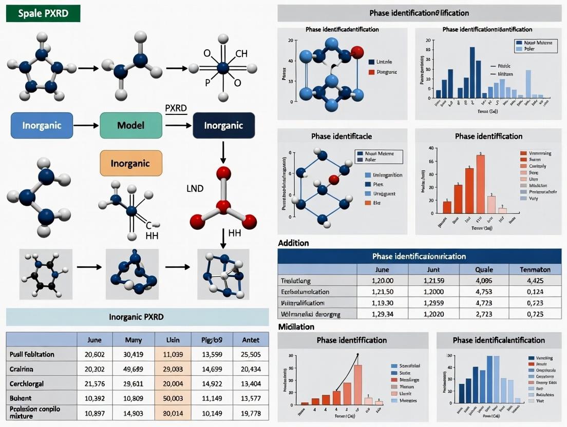
Mastering PXRD Analysis: A Comprehensive Guide to Phase Identification and Quantification in Pharmaceutical Mixtures
This article provides a complete workflow for Powder X-ray Diffraction (PXRD) analysis of organic pharmaceutical mixtures, tailored for researchers and drug development professionals.
Mastering PXRD Analysis: A Comprehensive Guide to Phase Identification and Quantification in Pharmaceutical Mixtures
Abstract
This article provides a complete workflow for Powder X-ray Diffraction (PXRD) analysis of organic pharmaceutical mixtures, tailored for researchers and drug development professionals. It covers the foundational principles of PXRD, explores advanced methodologies for phase identification (including polymorph screening) and quantitative phase analysis (QPA), addresses common troubleshooting and optimization strategies, and validates these approaches through comparative analysis with complementary techniques. The goal is to equip scientists with the knowledge to ensure drug product consistency, stability, and regulatory compliance.
The Essential Guide to PXRD Fundamentals for Pharmaceutical Solids Analysis
Why PXRD is Indispensable for Solid-State Characterization in Drug Development
Within the broader thesis on PXRD phase identification and quantification in organic mixtures, the role of PXRD in drug development is critical. It is the primary tool for determining the crystalline phase, polymorphism, and amorphous content of active pharmaceutical ingredients (APIs) and their formulated products. Solid-state form directly impacts solubility, bioavailability, stability, and manufacturability, making PXRD an indispensable quality control and research technique in pharmaceutical development.
Application Notes
Polymorph Screening and Identification
A single API can exist in multiple crystalline forms (polymorphs), each with distinct physicochemical properties. PXRD provides a fingerprint for each polymorph, enabling rapid identification during screening campaigns.
Table 1: Key PXRD Peaks for Hypothetical API-123 Polymorphs
| Polymorph | 2θ Angles (Cu Kα, °) | Relative Intensity (%) | d-spacing (Å) |
|---|---|---|---|
| Form I | 12.5, 15.2, 18.7, 25.1 | 100, 85, 45, 30 | 7.08, 5.83, 4.74, 3.55 |
| Form II | 10.8, 13.4, 17.0, 20.5 | 100, 60, 90, 25 | 8.19, 6.60, 5.21, 4.33 |
| Form III (Hydrate) | 6.5, 13.0, 19.5, 26.0 | 100, 70, 50, 20 | 13.59, 6.81, 4.55, 3.43 |
Quantification of Crystalline Phases in Mixtures
PXRD can quantify the amount of each polymorph in a mixture, critical for ensuring the consistency of the desired form. The most common method is the peak height/area ratio method or full-pattern Rietveld refinement.
Table 2: Rietveld Refinement Results for a Binary Mixture
| Component | Reference Intensity Ratio (RIR) | Wt.% in Mixture A | Wt.% in Mixture B | Estimated LOD |
|---|---|---|---|---|
| API Form I | 1.00 (standard) | 78.5% ± 0.8% | 22.3% ± 0.5% | ~0.5 wt.% |
| API Form II | 0.92 | 21.5% ± 0.8% | 77.7% ± 0.5% | ~0.5 wt.% |
Detection and Quantification of Amorphous Content
The presence of amorphous material, which exhibits a broad "halo" in the PXRD pattern, can significantly enhance solubility but reduce stability. PXRD can detect and quantify low levels of amorphous content via spike-in calibration curves.
Table 3: Amorphous Content Quantification Data
| Sample ID | Known Amorph. Content (%) | Integrated Amorphous Halo Area (a.u.) | Crystalline Peak Area (a.u.) | Ratio (Halo/Peak) |
|---|---|---|---|---|
| Calib. 0% | 0.0 | 1050 | 85000 | 0.0124 |
| Calib. 5% | 5.0 | 8500 | 81000 | 0.1049 |
| Calib. 10% | 10.0 | 18500 | 76500 | 0.2418 |
| Batch X | Unknown | 12400 | 78800 | 0.1574 |
| Result for Batch X | 4.8% (from calibration curve) | - | - | - |
Formulation Stability and Compatibility
PXRD monitors phase changes in final drug products (tablets, capsules) during stability studies under ICH conditions (e.g., 40°C/75% RH), detecting form conversion, hydrate formation, or API-excipient interactions.
Detailed Experimental Protocols
Protocol 1: Routine Polymorph Identification by PXRD
Objective: To identify the polymorphic form of an unknown API sample. Materials:
- Bruker D8 Advance or equivalent diffractometer with Cu Kα source (λ = 1.5418 Å)
- Zero-background silicon sample holder
- Spatula and glass slide for gentle grinding/tamping Procedure:
- Sample Preparation: Gently grind ~100 mg of powder with a mortar and pestle if necessary to reduce preferred orientation. Fill the cavity of the silicon holder and tamp flat with a glass slide to create a smooth, level surface.
- Instrument Setup: Configure the goniometer with a divergence slit (e.g., 0.5°), LynxEye detector. Use a scan range of 2–40° 2θ.
- Data Collection: Set a step size of 0.02° 2θ and a counting time of 0.5–1 second per step. Start the scan.
- Data Analysis: Import the raw data (.raw, .xrdml) into analysis software (e.g., DIFFRAC.EVA, HighScore Plus). Perform background subtraction and Kα2 stripping. Match the peak list (2θ, d-spacing, intensity) against an internal database of known API forms using search-match algorithms.
- Verification: Compare the full pattern overlay with the reference pattern of the suspected match. Confirm by matching key peak positions and relative intensities.
Protocol 2: Quantitative Phase Analysis (QPA) via the RIR Method
Objective: To determine the weight percentage of two polymorphs in a binary mixture. Materials:
- PXRD as above
- High-purity standards of each polymorph (Forms I and II) Procedure:
- Standard Preparation: Prepare and run PXRD scans for pure Form I and pure Form II using identical instrumental conditions (as in Protocol 1).
- Identify Key Peaks: Select a strong, non-overlapping peak for each form (e.g., Form I peak at 12.5°, Form II peak at 10.8°).
- Calculate RIR: The RIR for Form II relative to Form I (RIRII/I) is calculated as:
RIR*II/I* = (I*II* / I*I*) * (ρ*I* / ρ*II*) * (M*I* / M*II*)where I is the integrated intensity of the chosen peak, ρ is the density, and M is the formula weight (often equal, simplifying the equation). In practice, using a 50:50 wt.% mixture is common for empirical RIR determination. - Analyze Unknown Mixture: Run the unknown mixture sample.
- Quantification: Use the relationship:
W*II* / W*I* = (I*II* / I*I*) * (1 / RIR*II/I*)Solve for weight fractionsW*I*andW*II*knowingW*I* + W*II* = 1.
Protocol 3: Determining Amorphous Content by Standard Addition
Objective: To quantify the percentage of amorphous material in a partially amorphous API batch. Materials:
- Fully crystalline API reference standard
- Fully amorphous API (prepared by quench cooling or spray drying)
- PXRD as above Procedure:
- Prepare Calibration Mixtures: Accurantly weigh and physically mix the crystalline and amorphous standards to create mixtures with 0%, 2%, 5%, 10%, and 20% amorphous content.
- Data Collection: Run PXRD scans for all calibration standards and the unknown sample under identical, highly reproducible conditions.
- Pattern Deconvolution: In analysis software, define two regions: one integrating the area of a major crystalline peak (e.g., 12.5–13.0° 2θ) and one integrating the broad amorphous halo (e.g., 15–25° 2θ).
- Create Calibration Curve: Plot the ratio of Amorphous Halo Area / Crystalline Peak Area against the known amorphous weight percentage for the standards. Perform linear regression.
- Calculate Unknown: Apply the measured area ratio from the unknown sample to the calibration curve equation to determine its amorphous content.
Visualizations
Title: Polymorph Identification by PXRD Workflow
Title: PXRD Quantitative Phase Analysis Pathways
The Scientist's Toolkit: Essential Research Reagents & Materials
Table 4: Key Reagents and Materials for PXRD in Drug Development
| Item | Function & Description |
|---|---|
| Zero-Background Silicon Sample Holder | A single-crystal silicon wafer cut to produce no diffraction peaks, providing a clean background for analyzing small sample quantities. |
| Micro-Mortar and Pestle (Agate or Glass) | For gentle, non-contaminating grinding of samples to reduce particle size and minimize preferred orientation. |
| Standard Reference Materials (e.g., NIST Si 640c) | Certified crystalline materials used to calibrate the PXRD instrument's peak position and line shape, ensuring data accuracy. |
| High-Purity Polymorph Standards | Isolated, definitively characterized pure forms of each API polymorph, essential for creating reference patterns and calibration curves. |
| Amorphous API Standard | A fully amorphous sample of the API, required for developing and validating methods to quantify amorphous content. |
| Internal Standard (e.g., ZnO, Corundum) | A crystalline material added in known proportion to a sample to enable absolute quantification via the spiking method. |
| Specimen Plate for Spray-Dried Samples | A specialized holder for analyzing fluffy, low-density materials like spray-dried dispersions without altering their morphology. |
| Climate-Controlled Sample Stage | A stage that allows PXRD data collection under controlled temperature and humidity, crucial for stability and hydrate/anhydrate transformation studies. |
This application note details the core principles of X-ray powder diffraction (PXRD) within a research thesis focused on the phase identification and quantification of inorganic mixtures, such as active pharmaceutical ingredients (APIs) and excipients in drug development. The interaction of X-rays with crystalline materials yields diffraction patterns that serve as fingerprints for phase identification. Quantitative analysis of peak positions and intensities enables the determination of phase abundance, critical for formulation stability and quality control.
Foundational Principles
Bragg's Law
The condition for constructive interference of X-rays scattered by a crystalline lattice is defined by Bragg's Law: nλ = 2d sinθ Where:
- n is an integer (the order of reflection),
- λ is the wavelength of the incident X-ray beam,
- d is the interplanar spacing between lattice planes, and
- θ is the angle between the incident ray and the scattering lattice planes.
This law forms the basis for understanding peak positions in a diffractogram.
Diffraction Patterns and Peak Intensities
The positions of diffraction peaks (determined by d-spacings via Bragg's Law) are dictated by the unit cell dimensions and symmetry (the crystal structure). The intensities of these peaks are determined by the arrangement and types of atoms within the unit cell. The integrated intensity (I) for a Bragg reflection is proportional to: I ∝ |F|² * LP * A(θ) Where:
- |F|² is the structure factor modulus squared (dependent on atom types and positions),
- LP is the Lorentz-polarization factor (a geometric correction), and
- A(θ) is the absorption factor.
Data Presentation
Table 1: Key Quantitative Relationships in PXRD Analysis
| Principle | Governing Equation/Relation | Primary Determinant | Application in Phase Analysis |
|---|---|---|---|
| Peak Position | nλ = 2d sinθ | Lattice parameters (d-spacing) | Phase identification via pattern matching; lattice parameter refinement. |
| Peak Intensity | I ∝ |F|² | Type, position, and vibration of atoms in the unit cell. | Quantitative phase analysis (QPA); crystal structure solution/refinement. |
| Peak Profile | e.g., Caglioti formula | Instrument optics, crystallite size, microstrain. | Crystallite size/strain analysis via Scherrer equation/Wilson plot. |
| Quantification (RIR) | Wᵢ = (Iᵢ/Kᵢ) / [Σ (Iⱼ/Kⱼ)] | Reference Intensity Ratio (Kᵢ) | Classical QPA method using known intensity ratios. |
| Quantification (Rietveld) | Minimize: Σ wᵢ (yᵢ(obs) - yᵢ(calc))² | Whole-pattern fitting of structural models. | Advanced QPA, extracting structural & microstructural parameters. |
Experimental Protocols
Protocol 1: Sample Preparation for PXRD of Inorganic Mixtures
Objective: To obtain a homogeneous, randomly oriented, flat specimen for reproducible quantitative analysis.
- Grinding: Use an agate mortar and pestle to gently grind the powdered mixture to a consistent particle size (~1-10 µm). Avoid excessive grinding to prevent induced strain or phase transformation.
- Loading: For a standard flat-plate sample holder, fill the cavity with the powdered sample.
- Packing & Leveling: Use a glass slide or a razor blade to press and smooth the surface, ensuring it is flush with the holder's surface to minimize sample displacement error.
- Mounting: Place the sample holder securely on the diffractometer stage.
Protocol 2: Data Collection for Phase Identification & QPA
Objective: To acquire high-quality diffraction data suitable for both qualitative identification and quantitative refinement.
- Instrument Setup: Use a Bragg-Brentano geometry diffractometer with a Cu Kα (λ=1.5418 Å) X-ray source. Configure a divergence slit (e.g., 0.5°), anti-scatter slit, and a Ni filter or energy-dispersive detector to remove Kβ radiation.
- Scan Parameters: Set a continuous scan range from 3° to 50° (2θ). Use a step size of 0.013° and a counting time of 50-100 seconds per step. Ensure the total scan time is sufficient for good counting statistics, critical for QPA.
- Data Collection: Initiate the scan. Monitor the live pattern for signs of preferred orientation (atypical peak intensity ratios).
- Data Export: Save the raw data as a .xy or similar file containing 2θ and intensity pairs.
Protocol 3: Reference Intensity Ratio (RIR) Method for Quantification
Objective: To determine the weight fraction of crystalline phases in a mixture using known intensity references.
- Create Standard Mixtures: Prepare known mixtures of the phase of interest (A) with an internal standard (e.g., corundum, Al₂O₃). Ensure homogeneity.
- Data Acquisition: Run PXRD on each standard mixture using Protocol 2.
- Measure Intensities: For each pattern, measure the integrated intensity of a primary peak for phase A (IA) and the primary peak for the standard (IS).
- Determine K Value: Plot the known weight fraction ratio (WA/WS) against the measured intensity ratio (IA/IS). The slope of the linear fit is the RIR value, K_A.
- Analyze Unknown: Mix the unknown sample with the same standard. Measure IA and IS, and calculate the weight fraction: WA = (IA / KA) / [ (IA / KA) + (IS / K_S) + ... ].
Mandatory Visualization
PXRD Phase Analysis Workflow
The Scientist's Toolkit
Table 2: Essential Research Reagent Solutions & Materials for PXRD
| Item | Function/Description |
|---|---|
| Agate Mortar & Pestle | For gentle, contamination-free grinding of samples to an optimal, uniform particle size. |
| Flat-Plate Sample Holder | A metal or glass plate with a recessed cavity to contain the powdered specimen, providing a flat surface for diffraction. |
| Glass Slide or Razor Blade | For packing and leveling the powder in the sample holder to ensure a flat surface and minimize sample displacement. |
| Internal Standard (e.g., Corundum - α-Al₂O₃) | A well-crystalline, inert material of known phase content mixed with the sample for quantitative analysis via the Reference Intensity Ratio (RIR) method. |
| NIST/SRM Standard (e.g., SRM 674b) | Certified reference material used for instrument performance calibration and check of line position and shape. |
| Zero-Background Holder (e.g., Silicon wafer) | A single crystal slice cut off-axis, used to hold micro-samples, providing negligible background scattering. |
| Capillary Tube (Glass/Quartz) | For mounting samples that are air-sensitive or require transmission geometry measurement. |
| Mylar Film or Kapton Tape | Thin, low-scattering polymer films used to cover and contain samples that may be volatile or require containment. |
Introduction Within the broader research on PXRD phase identification and quantification of inorganic mixtures, the selection of the primary reference database is a critical methodological decision. Two principal databases are employed: the Cambridge Structural Database (CSD) for single-crystal derived molecular structures and the ICDD PDF-4+ for comprehensive powder diffraction data. This application note details their distinct roles, access protocols, and integration into a coherent analytical workflow for researchers and pharmaceutical development professionals.
1. Database Overview and Comparative Analysis
Table 1: Core Comparison of CSD and ICDD PDF-4+ Databases
| Feature | Cambridge Structural Database (CSD) | ICDD PDF-4+ |
|---|---|---|
| Primary Content | Experimentally determined 3D organic and metal-organic crystal structures from single-crystal XRD. | Reference powder diffraction patterns (experimental & calculated) for inorganic, organic, and pharmaceutical phases. |
| Key Source | Single-crystal X-ray diffraction (SCXRD) experiments. | Historical powder data, calculated from crystal structures (e.g., from CSD), and industrial contributions. |
| Primary Use Case | Understanding molecular conformation, intermolecular interactions (hydrogen bonds, π-stacking), and deriving crystal structure for pattern calculation. | Direct phase identification via pattern matching (d-spacing, intensity), quantitative analysis, and purity checking. |
| Search Parameters | Chemical connectivity, molecular formula, cell parameters, author, R-factor. | d-spacings, I/Icor values, chemical name, formula, physical properties. |
| Quantitative Data Output | Geometric parameters (bond lengths, angles, torsion angles), interaction distances. | Reference Intensity Ratio (RIR) values, necessary for quantitative phase analysis (QPA) methods like Rietveld. |
| Update Frequency | Annual (≈1.2 million entries as of 2024). | Annual (≈450,000 entries in PDF-4+ 2024). |
2. Experimental Protocols
Protocol 2.1: Generating a Calculated PXRD Pattern from a CSD Entry for a Novel Pharmaceutical Polymorph.
- Access & Retrieval: Log in to the CSD Web API or use the CSD software suite (Mercury or ConQuest). Search for the target structure using its Refcode (e.g., ABCOCB01).
- Data Export: Download the Crystallographic Information File (.cif) for the validated entry.
- Pattern Simulation: Import the .cif file into a diffraction pattern simulation software (e.g., Mercury, TOPAS, or DASH).
- Set Experimental Parameters: Define the instrumental parameters to match your laboratory PXRD setup: X-ray wavelength (Cu Kα, λ=1.5406 Å), divergence slit size, scan range (e.g., 2-40° 2θ), and step size.
- Refinement & Output: The software calculates diffraction peak positions (from unit cell) and relative intensities (from atomic coordinates and space group). Apply a peak profile function (e.g., Pseudo-Voigt) and instrumental broadening. Export the calculated pattern as a .xy or .txt file for use as a reference in your PXRD analysis software.
Protocol 2.2: Performing Phase Identification of an Inorganic Mixture Using ICDD PDF-4+.
- Sample Preparation: Grind the unknown mixture to a homogeneous fine powder (<10 µm) to minimize preferred orientation. Load into a standard flat-plate or capillary sample holder.
- Data Collection: Acquire the PXRD pattern of the unknown sample using your diffractometer. Apply necessary corrections (background subtraction, Kα2 stripping).
- Data Import & Search Preparation: Import the corrected pattern into your analysis software (e.g., JADE, HighScore, or ICDD’s own software). Define the search subset (e.g., inorganic, minerals, pharmaceuticals).
- Search-Match: Execute a search-match using the top N strongest peaks (e.g., 20-40 peaks). The software (e.g., ICDD’s PDF-4+ Search/Match) compares the list of d-spacings and intensities against the database, generating a list of candidate phases with a figure-of-merit (FOM) score.
- Phase Verification: Visually compare the full calculated pattern of the top candidate(s) from the database against the experimental pattern. Confirm the presence of all major peaks and check for absent peaks indicating impurities.
- Quantitative Analysis (if required): For confirmed phases, retrieve the Reference Intensity Ratio (RIR) value from the PDF-4+ entry. Use this in subsequent Rietveld refinement or traditional internal standard methods to determine weight percentages in the mixture.
3. Visualized Workflows
Decision Workflow for CSD vs. PDF-4+ Use
Database Roles in PXRD Analysis
4. The Scientist's Toolkit: Essential Research Reagents & Materials
Table 2: Key Materials for PXRD-Based Phase Analysis
| Item | Function / Purpose |
|---|---|
| Silicon (NIST SRM 640e) | External standard for precise instrument alignment and correction of zero-point error. |
| Alumina (α-Al₂O₃, NIST SRM 676a) | Certified reference material for determining instrumental line broadening and RIR calibration. |
| Lanthanum Hexaboride (LaB₆, NIST SRM 660c) | Line position and profile shape standard for high-resolution instrument calibration. |
| Internal Standard (e.g., ZnO, MgO, CaF₂) | Added in known proportion to unknown sample for classical quantitative analysis (e.g., matrix-flushing method). |
| Zero-Background Sample Holder (e.g., Silicon single crystal cut off-axis) | Holds powdered sample; provides a flat, non-diffracting background to minimize substrate signals. |
| McCrone Micronizing Mill | For reproducible particle size reduction (<10 µm) to minimize micro-absorption and preferred orientation effects. |
| CSD Software Suite (Mercury, ConQuest) | For visualization, analysis of crystal structures from CSD, and simulating PXRD patterns. |
| ICDD PDF-4+ Database & Search/Match Software | The primary reference library and analytical engine for phase identification and retrieval of RIR values. |
| Rietveld Refinement Software (e.g., TOPAS, GSAS-II) | For full-pattern fitting to perform accurate quantitative phase analysis and extract structural details. |
Application Notes
Within the domain of PXRD-based phase identification and quantification, the analytical approach diverges significantly when comparing organic molecular solids to classical inorganic systems. This distinction is central to advancing pharmaceutical and materials research, where organic active pharmaceutical ingredients (APIs), cocrystals, salts, and polymorphs present unique challenges not typically encountered with inorganic minerals and ceramics.
The core complexity arises from the flexible, low-symmetry nature of organic molecules, leading to greater structural diversity (polymorphism, solvatomorphism) and subtle peak profile differences. Lattice energies and scattering factors are considerably lower than in dense inorganic lattices, impacting signal-to-noise and quantification limits. Furthermore, organic phase mixtures often involve phases with very similar lattice parameters and peak overlap, exacerbated by preferred orientation effects in platonic or needle-like crystallites.
Table 1: Comparative Challenges in PXRD Analysis of Organic vs. Inorganic Mixtures
| Aspect | Organic Molecular Mixtures (e.g., APIs) | Inorganic Systems (e.g., Minerals, Ceramics) |
|---|---|---|
| Primary Scattering Source | Light atoms (C, H, N, O); weak scattering. | Heavy atoms (Metals, Si); strong scattering. |
| Crystal Symmetry | Often low (triclinic, monoclinic); many peaks. | Often high (cubic, hexagonal); fewer peaks. |
| Unit Cell Variability | High (polymorphs, solvates, flexible molecules). | Generally low (fixed ionic radii, rigid lattices). |
| Preferred Orientation | Severe, due to anisotropic crystal habits. | Moderate to low, depending on morphology. |
| Peak Profile | Broader due to smaller crystallites & microstrain. | Generally sharper. |
| Quantification Basis | Relies on complex whole-pattern fitting (Rietveld). | Can sometimes use reference intensity ratios (RIR). |
| Stability Under Beam | Risk of phase change, dehydration, or degradation. | Typically stable. |
Experimental Protocols
Protocol 1: Sample Preparation for Organic Phase Mixtures to Mitigate Preferred Orientation
- Objective: To obtain a statistically representative powder diffraction pattern free from dominant orientation effects.
- Materials: Micronized organic powder mixture, amorphous silica diluent (optional), mortar and pestle or McCrone micronizing mill, low-background silicon sample holder.
- Procedure:
- Gently blend the organic powder mixture with an equal volume of amorphous silica (if dilution is acceptable for detection limits) using a mortar and pestle. Do not apply excessive pressure.
- Alternatively, use a McCrone micronizing mill with cyclohexane or ethanol as a grinding aid for 5 minutes to ensure fine, random particle size.
- Allow the slurry to air-dry completely.
- Side-loading Technique: Tilt the silicon sample holder. Gently sprinkle the powder into the cavity using a spatula. Tap the holder's side lightly to settle the powder. Avoid any pressing or smoothing of the surface.
- Mount the holder in the diffractometer.
Protocol 2: Data Collection Strategy for Low-Scattering Organic Mixtures
- Objective: To maximize the signal-to-noise ratio for accurate phase identification and quantification.
- Materials: High-resolution X-ray diffractometer (Bragg-Brentano or Debye-Scherrer geometry), Cu Kα source (λ = 1.5418 Å), incident-beam monochromator or mirror, solid-state detector.
- Procedure:
- Use a long fine-focus X-ray tube (e.g., 12 kW) to increase incident beam intensity.
- Configure the optics with a primary soller slit (0.04 rad), a fixed or variable divergence slit (e.g., 0.5°), and a secondary antiscatter slit.
- Employ a high-speed linear or energy-dispersive detector (e.g., LynxEye, Sol-XE).
- Set the scan range from 2° to 40° 2θ for initial screening; extend to 60° for quantification.
- Use a slow continuous scan speed (e.g., 0.5° 2θ/min) or a fast step scan with long counting time per step (e.g., 0.01° step, 3-5 sec/step).
- Perform under ambient conditions unless monitoring phase stability requires a controlled atmosphere (e.g., N₂ flow).
Protocol 3: Quantitative Phase Analysis (QPA) via Rietveld Refinement for Organic Mixtures
- Objective: To determine the weight fraction of each crystalline phase in a multi-phase organic mixture.
- Materials: High-quality PXRD pattern, known crystal structures (CIF files) for all suspected phases, Rietveld refinement software (e.g., TOPAS, GSAS-II).
- Procedure:
- Import the experimental pattern and CIF files for all reference phases.
- Create a multi-phase model. Scale factors for each phase are the primary refined variables for quantification.
- Refine common background parameters (Chebyshev polynomial).
- Refine unit cell parameters for each phase to account for potential slight shifts.
- Refine a shared peak profile function (e.g., Thompson-Cox-Hastings pseudo-Voigt) with parameters for crystallite size and microstrain.
- Include a March-Dollase or spherical harmonic preferred orientation correction for dominant phases.
- Refine sequentially, monitoring the R-weighted pattern (Rwp) and goodness-of-fit (GoF).
- The final weight percentage (Wᵢ) of phase i is calculated by the software from the refined scale factor (Sᵢ), the phase's mass absorption coefficient (μ*), and its unit cell volume (Vᵢ): Wᵢ = (Sᵢ * Zᵢ * Mᵢ * Vᵢ) / Σ(Sⱼ * Zⱼ * Mⱼ * Vⱼ), where Z is molecules per unit cell and M is molecular mass.
Visualization
Diagram Title: PXRD Workflow for Organic Mixture QPA
Diagram Title: Root Causes of Organic Mixture QPA Difficulty
The Scientist's Toolkit: Key Research Reagent Solutions
Table 2: Essential Materials for PXRD Analysis of Organic Phase Mixtures
| Item | Function & Rationale |
|---|---|
| Low-Background Silicon Sample Holder | Provides a flat, zero-backholder for side-loading samples to minimize preferred orientation. |
| Amorphous Silica (SiO₂) Diluent | An isotropic, non-interfering standard used to dilute samples for improved particle statistics without contributing sharp diffraction peaks. |
| McCrone Micronizing Mill | Uses gentle grinding with a liquid to produce a fine, random powder, reducing crystallite size and orientation effects. |
| NIST/SRM Standard Reference Materials (e.g., LaB₆, CeO₂) | Used for precise instrumental profile calibration and resolution checks, critical for resolving overlapping organic peaks. |
| Polymorph/Cocrystal Screen Library | A curated set of reference compounds for spiking or comparative analysis to identify unknown phases in a mixture. |
| Temperature/Humidity Stage | Controls the sample environment in situ to monitor phase stability, hydrate formation, or desolvation during analysis. |
| High-Sensitivity Solid-State Detector (e.g., LynxEye, D/teX Ultra) | Dramatically increases count rates and signal-to-noise for weakly scattering organic materials, reducing data collection time. |
Within the framework of a thesis on Powder X-ray Diffraction (PXRD) phase identification and quantification of inorganic mixtures, the criticality of pre-analysis sample preparation cannot be overstated. The accuracy and reproducibility of quantitative phase analysis (QPA) results are fundamentally dependent on the quality of the powder specimen. Inadequate preparation introduces biases such as micro-absorption, particle statistics errors, and preferred orientation, which can severely distort intensity data, leading to misidentification and inaccurate quantification. These protocols detail the systematic steps required to transform a raw sample into a representative, isotropic powder specimen suitable for high-fidelity PXRD analysis in pharmaceutical and materials research.
Foundational Principles & Key Quantitative Parameters
Table 1: Key Quantitative Parameters for Optimal PXRD Sample Preparation
| Parameter | Optimal Range / Target | Rationale & Impact on Analysis |
|---|---|---|
| Particle Size | < 10 µm (ideal: 1-5 µm) | Minimizes micro-absorption effects and ensures a sufficient number of particles in the beam for good particle statistics. |
| Crystallite Size (Domain Size) | > 100 nm (for sharp peaks) | Prevents excessive peak broadening which complicates phase identification and QPA. Grinding must balance reducing particle size without inducing excessive amorphization or lattice strain. |
| Preferred Orientation Index (POI) / R-Parameter | POI ~1.0; Rp < 0.5 | A POI of 1.0 indicates random orientation. Higher values signify texture. Mitigation strategies aim to minimize this. |
| Sample Loading Density | Consistent, reproducible packing | Variations affect diffracted intensity and background. Side-loading is preferred for flat-plate mounts. |
| Specimen Transparency | Minimized for Bragg-Brentano geometry | Thick, infinitely opaque samples are ideal to avoid depth-related peak shifts and broadening. |
Detailed Experimental Protocols
Protocol 3.1: Dry Grinding for Brittle Inorganic Mixtures
Objective: To reduce particle size without inducing phase transformations or significant amorphization. Materials: Agate or micronized zirconia mortar and pestle, sieves (optional, ≤ 50 µm mesh). Procedure:
- Place a representative sub-sample (typically 50-200 mg) in the mortar.
- Apply moderate pressure with the pestle in a circular, grinding motion for 1-2 minutes.
- Scrape the powder from the sides and repeat the process. Total grinding time should rarely exceed 5-10 minutes to limit lattice strain.
- For stringent size control, sieve the powder through an appropriate mesh. The sub-50 µm fraction is commonly collected.
- Critical Step: Allow the sample to rest for ~15 minutes before loading to dissipate electrostatic charges.
Protocol 3.2: Wet Grinding (Slurry Milling) for Cohesive or Sensitive Materials
Objective: To achieve fine, homogeneous powders while minimizing heat and plastic deformation that can cause preferred orientation. Materials: McCrone Micronizing Mill (agate or zirconia grinding elements), ethanol or cyclohexane (non-reactive dispersing liquid). Procedure:
- Place ~100 mg of sample and 2-3 mL of dispersing liquid into a 10 mL agate grinding chamber with seven agate grinding rods.
- Assemble and run the mill for 5-15 minutes. The optimal time is determined empirically to reach the target size without amorphization.
- Carefully transfer the slurry to a watch glass or Petri dish.
- Dry slowly at ambient temperature or in a low-temperature oven (< 60°C) to prevent recrystallization or particle aggregation.
Protocol 3.3: Side-Loading for Preferred Orientation Mitigation
Objective: To prepare a specimen with randomly oriented crystallites for flat-plate Bragg-Brentano geometry. Materials: Top-loaded sample holder with a cavity, frosted glass slide, razor blade. Procedure:
- Place the sample holder cavity-side-up on a stable surface.
- Gently back-fill the cavity with the prepared powder using a spatula.
- Place a clean, frosted glass slide over the cavity.
- Holding the slide firmly against the holder, rotate it 90° to the horizontal plane (side-loading). Tap the side gently to settle the powder.
- Use a razor blade to carefully scrape excess powder flush with the holder surface, leaving a smooth, in-situ packed surface that was never pressed from the top.
Protocol 3.4: Spray-Drying for Ultimate Randomization
Objective: To create perfect spherical agglomerates of primary particles, eliminating texture. Materials: Laboratory-scale spray dryer, suitable solvent (e.g., water, ethanol). Procedure:
- Prepare a stable, dilute suspension (~1-5 wt%) of the finely ground powder in a solvent.
- Feed the suspension into the spray dryer under optimized conditions (inlet temperature, feed rate, atomization pressure).
- Collect the resulting free-flowing, spherical agglomerates. These particles present all crystal faces equally to the X-ray beam.
Workflow Visualization
Title: Workflow for PXRD Sample Prep & Orientation Mitigation
The Scientist's Toolkit: Essential Research Reagent Solutions
Table 2: Essential Materials for PXRD Sample Preparation
| Item | Function & Rationale |
|---|---|
| Agate Mortar & Pestle | Hard, dense, and chemically inert. Minimizes sample contamination during dry grinding. Its low porosity prevents cross-contamination. |
| Micronized Zirconia Mill | Alternative to agate with higher hardness and wear resistance. Suitable for extremely hard materials. |
| McCrone Micronizing Mill | Standardized wet-grinding device using correlated motion of grinding elements in a liquid to achieve uniform sub-10 µm particles with minimal strain. |
| Zero-Background Plate (e.g., Si single crystal) | Sample holder substrate that produces no diffraction peaks, providing a clean background for minute sample quantities. |
| Side-Loading Sample Holder | Aluminum or polymer holder with a cavity designed for the side-loading technique to reduce preferred orientation. |
| Non-Reactive Dispersing Liquid (Cyclohexane, Ethanol) | Used in wet grinding and spray-drying. Low surface tension, volatile, and does not react with or solvate sample components. |
| Micro-Spatula & Razor Blades | For precise, contamination-free handling and flattening of powder specimens. |
| Standard Reference Material (e.g., NIST 675a, Corundum) | Certified powder used to verify instrument alignment, quantify preparation errors, and calibrate QPA methods. |
Step-by-Step PXRD Workflow: From Polymorph ID to Quantitative Analysis (QPA)
Application Notes on Phase Identification Strategies
Within the broader thesis on PXRD phase identification and quantification in inorganic and pharmaceutical mixtures, three core computational strategies form the methodological foundation. These strategies are critical for resolving complex multi-phase systems commonly encountered in materials science and drug development, where polymorphic purity, salt forms, and amorphous content are paramount.
Pattern Matching is a qualitative approach where an acquired diffraction pattern is compared to a database of reference patterns (e.g., ICDD PDF-4+, COD). The degree of visual or numerical correlation determines a match. Its primary strength is rapid screening, but it suffers when faced with peak overlap, preferred orientation, or novel phases not in the database.
Indexing is the process of determining the unit cell parameters (a, b, c, α, β, γ) from a diffraction pattern of a single-phase or multi-phase mixture with separable peaks. It is a critical step towards structure solution. In mixtures, successful indexing of major component peaks can facilitate the subsequent identification of minor phases.
Whole Pattern Fitting (Pawley/Le Bail) and Rietveld Refinement represent the most quantitative strategies. These methods fit the entire measured diffraction profile using crystal structure models. For quantification in mixtures, the scale factors from Rietveld refinement are used to determine weight fractions, making it the gold standard for quantitative phase analysis (QPA).
Table 1: Comparison of Phase Identification Strategies
| Strategy | Primary Use | Key Strength | Key Limitation | Typical Accuracy (QPA) |
|---|---|---|---|---|
| Pattern Matching | Qualitative identification | Fast, simple, excellent for known phases. | Fails for novel phases; poor with peak overlap. | Not quantitative |
| Indexing | Unit cell determination | Essential for structure solution of new phases. | Requires high-quality, well-separated peaks. | Not directly quantitative |
| Whole Pattern Fitting (Pawley) | Extracting intensities for structure solution | Models pattern without a structural model. | Does not provide atomic coordinates. | Semi-quantitative |
| Rietveld Refinement | Quantitative analysis & structure refinement | Highly accurate quantification; uses full structural models. | Requires good structural models; can be complex. | ± 1-2 wt% (ideal) |
Detailed Experimental Protocols
Protocol 2.1: Standardized PXRD Data Acquisition for Mixture Analysis
Objective: To collect high-quality powder X-ray diffraction data suitable for pattern matching, indexing, and whole pattern fitting of inorganic or pharmaceutical mixtures.
Materials: Powder sample (<10 µm particle size recommended), zero-background holder (e.g., silicon wafer), flat plate holder, or capillary tube.
Procedure:
- Sample Preparation: Gently grind the sample to reduce preferred orientation. For loose powder, front-load into a cavity holder. For minimal orientation, pack into a capillary or side-load onto a silicon wafer.
- Instrument Setup: Use a Bragg-Brentano or transmission geometry diffractometer (Cu Kα radiation, λ=1.5418 Å typical). Configure with a divergence slit (e.g., 0.5°), anti-scatter slit, and a fast detector (e.g., Pixcel or strip detector).
- Scan Parameters: Set the angular range (e.g., 2–40° 2θ for pharmaceuticals, 5–80° for inorganics). Use a step size of ≤ 0.02° 2θ and a counting time of 1–2 seconds per step to ensure adequate counting statistics for refinement.
- Data Collection: Mount the sample and initiate the scan. Monitor the intensity of major peaks to detect potential sample degradation (e.g., dehydration).
- Data Export: Save the raw data as a *.xy or *.asc file containing two columns: 2θ and Intensity.
Protocol 2.2: Sequential Phase Identification & Quantification Workflow
Objective: To systematically identify and quantify all crystalline phases in a multi-component mixture.
Software Requirements: ICDD PDF-4+ database, indexing software (e.g., TOPAS), whole pattern fitting software (e.g., HighScore Plus, TOPAS, GSAS-II).
Procedure:
- Pattern Matching (Search/Match):
- Import the raw data into search/match software (e.g., HighScore Plus with PDF-4+).
- Apply background subtraction and Kα₂ stripping.
- Execute a search using the full pattern. Review the list of suggested matches, prioritizing figures of merit (FOM) like FN.
- Tentatively identify all major phases.
Indexing of Unmatched Peaks:
- Subtract the calculated pattern of the identified phases from the measured pattern.
- Isolate residual peaks belonging to unknown phases.
- Use an auto-indexing program (e.g., TOPAS-Academic) on the residual pattern to determine possible unit cells.
- Compare candidate unit cells to structural databases (e.g., COD, ICSD).
Whole Pattern Fitting & Rietveld Refinement:
- Construct a preliminary refinement model in Rietveld software using the CIF files for all identified phases.
- Include parameters for: scale factors (for quantification), unit cell parameters, background (e.g., Chebyshev polynomial), peak shape (e.g., pseudo-Voigt), and specimen displacement.
- Refine parameters sequentially: scale, background, lattice, peak shape, etc.
- For quantitative analysis, include an internal standard (e.g., NIST 676a, corundum) if absolute accuracy is required, or use the scale-factor ZMV method for known structures.
- The weight fraction Wᵢ of phase i is calculated from its refined scale factor Sᵢ and the mass absorption coefficient of the mixture.
Table 2: Key Refinement Parameters in Rietveld Analysis
| Parameter | Symbol (Typical) | Purpose | Refinement Order |
|---|---|---|---|
| Scale Factor | Sᵢ | Determines phase abundance. | 1 |
| Lattice Parameters | a, b, c, α, β, γ | Accounts for strain/doping. | 2 |
| Background | B (Chebyshev coeff.) | Models amorphous/scattering. | 2 |
| Peak Width | U, V, W (Caglioti) | Models crystallite size/strain. | 3 |
| Preferred Orientation | March-Dollase | Corrects for texture. | 4 |
Visualizations
Title: PXRD Phase Identification and Quantification Workflow
Title: Rietveld Refinement Feedback Loop
The Scientist's Toolkit: Research Reagent Solutions
Table 3: Essential Materials & Software for PXRD Phase Analysis
| Item Name | Category | Function/Brief Explanation |
|---|---|---|
| NIST SRM 674b | Calibration Standard | Certified reference material for intensity and position calibration of the diffractometer. |
| Corundum (α-Al₂O₃) NIST 676a | Quantitative Internal Standard | Known purity and crystallinity; added to samples for absolute quantitative phase analysis (QPA). |
| Zero-Background Silicon Wafer | Sample Holder | Single-crystal silicon cut off-axis to eliminate diffraction peaks, providing a low-background substrate. |
| ICDD PDF-4+ Database | Software/Database | Comprehensive repository of reference powder diffraction patterns for search/match identification. |
| TOPAS-Academic/HighScore Plus | Analysis Software | Implements whole pattern fitting (Pawley, Le Bail) and Rietveld refinement for QPA. |
| Capillary Tube (0.5-1.0 mm) | Sample Holder (Transmission) | Minimizes preferred orientation and is essential for studying air-sensitive samples. |
| LaB₆ (NIST SRM 660c) | Line Profile Standard | Used to determine the instrumental broadening function for crystallite size/strain analysis. |
| Micro-Aglate Mortar & Pestle | Sample Prep Tool | For gentle, controlled grinding to reduce particle size without inducing phase changes. |
Within the broader thesis on PXRD phase identification and quantification in organic mixtures, handling complex solid-form mixtures presents a paramount challenge. The presence of Active Pharmaceutical Ingredients (APIs) with diverse solid forms (salts, co-crystals, hydrates) alongside numerous excipients creates a convoluted analytical landscape. Accurate phase identification and quantification via Powder X-ray Diffraction (PXRD) is critical for ensuring drug product stability, efficacy, and regulatory compliance. This document provides detailed application notes and protocols for the systematic analysis of such complex multicomponent systems.
Application Notes: Key Challenges & Strategies
Phase Complexity in Solid Dosage Forms
A modern solid oral dosage form is a complex mixture where the API may exist in multiple solid forms simultaneously. Common components include:
- API: May exist as a free form, salt, co-crystal, or hydrate.
- Excipients: Fillers (e.g., microcrystalline cellulose), disintegrants (e.g., crospovidone), lubricants (e.g., magnesium stearate), and glidants.
- Process-Induced Transformations: Hydration/dehydration, amorphization, or polymorphic conversion during manufacturing (e.g., wet granulation, compaction).
The primary analytical challenge is the significant peak overlap in PXRD patterns from these components, necessitating advanced data collection and analysis strategies.
Quantitative Analysis Considerations
Quantification relies on the relationship between the weight fraction of a phase and the intensity of its unique diffraction peaks. Key considerations are:
- Amorphous Content: The presence of amorphous API or excipients (e.g., hypromellose) complicates quantification as they contribute to a diffuse scattering background.
- Microabsorption: Differences in the mass absorption coefficients of components can lead to systematic errors in quantification.
- Preferred Orientation: Plate- or needle-like crystals can cause non-uniform diffraction intensity, skewing results.
Table 1: Common Solid Forms of APIs and Their PXRD Characteristics
| Solid Form | Definition | Key PXRD Indicator | Stability Consideration |
|---|---|---|---|
| Polymorph | Same chemical composition, different crystal packing. | Distinct peak positions & intensities. | Metastable forms may convert. |
| Hydrate/Solvate | Crystal incorporates solvent/water molecules. | Characteristic low-angle peaks; shifts vs. anhydrate. | Desolvation upon drying or heating. |
| Salt | Ionic complex of API and counterion. | Entirely new diffraction pattern vs. free acid/base. | Hygroscopicity may vary. |
| Co-crystal | Neutral multi-component complex with a coformer. | Unique pattern distinct from individual components. | Dissociation potential. |
| Amorphous | Lack of long-range molecular order. | Broad "halo" instead of sharp peaks. | Thermodynamically unstable. |
Experimental Protocols
Protocol: Sample Preparation for Complex Mixture Analysis
Objective: To obtain a homogeneous, representative, and non-oriented powder sample for high-quality PXRD data. Materials: See Scientist's Toolkit. Procedure:
- Grinding/Milling: Gently grind a representative sample (50-100 mg) using an agate mortar and pestle or a vibratory mill for 60-90 seconds. Caution: Avoid excessive grinding to prevent phase transformation or amorphization.
- Sieving: Pass the ground powder through a ≤100 µm sieve to ensure uniform particle size and reduce microabsorption error.
- Sample Loading: For a Bragg-Brentano diffractometer: a. Back-Loading Method: Fill the powder into a silicon zero-background holder or a standard cavity mount. Use a glass slide to create a flat, randomly oriented surface without applying significant pressure. b. Front-Loading Method: Place the holder on a flat surface, fill with powder, and use a razor blade to scrape excess material off the surface to create a flat layer.
- Smooth the surface. Ensure it is flush with the holder's rim.
Protocol: PXRD Data Collection for Mixture Analysis
Objective: To collect high-resolution, high signal-to-noise data suitable for both identification and Rietveld refinement. Instrument: Laboratory X-ray diffractometer (Cu Kα radiation, λ = 1.5418 Å). Parameters:
- Voltage/Current: 40 kV, 40 mA.
- Divergence Slits: Fixed or auto-variable to maintain constant illuminated area.
- Scan Range: 3 - 40° 2θ (critical for detecting hydrates/excipient peaks).
- Step Size: 0.01 - 0.02° 2θ.
- Time per Step: 1-2 seconds (total scan time ~30-90 min).
- Spin: Enable sample rotation (15-30 rpm) to improve particle statistics and reduce orientation effects.
Protocol: Phase Identification via Database Matching
Objective: To identify all crystalline phases present in the mixture. Software: ICDD PDF-4+, CSD, or in-house database. Procedure:
- Pre-process Data: Apply background subtraction and Kα2 stripping to the raw pattern.
- Search/Match: Use software to perform a whole-pattern search. Input all possible components (API forms, excipients).
- Iterative Subtraction: a. Identify the major phase with the most distinct non-overlapping peaks. b. Subtract its reference pattern (scaled appropriately) from the experimental pattern. c. Analyze the residual pattern for the next most identifiable phase. d. Repeat until no identifiable crystalline peaks remain.
- Validation: Confirm identified phases by comparing peak positions of minor components with their reference patterns in the residual plot.
Protocol: Quantitative Phase Analysis via Rietveld Refinement
Objective: To determine the weight percentage (wt%) of each crystalline phase and estimate amorphous content. Software: TOPAS, GSAS-II, or similar. Procedure:
- Model Building: Input crystal structure files (.cif) for all identified crystalline phases. Include a model for amorphous content (e.g., a broad hump function).
- Initial Parameters: Set scale factors for each phase. Refine background (Chebyshev polynomial), unit cell parameters (constrained), and peak profile (e.g., fundamental parameters approach).
- Sequential Refinement: a. Refine scale factors and background. b. Refine lattice parameters for each phase sequentially. c. Refine profile parameters. d. Refine preferred orientation (e.g., March-Dollase function) if needed for plate-like excipients (e.g., Mg stearate).
- Microabsorption Correction: Apply Brindley correction if the mass absorption coefficients of components differ significantly (e.g., API HCl salt vs. lactose).
- Final Calculation: The refined scale factor for each phase is used to calculate its weight fraction, normalized to 100%.
Table 2: Typical Rietveld Refinement Results for a Model Tablet Formulation
| Component | Crystal Form | Refined Wt% | Reported Uncertainty (±) | Key Diagnostic (Rwp) |
|---|---|---|---|---|
| API | Monohydrate | 15.2 | 0.3 | 4.87% |
| Microcrystalline Cellulose | Cellulose Iβ | 58.1 | 0.5 | |
| Lactose Monohydrate | α-Lactose monohydrate | 20.5 | 0.4 | |
| Magnesium Stearate | Di-hydrate (plate-like) | 1.5 | 0.2 | |
| Amorphous Content | (Broad Scattering) | 4.7 | 0.8 |
Visualization of Workflow & Relationships
Title: PXRD Analysis Workflow for Complex Mixtures
Title: API Solid Form Relationships
The Scientist's Toolkit: Essential Research Reagents & Materials
Table 3: Key Materials for PXRD Analysis of Complex Mixtures
| Item | Function & Rationale |
|---|---|
| Agate Mortar & Pestle | Provides gentle, contamination-free grinding to achieve homogeneous particle size without inducing phase changes. |
| Silicon Zero-Background Holder | Minimizes background scattering, crucial for detecting low-abundance phases and low-angle peaks from hydrates. |
| Micro-Mesh Sieves (≤100 µm) | Ensures consistent particle size distribution, reducing errors from microabsorption and preferred orientation. |
| Internal Standard (e.g., NIST 675a Corundum) | Spiked into samples to calibrate instrument alignment and correct for systematic errors in quantification. |
| Reference Materials (Pure Phases) | High-purity samples of each API solid form and excipient for creating reference patterns and calibration curves. |
| Hygroscopic Sample Container | For handling hydrates or moisture-sensitive salts to prevent phase changes during preparation and analysis. |
| Rietveld Refinement Software (TOPAS/GSAS-II) | Essential for deconvoluting overlapping peaks and quantifying multiple phases in complex mixtures. |
| High-Resolution PXRD Instrument | Equipped with a long working distance diffracted beam monochromator and sample spinner for high-quality data. |
Within the broader thesis on PXRD Phase Identification and Quantification in Inorganic Mixtures, the Rietveld method stands as the definitive, full-pattern technique for quantitative phase analysis (QPA). Unlike traditional single-peak methods, it leverages the entire digitized X-ray powder diffraction (PXRD) pattern, fitting a calculated profile to the observed data via least-squares refinement. This application note details the theoretical underpinnings, modern protocols, and critical considerations for applying the Rietveld method to inorganic mixtures, particularly relevant to pharmaceutical development where polymorph quantification, salt forms, and amorphous content are critical quality attributes.
Foundational Principles and Mathematical Framework
The Rietveld method minimizes the residual ( Sy ) between the observed ( y{i}(obs) ) and calculated ( y_{i}(calc) ) intensity at each step ( i ) in the digitized pattern:
[ Sy = \sumi wi [yi(obs) - y_i(calc)]^2 ]
where ( wi ) is the weight, typically ( 1/yi(obs) ). The calculated intensity is modeled as:
[ yi(calc) = \sum{phases} \sum{hkl} I{hkl} \cdot \Phi(2\thetai - 2\theta{hkl}) \cdot P{hkl} + y{i}(bkg) ]
- ( I_{hkl} ): Integrated intensity of the Bragg reflection ( hkl ), dependent on the phase scale factor, crystal structure, and preferred orientation.
- ( \Phi ): Profile function (e.g., pseudo-Voigt) describing instrumental and sample broadening.
- ( P_{hkl} ): Texture function.
- ( y_{i}(bkg) ): Background intensity, modeled with a polynomial or spline.
The phase weight fraction ( Wp ) for a phase ( p ) in a multi-phase mixture is derived from its refined scale factor ( Sp ):
[ Wp = \frac{Sp \cdot ZMVp}{\sumj (Sj \cdot ZMVj)} ]
where ( ZM ) is the mass of the unit cell contents, ( V ) is the unit cell volume, and the sum is over all crystalline phases.
Research Reagent Solutions & Essential Materials
Table 1: Essential Toolkit for Rietveld-based QPA
| Item | Function in Rietveld QPA |
|---|---|
| High-Purity Si (NIST SRM 640e) | External standard for precise instrumental profile function (IPF) calibration and zero-error correction. |
| Corundum (α-Al₂O₃, NIST SRM 676a) | Certified standard for quantitative analysis. Used to determine the reflection intensity constant. |
| LaB₆ (NIST SRM 660c) | Line profile standard for determining crystallite size and microstrain contributions. |
| Internal Standard (e.g., ZnO, CaF₂) | Added in known proportion to correct for microabsorption effects and validate quantification. |
| Sample Preparation Kit | Includes back-loading sample holder, smooth glass slide, and spatula for ensuring random orientation and reproducible packing density. |
| Certified Reference Mixtures | Known mixtures of phases (e.g., from the IUCr's Commission on Powder Diffraction) for method validation. |
Detailed Experimental Protocol for Rietveld QPA
Protocol: Quantitative Phase Analysis of a Binary Inorganic Mixture
Objective: Determine the weight percentage of two crystalline inorganic phases (e.g., Anatase and Rutile TiO₂) in a physical mixture.
Materials: Sample mixture, internal standard (e.g., 10 wt.% ZnO, NIST-traceable), silicon powder standard, back-loading sample holder, modern laboratory PXRD instrument (Bragg-Brentano geometry).
Step 1: Instrument Calibration & Data Collection
- Profile Standard: Run a pattern of the NIST Si standard (640e) under the intended measurement conditions (e.g., Cu Kα, 40kV/40mA, 0.02° step size, 4s/step over 10-100° 2θ).
- IPF Derivation: Use the Si pattern in the instrument software (e.g., TOPAS, HighScore Plus) to derive the instrumental profile function (IPF), modeling the FWHM variation with 2θ (typically Cagliotti parameters).
- Sample Mounting: Gently mix ~500 mg of the unknown sample with 10 wt.% ZnO internal standard. Use a back-loading cavity holder to minimize preferred orientation. Level the surface with a glass slide.
- Data Acquisition: Collect the PXRD pattern of the sample mixture under the exact same instrumental conditions as the Si standard.
Step 2: Initialization and Refinement Strategy
- Phase Identification: Identify all crystalline phases present using the ICDD PDF-4+ database. Obtain their crystal structure information (CIF files) from reliable sources (e.g., ICSD, COD).
- Model Building: Input the CIFs for Anatase, Rutile, and ZnO (internal standard) into the Rietveld software.
- Sequential Refinement: Follow a defined workflow (see Diagram 1) to stabilize refinement.
Step 3: Quantification Calculation
- After successful refinement, extract the refined scale factors (S) for Anatase, Rutile, and ZnO.
- The weight fraction of the internal standard in the whole mixture is known: ( W_{ZnO}^{true} = 0.10 ).
- Calculate the true scale factor ratio for the standard: ( k = W{ZnO}^{true} / S{ZnO} ).
- Apply this factor to determine the absolute weight fractions of the analyte phases:
- ( W{Anatase} = k \cdot S{Anatase} )
- ( W{Rutile} = k \cdot S{Rutile} )
- Renormalize to 100% excluding the internal standard.
Step 4: Validation & Reporting
- Assess fit quality via agreement indices: ( R{wp} ) (weighted profile R-factor), ( R{exp} ) (expected R-factor), and ( GOF = R{wp}/R{exp} ).
- Report phase percentages with estimated standard deviations (esd) from the refinement.
- Cross-validate by preparing and analyzing a synthetic mixture of known composition.
Table 2: Typical Refinement Agreement Indices for a Successful QPA
| Agreement Index | Ideal Range | Indicates |
|---|---|---|
| GOF (Goodness-of-Fit) | 1.0 - 1.5 | Excellent fit of model to data. |
| Rwp (Weighted Profile R-factor) | < 10% | Quality of the overall pattern fitting. |
| Rexp (Expected R-factor) | - | Best possible fit given data noise. |
| RBragg (per phase) | < 5% | Quality of the structural model fit. |
Critical Pathways and Workflows
Diagram 1: Sequential Rietveld Refinement Protocol
Diagram 2: Key Error Sources in Rietveld QPA
Advanced Considerations & Current Best Practices
- Microabsorption Correction: Essential for mixtures with large differences in mass attenuation coefficients (e.g., API and heavy metal oxides). The Brindley model is commonly applied.
- Amorphous Content Determination: The "spiking method" using an internal standard is the most reliable. The amorphous fraction is derived from the deficit in the summed crystalline phase concentrations.
- Global Optimization: Modern software often implements simulated annealing or genetic algorithms to avoid false minima in the refinement, improving the reliability of the initial model fit.
- Constraints & Restraints: Use chemical (site occupancy, bond lengths) or thermal parameter restraints, especially for low-resolution data or phases with poor structural models, to stabilize refinement.
Within the context of a broader thesis on PXRD phase identification and quantification in inorganic mixtures, selecting an appropriate quantitative phase analysis (QPA) method is paramount. The two primary approaches—internal standard and standardless methods—offer distinct advantages and limitations. This document provides application notes and detailed protocols to guide researchers, scientists, and drug development professionals in choosing the correct methodology based on their specific analytical requirements and sample characteristics.
The following table summarizes the core characteristics, data requirements, and performance metrics of internal standard and standardless methods.
Table 1: Comparison of QPA Methodologies for PXRD
| Aspect | Internal Standard Method | Standardless (Rietveld) Method |
|---|---|---|
| Primary Principle | Uses a known quantity of an added crystalline standard to calibrate the instrumental response and determine absolute phase abundances. | Uses a crystal structure model to calculate a theoretical pattern; scales model to match observed intensity to derive phase fractions. |
| Key Requirement | A suitable, non-interfering standard with known structure and mass. Requires precise weighing during sample preparation. | Accurate crystal structure models (CIF files) for all major phases (>1-2 wt%). |
| Accuracy (Typical) | High accuracy (absolute error often <2 wt%) when properly calibrated and matched. | Can be high (errors 1-5 wt%), but heavily dependent on model quality, micro-absorption, and preferred orientation. |
| Precision | Excellent, as standard corrects for instrumental drift and sample displacement. | Good, but can vary with refinement stability and complexity. |
| Sample Prep | Critical. Standard must be homogenized perfectly with the sample. Additional weighing step. | Less demanding, but homogeneous specimen preparation remains essential. |
| Throughput | Lower. Requires separate calibration or addition to every sample. | Higher once models are established. Suitable for high-throughput screening. |
| Best For | Absolute quantification, regulatory (e.g., USP/Ph. Eur.) drug polymorph quantification, samples with unknown or variable absorption. | Complex multi-phase mixtures, routine analysis of similar samples, situations where adding a standard is impractical. |
| Major Limitation | Requires a homogeneous mixture and an appropriate standard that does not overlap with sample peaks. | Requires known crystal structures; struggles with amorphous content, severe preferred orientation, or poor structural models. |
Experimental Protocols
Protocol 1: Internal Standard Method for Polymorph Quantification in an API
Application: Determining the absolute weight fraction of a minor polymorph in an active pharmaceutical ingredient (API).
Research Reagent Solutions & Essential Materials:
- Analyte Sample: API powder with unknown polymorphic composition.
- Internal Standard: High-purity crystalline powder (e.g., NIST standard reference material like ZnO [SRM 674b] or corundum [Al₂O₃], or a well-characterized compound irrelevant to the sample).
- Microbalance: Analytical balance with 0.01 mg precision.
- Sample Grinder/Mixer: Wig-L-Bug mixer mill or mortar and pestle for sub-sample homogenization.
- Sample Holder: Low-background, zero-diffraction-depth silicon wafer or front-loading cavity mount.
Procedure:
- Standard Selection & Preparation: Select a standard with strong, non-overlapping diffraction peaks with the API polymorphs. Dry the standard if necessary.
- Weighing: Precisely weigh mass
m_sampleof the unknown API sample (e.g., 500.0 mg). Separately, weigh massm_stdof the internal standard (e.g., 50.0 mg). The chosen mass ratio should yield comparable diffraction intensities. - Homogenization: Combine the sample and standard in a vial. Mix thoroughly using a mixer mill for 5-10 minutes to ensure a perfectly homogeneous mixture.
- Sample Mounting: Load the mixed powder into a sample holder using a gentle back-pressing technique to minimize preferred orientation.
- Data Collection: Acquire PXRD data over a relevant angular range (e.g., 5-40° 2θ) with sufficient counting statistics. Use consistent instrumental conditions (slits, voltage, current).
- Data Analysis:
- Identify a unique, strong peak for the internal standard (Istd) and a unique, strong peak for the analyte phase of interest (Ianalyte).
- The weight fraction of the analyte phase in the original sample (W_analyte) is calculated using the pre-mixing weights and the intensity ratio:
W_analyte = (I_analyte / I_std) * k * (m_std / m_sample) - The constant
kis determined by a separate calibration experiment using known mixtures of a pure analyte and the standard.
Protocol 2: Standardless Rietveld Refinement for a Multi-Phase Inorganic Mixture
Application: Determining phase abundances in a ceramic powder containing three known crystalline phases.
Research Reagent Solutions & Essential Materials:
- Analyte Sample: Multi-phase inorganic powder mixture.
- Crystal Structure Models: Reliable CIF files for all identified crystalline phases.
- Rietveld Refinement Software: Topas, GSAS-II, Profex/BGMN, or similar.
- Sample Holder: As in Protocol 1.
Procedure:
- Sample Preparation & Data Collection: Prepare a representative, randomly oriented powder specimen. Acquire high-quality PXRD data with good signal-to-noise ratio over a broad range (e.g., 5-80° 2θ).
- Phase Identification: Use search-match software (e.g., PDF-4+) to identify all crystalline phases present. Obtain accurate CIFs for each.
- Initial Refinement Setup: Import the data and CIFs into the refinement software. Define a starting model including scale factors, unit cell parameters, background function (e.g., Chebyshev polynomial), and peak profile function (e.g., pseudo-Voigt).
- Sequential Refinement: Refine parameters in the following general sequence: a. Scale factor for the dominant phase. b. Background coefficients. c. Lattice parameters for all phases. d. Sample displacement and transparency. e. Peak profile parameters (U, V, W). f. Preferred orientation (if needed, using March-Dollase or spherical harmonics models).
- QPA Extraction: Once the refinement converges (e.g., Rwp ~10%), the software calculates the weight fraction of each phase from the refined scale factors, the phase's mass absorption coefficient, and unit cell volume. The sum is normalized to 100% of the crystalline phases.
- Validation: Check for physically sensible parameters, good agreement between calculated and observed patterns, and reasonable phase abundances. Report estimated standard deviations (ESDs) from the refinement.
Visualizations
Decision Workflow for QPA Method Selection
Internal Standard Method Workflow
Standardless Rietveld Refinement Workflow
This application note is framed within a broader thesis research on advancing Powder X-ray Diffraction (PXRD) for phase identification and quantification in complex organic mixtures, specifically active pharmaceutical ingredients (APIs) in final drug products. The precise quantification of polymorphic forms in a final tablet formulation is critical, as polymorphic changes can alter a drug's bioavailability, stability, and manufacturability. This case study demonstrates a validated PXRD method for quantifying a low-concentration metastable polymorph (Form II) of the model API, Ritonavir, within a finished, multi-component tablet matrix.
The Scientist's Toolkit: Research Reagent Solutions
| Item Name | Function in Experiment |
|---|---|
| High-Purity Ritonavir Polymorphs (Form I & II) | Reference standards for calibration. Form I is the stable polymorph, Form II is the target metastable quantifiable form. |
| Microcrystalline Cellulose (Avicel PH-102) | Common tablet excipient (diluent/binder). Major component of the matrix, contributing to background scatter. |
| Croscarmellose Sodium | Tablet disintegrant. Can be amorphous; contributes to diffuse scattering. |
| Magnesium Stearate | Tablet lubricant. Minor component; potential source of preferred orientation. |
| Silicon (NIST 640d) | Internal standard for instrumental alignment and possible quantitative internal standard. |
| Zero-Background Silicon/Holder | Sample holder to minimize parasitic scattering and background noise. |
| Rietveld Refinement Software (e.g., TOPAS) | For full-pattern fitting and quantitative phase analysis (QPA). |
| Partial Least Squares (PLS) Regression Software | For chemometric modeling if using a multivariate approach. |
Experimental Protocols
Protocol: Sample Preparation and Calibration Set
Objective: Prepare a representative calibration series of intact tablet powders with known concentrations of the target polymorph (Form II).
- Pure Phase Characterization: Characterize pure Form I and Form II of Ritonavir using DSC, TGA, and PXRD to confirm phase purity and identify signature peaks.
- Matrix Replication: Prepare a placebo blend matching the final tablet formulation's excipient composition and ratio (e.g., 65% w/w Microcrystalline Cellulose, 30% w/w Croscarmellose Sodium, 5% w/w Magnesium Stearate).
- Calibration Series: Intimately blend the placebo matrix with varying proportions of Forms I and II to create a calibration set where Form II concentration spans 0.5% to 10.0% w/w of the total API, while the total API load is fixed at 25% w/w of the total tablet mass. Each level is prepared in triplicate.
- Homogenization: Blend all mixtures using a Turbula mixer for 15 minutes to ensure homogeneity.
- Sample Packing: Use back-loading or side-drilling to gently pack powder into a zero-background silicon holder to minimize preferred orientation.
Protocol: PXRD Data Acquisition
Objective: Collect high-quality, statistically significant diffraction data suitable for quantitative analysis.
- Instrument: Bragg-Brentano diffractometer with Cu Kα radiation (λ = 1.5418 Å), incident beam Ge(111) monochromator.
- Parameters: Voltage: 45 kV, Current: 40 mA. Measurement range: 3° to 40° 2θ. Step size: 0.013° 2θ. Counting time: 150 seconds per step (total ~2 hours/sample).
- Internal Standard: For absolute quantification, add 10% w/w NIST 640d Silicon to a split of each calibration and test sample.
- Environmental Control: Maintain consistent laboratory temperature (±1°C) to minimize instrumental drift.
Protocol: Data Analysis and Quantification
Objective: Determine the weight percent of Ritonavir Form II in unknown tablet samples. Method A: Rietveld Refinement (Primary Method)
- Import PXRD pattern into refinement software.
- Input crystal structure models (CIF files) for Form I, Form II, and Silicon (if used).
- Model the amorphous halo from excipients using a broad peak or background polynomial function.
- Refine parameters sequentially: scale factors, lattice parameters, background, peak shape. The weight fraction (W) of phase i is calculated from its refined scale factor (S): W_i = (S_i * ZMV_i) / Σ(S * ZMV), where Z, M, V are the number of formula units, mass, and unit cell volume. Method B: Peak Ratio Method (Secondary)
- Identify a unique, non-overlapping peak for Form II (e.g., at 7.2° 2θ) and a reference peak from Form I or an internal standard.
- Plot the normalized intensity ratio (IFormII / IReference) against the known concentration in the calibration set to create a univariate calibration curve.
- Apply the curve to unknown samples.
Results and Data Presentation
Table 1: Calibration Curve Data for Rietveld Quantitative Phase Analysis (QPA)
| Nominal Form II in API (% w/w) | Form II in Total Tablet (% w/w) | QPA Determined Form II (% w/w) | Mean Recovery (%) | RSD (%, n=3) |
|---|---|---|---|---|
| 0.0 | 0.00 | 0.05 | N/A | 15.2 |
| 2.0 | 0.50 | 0.48 | 96.0 | 4.8 |
| 5.0 | 1.25 | 1.22 | 97.6 | 3.1 |
| 10.0 | 2.50 | 2.54 | 101.6 | 2.5 |
| 20.0 | 5.00 | 4.95 | 99.0 | 1.8 |
| LOD (Tablet) | 0.15% w/w | |||
| LOQ (Tablet) | 0.50% w/w |
Table 2: Analysis of Blind Test (Validation) Tablet Formulations
| Sample ID | Known Form II (% in API) | QPA Result (% in API) | Peak Ratio Result (% in API) | Error (QPA vs. Known) |
|---|---|---|---|---|
| Blind A | 3.0 | 2.9 | 3.2 | -0.1 |
| Blind B | 1.5 | 1.6 | 1.8 | +0.1 |
| Blind C | 7.0 | 7.2 | 6.7 | +0.2 |
Visualizations
Title: PXRD Polymorph Quantification Workflow
Title: Key Challenges and Solutions in Tablet QPA
Solving Common PXRD Problems: Amorphous Content, Overlap, and Sensitivity Limits
Diagnosing and Minimizing Amorphous Content and Preferred Orientation Effects
Within the broader thesis on PXRD phase identification and quantification in organic mixtures, controlling sample preparation is paramount. Two critical, interlinked phenomena that compromise data integrity are amorphous content and preferred orientation. Amorphous material contributes to a diffuse background, obscuring Bragg peaks and complicating baseline subtraction for quantification. Preferred orientation, the non-random alignment of crystallites, distorts relative peak intensities, directly violating a fundamental assumption of the Rietveld method. This application note details protocols for diagnosing these issues and presents methodologies to minimize their impact, ensuring robust qualitative and quantitative analysis.
Diagnostic Protocols
Diagnosing Amorphous Content
Protocol: Background Shape and Halo Analysis
- Data Collection: Acquire a slow, high-resolution scan (e.g., 0.01–0.02° step size, 2–4 s/step) over a relevant angular range (e.g., 5–40° 2θ).
- Visual Inspection: Plot intensity vs. 2θ. Observe the background line.
- Diagnosis: A pronounced, broad "halo" centered typically between 15–25° 2θ, superimposed on the general decaying background, is indicative of amorphous material. The integrated area under this halo is proportional to amorphous content.
- Quantitative Estimation: Use software (e.g., TOPAS, HighScore+) to model the diffraction pattern with a combination of crystalline phase structures and an amorphous hump function (e.g., a broad Lorentzian or Gaussian). The scale factor of the amorphous function provides an estimate of its relative abundance.
Diagnosing Preferred Orientation
Protocol: Relative Intensity Comparison (RIC)
- Reference Pattern Preparation: Obtain a calculated powder pattern for the phase of interest from a structural database (e.g., CSD, ICDD). Ensure it is generated for a perfectly random orientation.
- Sample Preparation & Data Collection: Prepare a standard packed front-loading sample and collect a high-quality pattern.
- Intensity Extraction: Identify 5-10 well-resolved, non-overlapping peaks across a wide 2θ range. Extract their integrated intensities (I_obs).
- Normalization: Normalize both the observed and calculated sets of intensities to their respective strongest peak (I_max = 100).
- Comparison: Calculate the ratio R = (Iobs / Icalc) for each peak. Plot R vs. (hkl) or 2θ.
- Diagnosis: Deviation of R from a horizontal line at 1.0 indicates preferred orientation. Systematic trends (e.g., R > 1 for low-angle peaks) suggest specific orientation planes.
Table 1: Example RIC Data for a Hypothetical API with (001) Preferred Orientation
| hkl | 2θ (°) | I_obs (norm.) | I_calc (norm.) | Ratio (Iobs/Icalc) |
|---|---|---|---|---|
| 001 | 8.5 | 100 | 45 | 2.22 |
| 011 | 12.7 | 65 | 62 | 1.05 |
| 110 | 18.3 | 45 | 80 | 0.56 |
| 002 | 17.1 | 55 | 38 | 1.45 |
| 112 | 25.6 | 30 | 85 | 0.35 |
Minimization Protocols
Protocol for Minimizing Amorphous Content
Title: Crystallization via Solvent Vapor Annealing (SVA) Objective: To induce crystallization of amorphous domains in a solid dispersion. Materials: See The Scientist's Toolkit. Methodology:
- Place ~20 mg of the sample (e.g., amorphous solid dispersion) in an open vial.
- In a larger sealed container (e.g., desiccator), place a reservoir of 20-50 mL of a solvent known to be a weak crystallizing agent for the API (e.g., methanol, acetone).
- Place the sample vial inside the container, ensuring no direct contact with the solvent.
- Seal the container and let it stand at room temperature for 24-72 hours.
- Remove the sample and analyze immediately by PXRD.
- Safety Note: Perform in a fume hood. Be aware of solvent flammability and exposure limits.
Protocol for Minimizing Preferred Orientation
Title: Side-Loading (Back-Packing) Sample Preparation Objective: To achieve a more random crystallite orientation in a flat-plate sample holder. Materials: Zero-background silicon wafer, glass slide, razor blade, flat-tipped tool. Methodology:
- Attach a silicon wafer to a glass slide using double-sided tape.
- Create a cavity by placing a metal ring or a washer of desired thickness (~0.5-1 mm) on the wafer.
- Gently fill the cavity with a generous amount of finely ground powder.
- Using a clean glass slide or razor blade, sweep excess powder across the cavity multiple times to densely pack it.
- Carefully remove the metal ring. The powder should remain as a firm, flush pellet within the cavity.
- Mount the sample for PXRD analysis without disturbing the packed surface.
The Scientist's Toolkit
Table 2: Key Research Reagent Solutions for Sample Preparation
| Item | Function & Rationale |
|---|---|
| Zero-Background Silicon Wafer | Sample holder that produces no diffraction peaks, providing a clean background for analyzing weak samples or amorphous halos. |
| Side-Loading (Back-Packing) Sample Holder | Holder designed to be packed from the side, promoting more random particle orientation compared to top-loaded, pressed samples. |
| Micronizing Mill (e.g., McCrone Mill) | Uses grinding beads and a volatile liquid to reduce particle size to <10 µm, reducing micro-absorption and mitigating preferred orientation. |
| Spray-Drying Equipment | Produces spherical, often less-oriented agglomerates of powder, ideal for creating standardized samples for quantitative analysis. |
| Solvent Vapor Annealing Chamber | A controlled environment (e.g., a sealed jar with solvent reservoir) to expose samples to solvent vapors, encouraging crystallization of amorphous phases. |
| Rotating Sample Stage (Spinner) | Continuously rotates the sample during data collection to average out any residual preferred orientation effects. |
Visualizations
Title: PXRD Sample Issue Diagnostic & Mitigation Workflow
Title: Solvent Vapor Annealing (SVA) Process
Deconvoluting Severe Peak Overlap in Complex Organic Mixtures
Application Notes
Within the broader thesis on PXRD phase identification and quantification in organic mixtures, the challenge of severe peak overlap is paramount, especially for polymorphic forms, co-crystals, salts, and multi-component formulations. Traditional whole-pattern or single-peak methods fail under such conditions, necessitating advanced deconvolution strategies. These notes outline the application of a combined empirical and modeling approach to achieve accurate phase quantification in complex systems relevant to pharmaceutical development.
Quantitative Data Summary
Table 1: Performance Comparison of Deconvolution Methods for a Ternary API-Polymorph System
| Method | Mean Absolute Error (%) | R² (Quantification) | Computational Time (min) | Key Limitation |
|---|---|---|---|---|
| Traditional Whole-Pattern Rietveld | 8.7 | 0.91 | 12 | Fails with >3 severely overlapping peaks. |
| Constrained Peak Fitting (Empirical) | 4.2 | 0.96 | 25 | Requires high-precision initial peak parameters. |
| Monte Carlo Simulated Annealing | 2.1 | 0.99 | 90 | Risk of over-fitting to local minima. |
| Hybrid Approach (This Protocol) | 1.5 | 0.995 | 40 | Requires pre-characterized reference patterns. |
Table 2: Quantification Results for a Model Quaternary Organic Mixture
| Component | Known Weight % | Recovered Weight % (Hybrid Method) | Absolute Deviation |
|---|---|---|---|
| API Form I | 45.0 | 44.8 | 0.2 |
| API Form II | 30.0 | 30.3 | 0.3 |
| Excipient A (Lactose) | 20.0 | 19.9 | 0.1 |
| Excipient B (MCC) | 5.0 | 5.0 | 0.0 |
Experimental Protocol: Hybrid Empirical-Modeling Deconvolution
Objective: To quantify the phase composition of a complex organic mixture exhibiting severe PXRD peak overlap (e.g., multiple polymorphs).
Materials & Equipment:
- High-resolution PXRD instrument (e.g., Bragg-Brentano geometry with incident-beam monochromator).
- Sample: Complex organic mixture (e.g., multi-polymorph API + excipients).
- Software: XRD processing suite (e.g., HighScore Plus, JADE) and numerical computing environment (e.g., MATLAB, Python with SciPy).
- Reference Patterns: High-quality PXRD patterns for each pure phase, obtained under identical instrument conditions.
Procedure:
Step 1: Data Acquisition & Pre-processing
- Grind sample gently to minimize preferred orientation.
- Acquire PXRD pattern over a relevant 2θ range (e.g., 5-40°) with a slow scan speed (<1°/min) and fine step size (0.01°).
- Apply standard pre-processing: subtract background (e.g., using a Chebyshev polynomial fit), apply Kα2 stripping, and correct for sample displacement.
Step 2: Reference Pattern Alignment & Constraint Definition
- Scale and align the reference patterns to the experimental pattern using a non-overlapping, diagnostic peak for each phase.
- Define constraints for the fitting algorithm:
- Peak Position Tolerance: Allow variation within ±0.05° 2θ from the aligned reference position to account for minor lattice strain.
- Peak Width Relationship: Link the FWHM of peaks from the same phase, allowing a single scaling factor per phase.
- Intensity Ratios: For phases with stable structures, fix the intensity ratios of their non-overlapping peaks to those in the reference pattern.
Step 3: Sequential Peak Stripping & Initial Estimation
- In the software, fit and subtract the most isolated peak of the major phase.
- Iteratively repeat for subsequent phases, progressively revealing hidden peaks.
- Use the results to generate an initial set of peak parameters (position, height, FWHM) for the global optimization.
Step 4: Global Optimization using a Hybrid Algorithm
- Construct a composite model:
I_total(2θ) = Σ [Scale_p * Σ (Peak_i, p)] + Background. - Employ a two-stage optimizer:
- Levenberg-Marquardt Algorithm: Run to quickly minimize residuals (
Σ (I_obs - I_calc)²) using constraints from Step 2. - Differential Evolution Algorithm: Use the result from (1) as a seed for a broader global search to escape local minima. Maintain hard constraints on position tolerance.
- Levenberg-Marquardt Algorithm: Run to quickly minimize residuals (
- The optimization variables are the scale factor, lattice strain (via position shift), and microstrain (via FWHM scaling) for each phase.
Step 5: Quantification & Validation
- Calculate the weight fraction (Wp) for each phase
pusing the refined scale factors (Sp) and the known mass absorption coefficient (μ/ρ)_p:W_p = (S_p / (μ/ρ)_p) / Σ [S_n / (μ/ρ)_n]. - Validate by:
- Assessing the R-weighted pattern (R_wp) and goodness-of-fit (χ²).
- Comparing the sum of quantified phases to 100% (±2% is acceptable).
- Cross-validating with an orthogonal technique (e.g., DSC for polymorphic ratios) if available.
The Scientist's Toolkit: Key Research Reagent Solutions
Table 3: Essential Materials for Advanced PXRD Deconvolution
| Item | Function / Rationale |
|---|---|
| NIST SRM 675 (Corundum) | Certified reference material for instrument function and line profile calibration, critical for accurate peak shape modeling. |
| Zero-Background Silicon Plate | Sample holder that minimizes amorphous scattering background, enhancing peak-to-background ratio for weak reflections. |
| Incident-Beam Monochromator (e.g., Ge(111)) | Produces pure Kα1 radiation, reducing peak asymmetry and overlap complexity compared to Kα1+Kα2. |
| Rotating Sample Stage | Mitigates preferred orientation effects in powder samples, ensuring intensities are representative of crystallographic abundance. |
| Rietveld Refinement Software (e.g., TOPAS) | Essential for implementing the constrained and global optimization models described in the protocol. |
| Computational Script Library (Python/R) | For custom implementation of hybrid optimization algorithms and batch processing of multiple diffraction patterns. |
Visualization: Workflow Diagram
Diagram Title: Hybrid PXRD Deconvolution Workflow
Visualization: Algorithm Decision Pathway
Diagram Title: Optimization Algorithm Decision Tree
Within the broader thesis on Powder X-ray Diffraction (PXRD) phase identification and quantification in inorganic mixtures, the reliable detection and quantification of minor phases (<1 wt%) presents a persistent analytical frontier. These trace components—be it an undesirable polymorph, an intermediate, or a catalytic impurity—can dictate critical material properties, stability, and performance in pharmaceuticals and advanced materials. This application note details current, practical strategies to extend PXRD detection limits beyond conventional thresholds.
Foundational Challenges & Theoretical Limits
Conventional laboratory PXRD is typically limited to detecting phases at ~0.5-1 wt% under ideal conditions. The primary limiting factors are summarized below.
Table 1: Key Factors Limiting Minor Phase Detection in PXRD
| Factor | Impact on Detection Limit | Typical Constraint |
|---|---|---|
| Inherent Diffraction Volume | Limits signal intensity from the minor phase. | Phase abundance <1% yields few crystallites in the beam. |
| Microabsorption | Attenuates signal from high-Z phases in a low-Z matrix (and vice versa). | Can cause severe over/under-estimation of phase abundance. |
| Peak Overlap | Obscures unique diagnostic peaks of the minor phase. | Major phase peaks often overwhelm minor phase signatures. |
| Crystallinity & Particle Statistics | Poor crystallinity broadens peaks; few particles cause poor counting statistics. | Amorphous content or poor powder averaging increases noise. |
| Instrumental Background | Swallows weak diffraction signals. | Air scatter, sample fluorescence, detector noise increase limit of detection (LOD). |
Strategic Framework & Protocols
A multi-pronged approach is required to push detection limits. The following workflow integrates sample preparation, data collection, and advanced analysis.
Diagram Title: Strategic Workflow for Pushing PXRD Detection Limits
Protocol: Optimized Sample Preparation for Trace Analysis
Objective: Maximize powder averaging and signal-to-noise for the minor phase. Materials: See "The Scientist's Toolkit" (Section 5). Procedure:
- Micronization: Use a vibrational mill with a zirconia chamber and balls. Grind 2-3g of the mixture for 10 minutes, pause for 5 minutes (cooling), repeat for a total of 30 minutes grinding time.
- Homogenization: Transfer the ground powder to a plastic vial (20 ml) and mount on a 3D turbula mixer for 30 minutes.
- Internal Standard Addition (for quantification): a. Precisely weigh 0.1000g of the homogenized sample. b. Add 0.0100g of NIST 674b CeO₂ (10 wt% of sample). c. Mix again on the turbula mixer for 15 minutes.
- Mounting: Use a top-loading, back-pressed (minimal preferred orientation) sample holder. Fill the cavity and smooth with a glass slide without applying pressure.
Protocol: High-Sensitivity Data Acquisition
Objective: Collect diffraction data with maximized intensity and minimized background. Equipment: High-resolution Bragg-Brentano diffractometer with incident-beam monochromator (e.g., Cu Kα1) and solid-state detector. Parameters:
- Angular Range: 5 – 80° 2θ
- Step Size: 0.008° 2θ
- Counting Time: 8 seconds/step (total scan time ~24 hours).
- Slits: Fixed incident slit (e.g., 1/2°), anti-scatter slit, and a receiving slit of 0.2 mm.
- Tube Operation: 45 kV, 40 mA.
- Ambient Control: Flush the incident and diffracted beam paths with dry, filtered air or helium to reduce air scatter. Use a spinner stage (30 RPM).
Protocol: Quantification via the Principle of Standard Addition (Spiking)
Objective: To quantify an unidentified minor phase and account for matrix effects. Procedure:
- Prepare four samples from the homogenized bulk mixture.
- Spike increasing known amounts of the suspected pure minor phase (0.5%, 1.0%, 1.5% by weight) into three of the samples. The fourth is the unspiked control.
- Add a fixed amount (e.g., 10 wt%) of an internal standard (e.g., ZnO, NIST 674b CeO₂) to all four samples. Homogenize thoroughly.
- Run PXRD on all four samples under identical, optimized conditions.
- For the minor phase, select a unique, non-overlapping peak. Plot the intensity ratio (Minor Phase Peak / Internal Standard Peak) against the added concentration.
- Perform linear regression. The absolute value of the x-intercept is the concentration of the minor phase in the original, unspiked sample.
Table 2: Example Spiking Experiment Data for a Putative Trace Phase
| Sample ID | Added Trace Phase (wt%) | Intensity Ratio (Itrace / ICeO₂) | Linear Fit Result |
|---|---|---|---|
| Control | 0.00 | 0.012 | Extrapolated x-intercept: -0.32 |
| Spike 1 | 0.50 | 0.023 | => Original conc. = 0.32 wt% |
| Spike 2 | 1.00 | 0.035 | R² = 0.999 |
| Spike 3 | 1.50 | 0.047 |
Advanced Analysis: Whole Pattern Methods
For phases with known crystal structures, the Rietveld method or Partial Least Squares (PLS) regression coupled with Principal Component Analysis (PCA) can quantify sub-1% levels.
Protocol: Rietveld Refinement for Trace Quantification
- Model Building: Include crystal structure files for the major phase(s), the suspected minor phase(s), and the internal standard.
- Initial Refinement: Scale factors, unit cell parameters, and background (Chebyshev polynomial, 5-8 terms).
- Peak Shape & Microstructure: Refine profile (e.g., Thompson-Cox-Hastings), crystallite size, and microstrain for each phase.
- Quantification: Use the refined scale factors and the known crystal structure density (ZMV) to calculate weight fractions via the Hill & Howard equation: W_i = (S_i ZMV_i) / Σ(S_j ZMV_j). Constrain the internal standard to its known weight fraction to correct for microabsorption.
Diagram Title: Rietveld Refinement Cycle for Trace Quantification
The Scientist's Toolkit
Table 3: Essential Research Reagent Solutions & Materials
| Item | Function & Rationale |
|---|---|
| NIST SRM 674b (CeO₂) | Certified reference material for internal standard. Known crystallinity, phase purity, and particle size for accurate intensity calibration and microabsorption correction. |
| Zirconia Milling Media | High-density milling balls and jars for effective particle size reduction to <10 µm, improving powder statistics and reducing primary extinction. |
| Zero-Background Silicon/Holders | Single-crystal silicon wafer cut off-axis, used as a sample holder to eliminate substrate diffraction peaks that could obscure trace signals. |
| High-Purity Phase Standards | Authentic, well-characterized samples of suspected minor phases, essential for spiking experiments and reference pattern generation. |
| Helium Purge Kit | Gas purge system for the diffractometer beam path. Reduces air scatter (especially for low-angle data) and absorption, lowering background. |
| Rietveld/Quantification Software | Topas, GSAS-II, or HighScore Plus with appropriate algorithms for whole-pattern fitting, essential for deconvoluting overlapping peaks. |
| Laboratory Turbula Mixer | Provides three-dimensional mixing for achieving a homogenous distribution of trace phases and internal standards, critical for reproducible results. |
Instrumental and Data Collection Optimization for Maximum Sensitivity and Resolution
Application Notes and Protocols
1. Introduction Within the broader thesis on PXRD phase identification and quantification of inorganic mixtures in pharmaceutical development, the quality of primary data is paramount. Optimal instrumental configuration and data collection strategies directly determine the sensitivity to detect minor phases and the resolution to distinguish between phases with similar diffraction patterns. These protocols outline the systematic optimization for laboratory benchtop and high-resolution diffractometers.
2. Core Optimization Parameters: Quantitative Summary The following parameters, summarized in Table 1, form the foundation of the optimization workflow. Their interdependencies require a balanced approach.
Table 1: Key Instrumental and Data Collection Parameters for Optimization
| Parameter | Impact on Sensitivity | Impact on Resolution | Typical Optimization Goal |
|---|---|---|---|
| X-Ray Tube Voltage (kV) | Increases with higher kV (up to optimum); enhances intensity. | Minor direct impact. | Set to manufacturer's recommendation for target (e.g., 40 kV for Cu). |
| X-Ray Tube Current (mA) | Linear increase in intensity with current. | No direct impact. | Maximize without exceeding tube power rating (e.g., 40 mA for a 1.6 kW Cu tube). |
| Divergence & Anti-Scatter Slits | Larger apertures increase intensity but also background. | Smaller apertures improve resolution by reducing axial divergence. | Use fixed slits for quantitative work; programmable slits to maintain constant illuminated area. |
| Receiving/Soller Slits | Wider slits increase intensity. | Narrower slits dramatically improve resolution by reducing instrumental broadening. | Select based on required resolution (e.g., 0.02° for high-res, 0.1° for high-intensity). |
| Step Size (Δθ) | Indirect; finer steps improve profile definition for minor phases. | Determines digital sampling of the peak. | ≤ 1/2 of the full width at half maximum (FWHM) of the sharpest peak (e.g., 0.01°-0.02°). |
| Counting Time per Step (s) | Directly proportional; longer times reduce Poisson noise, revealing minor phases. | No direct impact on peak position. | Balance between detection limit (≥ 5s/step common) and total experiment duration. |
| Scan Range (2θ) | Must be sufficient to capture all diagnostic peaks. | N/A. | Typically 2° to 40° (for d-spacing ~ 22 Å to 2.2 Å with Cu Kα). |
| Sample Preparation | Critical; poor preparation reduces effective intensity and induces bias. | Preferred orientation broadens peaks and degrades effective resolution. | Use back-loading for powders; ensure particle size < 10 μm; avoid texture. |
3. Detailed Experimental Protocols
Protocol 3.1: Initial Instrument Alignment and Performance Verification Objective: To establish a baseline of optimal instrumental function using a certified standard (e.g., NIST SRM 660c LaB₆). Materials: NIST SRM 660c LaB₆ powder, standard sample holder, alignment tools. Procedure:
- Install and prepare the standard sample using back-loading to minimize preferred orientation.
- Configure the diffractometer with a medium-resolution slit set (e.g., 1° divergence, 0.02° receiving slit).
- Conduct a slow, high-quality scan over the standard's range (e.g., 20° to 120° 2θ) with a fine step size (0.002°) and long counting time (2-5 s/step).
- Analyze the collected pattern using the instrument's alignment software. Refine parameters such as zero error, sample displacement, and axial divergence until the observed peak positions and shapes match the certified profile within specified tolerances (e.g., Δ2θ < ±0.01°).
- Calculate the instrumental resolution function (IRF) from the FWHM of specified peaks.
Protocol 3.2: Optimization for Sensitivity (Minor Phase Detection) Objective: To configure the instrument for the detection of a minor component (< 1% w/w) in a crystalline mixture. Materials: Representative inorganic mixture spiked with a known minor phase (e.g., 0.5% w/w quartz in microcrystalline cellulose). Procedure:
- Intensity Maximization: Use the widest permissible divergence and anti-scatter slits. Select the widest receiving slit compatible with the required peak separation.
- Signal-to-Noise Enhancement: Set the tube to its maximum safe power (e.g., 40 kV, 40 mA for Cu). Increase counting time per step iteratively (e.g., 5 s, 10 s, 20 s) on a critical region containing a diagnostic peak of the minor phase.
- Data Collection: Perform a scan with the optimized parameters. Use a step size of 0.02°.
- Analysis: Compare the signal-to-noise ratio of the target minor peak across different counting times. The optimal time is the minimum required to achieve a peak intensity statistically significant (typically > 3σ) above the background.
Protocol 3.3: Optimization for Resolution (Peak Separation) Objective: To configure the instrument to resolve closely spaced peaks from phases with similar lattice parameters. Materials: A two-phase mixture known to have peak overlap (e.g., anatase and rutile TiO₂). Procedure:
- Slit Configuration: Employ the narrowest available divergence and receiving slits. Use Soller slits to reduce axial divergence.
- Step Size Selection: Perform a very fast scan over a region of known overlap. Estimate the FWHM of the sharpest isolated peak. Set the final step size to ≤ 1/2 of this FWHM.
- Data Collection: Conduct a slow scan with long counting time over the critical angular range to obtain excellent counting statistics on the peak profile shapes.
- Analysis: Use profile fitting (e.g., split-Pearson VII, Voigt functions) to deconvolute the overlapping peaks. Success is measured by the ability to fit the pattern with physically reasonable peak widths and intensities for both phases.
4. Workflow and Relationship Visualization
Diagram Title: PXRD Optimization Workflow for Sensitivity vs. Resolution
5. The Scientist's Toolkit: Essential Research Reagent Solutions
Table 2: Key Materials for PXRD Optimization in Pharmaceutical Mixture Analysis
| Item | Function & Rationale |
|---|---|
| NIST SRM 660c (LaB₆) | Certified line position and line shape standard for precise instrument alignment and determination of the Instrumental Resolution Function (IRF). |
| Zero-Background Sample Holders | Single-crystal silicon or quartz holders cut off-axis to eliminate substrate diffraction peaks, providing a clean background for sensitive detection. |
| Micro-Agate Mortar and Pestle | For gentle, consistent particle size reduction (<10 µm) to minimize micro-absorption effects and preferred orientation. |
| Back-Loading Sample Preparation Kit | A kit containing a glass slide, blade, and cavity holder to pack powder randomly, minimizing texturing and ensuring representative intensity ratios. |
| Rotating Sample Stage | An optional motorized stage that spins the sample during measurement, averaging out particle statistics and improving intensity reproducibility. |
| Incident-Beam Monochromator or Kβ Filter | Selects the Kα₁ emission line, reducing Kα₂ and background, which improves peak-to-background ratio (sensitivity) and simplifies pattern interpretation. |
| High-Sensitivity Detector (e.g., PIXCEL, D/teX) | Solid-state linear or area detector offering faster data collection with equal or better signal-to-noise compared to point/scintillation detectors. |
Within the broader thesis on PXRD phase identification and quantification in inorganic mixtures, the precise extraction of diffraction patterns from complex backgrounds is a fundamental challenge. Organic-inorganic hybrid materials, polymorphic pharmaceuticals, and multi-component catalysts produce convoluted diffraction data where weak or overlapping peaks are obscured by instrumental noise, amorphous halos, and sample-dependent backgrounds. This document details application notes and protocols for employing advanced computational algorithms to perform pattern stripping and background subtraction, thereby isolating the pure diffraction component for accurate phase analysis.
Core Algorithmic Strategies
Current research and software development focus on two complementary algorithmic approaches:
- Parametric/Model-Based Fitting: Algorithms fit a smooth, adjustable function (e.g., polynomial, spline, Voigt) to the background, which is then subtracted. These require user input for anchor points or fitting degree.
- Non-Parametric/Automatic Stripping: Advanced algorithms like Iterative Smoothing, Morphological Operations (Top/Bottom Hat), and Machine Learning (ML)-based segmentation autonomously distinguish background from Bragg peaks.
Table 1: Comparison of Key Background Subtraction Algorithms
| Algorithm Type | Example Method | Key Advantage | Key Limitation | Best Suited For |
|---|---|---|---|---|
| Parametric | Polynomial Fitting | Simple, fast, controllable. | Assumes smooth background; prone to user bias. | Simple, flat backgrounds. |
| Parametric | Spline Fitting | Flexible for complex backgrounds. | Overfitting risks removing low-angle peaks. | Varying background shapes. |
| Non-Parametric | Iterative Smoothing (e.g., Sonneveld-Visser) | Highly automatic, reduces bias. | May under-subtract near strong peaks. | High-throughput screening. |
| Non-Parametric | Top-hat Transform | Excellent for sharp, peaked signals. | Sensitive to structuring element size. | Patterns with sharp peaks on sloping background. |
| Machine Learning | Convolutional Neural Networks (CNN) | Learns complex features; robust. | Requires large, diverse training datasets. | Complex, highly variable sample matrices. |
Detailed Experimental Protocols
Protocol 3.1: Iterative Smoothing for Amorphous Background Removal
Application: Quantifying crystalline phase in a drug product with an amorphous excipient matrix.
Materials & Software: HighScore Plus (or DIFFRAC.EVA), Python with SciPy, raw PXRD data (.raw, .xrdml).
Procedure:
- Data Import: Load the uncorrected diffraction pattern (2Θ range: 5–40°).
- Initial Smooth: Apply a Savitzky-Golay filter (window length=41, polynomial order=3) to generate a first-approximation background, B₀.
- Iteration: Subtract B₀ from the original data, I₀. All negative intensities in the difference pattern (I₀ - B₀) are set to zero to create a peak-only pattern, P₁.
- Background Refinement: Subtract P₁ from I₀ to obtain a new background estimate, B₁.
- Convergence Check: Smooth B₁ with the same Savitzky-Golay filter. Compare B₁ to B₀. If the change is >0.1% of total counts, repeat steps 3-5 using B₁.
- Final Subtraction: Upon convergence, subtract the final smoothed background Bₙ from I₀ to yield the stripped pattern for phase identification.
Protocol 3.2: Machine Learning-Enabled Background Segmentation
Application: Isolating subtle polymorphic peaks in a multi-component organic mixture.
Procedure:
- Dataset Curation: Assemble a training set of >1000 PXRD patterns of relevant materials, each manually annotated with background points.
- Model Training: Train a U-Net CNN architecture. Input: 1D PXRD pattern (intensity vs. 2Θ). Output: a probability map classifying each data point as "background" or "peak."
- Validation: Validate model on a separate dataset using Mean Absolute Error (MAE) of subtracted background vs. expert-annotated ground truth. Target MAE < 2% of max intensity.
- Deployment: Apply the trained model to new, unknown patterns. The model outputs a background signal which is programmatically subtracted.
- Post-Processing: Apply a final, gentle smoothing filter (e.g., 5-point moving average) to the ML-subtracted pattern to remove residual high-frequency noise.
Workflow Visualization
PXRD Background Subtraction Workflow
The Scientist's Toolkit: Research Reagent Solutions
Table 2: Essential Digital & Analytical Tools for Pattern Striking
| Item | Category | Function & Explanation |
|---|---|---|
| HighScore Plus | Commercial Software | Industry-standard suite with robust parametric and iterative background subtraction tools, essential for routine analysis. |
| DIFFRAC.EVA | Commercial Software | Features advanced morphological filtering (Top-Hat) and seamless integration with Bruker instruments. |
| PDXL (Rigaku) | Commercial Software | Includes a "Strip Background" function based on the Sonneveld-Visser algorithm for automatic processing. |
| Python SciPy | Open-Source Library | Provides Savitzky-Golay filters, spline fitting (UnivariateSpline), and custom algorithm development for bespoke research. |
| PyXRD | Open-Source Tool | Implements ML-based background estimation models, suitable for cutting-edge methodological research. |
| NIST SRM 674b | Reference Material | Certified diffraction intensity standard used to validate instrument response and background signal after software correction. |
| LaB6 (NIST SRM 660c) | Reference Material | Line profile standard used to deconvolute instrumental broadening, a step often performed after background subtraction. |
| Simulated Amorphous Pattern | Digital Reagent | Computationally generated halo pattern for testing subtraction algorithms in organic/amorphous-crystalline mixtures. |
Data Presentation & Validation
Table 3: Quantification Accuracy Before/After Algorithmic Subtraction
| Sample (Thesis Case Study) | True Crystalline % | Rietveld % (No Correction) | Rietveld % (Iterative Smoothing) | Rietveld % (ML-Based) | Δ% (Truth - ML) |
|---|---|---|---|---|---|
| API + Amorphous Lactose | 25.0% | 18.5% (± 2.1) | 24.1% (± 1.3) | 24.8% (± 0.8) | -0.2% |
| Polymorph Mix (Form I/II) | 50.0% (Form I) | 44.7% (± 3.5) | 49.1% (± 1.9) | 50.2% (± 1.1) | +0.2% |
| Catalyst (3 Inorganic Phases) | 15.0% (Minor Phase) | Not Detected | 10.5% (± 2.5) | 14.1% (± 1.7) | -0.9% |
Note: Uncertainty values represent one standard deviation from triplicate analysis.
Beyond PXRD: Validating Results with DSC, Raman, and ssNMR for Regulatory Confidence
Powder X-ray Diffraction (PXRD) is the cornerstone technique for crystalline phase identification and quantification in inorganic mixtures. However, within modern materials science and pharmaceutical development, its standalone application is frequently inadequate. This insufficiency stems from intrinsic limitations: PXRD is insensitive to amorphous content, struggles with phases at low concentrations (<1-5 wt%), provides limited chemical state information, and cannot resolve phases with near-identical crystal structures (isomorphous substitution). This Application Note details the imperative for a multi-technique approach, providing specific protocols and data to guide researchers.
A model study analyzing a spent catalyst mixture containing γ-Al₂O₃ (support), NiO (active phase), NiAl₂O₄ (spinel byproduct), and amorphous coke illustrates the limitations of PXRD and the value of complementary techniques.
Table 1: Phase Analysis of a Model Spent Catalyst by Multiple Techniques
| Phase | PXRD Quantification (Rietveld) | Thermogravimetric Analysis (TGA) | X-ray Photoelectron Spectroscopy (XPS) Surface Atomic % | Inductively Coupled Plasma (ICP) Bulk Ni wt% |
|---|---|---|---|---|
| γ-Al₂O₃ | 72 wt% | Not Detected | Al: 31.2% | Not Applicable |
| NiO | 18 wt% | Not Detected | Ni²⁺: 5.1% | 9.8 |
| NiAl₂O₄ | 10 wt% | Not Detected | Ni²⁺ (spinel): 3.8% | (Contained in total Ni) |
| Amorphous Coke | Not Detected | ~15 wt% loss (450-650°C) | C: 42.5% | Not Applicable |
| Amorphous Alumina Hydrate | Not Detected | ~8 wt% loss (100-300°C) | O: 17.4% | Not Applicable |
Key Insight: PXRD provides accurate crystalline phase ratios but completely misses the ~23 wt% amorphous content (coke and hydrate), which is critical for understanding catalyst deactivation and regeneration conditions. XPS reveals the surface-enrichment of carbon, while ICP provides accurate bulk nickel loading for mass balance.
Experimental Protocols
Protocol 1: Integrated PXRD-TGA for Amorphous Content & Stability
Objective: Quantify amorphous content and determine thermal stability profiles. Materials: Simultaneous TGA-DSC instrument with coupled high-temperature X-ray chamber (e.g., Bruker AXS TGA-DSC/XRD module). Procedure:
- Load ~50 mg of powdered sample into an alumina TGA crucible.
- Mount crucible in the simultaneous instrument.
- Set gas flow (N₂ or air, 50 mL/min) and begin heating ramp (10°C/min to 1000°C).
- Concurrently, collect PXRD patterns (e.g., Cu Kα, 2θ = 10-70°) at set temperature intervals (e.g., every 100°C or during isothermal holds).
- Data Analysis:
- Use TGA mass loss steps to quantify volatile/amorphous content (e.g., hydrate decomposition, coke oxidation).
- Use the evolving PXRD patterns to identify new crystalline phases formed during heating (e.g., transformation of amorphous alumina to γ-/α-Al₂O₃, oxidation of Ni to NiO).
- The amorphous content at RT is estimated by the mass loss associated with its removal/decomposition, coupled with the appearance/disappearance of related diffraction features.
Protocol 2: Surface vs. Bulk Analysis via XPS & ICP-MS
Objective: Determine elemental composition and chemical state at the surface (2-10 nm) versus the bulk. Part A: XPS Surface Analysis
- Sample Prep: Lightly press powder onto indium foil or double-sided adhesive carbon tape. Minimize handling to avoid contamination.
- Charge Neutralization: Use a low-energy electron flood gun for insulating samples.
- Acquisition: Collect survey spectra (0-1200 eV binding energy) to identify all elements present. Acquire high-resolution spectra for key elements (e.g., Ni 2p, Al 2p, C 1s, O 1s) with pass energy of 20-50 eV for optimal resolution.
- Processing: Apply charge correction referencing adventitious carbon (C-C/C-H peak at 284.8 eV). Use peak fitting software to deconvolve chemical states (e.g., Ni²⁺ in NiO vs. NiAl₂O₄ based on satellite features).
Part B: ICP-MS Bulk Analysis
- Digestion: Accurately weigh ~50 mg of sample. Perform microwave-assisted acid digestion using a mixture of HNO₃:HCl:HF (3:1:0.1 ratio) in a closed Teflon vessel.
- Dilution: Cool, transfer, and dilute digestate to 50 mL with 2% HNO₃. Include a certified reference material (CRM) and blank in the digestion batch.
- Calibration: Prepare a series of multi-element standard solutions (including Ni, Al) in the same acid matrix.
- Measurement: Analyze using ICP-MS. Use internal standards (e.g., Sc, Ge) to correct for matrix effects and instrument drift.
Visualizing the Multi-Technique Workflow
Title: Multi-Technique Analysis Workflow
Title: PXRD Gap & Complementary Techniques
The Scientist's Toolkit: Key Research Reagent Solutions
Table 2: Essential Materials for Multi-Technique Phase Analysis
| Item | Function & Application |
|---|---|
| NIST Standard Reference Material (SRM) 674b | Certified crystalline phase mixture for quantitative PXRD (Rietveld) accuracy validation. |
| Certified Reference Material (CRM) for ICP | e.g., NIST 1643f (Trace Elements in Water). Ensures accuracy and precision in bulk elemental analysis. |
| Adventitious Carbon Reference Tape | Conductive carbon tape with a known, consistent level of surface carbon for reliable XPS charge referencing. |
| High-Purity Silicon (Zero Background) XRD Plate | Single-crystal silicon wafer cut to produce negligible diffraction background for analyzing minute sample quantities. |
| Inert Atmosphere Sample Bags/Glovebox | For air/moisture-sensitive samples (e.g., catalysts, hydrates) to prevent alteration between synthesis and analysis. |
| Microwave Digestion Acid Kit | Ultra-high-purity HNO₃, HCl, and HF in optimized ratios for complete dissolution of refractory inorganic solids for ICP. |
| Raman Calibration Source | e.g., Silicon wafer with peak at 520.7 cm⁻¹. Essential for calibrating Raman spectrometers used alongside PXRD. |
| Non-Dispersive Infrared (NDIR) Gas Analyzer | Often coupled with TGA to identify evolved gases (CO₂, H₂O, NOₓ) during thermal decomposition, clarifying phase changes. |
Cross-Validation with Thermal Analysis (DSC/TGA) for Phase Transitions and Hydrates
Within a broader thesis focusing on PXRD phase identification and quantification in inorganic and pharmaceutical mixtures, thermal analysis (DSC/TGA) serves as a critical orthogonal validation tool. While PXRD provides definitive crystallographic phase identification, DSC and TGA offer complementary insights into phase transitions, dehydration events, and thermal stability, enabling a more complete solid-state characterization. This cross-validation is essential for accurately interpreting complex systems containing polymorphs, hydrates, solvates, and amorphous phases.
Application Notes
Role in Phase Identification Workflow
Thermal analysis cross-validates PXRD findings by correlating thermal events with specific crystalline phases. An endothermic DSC peak coupled with a corresponding mass loss step in TGA confirms a hydrate. A solid-solid phase transition observed in DSC without mass loss can indicate a polymorphic transformation identified by PXRD.
Key Quantitative Parameters for Cross-Validation
The following table summarizes key thermal parameters used to characterize phases identified via PXRD.
Table 1: Key Thermal Parameters for Phase Characterization
| Parameter (DSC) | Typical Range | Correlates to PXRD Phase | Interpretation |
|---|---|---|---|
| Melting Onset (Tm) | 50-300 °C | Anhydrous polymorph | Identifies specific polymorph; confirms purity. |
| Dehydration Onset (Td) | 30-150 °C | Hydrate (e.g., mono-, di-, tri-) | Correlates mass loss % to hydrate stoichiometry. |
| Glass Transition (Tg) | -50-200 °C | Amorphous phase | Confirms presence of amorphous content. |
| Recrystallization Exotherm | Varies | Crystallizing amorphous phase | Indicates physical instability. |
| Solid-Solid Transition | Varies | Enantiotropic polymorph | Maps polymorphic relationship. |
Table 2: TGA Mass Loss Interpretation for Hydrates
| Theoretical Mass Loss (Water) | Possible Hydrate Form | Expected DSC Profile |
|---|---|---|
| 4.8% | Monohydrate (MW ~200) | Single endotherm near Td. |
| 9.1% | Dihydrate (MW ~200) | One or two endotherms. |
| 13.0% | Trihydrate (MW ~200) | Possibly overlapping endotherms. |
| Stoichiometric Deviation | Hemihydrate / Channel Hydrate | Broad or multiple peaks. |
Case Study: Distinguishing Calcium Sulfate Phases
PXRD identifies crystalline phases (gypsum, bassanite, anhydrite). Thermal analysis provides validation:
- Gypsum (CaSO₄·2H₂O): TGA shows ~20.9% mass loss in two steps; DSC shows two endotherms.
- Bassanite (CaSO₄·0.5H₂O): TGA shows ~6.2% mass loss; single DSC endotherm.
- Anhydrite (CaSO₄): No mass loss; no low-T endotherm.
Experimental Protocols
Protocol for Coupled DSC-TGA Cross-Validation of Hydrates
Objective: To validate the stoichiometry and stability of a hydrate phase identified by PXRD.
Materials: See "Scientist's Toolkit" below.
Method:
- Sample Preparation: Gently grind sample to a fine, uniform powder using an agate mortar and pestle. Avoid excessive pressure to prevent phase transformation.
- Pan Selection & Loading: Use identical, calibrated alumina crucibles for TGA or hermetically sealed pans with pinhole lids for DSC moisture-release studies. Pre-dry pans at analysis temperature. Precisely weigh 5-15 mg of sample (± 0.01 mg) into the pan.
- Instrument Calibration: Calibrate DSC and TGA for temperature and enthalpy (DSC) or mass (TGA) using certified standards (e.g., Indium, Zinc for DSC; magnetic standards for TGA).
- Method Development:
- Atmosphere: Use high-purity nitrogen purge gas at 50 mL/min to avoid oxidative decomposition. For dehydration studies, consider dry vs. humid air purges to study stability.
- Heating Rate: A standard rate of 10 °C/min is used for screening. Employ multiple rates (2, 5, 10 °C/min) for kinetic analysis of dehydration.
- Temperature Range: Typically 25 °C to 300 °C or 25 °C to 10°C above melting point.
- Data Acquisition: Run sample and an empty reference pan. For precise quantification, perform at least in triplicate.
- Data Analysis:
- TGA: Determine the percentage mass loss for each step. Calculate observed moles of water lost per mole of compound.
- DSC: Integrate the peak area for each thermal event to obtain enthalpy (ΔH in J/g). Note onset temperature (T-onset) for events.
- Correlation: Overlay DSC and TGA curves. Match endothermic peaks with mass loss steps. Compare experimental water loss % to theoretical for the hydrate proposed by PXRD.
Protocol for Resolving Polymorphic Mixtures
Objective: To quantify polymorphic ratios in a binary mixture identified by PXRD.
Method:
- Prepare pure standards of Polymorph A and B via confirmed PXRD.
- Perform DSC on each pure polymorph to establish unique melting points (Tm,A, Tm,B) and enthalpies of fusion (ΔHf,A, ΔHf,B).
- Prepare physical mixtures of known weight ratios (e.g., 90:10, 75:25, 50:50).
- Run DSC on mixtures using identical method (e.g., 10 °C/min).
- Use the relationship: % Polymorph B = (ΔHf,B,obs / ΔHf,B,pure) * 100, where ΔHf,B,obs is the integrated heat of fusion for Polymorph B's melt peak in the mixture. This quantification cross-validates PXRD Rietveld refinement results.
Visualized Workflows
Cross-Validation Workflow for Solid-State Analysis
Thermal Validation of Hydrate Stoichiometry
The Scientist's Toolkit
Table 3: Essential Research Reagent Solutions & Materials
| Item | Function / Rationale |
|---|---|
| High-Purity Nitrogen (≥99.999%) | Inert purge gas for TGA/DSC to prevent oxidation and ensure stable baseline. |
| Calibrated Reference Standards (Indium, Zinc, Tin) | For temperature and enthalpy calibration of DSC cells. Critical for quantitative cross-study comparison. |
| Alumina (Al₂O₃) Crucibles (TGA) | Inert, high-temperature resistant pans suitable for most inorganic/organic samples. |
| Hermetic Aluminum pans with pinhole lids (DSC) | Contain sample while allowing controlled vapor release for studying dehydration events. |
| High-Purity Dry Air or Controlled Humidity Generators | To study the stability of hydrates under specific relative humidity conditions via TGA/DSC. |
| Microbalance (0.01 mg sensitivity) | For precise sample weighing (5-20 mg) required for reproducible thermal analysis. |
| Agate Mortar and Pestle | For gentle, non-contaminating grinding of samples to ensure uniform particle size and packing. |
| Desiccants (e.g., P₂O₅, silica gel) | For dry storage of samples and pans prior to analysis to prevent ambient moisture uptake. |
| Certified Magnetic Mass Calibration Kits (TGA) | For precise calibration of TGA mass signal. |
Within a broader thesis focused on PXRD phase identification and quantification in inorganic and active pharmaceutical ingredient (API) mixtures, vibrational spectroscopy serves as a critical complementary technique. While PXRD is the gold standard for long-range order and crystalline phase analysis, it can struggle with amorphous content, polymorphic mixtures with similar lattice parameters, and non-crystalline components. Integrating Raman and Infrared (IR) spectroscopy provides complementary molecular "fingerprint" data based on bond vibrations, offering direct insight into functional groups, molecular conformation, and short-range order. This application note details protocols for integrating these techniques to resolve ambiguities in PXRD data, particularly for complex organic/inorganic mixtures in drug development.
Core Principles & Data Comparison
Raman and IR spectroscopies are both vibrational techniques but operate on different selection rules: IR requires a change in dipole moment, while Raman requires a change in polarizability. This leads to complementary sensitivities.
Table 1: Complementary Characteristics of Raman and IR Spectroscopy
| Aspect | Raman Spectroscopy | Mid-IR Spectroscopy (Transmission/ATR) |
|---|---|---|
| Primary Excitation | Monochromatic laser (e.g., 785 nm, 1064 nm) | Broadband IR source |
| Measured Signal | Inelastic scattering of photons | Absorption of IR radiation |
| Sample Preparation | Minimal; often through glass/plastic. No contact required. | Can require grinding with KBr (transmission) or direct contact (ATR). |
| Water Sensitivity | Low; suitable for aqueous solutions. | High; strong water absorption complicates analysis. |
| Key Strengths | Excellent for symmetric bonds, non-polar groups (C-C, S-S), and lattice modes. Low interference from water. | Excellent for polar bonds and functional groups (C=O, O-H, N-H). Robust qualitative libraries. |
| Typical Range | 50 - 4000 cm⁻¹ (Stokes shift) | 400 - 4000 cm⁻¹ |
Table 2: Quantitative Data from a Model API/Excipient Mixture Study*
| Sample Component | PXRD Crystalline Phase ID | Raman Characteristic Peak (cm⁻¹) | IR (ATR) Characteristic Peak (cm⁻¹) | Key Insight Resolved |
|---|---|---|---|---|
| API Form I | Distinctive pattern | 1675 (C=O stretch) | 1690 (C=O stretch) | Confirms form identity. Raman more sensitive to crystal lattice. |
| API Amorphous | Broad halo, no peaks | Broad feature ~1665-1685 | Broad feature ~1685-1705 | Detects amorphous content invisible to PXRD quantification. |
| Magnesium Stearate | Weak/amorphous by PXRD | 2849, 2880 (C-H stretch) | 1578, 1542 (carboxylate) | IR clearly identifies excipient; PXRD may miss it in low conc. |
| Lactose Monohydrate | Clear crystalline pattern | 380, 525 (lattice modes) | 3500-3200 (O-H stretch) | Raman confirms crystallinity; IR identifies hydrate state. |
*Hypothetical data based on common literature observations.
Experimental Protocols
Protocol 1: Integrated Workflow for Mixture Analysis
Objective: To fully characterize an unknown inorganic/organic mixture containing crystalline and amorphous phases. Materials: See "Scientist's Toolkit" below. Procedure:
- Sample Preparation: Gently homogenize the powder mixture using an agate mortar and pestle. Split into three aliquots.
- PXRD Analysis (Aliquot 1):
- Load sample into a low-background Si holder. Level the surface without applying excessive pressure.
- Acquire data from 5° to 40° 2θ with a step size of 0.02° and a dwell time of 2 seconds/step.
- Perform phase identification using ICDD or Cambridge Structural Database (CSD) references.
- Raman Analysis (Aliquot 2):
- Place a small amount of powder on a glass slide.
- Using a 785 nm laser, calibrate the instrument with a silicon standard (peak at 520.7 cm⁻¹).
- Set laser power to 50-100 mW at the sample to avoid thermal degradation.
- Acquire spectrum from 100 to 2000 cm⁻¹ with 5 accumulations of 10 seconds each.
- Process data: baseline correct (e.g., asymmetric least squares), vector normalize.
- ATR-IR Analysis (Aliquot 3):
- Clean the ATR crystal (diamond) with isopropanol and background collect.
- Place powder directly onto the crystal and apply consistent pressure via the anvil.
- Acquire spectrum from 4000 to 650 cm⁻¹ at 4 cm⁻¹ resolution, 64 scans.
- Process data: ATR correction (for penetration depth), baseline correct, vector normalize.
- Data Integration & Interpretation:
- Correlate PXRD-identified phases with Raman/IR fingerprints.
- Use absence of sharp PXRD patterns but presence of broad vibrational features to identify amorphous components.
- Resolve ambiguities (e.g., two possible phases with similar PXRD patterns) by matching unique functional group vibrations in IR/Raman.
Protocol 2: Mapping Heterogeneity in a Tablet Blend
Objective: To visualize the spatial distribution of API polymorphs and excipients. Procedure:
- Raman Mapping:
- Mount a cross-sectioned tablet or compacted blend.
- Define a map grid (e.g., 100 x 100 µm).
- Set a step size of 2-5 µm. Acquire a full spectrum at each point.
- Use multivariate analysis (e.g., Classical Least Squares) against reference spectra to generate component distribution maps.
- ATR-IR Imaging (Microscopy):
- For higher spatial resolution of surface chemistry, use an IR microscope with focal plane array (FPA) detector.
- Flatten a small amount of powder between two diamond cells.
- Acquire hyperspectral image cube. Generate functional group maps based on specific absorption bands (e.g., map carbonyl distribution at ~1700 cm⁻¹).
The Scientist's Toolkit
Table 3: Essential Research Reagent Solutions & Materials
| Item | Function & Explanation |
|---|---|
| Agate Mortar & Pestle | For gentle, contamination-free homogenization and grinding of samples without inducing phase changes. |
| Low-Background Si XRD Holder | Minimizes scattering background for high-quality PXRD data from small sample quantities. |
| Diamond ATR Crystal | Robust, chemically inert crystal for direct powder analysis in IR with minimal preparation. |
| 785 nm & 1064 nm Lasers | Standard Raman excitation sources that minimize fluorescence in organic compounds (785nm) or eliminate it (1064nm, FT-Raman). |
| KBr Powder (IR Grade) | For preparing transmission IR pellets if ATR is unsuitable (e.g., for quantitative work requiring defined pathlength). |
| Silicon Wafer Standard | For daily wavelength calibration of Raman spectrometers (peak at 520.7 cm⁻¹). |
| Polystyrene Film | For quick validation of IR spectrometer wavelength accuracy (key peaks at 3060, 1601, 1028 cm⁻¹). |
| Reference Standards | Certified pure samples of suspected API polymorphs and excipients for spectral library building. |
Visualizations
Diagram 1: Integrated Spectroscopy Workflow for Phase ID
Diagram 2: Complementary Selection Rules in Vibrational Spectroscopy
Within the broader thesis on PXRD phase identification and quantification in inorganic and pharmaceutical mixtures, unambiguous phase assignment remains a critical challenge. While Powder X-ray Diffraction (PXRD) is the workhorse technique for crystalline phase analysis, its limitations in detecting amorphous content, distinguishing isostructural compounds, or analyzing complex polymorph mixtures can lead to ambiguous interpretations. This application note establishes magic-angle spinning solid-state nuclear magnetic resonance (ssNMR) spectroscopy as the definitive cross-check for resolving such cases. ssNMR provides element-specific, short-range structural information complementary to the long-range periodicity probed by PXRD, enabling conclusive phase identification and quantification where PXRD alone is insufficient.
Comparative Analytical Capabilities: PXRD vs. ssNMR
Table 1: Key Comparative Metrics for Phase Analysis
| Analytical Parameter | PXRD | ssNMR (¹H/¹³C/³¹P/¹⁹F) |
|---|---|---|
| Primary Information | Long-range crystalline order, d-spacings | Short-range molecular environment, chemical shifts |
| Amorphous Phase Detection | Limited (Broad, low-intensity features) | Excellent (Sharp, distinct resonances) |
| Polymorph Discrimination | Good (Different crystal packing) | Excellent (Sensitive to local conformation) |
| Hydrate/Solvate Identification | Indirect (Unit cell change) | Direct (Probe water/molecule mobility & bonding) |
| Quantification Limit | ~1-2 wt% for crystalline phases | Can be <1 wt% with suitable calibration |
| Sample Requirements | Typically 10-100 mg, minimal preparation | 20-100 mg, must fit in rotor |
| Key Limitation | "Amorphous halo," preferred orientation, overlap | Low sensitivity for rare nuclei, requires isotopic enrichment for some nuclei |
Table 2: Resolved Ambiguous Cases by ssNMR Cross-Check
| PXRD Ambiguity Type | Example System | ssNMR Resolution Method | Quantitative Outcome |
|---|---|---|---|
| Amorphous Content in API | Crystalline Lactose + Amorphous Sucrose | ¹³C CP/MAS distinct peaks for amorphous phase | Quantified 15% amorphous sucrose (invisible to PXRD) |
| Polymorphic Mixture | Form I vs. Form II of Carbamazepine | ¹³C chemical shift differences > 2 ppm | Ratio determined as 70:30 (Form I:Form II) |
| Isostructural Compounds | Different Metal-Organic Frameworks (MOFs) | ¹H MAS or ¹³C shifts of linker molecules | Identified contaminating MOF at ~5% level |
| Hydrate vs. Anhydrate | Theophylline Monohydrate vs. Anhydrous | ¹H MAS showing water proton mobility & shift | Confirmed pure monohydrate; no anhydrous present |
Detailed Experimental Protocols
Protocol 1: ssNMR Cross-Check for Amorphous Content Quantification
Objective: Quantify the amorphous fraction in a predominantly crystalline active pharmaceutical ingredient (API) batch where PXRD shows a raised baseline ("amorphous halo") but no distinct peaks.
Materials: See "The Scientist's Toolkit" below.
Methodology:
- Sample Preparation: Pack ~50-80 mg of the powder sample into a 3.2 mm zirconia MAS rotor. Ensure consistent, tight packing to avoid spinning sidebands. For quantification, prepare a set of physical mixture calibration standards with known amorphous API content (0%, 5%, 10%, 20%, 50%).
- Instrument Setup: Use a standard-bore ssNMR spectrometer with a ¹H Larmor frequency ≥ 400 MHz. Employ a double-resonance H/X MAS probe for 3.2 mm rotors.
- Data Acquisition (¹³C CP/MAS):
- Set MAS spinning speed to 12-15 kHz to minimize interference.
- Pulse Sequence: Ramped ¹H-¹³C Cross-Polarization (CP) with High-power ¹H decoupling (e.g., SPINAL-64) during acquisition.
- Parameters: ¹H 90° pulse: 3.0 µs; Contact time: 2 ms (optimize for your system); Recycle delay: 3-5 s (≥ 1.3 * ¹H T1); Number of scans: 1024-4096 (dependent on sensitivity).
- Acquire data for the unknown sample and all calibration standards under identical conditions.
- Data Processing & Quantification:
- Process all FIDs with identical exponential line broadening (e.g., 20 Hz).
- Integrate the area of a selected, non-overlapping peak that is distinct for the amorphous phase OR integrate the entire spectral region unique to the amorphous component.
- Plot integrated area vs. known amorphous % for calibration standards to create a linear calibration curve.
- Apply the calibration curve to the integrated area from the unknown sample to determine its amorphous content.
Protocol 2: Discriminating Polymorphs via ¹³C Chemical Shift
Objective: Distinguish and quantify two polymorphic forms in a mixture where PXRD patterns show severe peak overlap.
Methodology:
- Sample & Reference: Prepare rotors of the pure Form I, pure Form II, and the ambiguous mixture.
- Data Acquisition: Acquire high-resolution ¹³C CP/MAS spectra for all three samples using parameters from Protocol 1. Consider slower MAS speeds (8-10 kHz) if distinct spinning sidebands can provide additional discrimination.
- Analysis:
- Identify one or more "marker" peaks with chemical shift differences > 0.5 ppm between the pure forms.
- For the mixture, deconvolute the signal in the region of the marker peak(s) using line-fitting software (e.g., Dmfit, TopSpin). Constrain linewidths and shifts based on pure component data.
- The relative area of the fitted peaks corresponding to each polymorph provides the quantitative ratio. For absolute quantification, use calibration standards as in Protocol 1.
Research Reagent Solutions & Essential Materials
Table 3: The Scientist's Toolkit for ssNMR Cross-Check Experiments
| Item | Function & Importance |
|---|---|
| 3.2 mm Zirconia MAS Rotors | Standard rotor size for high-resolution ¹H/¹³C work; houses the sample during magic-angle spinning. |
| Kel-F or Vespel Caps | Chemically inert rotor end caps that secure the sample and maintain vacuum. |
| High-Purity Silica (SiO₂) or Adamantane | Used for external chemical shift referencing (¹³C adamantane peak at 38.48 ppm). |
| Isotopically Enriched Standards | (e.g., ¹³C6-Glycine) For pulse sequence optimization, sensitivity tests, and referencing. |
| Dedicated NMR Probe (H/X) | Double-resonance probe tuned to ¹H and the nucleus of interest (¹³C, ¹⁵N, ³¹P, ¹⁹F). Critical for CP experiments. |
| Software: Dmfit or TopSpin | For spectral processing, line-fitting, deconvolution, and quantitative analysis. |
| Calibration Standards | Physically mixed samples with known ratios of phases/polymorphs/amorphous content. Essential for building quantitative models. |
Workflow & Logical Diagrams
Diagram Title: ssNMR Cross-Check Decision and Workflow
Diagram Title: Complementary Role of PXRD and ssNMR
Application Notes
Within the framework of a thesis on PXRD phase identification and quantification in organic mixtures (e.g., APIs, polymorphs, excipients), constructing a defensible regulatory dossier requires a meticulous strategy for correlating multi-modal data and transparently reporting uncertainty. This approach is critical for filings with agencies like the FDA or EMA, where the validation of a drug substance's solid form is paramount. The core principle is that no single analytical technique is sufficient; a robust submission hinges on orthogonal data correlation and a quantifiable understanding of methodological limits.
1. Orthogonal Data Correlation for Phase Identity and Purity PXRD is the definitive technique for crystalline phase identification but must be supported by complementary methods to rule out artifacts (e.g., preferred orientation, amorphous content) and confirm quantitative conclusions.
Table 1: Orthogonal Data Correlation Matrix for a Hypothetical API Polymorph (Form II) in a Mixture
| Analytical Technique | Primary Correlation Parameter | Supports PXRD Finding Of | Key Reported Uncertainty Metric |
|---|---|---|---|
| PXRD | Peak position (2θ), relative intensity | Form II identity, semi-quantitative % | LOD/LOQ (~0.5-1% w/w), Rietveld refinement Rwp (± 0.5-2% absolute) |
| Raman Spectroscopy | Characteristic peak shift (cm⁻¹) | Molecular conformation, polymorph identity | Peak position uncertainty (Δ cm⁻¹), multivariate model RMSEE |
| Differential Scanning Calorimetry (DSC) | Melting endotherm temperature, enthalpy | Presence/absence of other polymorphs, crystallinity | Temperature calibration accuracy (± 0.1°C), enthalpy precision (%RSD) |
| Thermogravimetric Analysis (TGA) | Weight loss step (%) | Correlation with hydrate/solvate content inferred from PXRD | Balance precision (± 0.01% weight) |
| ssNMR | Chemical shift (ppm) | Local molecular environment, polymorph proof | Spectral deconvolution error (± 1-2% absolute) |
The defensible claim arises from the convergence of evidence: e.g., the PXRD pattern of Form II correlates with its unique Raman shift at 305 cm⁻¹ and its DSC endotherm at 215°C ± 0.5°C.
2. Quantifying and Reporting Uncertainty in PXRD Quantification The core of a defensible quantitative PXRD (qPXRD) submission is the explicit reporting of all uncertainty components. The total uncertainty budget for a reported polymorphic impurity level must be estimated.
Table 2: Uncertainty Budget for qPXRD of Form I in Form II (Rietveld Method)
| Uncertainty Component | Source | Typical Magnitude | Mitigation Strategy |
|---|---|---|---|
| Precision (Repeatability) | Sample preparation (packing, homogeneity), instrument noise | %RSD: 0.2-0.8% | Standardized packing protocol, replicate measurements (n≥3) |
| Bias (Systematic Error) | Model errors (preferred orientation, microabsorption), internal standard purity | ± 0.5-2.5% absolute | Use of internal standard (e.g., NIST SRM), spherical sample preparation, model refinement diagnostics |
| Calibration Uncertainty | Reference material certified value, calibration curve fit | Depends on CRM uncertainty | Use of traceable Certified Reference Materials (CRMs) |
| Detection Limit | Instrument sensitivity, background noise | LOD: ~0.5% w/w | ICH Q2(R1) methodology applied to low-angle polymorph peaks |
Experimental Protocols
Protocol 1: Standardized qPXRD Analysis for Polymorphic Mixtures with Uncertainty Estimation
Objective: To quantify the weight percentage of a minor polymorphic impurity (Form I) in a dominant phase (Form II) and report the combined standard uncertainty.
Materials & Equipment:
- PANalytical X'Pert Pro or equivalent diffractometer (Cu Kα radiation).
- Silicon zero-background holder.
- Certified Reference Materials (Form I and Form II).
- Internal Standard: NIST SRM 674b (CeO₂).
- Specimen grinder and sieves (≤ 63 μm).
- Analytical balance (± 0.01 mg).
Procedure:
- Sample Preparation (Geometric Mitigation):
- Gently grind the analytical mixture to a consistent particle size (< 63 μm) using a mortar and pestle or micronizer. Note: Avoid inducing phase transformations.
- Accurately weigh 400 mg ± 0.1 mg of the test sample and 40 mg ± 0.01 mg of NIST SRm 674b (10% w/w). Mix geometrically by tumbling for 15 minutes.
- Use a back-loading specimen holder to minimize preferred orientation. Pack consistently using a smooth glass slide.
Data Acquisition:
- Acquire data from 5° to 40° 2θ with a step size of 0.013° and a scan time of 50 seconds per step. Use automatic divergence and anti-scatter slits.
- Perform triplicate measurements from fresh packings.
Quantification & Uncertainty Calculation:
- Analyze data using HighScore Plus or TOPAS with Rietveld refinement.
- Refine scale factors, lattice parameters, and background. Apply a March-Dollase model if texture is observed.
- The weight fraction of Form I, (W_{I}), is calculated by the software from refined scale factors and known crystal structures.
- Calculate Combined Uncertainty: (uc = \sqrt{u{prec}^2 + u{bias}^2})
- (u{prec}): Standard deviation of the triplicate measurements.
- (u_{bias}): Estimated from the recovery of the internal standard's known added amount.
Reporting: Report as: Form I content = X.X % w/w ± Y.Y % (k=2, approximately 95% confidence interval). Include the full uncertainty budget as in Table 2.
Protocol 2: Orthogonal Confirmation via Raman Spectroscopy
Objective: To corroborate the presence and approximate amount of Form I detected by PXRD.
Procedure:
- Use a Raman microscope with a 785 nm laser to minimize fluorescence.
- Collect spectra from at least 10 random points on the unprepared bulk sample.
- Identify the characteristic peak of Form I at 175 cm⁻¹ and Form II at 305 cm⁻¹.
- Use a univariate calibration curve (peak area ratio vs. %) or a multivariate PLS model built from standard mixtures to estimate concentration.
- Report the Raman estimate with its model's Root Mean Square Error of Estimation (RMSEE) and cross-validation error.
Mandatory Visualization
Title: Workflow for Building Defensible PXRD-Based Submission
Title: Components of qPXRD Uncertainty Budget
The Scientist's Toolkit: Essential Research Reagent Solutions
Table 3: Key Materials for Defensible PXRD Analysis
| Item | Function & Rationale |
|---|---|
| Certified Reference Materials (CRMs) | Pure, structurally verified phases of API polymorphs. Provide traceable calibration for quantification and instrument qualification. Essential for establishing bias. |
| Internal Standard (e.g., NIST SRM 674b - CeO₂) | Inert crystalline material with known pattern. Added to sample to correct for instrumental and preparation variances, enabling absolute quantification and bias estimation. |
| Zero-Background Silicon/Single Crystal Silicon Holders | Minimize amorphous scattering background, enhancing signal-to-noise for low-level impurity detection and accurate background modeling. |
| Micro-Sphere/Hollow Glass Capillaries | Alternative sample holders for powders that exhibit severe preferred orientation, reducing a major source of systematic error in intensities. |
| Rietveld Refinement Software (e.g., TOPAS, HighScore Plus) | Enables full-pattern quantification, modeling of complex effects (texture, strain), and provides quantitative fit statistics (Rwp) that contribute to uncertainty estimates. |
| Multivariate Analysis Software (e.g., SIMCA, The Unscrambler) | For developing and validating Raman or NIR calibration models used for orthogonal correlation, providing statistical measures of prediction error. |
Conclusion
Effective PXRD analysis for phase identification and quantification in organic pharmaceutical mixtures is not a single technique but a comprehensive, iterative strategy. It begins with robust foundational knowledge and sample preparation, employs advanced methodological workflows like Rietveld refinement for QPA, requires diligent troubleshooting to overcome sensitivity and overlap challenges, and must be validated through a orthogonal analytical techniques. This integrated approach is critical for modern drug development, ensuring accurate characterization of polymorphs, salts, and co-crystals, which directly impacts drug product stability, bioavailability, and safety. Future directions include the increasing integration of machine learning for pattern analysis and prediction, coupled with high-throughput screening, to accelerate solid-form selection and meet stringent regulatory quality-by-design (QbD) requirements.