Development and Validation of a Novel UFLC-DAD Method for Inorganic Pharmaceuticals: A Comprehensive Guide from Optimization to Application
This article provides a comprehensive framework for the development, optimization, and validation of Ultra-Fast Liquid Chromatography with Diode Array Detection (UFLC-DAD) methods specifically for inorganic pharmaceuticals.
Development and Validation of a Novel UFLC-DAD Method for Inorganic Pharmaceuticals: A Comprehensive Guide from Optimization to Application
Abstract
This article provides a comprehensive framework for the development, optimization, and validation of Ultra-Fast Liquid Chromatography with Diode Array Detection (UFLC-DAD) methods specifically for inorganic pharmaceuticals. Tailored for researchers, scientists, and drug development professionals, the content spans from foundational principles and column chemistry selection to robust method development, systematic troubleshooting, and rigorous validation following ICH and regulatory guidelines. It further explores practical applications in complex matrices and offers comparative analyses with other analytical techniques, serving as an essential resource for ensuring accuracy, precision, and regulatory compliance in pharmaceutical analysis.
Fundamentals of UFLC-DAD for Inorganic Pharmaceuticals: Principles, Scope, and Column Chemistry
Core Technology and Principle of UFLC-DAD
Ultra-Fast Liquid Chromatography (UFLC), often used interchangeably with UPLC and UHPLC, represents a significant evolution in chromatographic separation technology. The core advancement lies in its use of smaller particle sizes (sub-2 µm) in the stationary phase compared to the 3–5 µm particles typical in conventional High-Performance Liquid Chromatography (HPLC). This reduction in particle size increases the surface area for interaction, enhancing separation efficiency and resolution. To propel mobile phases through these densely packed columns, UFLC systems operate at significantly higher pressures (up to 15,000–20,000 psi), unlike HPLC systems which typically max out around 6,000 psi [1].
Coupled with this is the Diode Array Detector (DAD), a sophisticated detection system. A DAD functions by passing a broad-spectrum light (UV-Vis) through the sample as it flows through a cell. Instead of detecting at a single wavelength, it uses an array of diodes to simultaneously measure light absorption across a range of wavelengths (e.g., 190–900 nm) [2]. This provides a full absorption spectrum for each data point in the chromatogram, enabling not just quantification but also peak purity analysis and verification of analyte identity based on spectral characteristics [2].
Comparative Advantages of UFLC-DAD over HPLC
The combination of UFLC and DAD offers compelling advantages for high-throughput analytical laboratories, particularly in pharmaceutical research.
Table 1: Key Performance Comparison between HPLC and UFLC-DAD
| Performance Factor | Conventional HPLC | UFLC-DAD |
|---|---|---|
| Operating Pressure | Up to 6,000 psi [1] | Up to 15,000–20,000 psi [1] |
| Particle Size | 3–5 µm [1] | Sub-2 µm [1] |
| Typical Analysis Time | Standard (e.g., 60-100 min) [3] [4] | Up to 80% faster (e.g., 21-40 min) [3] [4] [1] |
| Flow Rate | 1–2 mL/min [1] | 0.2–0.7 mL/min [1] |
| Solvent Consumption | Higher | Significantly reduced (up to 4x less) [5] |
| Sensitivity & Resolution | Standard | Increased due to sharper peaks [1] |
| Data Richness | Single-wavelength data | Full spectral data for peak purity and identity confirmation [2] |
A practical demonstration of these advantages is evident in method conversion. One study developed a UFLC-DAD method to separate 38 polyphenols in just 21 minutes, a task that previously required 60 minutes using a traditional HPLC approach [3]. Similarly, a fingerprint analysis of a traditional Chinese medicine was shortened from 75 minutes on HPLC to 40 minutes on UFLC [4]. Furthermore, UFLC-DAD methods consume substantially less solvent, making them more cost-effective and environmentally friendly over time [5].
Detailed Experimental Protocol: Method Validation for Pharmaceutical Analysis
The following protocol outlines the development and validation of a UFLC-DAD method for quantifying active pharmaceutical ingredients (APIs) and related compounds, ensuring reliability, accuracy, and reproducibility.
Materials and Equipment
- UFLC System: Binary pump, autosampler, and column oven capable of sustaining high back-pressures.
- Detector: Diode Array Detector (DAD).
- Data Station: Computer with chromatography data system (CDS) software.
- Analytical Column: Reversed-phase C18 column (e.g., 100 mm x 2.1 mm, 1.7 µm particle size) [6].
- Reference Standards: High-purity certified reference standards of the target analyte(s).
- Solvents: HPLC-grade or higher water, acetonitrile, and methanol. Additives like formic acid or ammonium salts may be required.
- Samples: Pharmaceutical formulations (tablets, capsules) or synthetic reaction mixtures.
Instrument and Method Setup
- Mobile Phase Preparation: Prepare the mobile phase as optimized. A typical example for a reversed-phase separation is 0.1% formic acid in water (Mobile Phase A) and 0.1% formic acid in methanol or acetonitrile (Mobile Phase B) [6]. Filter and degas all solvents before use.
- Column Equilibration: Install the suitable column and equilibrate it with the initial mobile phase composition (e.g., 8% A / 92% B) at the working flow rate (e.g., 0.2–0.4 mL/min for a 2.1 mm ID column) until a stable baseline is achieved [6].
- DAD Wavelength Configuration: Set the DAD to monitor at the specific wavelength of maximum absorbance for the analyte(s). Simultaneously, acquire spectral data across a wider range (e.g., 200–400 nm) for peak purity assessment [2].
- Gradient Program: Input the optimized gradient elution program. An example is shown in the diagram below.
Method Validation Procedure
After method development, validation is conducted per International Council for Harmonisation (ICH) or other relevant guidelines [3] [7] [6].
Table 2: Key Validation Parameters and Acceptance Criteria
| Validation Parameter | Experimental Procedure | Typical Acceptance Criteria |
|---|---|---|
| Linearity & Range | Analyze at least 5 concentrations of the standard in triplicate. Plot peak area vs. concentration. | Correlation coefficient (R²) > 0.999 [3] [6] |
| Precision | Repeatability (Intra-day): Inject 6 replicates of a standard solution within one day.Intermediate Precision (Inter-day): Inject the same standard over 3 different days. | Relative Standard Deviation (RSD) < 5% for retention time and peak area [3] [7] |
| Accuracy | Spike a known amount of standard into a placebo or sample matrix (e.g., at 80%, 100%, 120% of target). Calculate % recovery. | Recovery between 95% and 105% [3] [6] |
| Specificity/Selectivity | Analyze blank matrix, standard, and sample. Check for baseline resolution of the analyte peak and absence of interference from other components. | Resolution > 1.5; Peak purity index > 990 [5] |
| Limit of Detection (LOD) & Quantification (LOQ) | Based on signal-to-noise ratio (S/N). LOD: S/N ≈ 3:1. LOQ: S/N ≈ 10:1. | LOD and LOQ values are compound-specific but should be sufficiently low for intended use [3] [6] |
| Robustness | Deliberately vary method parameters (e.g., flow rate ±0.05 mL/min, temperature ±2°C, pH ±0.1). Monitor system suitability. | Method remains unaffected by small, deliberate variations [7] |
The Scientist's Toolkit: Essential Research Reagent Solutions
Table 3: Key Materials and Reagents for UFLC-DAD Methods
| Item | Function/Description | Considerations for UFLC-DAD |
|---|---|---|
| UFLC Column (C18, 1.7-1.8µm) | The core separation unit with sub-2µm particles for high efficiency. | Select pore size and surface chemistry (e.g., polar-embedded) suitable for your analyte polarity [6]. |
| HPLC-Grade Solvents & High-Purity Water | Constituents of the mobile phase; carry the sample through the column. | UFLC requires higher purity to prevent clogging and detector noise. Use LC-MS grade for sensitive applications [1]. |
| Acid/Additives (e.g., Formic Acid) | Modifies the mobile phase pH to improve peak shape and ionization. | Typically used at low concentrations (0.1%). Volatile additives are preferred if coupling to MS later [6]. |
| Certified Reference Standards | Highly pure compounds used for accurate identification and quantification. | Essential for method validation and ensuring analytical accuracy [7]. |
| D₂ and W Lamps for DAD | The light sources for the detector, covering UV and Visible ranges. | These are consumables with a finite lifespan and must be replaced when output degrades [2]. |
| Vial Inserts | Small volume inserts placed inside sample vials to minimize sample dead volume. | Crucial for reducing injection volume waste, especially with the low flow rates and sample volumes common in UFLC. |
The analysis of inorganic pharmaceuticals presents a unique set of challenges that complicate their quality control, stability assessment, and efficacy evaluation. These challenges primarily revolve around three core aspects: solubility limitations in common chromatographic solvents, complex degradation pathways under various stress conditions, and detection difficulties due to the absence of chromophores in many inorganic compounds. Within modern pharmaceutical research, Ultra-Fast Liquid Chromatography with Diode Array Detection (UFLC-DAD) has emerged as a powerful analytical technique to address these challenges, provided that methods are carefully developed and thoroughly validated to handle the specific properties of inorganic drug substances.
This application note outlines structured protocols for overcoming these analytical hurdles through validated UFLC-DAD methodologies. The framework presented here aligns with regulatory requirements while providing practical solutions for researchers and drug development professionals working with inorganic pharmaceuticals. By implementing these approaches, laboratories can establish robust analytical procedures that ensure drug safety, efficacy, and quality throughout the product lifecycle.
Experimental Design and Setup
Critical Considerations for Method Development
The analysis of inorganic pharmaceuticals requires careful consideration of several method parameters to address their specific challenges. The following aspects are particularly crucial for successful method development.
Solubility and Sample Preparation: Many inorganic pharmaceuticals exhibit poor solubility in common organic solvents, necessitating extensive investigation of solvent systems. The use of buffered aqueous solutions, pH adjustment, or minimal amounts of organic modifiers is often required to achieve adequate solubility without compromising the chromatographic separation or stability of the analyte.
Stability-Indicating Properties: The method must adequately separate the active pharmaceutical ingredient from its degradation products, which requires understanding the degradation pathways of the compound under various stress conditions. Forced degradation studies under acidic, alkaline, oxidative, thermal, and photolytic conditions are essential to demonstrate the method's stability-indicating capability [8] [9].
Detection Strategy: Many inorganic compounds lack chromophores, making UV detection challenging. For UFLC-DAD methods, this may require operation at low wavelengths (200-210 nm) where many inorganic compounds exhibit some absorbance, though with potentially reduced specificity and increased interference from mobile phase components [6].
Research Reagent Solutions
The following table details essential materials and reagents required for implementing the validated UFLC-DAD methods described in this application note.
Table 1: Key Research Reagent Solutions for UFLC-DAD Analysis of Inorganic Pharmaceuticals
| Reagent/Material | Function/Application | Considerations for Inorganic Pharmaceuticals |
|---|---|---|
| High-Purity Water | Aqueous component of mobile phase; sample reconstitution | Must be free of trace metals that could complex with inorganic pharmaceuticals; 18 MΩ resistivity recommended |
| Buffers (Phosphate, Acetate) | Mobile phase modifiers for pH control | Critical for maintaining stability of metal complexes; typically used at 10-50 mM concentration [8] |
| Organic Modifiers (Methanol, Acetonitrile) | Mobile phase components for retention control | Acetonitrile often preferred for lower UV cut-off; purity essential to reduce background noise |
| Acid/Base Modifiers (Formic, Phosphoric Acid) | pH adjustment; ion-pairing agents | Aid in solubility and peak shape; phosphoric acid preferred for low-UV detection [6] |
| Stationary Phases (C18, C8, Phenyl) | Chromatographic separation | C18 most common; specialized phases may enhance selectivity for metal complexes |
| Reference Standards | Method qualification and quantification | Certified reference materials with documented purity essential for accurate quantification |
Detailed Protocols
Protocol 1: Method Development and Optimization
Objective: To establish initial chromatographic conditions for the separation of inorganic pharmaceuticals from potential degradation products.
Materials:
- UFLC system with DAD detector
- C18 column (50-100 mm × 2.1-4.6 mm, 1.7-2.7 μm particle size)
- Mobile phase components: high-purity water, acetonitrile (HPLC grade), buffer salts
- Standard solutions of the target inorganic pharmaceutical
Procedure:
- Mobile Phase Selection:
- Prepare a simple mobile phase system consisting of 0.1% formic acid in water (A) and 0.1% formic acid in acetonitrile (B)
- Begin with a gradient from 5% B to 95% B over 3-5 minutes
- Adjust gradient slope to achieve adequate retention (typically 1-3 minutes for the active compound)
Column Temperature:
- Set column temperature to 25°C initially
- Evaluate temperatures between 25-40°C to optimize efficiency and resolution
Flow Rate:
- For columns with 2.1 mm internal diameter, use 0.2-0.4 mL/min
- For columns with 4.6 mm internal diameter, use 0.8-1.0 mL/min
Detection Wavelength:
- Perform DAD scanning from 200-400 nm
- Select wavelength with adequate absorbance for the target compound
- For compounds with weak chromophores, consider 205-210 nm with appropriate mobile phase transparency
System Optimization:
- Modify mobile phase pH (typically 2.5-4.5 for acidic compounds, 6.5-7.5 for basic compounds)
- Adjust organic modifier percentage to achieve k (retention factor) between 1-10
- Optimize gradient to achieve resolution >1.5 between all peaks of interest
Troubleshooting:
- If poor peak shape is observed, consider adding ion-pairing reagents or increasing buffer concentration
- If retention is too weak, decrease organic modifier or consider alternative stationary phases
- If sensitivity is inadequate, consider alternative detection strategies or sample concentration
Protocol 2: Forced Degradation Studies
Objective: To subject the inorganic pharmaceutical to various stress conditions and demonstrate the stability-indicating capability of the method.
Materials:
- Standard solution of the inorganic pharmaceutical (1 mg/mL)
- Stress reagents: 0.1M HCl, 0.1M NaOH, 3% H₂O₂, heat, light source
- Thermostated water bath or heating block
- Photostability chamber or controlled light source
Procedure:
- Acidic Degradation:
- Mix 1 mL of standard solution with 1 mL of 0.1M HCl
- Heat at 60°C for 1-8 hours or maintain at room temperature for 24 hours
- Neutralize with 0.1M NaOH before analysis
Alkaline Degradation:
- Mix 1 mL of standard solution with 1 mL of 0.1M NaOH
- Heat at 60°C for 1-8 hours or maintain at room temperature for 24 hours
- Neutralize with 0.1M HCl before analysis
Oxidative Degradation:
- Mix 1 mL of standard solution with 1 mL of 3% H₂O₂
- Maintain at room temperature for 24 hours or heat at 40°C for 4-8 hours
Thermal Degradation:
- Expose solid drug substance to 60°C for 48 hours [8]
- Prepare sample solution after stress exposure
Photolytic Degradation:
- Expose solid drug substance and sample solutions to visible and UV light (e.g., 1.2 million lux hours and 200 watt hours/m²)
- Use controlled photostability chamber or appropriate light sources
Analysis:
- Analyze stressed samples using the developed UFLC-DAD method
- Compare chromatograms with unstressed controls
- Confirm separation of degradation products from the main peak
Interpretation:
- The method is considered stability-indicating if it separates the active pharmaceutical ingredient from all degradation products
- Degradation under specific conditions provides insight into the compound's intrinsic stability
- Mass balance should be calculated to account for all degradation products
Protocol 3: Method Validation
Objective: To demonstrate that the analytical method is suitable for its intended purpose in accordance with regulatory guidelines.
Materials:
- Standard stock solutions of the inorganic pharmaceutical
- Placebo formulations (if available)
- Quality control samples at multiple concentration levels
Procedure:
- Specificity:
- Analyze blank matrix, placebo (if applicable), standard, and stressed samples
- Demonstrate that the analyte peak is free from interference
- Use peak purity algorithms from DAD data to confirm homogeneous peaks
Linearity:
- Prepare at least 5 concentrations spanning the expected range (e.g., 50-150% of target concentration)
- Inject each concentration in triplicate
- Plot peak area versus concentration and calculate regression statistics
- The correlation coefficient (r²) should be >0.999
Accuracy:
- Prepare quality control samples at three levels (e.g., 80%, 100%, 120% of target)
- Analyze at least three replicates at each level
- Calculate percent recovery; acceptable range is typically 98-102%
Precision:
- Repeatability: Analyze six replicates at 100% concentration
- Intermediate Precision: Perform analysis on different days, with different analysts, or different instruments
- %RSD should be ≤2% for the drug substance
Detection and Quantitation Limits:
- Prepare decreasing concentrations of the analyte
- Determine LOD as concentration giving signal-to-noise ratio of 3:1
- Determine LOQ as concentration giving signal-to-noise ratio of 10:1 with precision ≤5% RSD and accuracy 80-120%
Robustness:
- Deliberately vary method parameters (temperature ±2°C, flow rate ±10%, mobile phase pH ±0.2 units)
- Evaluate system suitability criteria to ensure method resilience
Table 2: Method Validation Parameters and Acceptance Criteria for Inorganic Pharmaceutical Analysis
| Validation Parameter | Experimental Approach | Acceptance Criteria | Reference |
|---|---|---|---|
| Specificity | Analysis of stressed samples; peak purity assessment | No interference; resolution >1.5 between analyte and nearest peak | [8] [9] |
| Linearity | Calibration curves with 5-8 concentration levels | Correlation coefficient (r²) > 0.999 | [8] [6] |
| Accuracy | Recovery studies at 3 concentration levels | Mean recovery 98-102% | [10] [11] |
| Precision | Repeatability (n=6) and intermediate precision | RSD ≤ 2% | [8] [10] |
| LOD/LOQ | Signal-to-noise ratio determination | LOD: S/N ≥ 3; LOQ: S/N ≥ 10 | [8] [12] |
| Robustness | Deliberate variation of method parameters | System suitability criteria maintained | [8] [6] |
Results and Data Interpretation
Analytical Data Presentation
The validation of a UFLC-DAD method for inorganic pharmaceuticals generates substantial quantitative data that must be systematically organized and interpreted. The following table summarizes typical results obtained during method validation for inorganic pharmaceutical compounds.
Table 3: Representative Validation Data for UFLC-DAD Analysis of Inorganic Pharmaceuticals
| Validation Parameter | Results | Comments |
|---|---|---|
| Linearity Range | 1-25 μg/mL | Covers expected concentration range for quality control |
| Correlation Coefficient (r²) | 0.9996 | Demonstrates excellent linear relationship [8] |
| Precision (Repeatability) | %RSD = 0.60-2.22% | Well within acceptable limits for pharmaceutical analysis [10] |
| Accuracy (% Recovery) | 100.08 ± 1.73% | Indicates minimal bias in the method [8] |
| LOD | 0.024 μg/mL | Suitable for detecting trace impurities [8] |
| LOQ | 0.081 μg/mL | Adequate for quantification of degradation products [8] |
| Robustness | System suitability criteria met across variations | Method resilient to minor operational changes |
Case Study: Stability-Indicating Method for an Inorganic Pharmaceutical
To illustrate the practical application of these protocols, consider a case study where a UFLC-DAD method was developed for an inorganic pharmaceutical compound. The method utilized a C18 column (50 mm × 2.1 mm, 1.7 μm) with a mobile phase consisting of 0.1% formic acid in water (A) and 0.1% formic acid in acetonitrile (B) with a gradient elution. The flow rate was 0.3 mL/min, column temperature was maintained at 25°C, and detection was performed at 210 nm.
Forced degradation studies revealed that the compound was susceptible to acidic degradation but stable under alkaline, oxidative, thermal, and photolytic conditions [8]. The method successfully separated the main peak from the degradation product with a resolution of >2.0, demonstrating its stability-indicating properties.
The method was validated over the concentration range of 1-25 μg/mL, with a correlation coefficient of 0.9996, precision of <2% RSD, and accuracy of 100.08% [8]. The LOD and LOQ were determined to be 0.024 μg/mL and 0.081 μg/mL, respectively, indicating adequate sensitivity for the detection and quantification of degradation products.
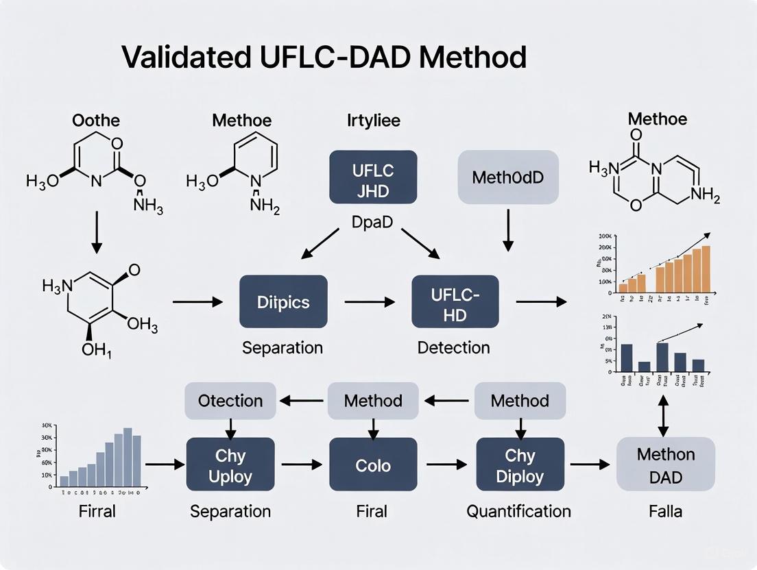
Visualization of Workflows
To enhance understanding of the experimental workflows and logical relationships in UFLC-DAD method development for inorganic pharmaceuticals, the following diagrams provide visual representations of key processes.
Diagram 1: UFLC-DAD Method Development Workflow
Diagram 2: Forced Degradation Study Protocol
Applications and Implications
The validated UFLC-DAD methods for inorganic pharmaceuticals have far-reaching applications across the drug development lifecycle. In pharmaceutical development, these methods facilitate formulation optimization by providing insights into drug-excipient compatibility. For quality control, they ensure the identity, potency, and purity of drug substances and products. In stability studies, they monitor changes in drug quality over time under various environmental conditions, establishing appropriate storage conditions and shelf life [8].
The implications of robust analytical methods extend beyond regulatory compliance. They contribute to patient safety by ensuring that pharmaceuticals maintain their quality, efficacy, and safety throughout their shelf life. Furthermore, they support the development of generic drugs by enabling comparative studies with reference products.
The methodologies described in this application note align with the principles of Green Analytical Chemistry by emphasizing the reduction of organic solvent consumption through faster separations and method optimization [13]. The transfer of methods from conventional HPLC to UFLC represents not only a technical improvement but also an environmentally conscious approach to pharmaceutical analysis.
As pharmaceutical research continues to explore increasingly complex inorganic compounds, the development of sophisticated analytical methods will remain crucial. The UFLC-DAD platform provides a versatile foundation for addressing the unique challenges posed by these compounds, particularly when enhanced with mass spectrometric detection for structural elucidation of degradation products.
The development of a robust Ultra-Fast Liquid Chromatography with Diode Array Detection (UFLC-DAD) method for inorganic pharmaceuticals research hinges on the critical choice of stationary phase. This selection directly dictates the selectivity, efficiency, and overall success of the analytical method in separating and quantifying complex pharmaceutical compounds. While C18 and C8 columns are the workhorses of reversed-phase chromatography, PFP (pentafluorophenyl) and other specialty fluorinated phases offer unique selectivity for challenging separations where traditional alkyl phases fall short. This application note provides a detailed comparison of these stationary phases and offers structured protocols to guide scientists in selecting and validating the optimal column for their specific research needs, ensuring the integrity and reliability of data generated for pharmaceutical development.
Stationary Phase Fundamentals and Comparison
Chemical Composition and Mechanism
The "C" in C8 and C18 denotes the carbon chain length of the alkyl groups bonded to the silica support. C8 columns feature octyl (8-carbon) chains, while C18 columns feature octadecyl (18-carbon) chains [14] [15]. This difference in chain length is a primary factor influencing the hydrophobicity and retention characteristics of the column. In contrast, PFP columns are functionalized with a pentafluorophenyl group (-C6F5) attached via a propyl spacer to the silica surface. The presence of five highly electronegative fluorine atoms creates a unique electron-withdrawing environment that facilitates interactions beyond standard hydrophobic binding [16] [17].
Comparative Characteristics and Selection Table
The following table summarizes the key properties of C18, C8, and PFP stationary phases to guide initial selection.
Table 1: Comparative Overview of C18, C8, and PFP Stationary Phases
| Feature | C18 Column | C8 Column | PFP Column |
|---|---|---|---|
| Chemical Structure | Octadecyl (C18) chains [15] | Octyl (C8) chains [15] | Pentafluorophenyl group [17] |
| Primary Mechanism | Hydrophobic interactions [15] | Hydrophobic interactions [15] | π-π, dipole-dipole, ion-exchange [16] [17] |
| Hydrophobicity | High [15] | Moderate [15] | Variable (low to moderate) |
| Retention of Non-polar Analytes | Strongest [15] | Moderate [15] | Moderate to Low [16] |
| Retention of Aromatics | Moderate | Low | Enhanced [17] |
| Shape Selectivity | Low | Low | Yes [17] |
| Typical Applications | Lipids, steroids, fatty acids [15] | Pharmaceuticals, natural products [15] | Isomers, aromatics, basic analytes [16] [17] |
The retention mechanism of C18 and C8 is predominantly reversed-phase, relying on hydrophobic interactions, where longer C18 chains provide greater retention for non-polar molecules [15]. It is crucial to note that factors such as carbon loading and silica purity can significantly influence performance; a heavily loaded C8 column can sometimes exhibit greater retention than a lightly loaded C18 column [14].
PFP columns operate through a multi-mechanistic retention process [16] [17]:
- π-π Interactions: The electron-deficient PFP ring strongly attracts the electron-rich pi-clouds of aromatic analytes.
- Dipole-Dipole Interactions: The polar C-F bonds can interact with polarizable regions in analyte molecules.
- Ion-Exchange: Residual silanols and the PFP ligand itself can exhibit cation-exchange behavior, particularly for basic compounds at higher organic modifier concentrations [16].
Quantitative Data and Selection Workflow
Key Performance Data
Practical method development requires consideration of quantitative performance metrics and operational parameters.
Table 2: Quantitative Method Development and Performance Data
| Parameter | C18 Column | C8 Column | PFP Column |
|---|---|---|---|
| Relative Retention Time | Longest for non-polar analytes [15] | Shorter than C18 [14] [15] | Variable; can be shorter for neutrals, longer for bases [16] |
| Analysis Time | Longer [15] | Shorter (e.g., reduction from 28 min to 9 min reported) [14] | Variable, method-dependent |
| Peak Tailing (for basic analytes) | Can be pronounced if not end-capped [14] | Potentially less tailing [14] | Can be significant due to ion-exchange; manageable with buffers [16] |
| Optimal pH Range | ~2-8 (silica dependent) | ~2-8 (silica dependent) | ~2.5-8.0 [17] |
| Ion-Exchange Capacity | Low (with modern end-capping) | Low (with modern end-capping) | High (can be exploited for selectivity) [16] |
Stationary Phase Selection Workflow Diagram
The following diagram provides a logical workflow for selecting the most appropriate stationary phase based on the analyte characteristics and separation goals.
Experimental Protocols
Protocol 1: Scouting and Selecting a Stationary Phase
Objective: To rapidly identify the most suitable stationary phase (C18, C8, or PFP) for separating a mixture of guanylhydrazone compounds with anticancer activity using a UFLC-DAD system.
Materials:
- Research Reagent Solutions: See Table 3.
- Columns: C18, C8, and PFP columns of similar dimensions (e.g., 150 mm x 4.6 mm, 2.7 µm).
- Instrumentation: UFLC system equipped with DAD and binary pump, auto-sampler, and column oven.
Table 3: Research Reagent Solutions for Method Scouting
| Reagent/Material | Function/Description | Example/Note |
|---|---|---|
| Acetonitrile (HPLC Grade) | Organic mobile phase modifier | Provides strong elution power [5]. |
| Methanol (HPLC Grade) | Organic mobile phase modifier | Alternative to acetonitrile; different selectivity [5]. |
| Ammonium Acetate | Volatile buffer salt | For controlling mobile phase pH and ionic strength; MS-compatible [16]. |
| Formic Acid | Mobile phase additive & pH modifier | Improves peak shape for acidic/basic analytes; 0.1% common [16] [5]. |
| Acetic Acid | Mobile phase additive & pH modifier | Alternative to formic acid; used at pH ~3.5 [5]. |
| Analyte Standards | Reference compounds for testing | e.g., Guanylhydrazones LQM10, LQM14, LQM17 [5]. |
Procedure:
- Initial Mobile Phase Setup: Prepare a binary mobile phase system.
- Mobile Phase A: 10 mM Ammonium Acetate in Water, pH adjusted to 4.5 with acetic acid.
- Mobile Phase B: Acetonitrile.
- Scouting Gradient: Use the same linear gradient for all three columns. For example: 10% B to 95% B over 15 minutes, hold at 95% B for 2 minutes, with a flow rate of 1.0 mL/min, column temperature at 35°C, and DAD detection from 200-400 nm.
- Column Equilibration: Flush each new column with at least 10 column volumes of the starting mobile phase composition before injecting the sample.
- Sample Injection: Inject a standard solution containing all target analytes (e.g., 10 µg/mL of each guanylhydrazone in a solvent matching the initial mobile phase composition).
- Data Analysis: Evaluate chromatograms based on:
- Resolution (Rs): Ability to separate analyte peaks from each other and any impurities.
- Retention Factor (k): Ensure analytes are adequately retained (typically 2 < k < 10).
- Peak Symmetry: Assess tailing or fronting, especially for basic compounds.
- Analysis Time: Total runtime for the chromatogram.
Protocol 2: Optimizing a Method on a PFP Column
Objective: To refine a separation on a PFP column by exploiting its unique interaction mechanisms, specifically for challenging analytes like basic compounds or structural isomers.
Materials: As in Protocol 1, with a focus on the selected PFP column.
Procedure:
- Investigate Ionic Interactions:
- Run the initial gradient from Protocol 1 using the PFP column.
- Prepare a new Mobile Phase A containing 10 mM Ammonium Formate (pH 3.0) instead of Ammonium Acetate.
- Repeat the analysis and compare the chromatograms. The addition of a competing ion like ammonium+ can suppress ionic interactions with residual silanols, often sharpening peaks and changing selectivity for basic analytes [16].
- Optimize pH for Selectivity:
- Prepare mobile phases (A and B) at different pH levels (e.g., 3.0, 5.0, and 7.0) using appropriate buffers. Ensure the pH is within the column's specified range (typically 2.5-8.0 for PFP) [17].
- Run the scouting gradient at each pH and note changes in elution order and resolution, particularly for ionizable compounds.
- Fine-tune Organic Modifier:
- Test the scouting gradient using Methanol in place of Acetonitrile as Mobile Phase B. Methanol can alter the hydrogen bonding and dipole interactions, providing different selectivity [18].
- Final Method Development:
- Based on the findings from steps 1-3, design a final gradient that achieves baseline resolution for all critical peak pairs in the shortest possible runtime.
Advanced Applications and Specialized Phases
Exploiting Orthogonality with PFP Phases
PFP phases are celebrated for their orthogonal selectivity compared to C18. This property is invaluable in pharmaceutical analysis for:
- Impurity Profiling: Confirming the purity of a main active ingredient by showing that potential impurities, which may co-elute on a C18 column, are resolved on a PFP column [18].
- Forced Degradation Studies: Identifying and resolving degradation products that are structurally similar to the parent drug molecule, often isomers [19].
- Metabolite Identification: Separating drug metabolites that may differ only in the position of a hydroxyl group on an aromatic ring, leveraging the shape selectivity of the PFP phase [17].
Retention Mechanism Diagram for PFP Phases
Understanding the multiple interaction mechanisms of PFP stationary phases is key to applying them effectively.
Selecting the optimal stationary phase is a foundational step in developing a validated UFLC-DAD method for inorganic pharmaceuticals. C18 columns provide robust, general-purpose methods for non-polar analytes, while C8 columns offer a balanced option for faster analysis of moderately polar compounds. For complex separations involving aromatic compounds, structural isomers, or basic molecules, PFP and other fluorinated columns provide a powerful orthogonal tool with unique selectivity. The experimental protocols outlined herein provide a systematic approach to column scouting and method optimization. By understanding the fundamental interaction mechanisms and applying a structured selection workflow, researchers can significantly enhance the efficiency, selectivity, and robustness of their chromatographic methods, thereby accelerating drug development and ensuring the highest quality of analytical data.
In the development of Ultra-Fast Liquid Chromatography with Diode Array Detection (UFLC-DAD) methods for inorganic pharmaceuticals, the composition of the mobile phase is a critical determinant of success. The mobile phase governs the entire separation process, influencing analyte retention, peak shape, selectivity, and detection sensitivity. For inorganic compounds, which often exhibit unique coordination chemistries and charge characteristics, a systematic approach to mobile phase optimization is particularly vital. This application note details the strategic use of buffers, pH control, and organic modifiers to develop robust, validated UFLC-DAD methods for inorganic pharmaceutical analysis, providing researchers with practical protocols and frameworks.
Theoretical Foundations of Mobile Phase Design
The Mobile Phase as a Tunable Parameter
In reversed-phase liquid chromatography, the mobile phase is the liquid solvent or mixture that carries the sample through the chromatographic system. Its composition directly controls the equilibrium distribution of analytes between the mobile phase and the stationary phase. For inorganic compounds, this interaction is not solely based on hydrophobicity but is significantly influenced by the ionic character, coordination geometry, and ligand-exchange properties of the analytes. The mobile phase must therefore be engineered to manage these complex interactions through careful selection of buffer systems, pH, and organic modifiers [20].
Key Factors in Mobile Phase Optimization
Several interrelated factors must be balanced during mobile phase development:
- Solvent Polarity: Adjusting the relative proportions of aqueous and organic components to control elution strength. In reversed-phase chromatography, increasing organic solvent concentration generally decreases retention for most compounds [20].
- pH Control: Manipulating the ionization state of analytes and stationary phase silanols to modulate retention and selectivity. The pH can profoundly affect the stability and chromatographic behavior of metal complexes and ionizable inorganic species [20] [21].
- Buffer Composition and Ionic Strength: Establishing a stable ionic environment that maintains pH and governs secondary interactions through ion-pairing, complexation, or suppression of analyte ionization [22].
- Additive Selection: Incorporating specialized reagents such as ion-pairing agents or metal chelators to address specific chromatographic challenges posed by inorganic pharmaceuticals [20].
Core Components of the Mobile Phase
Buffer Systems and pH Control
Buffer selection is paramount for methods requiring precise pH control, which is common in inorganic pharmaceutical analysis where analytes may be pH-sensitive.
Table 1: Common Buffer Systems for Pharmaceutical Chromatography
| Buffer System | Effective pH Range | Common Applications | Key Considerations |
|---|---|---|---|
| Phosphate | 6.0 – 8.0 | Ophthalmic, nasal, and parenteral preparations; general pharmaceutical analysis | Versatile, close to physiological pH; may precipitate with organic solvents at high concentrations [22] |
| Acetate | 3.6 – 5.6 | Formulations requiring mild acidity; LC-MS compatible | Volatile, MS-friendly; limited buffering capacity at neutral pH [22] |
| Citrate | 2.5 – 6.5 | Internal and external formulations; broad range applications | Good chelating properties; may complicate MS detection [22] |
| Ammonium Bicarbonate | 7.0 – 9.0 | Basic pH applications; mass spectrometry | Volatile, suitable for MS; limited stability over time [23] |
The buffer capacity must be sufficient to resist pH changes during analysis, but buffer concentration should be optimized to avoid precipitation, especially when using high organic solvent proportions. For UFLC-DAD methods, it is critical to measure pH before adding organic modifiers, as pH meters are calibrated for aqueous solutions and readings in mixed solvents are inaccurate [20].
Organic Modifiers
Organic modifiers control elution strength and selectivity in reversed-phase chromatography. The choice of modifier affects solubility, backpressure, and UV transparency.
- Acetonitrile: Most commonly used due to its low viscosity, high UV cut-off, and strong eluting power. Provides sharp peaks and reduced backpressure [20] [24].
- Methanol: Alternative to acetonitrile with different selectivity. Has higher viscosity but is less expensive and can be useful for separating different compound classes [5] [25].
- Tetrahydrofuran (THF): A strong solvent that can provide unique selectivity for challenging separations, but has poor UV transparency and oxidative instability [20].
The organic modifier ratio is frequently optimized through gradient elution, where the organic proportion increases during the analysis to elute compounds with a wide range of hydrophobicities [20] [24].
Mobile Phase Additives for Inorganic Compounds
Inorganic pharmaceuticals often require specialized additives to achieve adequate chromatography:
- Ion-Pairing Reagents: Amphiphilic ions such as alkyl sulfonates (for bases) or tetraalkylammonium salts (for acids) can mask analyte charge and increase retention of ionic inorganic compounds on reversed-phase columns [20].
- Metal Chelators: Additives like EDTA can prevent analyte binding to metal surfaces within the HPLC system and improve peak shapes for metal-complexing compounds [20].
- Competing Ligands: Species that moderate the interaction between analytes and the stationary phase can be added to fine-tune selectivity for metal complexes [23].
Experimental Protocols
Protocol 1: Initial Mobile Phase Scouting with Factorial Design
A systematic approach to initial method development employs factorial design to efficiently explore the multidimensional parameter space [5].
Materials and Equipment:
- UFLC system with DAD detector
- Reversed-phase column (e.g., C18, 150 mm × 4.6 mm, 2.7 μm)
- Analytical standards of target inorganic pharmaceuticals
- Buffer components (potassium phosphate, ammonium acetate, etc.)
- Organic modifiers (HPLC-grade acetonitrile, methanol)
- pH meter and calibration standards
- Vacuum filtration apparatus with 0.45 μm membranes
Procedure:
- Define Factors and Ranges: Identify critical mobile phase factors (e.g., pH, organic modifier percentage, buffer concentration) and establish practical ranges based on column specifications and analyte properties.
- Prepare Mobile Phases: Prepare a series of mobile phases according to the experimental design, ensuring consistent buffer preparation and pH adjustment before organic modifier addition.
- Filter and Degas: Vacuum-filter all mobile phases through 0.45 μm membranes to remove particulates and degas to prevent bubble formation.
- System Equilibration: Equilibrate the chromatographic system with each mobile phase composition for at least 10 column volumes before analysis.
- Analyze Standards: Inject analyte standards and record retention times, peak asymmetry, resolution, and plate count.
- Statistical Analysis: Analyze results to identify significant factors and interaction effects using statistical software.
- Refine Conditions: Based on the analysis, narrow the experimental ranges and iterate toward optimal conditions.
Protocol 2: pH Scouting and Buffer Selection
This protocol determines the optimal pH and buffer system for separating ionizable inorganic pharmaceuticals.
Procedure:
- Select Buffer Candidates: Choose 2-3 buffer systems covering the pH range of interest (e.g., phosphate for 6.0-8.0, acetate for 4.0-5.5).
- Prepare Buffers: Prepare 20 mM solutions of each buffer at 3-4 pH values within their effective ranges. Adjust pH using NaOH or HCl before adding organic modifier.
- Prepare Mobile Phases: Mix each buffer with organic modifier at a fixed ratio (e.g., 80:20 aqueous:organic).
- Analyze Standards: Chromatograph analyte standards under each condition, monitoring retention factor (k), selectivity (α), and peak shape.
- Plot Retention vs. pH: Create plots of retention time versus pH for each analyte to identify the pH region providing optimal separation.
- Assess Buffer Effects: Compare different buffer systems at similar pH values to identify system-specific effects on retention or selectivity.
- Optimize Buffer Concentration: Once optimal pH is identified, evaluate buffer concentrations from 5-50 mM to determine the minimum concentration providing adequate pH control without risk of precipitation.
Protocol 3: Method Validation for Regulated Environments
After optimal mobile phase conditions are established, method validation follows international guidelines (ICH Q2) to confirm reliability [5] [26].
Validation Parameters and Procedures:
- Linearity: Prepare analyte standards at 5-7 concentration levels across the expected range. Inject each level in triplicate and plot peak area versus concentration. Calculate regression statistics with r² ≥ 0.999 [5] [24].
- Accuracy (Recovery): Spike placebo or matrix with known analyte quantities at three levels (e.g., 80%, 100%, 120% of target). Calculate percent recovery (98-102% acceptable) and relative standard deviation (RSD ≤ 2%) [5] [26].
- Precision:
- Robustness: Deliberately vary mobile phase parameters (pH ± 0.2 units, organic composition ± 2%, flow rate ± 0.1 mL/min) to assess method resilience [5].
- Solution Stability: Monitor standard and sample solutions over time (e.g., 0, 6, 12, 24, 48 hours) to establish acceptable storage conditions and timelines.
Results and Data Analysis
Quantitative Validation Parameters
Table 2: Exemplary Validation Data for a UFLC-DAD Method for Guanylhydrazone Analysis [5]
| Validation Parameter | LQM10 | LQM14 | LQM17 |
|---|---|---|---|
| Linearity (r²) | 0.9995 | 0.9999 | 0.9994 |
| Accuracy (% Recovery) | |||
| 8 μg/mL | 99.71 ± 1.67 | 98.69 ± 1.85 | 100.22 ± 1.86 |
| 10 μg/mL | 100.46 ± 1.34 | 101.47 ± 0.24 | 99.71 ± 1.36 |
| 12 μg/mL | 99.49 ± 1.79 | 98.71 ± 1.50 | 100.15 ± 1.25 |
| Precision (RSD, %) | |||
| Intra-day (n=6) | 1.48 | 2.00 | 1.24 |
| Inter-day (n=6) | 2.81 | 1.56 | 2.20 |
The data in Table 2 demonstrates the high quality of a validated chromatographic method, with excellent linearity, accuracy, and precision parameters meeting acceptance criteria for pharmaceutical analysis.
Impact of Mobile Phase Composition on Performance
Table 3: Comparison of HPLC vs. UHPLC Methods Showing Impact of Mobile Phase Optimization [5]
| Parameter | Conventional HPLC | Optimized UHPLC | Improvement |
|---|---|---|---|
| Analysis Time | ~10-15 minutes | ~5 minutes | ~50% reduction |
| Solvent Consumption | Baseline | 4x less | 75% reduction |
| Injection Volume | Baseline | 20x less | 95% reduction |
| Column Performance | Baseline | Improved efficiency | Higher plate count |
Optimized mobile phase conditions in UHPLC methods provide significant advantages in solvent consumption, analysis time, and operational efficiency, aligning with green chemistry principles [5].
The Scientist's Toolkit: Essential Research Reagents
Table 4: Key Reagents for Mobile Phase Preparation in UFLC-DAD Method Development
| Reagent Category | Specific Examples | Function in Mobile Phase |
|---|---|---|
| Buffer Salts | Potassium phosphate, Ammonium acetate, Sodium citrate | Maintain constant pH, affect ionization state of analytes [22] [24] |
| pH Adjusters | Phosphoric acid, Acetic acid, Ammonium hydroxide, Sodium hydroxide | Fine-tune mobile phase pH to optimal range for separation [20] [24] |
| Organic Modifiers | Acetonitrile, Methanol, Tetrahydrofuran | Control elution strength and selectivity; dissolve hydrophobic analytes [5] [20] [24] |
| Ion-Pairing Reagents | Alkane sulfonates, Tetraalkylammonium salts | Mask charge of ionic analytes to increase retention in reversed-phase systems [20] |
| Metal Chelators | EDTA, Citrate | Prevent metal-mediated degradation and improve peak shape for metal-complexing compounds [20] |
Method Implementation and Workflow
The following workflow diagram illustrates the systematic development of a mobile phase composition for inorganic pharmaceutical analysis:
Systematic Method Development Workflow
Troubleshooting Common Mobile Phase Issues
Even carefully developed methods may encounter implementation challenges:
- Buffer Precipitation: When using phosphate buffers with high organic content, salts may precipitate, damaging pumps and columns. Prevention includes using lower buffer concentrations (<25 mM) and ensuring the organic solvent percentage doesn't exceed the buffer's solubility limit [20].
- Retention Time Drift: Gradual changes in retention often stem from improper mobile phase preparation or pH instability. Standardize mixing procedures and measure pH after preparation but before organic addition [20].
- Peak Tailing: For basic inorganic compounds, tailing may result from residual silanol interactions. Remedies include using lower pH mobile phases, specialized end-capped columns, or adding competing amines to the mobile phase [23].
- High Backpressure: May indicate microbial growth in aqueous mobile phases or particulate contamination. Always filter mobile phases through 0.45 μm membranes and do not store for extended periods [20].
The role of mobile phase composition in UFLC-DAD method development for inorganic pharmaceuticals cannot be overstated. Through strategic selection of buffer systems, precise pH control, and optimization of organic modifiers, researchers can develop robust, validated methods suitable for regulated environments. The experimental protocols and troubleshooting guidance provided herein offer a systematic framework for navigating the complexities of inorganic pharmaceutical analysis. As demonstrated by the validation data presented, careful attention to mobile phase composition yields methods with excellent linearity, precision, accuracy, and robustness—the essential attributes of reliable analytical methods for pharmaceutical development and quality control.
The initial scoping phase is a critical determinant of success in developing a robust Ultra-Fast Liquid Chromatography with Diode Array Detection (UFLC-DAD) method for inorganic pharmaceuticals. This foundational stage establishes the analytical targets that guide all subsequent development, validation, and application activities. Proper scoping ensures the final method will satisfy both scientific requirements for reliable drug quantification and regulatory standards for pharmaceutical quality control. For inorganic pharmaceutical compounds, which often present unique challenges related to stability, polarity, and detection characteristics, a systematic approach to defining analytical targets is particularly vital. Early-stage planning must balance analytical performance with practical considerations including time, resource efficiency, and alignment with Green Analytical Chemistry (GAC) principles [13] [27].
This protocol provides a structured framework for establishing key parameters during the method scoping phase, incorporating both traditional analytical science metrics and modern assessment tools. By defining clear analytical targets at the outset, researchers can streamline method development, reduce costly iterations, and build quality into the analytical process from its inception—a core principle of Analytical Quality by Design (AQbD) [27].
Core Analytical Performance Parameters
The foundation of effective method scoping lies in establishing quantitative targets for analytical performance. These targets should reflect the intended application of the method while meeting or exceeding regulatory standards.
Table 1: Essential Analytical Performance Targets for UFLC-DAD Method Scoping
| Parameter | Target Range | Regulatory Consideration | Application-Specific Factors |
|---|---|---|---|
| Linearity Range | 50-150% of expected sample concentration [28] [29] | R² > 0.99 [28] [11] | Expected concentration in pharmaceutical dosage forms and biological matrices |
| Detection Limit (LOD) | Not more than 0.1 µg/mL for API quantification [27] | Signal-to-noise ratio ≥ 3:1 [27] | Sensitivity requirements for impurity profiling or low-dose formulations |
| Quantification Limit (LOQ) | Not more than 0.3 µg/mL for API quantification [27] | Signal-to-noise ratio ≥ 10:1 [27] | Sufficient for accurate quantification of the active and related substances |
| Precision (Repeatability) | RSD < 2% for retention time [5] [11] | RSD < 2% for system suitability [5] | Critical for method robustness and transferability |
| Accuracy (Recovery) | 98-102% for drug substance [28] [11] | Consistent across specified range [28] | Matrix effects for complex formulations or biological samples |
When scoping methods for inorganic pharmaceuticals, researchers must pay particular attention to the polarity and chromophore characteristics of the target analytes, as these will directly influence detection wavelength selection and chromatographic retention strategy. The diode array detector provides advantages for method scoping through spectral confirmation of peak purity and identity, which is especially valuable when analyzing compounds with potential degradation products or complex matrices [30] [31].
Systematic Scoping Workflow
A structured approach to analytical target definition ensures comprehensive consideration of all critical factors that influence method performance and applicability. The following workflow outlines a systematic process for establishing analytical targets during early-stage method scoping.
Diagram 1: Analytical target scoping workflow illustrates the systematic process for defining key parameters, incorporating decision points for critical performance metrics.
This workflow emphasizes iterative refinement of analytical targets, allowing researchers to balance competing priorities such as sensitivity, speed, and environmental impact. The process begins with a clear definition of the analytical need, which drives all subsequent decisions regarding instrument configuration, separation strategy, and validation requirements.
Chromatographic and Detection Parameters
Chromatographic conditions must be scoped with consideration to both the physicochemical properties of the target inorganic pharmaceuticals and the practical constraints of the analytical environment.
Table 2: Chromatographic Condition Targets for UFLC-DAD Methods
| Parameter | Recommended Targets | Rationale | Optimization Considerations |
|---|---|---|---|
| Stationary Phase | C18 (50-100 mm length) [27] [29] | Balance of efficiency and analysis time | Analyte polarity, pH stability, and backpressure constraints |
| Particle Size | 1.7-2.5 µm [27] | Enhanced efficiency for UFLC | System pressure capabilities and column lifetime |
| Mobile Phase | Aqueous buffer with acetonitrile or methanol [27] [11] | Compatibility with reversed-phase and DAD | pH selection based on analyte pKa; organic modifier strength |
| Flow Rate | 0.3-0.6 mL/min [27] [29] | Optimal linear velocity for narrow-bore columns | Analysis time requirements and system pressure |
| Detection Wavelength | 200-400 nm range [29] [11] | Coverage of common chromophores | Use of DAD for multi-wavelength monitoring and peak purity |
For UFLC-DAD methods targeting inorganic pharmaceuticals, the selection of detection wavelength should be based on the chromophoric properties of the analyte. The diode array detector enables simultaneous monitoring at multiple wavelengths, which is particularly valuable for methods that must quantify multiple compounds with different absorbance maxima [30] [11]. When scoping detection parameters, consider both the primary quantification wavelength and secondary wavelengths for peak identity confirmation.
Experimental Protocols for Parameter Definition
Protocol 1: Preliminary Detection Wavelength Scoping
Purpose: To determine the optimal detection wavelength(s) for target analytes using DAD detection.
Materials:
- Standard solutions of target analytes (approximately 10 µg/mL in suitable solvent)
- UFLC system with DAD capability
- Appropriate mobile phase for isocratic elution
- C18 column (50 mm × 2.1 mm, 1.7-2.5 µm)
Procedure:
- Prepare standard solutions of each target analyte in mobile phase or suitable solvent
- Set up UFLC-DAD method with isocratic elution (50% organic modifier, 50% aqueous phase)
- Inject standard solution and acquire spectra across 200-400 nm range
- Identify wavelength of maximum absorbance (λmax) for each compound
- Evaluate signal-to-noise ratio at potential detection wavelengths
- Select primary quantification wavelength that balances sensitivity and selectivity
- Document spectral characteristics for future peak identity confirmation
Validation Checkpoint: Verify that the selected wavelength provides a linear response across the anticipated concentration range (typically 50-150% of target concentration) [28].
Protocol 2: Initial Separation Scoping
Purpose: To establish preliminary chromatographic conditions for separating target analytes from potential interferents.
Materials:
- Standard solutions of target analytes and known impurities/degradation products
- UFLC system with DAD detection
- Selection of columns (varying chemistry and dimensions)
- Mobile phase components (buffers, organic modifiers)
Procedure:
- Prepare mixed standard solution containing all target analytes
- Begin with C18 column (100 mm × 2.1 mm, 1.7 µm) and temperature 40°C
- Develop gradient method: 5-95% organic modifier over 5-10 minutes
- Adjust pH of aqueous component to suppress ionization of acidic/basic analytes
- Evaluate separation based on resolution, peak symmetry, and analysis time
- If necessary, test alternative column chemistries (phenyl, CN, Aqua) [11]
- Optimize gradient profile to achieve baseline separation (Rs > 2.0)
- Document initial conditions for further optimization
Validation Checkpoint: Verify that the method demonstrates specificity for all target analytes in the presence of potential interferents [28] [27].
Protocol 3: Greenness Assessment
Purpose: To evaluate the environmental impact of the scoped analytical method using established green metrics.
Materials:
- Detailed description of proposed method conditions
- AGREE and BAGI assessment software/tools
- Solvent consumption calculations
Procedure:
- Document all method parameters: mobile phase composition, flow rate, run time, sample preparation
- Calculate total solvent consumption per analysis
- Identify hazardous reagents and potential alternatives
- Input parameters into AGREE (Analytical GREEnness) metric tool
- Calculate BAGI (Blue Applicability Grade Index) score
- Evaluate method against 12 principles of Green Analytical Chemistry [13]
- Identify opportunities to improve greenness while maintaining performance
- Set target scores for final method (e.g., AGREE > 0.65) [27]
Output: Quantitative greenness assessment to guide environmentally conscious method development decisions.
Assessment Tools and Compliance Frameworks
Modern analytical method scoping should incorporate standardized assessment tools to evaluate both performance and environmental impact. These tools provide quantitative metrics for comparing alternative approaches and demonstrating compliance with regulatory and sustainability guidelines.
Table 3: Method Assessment Tools for Analytical Target Profiling
| Assessment Tool | Application in Method Scoping | Target Values | Regulatory Relevance |
|---|---|---|---|
| Analytical Eco-Scale | Penalty point system for non-green practices [13] | >75 (Excellent greenness) [13] | Aligns with green chemistry principles |
| AGREE (Analytical GREEnness) | Comprehensive greenness assessment [13] [27] | >0.65 (Acceptable) [27] | Increasing regulatory expectation |
| BAGI (Blue Applicability Grade Index) | Practical applicability evaluation [13] | >85 (High practicality) [27] | Cost-effectiveness and transferability |
| ICH Q2(R2) Guidelines | Validation parameter definition [27] | Full compliance for regulated methods | Required for pharmaceutical applications |
The relationship between these assessment tools and the overall method development process can be visualized as an integrated framework where analytical targets are evaluated against multiple criteria throughout the scoping phase.
Diagram 2: Analytical target assessment framework shows the integration of multiple evaluation dimensions into a comprehensive target profile.
The Scientist's Toolkit: Essential Research Reagents and Materials
Proper selection of research reagents and materials during the scoping phase establishes the foundation for a robust and transferable analytical method.
Table 4: Essential Research Reagents and Materials for UFLC-DAD Method Scoping
| Item | Function | Selection Criteria | Scoping Considerations |
|---|---|---|---|
| Reference Standards | Method development and calibration | Highest available purity with certificate of analysis | Availability, stability, and cost for routine use |
| Chromatographic Columns | Analytical separation | Multiple chemistries for selectivity screening | Column lifetime, backpressure, and availability |
| Mobile Phase Solvents | Liquid chromatography elution | HPLC-grade with low UV cutoff | UV transparency at detection wavelength, purity |
| Buffer Salts | Mobile phase pH control | High purity with minimal UV absorbance | Volatility for LC-MS compatibility, buffering capacity |
| SPE Cartridges | Sample preparation optimization | Various sorbents for extraction efficiency | Recovery, selectivity, and reproducibility [29] |
Systematic definition of analytical targets during the initial scoping phase provides a strategic foundation for efficient development of UFLC-DAD methods for inorganic pharmaceuticals. By establishing clear, quantitative targets for chromatographic performance, detection parameters, and green chemistry metrics, researchers can streamline method development, reduce costly iterations, and ensure the final method meets both scientific and regulatory requirements. The protocols and frameworks presented in this application note offer a structured approach to analytical target definition that aligns with modern quality-by-design principles and sustainability initiatives in pharmaceutical analysis.
Strategic Method Development and Real-World Application in Complex Matrices
The choice of elution mode is a cornerstone of chromatographic method development, directly impacting the efficiency, speed, and success of the separation. Within the context of developing a validated UFLC-DAD method for inorganic pharmaceuticals research, this decision becomes paramount. Gradient elution involves a systematic change in the mobile phase composition during the analytical run, while isocratic elution maintains a constant mobile phase composition throughout [32] [33]. This application note provides a systematic comparison of these two techniques and offers detailed protocols for their optimization, enabling researchers and drug development professionals to make informed decisions that enhance method performance for complex pharmaceutical matrices.
Theoretical Background and Core Principles
Isocratic Elution
Isocratic elution is characterized by the use of a single solvent or a consistent solvent mixture for the entire duration of the separation [33] [34]. This simplicity is its primary advantage, leading to straightforward method development, consistent reproducibility, and lower operational costs due to reduced solvent management complexity [34]. It is ideally suited for routine analyses of samples with low complexity or for separating compounds with similar polarities and chemical properties [34].
A significant drawback of isocratic elution is band-broadening [32]. As a sample migrates through the column, compounds with higher affinity for the stationary phase take longer to elute. This extended migration time leads to a dilution effect, resulting in broader elution bands, which manifest as wider peaks on the chromatogram. This can reduce peak height and negatively impact detection sensitivity for later-eluting compounds [32] [33].
Gradient Elution
Gradient elution employs a dynamic change in the mobile phase composition, typically starting with a higher percentage of a "weaker" solvent and progressively increasing the percentage of a "stronger" solvent [32] [34]. This gradual increase in elution strength expedites the migration of compounds with greater affinity for the stationary phase.
The key advantages of gradient elution include:
- Reduced Band-Broadening: Later-eluting compounds experience sharper elution bands, which increases their concentration and improves detection sensitivity [32].
- Improved Peak Resolution: The technique often enhances separation between compounds, leading to higher purity fractions [32].
- Shorter Run Times: By accelerating the elution of strongly retained compounds, gradient elution reduces overall analysis time, which is beneficial for high-throughput applications [34] [35].
The two main types of gradients are linear gradients, where the solvent ratio changes linearly over time, and step gradients, which use a series of discrete isocratic steps to maximize separation and can reduce solvent consumption [32].
Systematic Comparison and Selection Criteria
The choice between gradient and isocratic elution is multi-faceted. The following table provides a structured comparison to guide the selection process.
Table 1: Comparative Analysis of Isocratic vs. Gradient Elution
| Parameter | Isocratic Elution | Gradient Elution |
|---|---|---|
| Mobile Phase | Constant composition [34] | Dynamically changing composition [34] |
| Complexity & Cost | Low; simpler equipment, lower operational cost [34] | High; requires sophisticated instrumentation [33] [34] |
| Ideal Sample Type | Simple mixtures, compounds with similar polarity [34] | Complex mixtures with a wide range of polarities [34] |
| Peak Shape | Broader peaks for later-eluting compounds [32] [33] | Consistently sharper peaks throughout the run [32] [33] |
| Analysis Speed | Can be slow for strongly retained analytes [34] | Faster overall for complex samples [34] [35] |
| Method Development | Simpler and faster [34] | More intricate, requires gradient profile optimization [34] |
| Primary Advantage | Simplicity, reproducibility, cost-effectiveness [34] | Enhanced resolution for complex samples, flexibility [34] |
The following decision flowchart synthesizes this information into a practical selection tool.
Figure 1: Decision workflow for elution mode selection, integrating key criteria from the literature [34] [35].
Experimental Protocols for Method Optimization
Protocol A: Initial Scouting and Column Equilibration
This initial protocol is critical for establishing baseline conditions for both isocratic and gradient methods.
- Column Selection: Begin with a suitable C18 column (e.g., 250 × 4.6 mm, 5 µm) as a robust starting point for reversed-phase chromatography of inorganic pharmaceuticals [36].
- Mobile Phase Preparation: Prepare aqueous and organic components. A common system is Water with 0.1% Trifluoroacetic Acid (TFA) as solvent A and Acetonitrile (ACN) with 0.1% TFA as solvent B [36]. Filter and degas all solvents.
- Temperature Control: Set the column oven temperature to 40 °C to ensure consistent retention times and backpressure [36] [37].
- Detection Wavelength: Configure the DAD detector according to the analytes' UV-Vis profiles. For multi-analyte methods, use a wavelength that offers a good compromise for all compounds of interest.
- System Equilibration:
- For Isocratic Elution: Pump the chosen constant solvent mixture (e.g., 85% A / 15% B) through the system until a stable baseline is achieved (typically 10-15 column volumes).
- For Gradient Elution: After a gradient run, flush the column with the initial mobile phase composition (e.g., 85% A) for a minimum of 5-10 column volumes to ensure full re-equilibration before the next injection [34] [35].
Protocol B: Isocratic Method Optimization
This protocol is designed for fine-tuning an isocratic method after initial scouting.
- Initial Condition Setup: Based on TLC or a preliminary gradient run, select a candidate solvent ratio (e.g., 70% NaH2PO4 buffer pH 4.95 : 30% Methanol) [37].
- Flow Rate Optimization: Inject the standard mixture and evaluate the separation. Adjust the flow rate (e.g., between 0.9 - 1.5 mL/min) to balance analysis time and backpressure [37].
- Composition Fine-Tuning: If resolution is inadequate, make small, incremental adjustments (± 2-5%) to the strong solvent percentage. The goal is to achieve a retention factor (k') between 2 and 10 for all analytes, with the last peak eluting at k' < 5 for optimal peak shape [35].
- Method Validation: Once optimal conditions are found, proceed with full method validation according to ICH guidelines, assessing linearity, accuracy, precision, LOD, and LOQ [36] [37].
Protocol C: Gradient Method Optimization Using Scanning Gradients
This protocol uses scanning gradients for efficient modeling and optimization of complex separations.
- Define Gradient Range: Establish the initial and final compositions for the gradient. A typical starting range is 5% to 95% of strong solvent (B) [34].
- Run Scanning Gradients: Perform at least two initial gradient runs with different slopes (e.g., 5-95% B in 10 minutes and in 30 minutes) to model analyte retention behavior. The accuracy of the final model depends more on the precision of these measurements than on a large difference in gradient slopes [38].
- Retention Modeling: Use chromatographic software to process the data from the scanning gradients and build a retention model for the key analytes. Simpler two-parameter models are often sufficient and statistically significant [38].
- Predict and Verify: Use the software to predict the optimal gradient profile (linear or multi-step) that will provide the best resolution within the desired runtime. The most accurate predictions are achieved by interpolating to a gradient slope that lies between those of the scanning gradients, rather than extrapolating beyond them [38].
- Experimental Confirmation: Run the predicted optimal gradient method experimentally and compare the results with the software prediction. Fine-tune the gradient program if necessary.
- Final Validation: Validate the optimized gradient method according to ICH guidelines for its intended application [36].
The Scientist's Toolkit: Essential Research Reagents and Materials
The following table lists key materials and reagents required for the development and execution of UFLC-DAD methods as discussed in this note.
Table 2: Essential Research Reagents and Materials for Chromatographic Method Development
| Item | Function / Application | Example from Literature |
|---|---|---|
| C18 Chromatography Column | Standard reversed-phase stationary phase for separating a wide range of compounds. | 250 × 4.6 mm, 5 µm column for separating antioxidants in a face mask [36]. |
| Buffering Agents (e.g., NaH₂PO₄, TFA) | Modify and control the pH of the mobile phase to improve peak shape and reproducibility. | NaH₂PO₄ buffer (pH 4.95) for vitamin B analysis; 0.1% TFA for antioxidant separation [36] [37]. |
| HPLC-Grade Solvents (Water, ACN, MeOH) | Constitute the mobile phase; high purity is critical to minimize baseline noise and ghost peaks. | Methanol/ACN mixture as diluent; ACN as strong solvent in gradients [36] [37]. |
| Standard Compounds | Used for method development, calibration, and validation to establish retention times and response factors. | Certified reference standards of target analytes (e.g., Benzoyl peroxide, Salicylic Acid, Vitamins B1, B2, B6) [36] [37]. |
| Solid Phase Extraction (SPE) Cartridges | For sample clean-up and pre-concentration of analytes from complex matrices like biological fluids. | SPE used for purifying gastrointestinal fluid samples in vitamin bioavailability studies [37]. |
Concluding Remarks
The systematic optimization of chromatographic conditions is a deliberate process that balances analytical requirements with practical constraints. Isocratic elution offers a straightforward, cost-effective solution for simpler separations, while gradient elution provides the power, speed, and resolution necessary for complex inorganic pharmaceutical mixtures. By applying the decision criteria and detailed experimental protocols outlined in this application note, scientists can develop robust, validated UFLC-DAD methods that ensure drug safety, efficacy, and quality.
The accurate determination of active pharmaceutical ingredients and their metabolites in complex matrices is a cornerstone of modern drug development and therapeutic drug monitoring (TDM). The complexity of biological samples, coupled with the typically low concentrations of target analytes, necessitates highly efficient sample preparation strategies alongside high-performance instrumentation to achieve the required sensitivity and selectivity in modern analytical chemistry. Sample preparation serves critical functions, including the selective isolation of target analytes from the matrix, minimization or elimination of interfering components, and analyte preconcentration when required. In the context of a validated Ultra-Fast Liquid Chromatography with Diode Array Detection (UFLC-DAD) method for inorganic pharmaceuticals research, effective sample preparation becomes paramount as it directly impacts the method's accuracy, precision, and reliability.
Solid-phase extraction (SPE) and related techniques have emerged as powerful tools for sample clean-up and preconcentration, effectively replacing many traditional liquid-liquid extraction methods. The evolution of these techniques has been marked by a trend toward miniaturization, leading to significant shifts in pharmaceutical analysis by offering greener and more ecologically friendly alternatives to traditional procedures. These miniaturized approaches align with the principles of Green Analytical Chemistry (GAC) and Green Sample Preparation (GSP), characterized by minimal sample requirements, high analytical sensitivity, shortened extraction duration, compatibility with eco-friendly solvents, and minimal waste generation. The fundamental principle underlying SPE involves the partitioning of analytes between a solid stationary phase and a liquid sample matrix, followed by selective elution of the retained analytes.
Table 1: Comparison of Major Sample Preparation Techniques
| Technique | Principle | Typical Sample Volume | Solvent Consumption | Key Advantages |
|---|---|---|---|---|
| Traditional SPE | Adsorption onto solid sorbent | 1-100 mL | Moderate | High analyte recovery, well-established protocols |
| Solid-Phase Microextraction (SPME) | Partition between sample and fiber coating | 1-10 mL | Very Low | Solventless, simple automation |
| Dispersive SPE (d-SPE) | Adsorption onto dispersed sorbent particles | 1-10 mL | Low | Rapid kinetics, high efficiency |
| Fabric Phase Sorptive Extraction (FPSE) | Hybrid extraction on sol-gel coated fabric | 1-5 mL | Low | Combines exhaustive and equilibrium extraction |
| Microextraction by Packed Sorbent (MEPS) | Miniaturized SPE in syringe barrel | 10-100 µL | Very Low | Minimal sample volume, reusable sorbents |
Advanced Sorbent Materials for Solid-Phase Extraction
The core of any SPE technique lies in the sorbent material, which determines the selectivity, capacity, and efficiency of the extraction process. Recent scientific advancements have focused on developing engineered materials with enhanced selectivity for targeted pharmaceutical analysis.
Molecularly Imprinted Polymers (MIPs) represent a significant breakthrough in selective sorbent technology. These "smart adsorbents" are synthetic polymers with specific recognition sites complementary in shape, size, and functional groups to the target molecule. Their creation involves polymerizing functional monomers around a template molecule (the target analyte), followed by template removal, leaving behind cavities with high affinity for the original molecule. The selectivity of MIPs makes them particularly valuable for extracting specific pharmaceuticals from complex biological matrices where structural analogs may be present. A particularly innovative development in this field is the emergence of stimuli-responsive MIPs, which exhibit tailored responses to various stimuli such as magnetic fields, pH, temperature, and light, allowing for controlled and reversible alteration of their chemical and physical properties for enhanced extraction and release of target analytes.
Metal-Organic Frameworks (MOFs) are crystalline porous materials consisting of metal ions or clusters coordinated to organic ligands to form one-, two-, or three-dimensional structures. Their exceptionally high surface areas, tunable porosity, and diverse functional groups make them excellent sorbents for pharmaceutical compounds. For instance, MIL-53(Al)-MOF has demonstrated remarkable adsorption capacities for penicillins—175 mg/g for piperacillin and 217 mg/g for penicillin V—attributed to electrostatic, hydrogen bonding, π-π, and hydrophobic interactions between the framework and the analyte molecules. The structural versatility of MOFs allows for precise customization to target specific pharmaceutical compounds based on their molecular properties.
Biopolymer-based sorbents offer sustainable extraction alternatives derived from natural sources such as chitosan, cellulose, and alginate. These materials are increasingly employed in sample preparation due to their biodegradability, low toxicity, and rich surface chemistry that can be further modified to enhance extraction capabilities. Fabric phase sorptive extraction (FPSE) utilizes sol-gel-coated fabric substrates (often 100% cellulose) as extraction devices, combining the benefits of solid-phase extraction and SPME. The sponge-like porous structure of sol-gel-derived hybrid sorbents enables rapid achievement of extraction equilibrium while providing high chemical, solvent, and thermal stability due to strong covalent bonding between the sorbent coating and fabric substrate.
Table 2: Advanced Sorbent Materials for Pharmaceutical Extraction
| Sorbent Category | Example Materials | Mechanism of Interaction | Typical Applications |
|---|---|---|---|
| Molecularly Imprinted Polymers (MIPs) | Non-stimuli responsive and stimuli-responsive polymers | Shape-selective recognition, hydrogen bonding, hydrophobic interactions | Therapeutic drug monitoring, selective analyte extraction |
| Metal-Organic Frameworks (MOFs) | MIL-53(Al), ZIF-8, UiO-66 | Electrostatic, π-π, hydrogen bonding, hydrophobic interactions | Antibiotic extraction, broad-spectrum pharmaceutical analysis |
| Carbon-based Materials | Graphene oxide, carbon nanotubes | π-π, hydrophobic, van der Waals interactions | Mixed pharmaceutical analysis |
| Biopolymer-based Sorbents | Cellulose, chitosan, sol-gel CW 20M | Hydrogen bonding, hydrophilic interactions | Green extraction approaches, fabric phase sorptive extraction |
| Mixed-mode Sorbents | Oasis PRiME HLB, Oasis MCX | Hydrophobic, cation/anion exchange | Phospholipid removal, broad-spectrum clean-up |
Experimental Protocols for Solid-Phase Extraction
Conventional Solid-Phase Extraction Protocol for Biological Fluids
This protocol outlines the SPE procedure for the determination of pharmaceuticals in biological samples such as plasma, serum, or urine, compatible with subsequent UFLC-DAD analysis.
Materials and Reagents:
- Oasis PRiME HLB 96-well elution plates (or equivalent cartridges)
- Sample: Plasma, serum, or urine (200 µL recommended)
- Conditioning solvents: Methanol (HPLC grade), deionized water
- Wash solution: 5% methanol in water (v/v)
- Elution solvent: Acetonitrile or methanol (HPLC grade) with 0.1% formic acid
- Internal standard solution (if applicable)
Procedure:
- Conditioning: Activate the sorbent by passing 1 mL of methanol through the SPE cartridge or well, followed by 1 mL of deionized water. Do not allow the sorbent to dry completely.
- Sample Loading: Dilute the biological sample (200 µL) with an equal volume of 0.1% formic acid in water. Mix thoroughly and load onto the conditioned SPE cartridge at a controlled flow rate (1-2 mL/min).
- Washing: Remove interfering matrix components by passing 1 mL of 5% methanol in water through the cartridge. This step effectively eliminates primary phospholipids from plasma samples (>99% removal).
- Elution: Elute the retained analytes with 0.5-1 mL of elution solvent (acetonitrile or methanol with 0.1% formic acid). Collect the eluate in a clean tube.
- Reconstitution: If necessary, evaporate the eluate under a gentle stream of nitrogen at 40°C and reconstitute in an appropriate mobile phase compatible with UFLC-DAD analysis (typically 100-200 µL).
- Analysis: Inject the purified sample into the UFLC-DAD system for separation and quantification.
Optimization Notes: The PRiME HLB sorbent is a innovative polymeric material that is water-wettable and does not require conditioning or equilibration, greatly simplifying the extraction procedure. This method has demonstrated extraction recoveries ranging from 96% to 106% for various pharmaceutical compounds.
Dispersive Micro-Solid Phase Extraction (d-μ-SPE) Protocol for Aqueous Samples
This miniaturized protocol is optimized for the extraction of pharmaceuticals from aqueous matrices, including biological fluids.
Materials and Reagents:
- Sorbent material: MIL-53(Al)-MOF or equivalent (50 mg)
- Sample: Aqueous solution or diluted biological fluid (10 mL)
- Elution solvent: Methanol or acetonitrile (1000 µL)
- pH adjustment: HCl or NaOH solutions (0.1 M)
- Centrifuge tubes (15 mL)
Procedure:
- pH Adjustment: Adjust the sample solution to pH 5.5 using 0.1 M HCl or NaOH.
- Sorbent Addition: Add 50 mg of MIL-53(Al)-MOF sorbent to the sample.
- Extraction: Mix the solution vigorously for 17.5 minutes using a vortex mixer or magnetic stirring to ensure proper interaction between the sorbent and analytes.
- Separation: Centrifuge the mixture at 5000 rpm for 5 minutes to separate the sorbent from the solution.
- Elution: Remove the supernatant and add 1000 µL of elution solvent to the sorbent pellet. Mix for 10 minutes to desorb the analytes.
- Analysis: Centrifuge again and collect the supernatant for direct injection into the UFLC-DAD system.
Optimization Notes: This d-μ-SPE approach has demonstrated high percentage recoveries (91-99%) for penicillin antibiotics from water matrices. The remarkable performance is attributed to electrostatic, hydrogen bonding, π-π, and hydrophobic interactions between the sorbent and target analytes.
Fabric Phase Sorptive Extraction (FPSE) Protocol for Urine Samples
This protocol describes the application of FPSE for the extraction of pharmaceuticals from urine, combining the benefits of exhaustive and equilibrium extraction techniques.
Materials and Reagents:
- FPSE membranes (carbowax 20 M coated on cellulose fabric)
- Sample: Urine (2000 μL)
- Elution solvent: Acetonitrile (500 μL)
- Phosphate buffer (0.05 M, pH 4.2)
Procedure:
- Sample Preparation: Use 2000 μL of undiluted urine. No pre-treatment such as filtration, protein precipitation, or centrifugation is required.
- Extraction: Add the FPSE membrane directly to the sample. Stir for optimized time (e.g., 30 minutes) at a controlled stirring rate to assist the extraction procedure.
- Desorption: Remove the membrane and immerse it in 500 μL of acetonitrile for analyte desorption. Agitate for 10-15 minutes.
- Analysis: Recover the eluent and inject directly into the UFLC-DAD system.
Optimization Notes: The simplified FPSE workflow eliminates the need for sample pre-treatment steps, allowing direct extraction from complex samples. The strong covalent bonding between the sol-gel sorbent coating and the fabric substrate provides high chemical, solvent, and thermal stability.
Integration with UFLC-DAD Analysis
The compatibility between sample preparation techniques and the subsequent chromatographic analysis is critical for method performance. SPE and related extraction methods effectively prepare samples for UFLC-DAD analysis by concentrating analytes and removing interfering matrix components that could compromise chromatography or detection.
Chromatographic Considerations: Following SPE clean-up, chromatographic separation can be performed using a C18 column (e.g., Kinetex C18 100 Å, 30 × 4.6 mm, 2.6 μm) maintained at 35°C. The mobile phase typically consists of a binary mixture with component A being an aqueous buffer (e.g., 0.05 M potassium dihydrogen phosphate buffer, pH adjusted to 4.2 with H3PO4) and component B being an organic modifier (acetonitrile or methanol). Gradient elution is often employed for complex samples, for instance: initial 70:30 (A:B, v/v) held for 1 min, changed to 40:60 at 1.5 min, 50:50 at 2 min, and 30:70 at 2.5 min, with constant composition for 1.5 min for column re-equilibration. The flow rate is typically 1.0 mL/min with injection volumes of 5-10 μL.
Detection Optimization: DAD detection parameters should be optimized for the specific pharmaceuticals of interest. For example, etoricoxib is monitored at 284 nm, while vitamins B1, B2, and B6 require different detection strategies—B1 needs pre-column oxidation/derivatization for fluorometric detection, while B2 and B6 can be detected directly by fluorescence. The elimination of matrix interferences through effective sample preparation enhances detection sensitivity and selectivity, particularly important for compounds lacking strong chromophores.
Method Validation: When coupled with UFLC-DAD, SPE-based methods should be validated according to ICH specifications, demonstrating linearity (R² > 0.999), accuracy (% Mean Recovery 100 ± 3-5%), precision (%RSD < 5-7%), limit of detection (LOD), and limit of quantitation (LOQ). The FPSE-HPLC-DAD method for etoricoxib, for instance, achieved LOD of 0.03 μg/mL and LOQ of 0.10 μg/mL, with relative standard deviation less than 7.2%.
The Scientist's Toolkit: Essential Research Reagents and Materials
Table 3: Essential Research Reagent Solutions for SPE and UFLC-DAD Analysis
| Item | Function/Application | Example Specifications |
|---|---|---|
| Oasis PRiME HLB Sorbent | Phospholipid removal from plasma samples | 96-well elution plates, 30 μm particle size |
| MIL-53(Al)-MOF | Selective extraction of antibiotics | Crystalline porous material, high surface area |
| Sol-gel CW 20M FPSE Membranes | Green extraction approach for urine samples | Carbowax 20M coated on cellulose fabric |
| C18 SPE Sorbent | Reversed-phase extraction of non-polar pharmaceuticals | Octadecylsilane-bonded silica, 40-60 μm |
| Mixed-mode Cation Exchange Sorbent | Simultaneous hydrophobic and ionic interactions | Oasis MCX, for basic compounds |
| Potassium Dihydrogen Phosphate Buffer | Mobile phase component for UFLC-DAD | 0.05 M, pH adjusted to 4.2 with H3PO4 |
| Formic Acid in Water (0.1% v/v) | Sample acidification for improved analyte retention | HPLC grade water with 0.1% formic acid |
| Acetonitrile (HPLC Grade) | Elution solvent for SPE, mobile phase component | UV transparency, low chemical interference |
Troubleshooting and Optimization Strategies
Reduced Recovery Rates: If analyte recovery is suboptimal, consider modifying the elution solvent composition or volume. Stronger solvents (higher organic content) or addition of modifiers (e.g., 0.1% formic acid or ammonium hydroxide) can improve elution efficiency. For ionizable compounds, adjusting sample pH to suppress ionization during loading can enhance retention.
Matrix Effects: When matrix interference persists despite SPE clean-up, incorporate additional washing steps or optimize wash solvent strength. For biological samples, protein precipitation prior to SPE may be necessary, though many modern sorbents like Oasis PRiME HLB can handle crude biological fluids directly.
Carry-over Issues: To address sorbent carry-over between extractions, implement rigorous cleaning steps with strong solvents between samples. For MEPS, the sorbents can typically be re-used 50-100 times with proper cleaning. Alternatively, use dedicated cartridges for samples with high analyte concentrations.
Column Protection: To extend the lifespan of UFLC-DAD columns, always use in-line filters (0.5 μm) after the SPE step to capture any particulate matter. Guard columns with the same stationary phase as the analytical column provide additional protection.
Method Development Workflow: Systematically optimize SPE conditions by first screening sorbent chemistry, then evaluating sample loading conditions (pH, volume), followed by wash optimization, and finally elution solvent selection. Statistical experimental designs such as fractional factorial design (FrFD) and Box-Behnken design (BBD) can efficiently investigate parameters influencing the extraction efficiency.
Method Development for Multi-Analyte Determination in Polypills and Complex Formulations
The rise of complex drug formulations, particularly polypills—single dosage forms containing multiple active pharmaceutical ingredients (APIs)—represents a significant advancement in managing multifactorial diseases like cardiovascular diseases (CVDs) [39]. CVDs remain a leading cause of mortality worldwide, accounting for approximately 17.9 million deaths annually [40]. Polypills simplify complex medication regimens, which has been shown to significantly improve patient adherence, a critical factor in the long-term management of chronic conditions [39]. However, the development of robust analytical methods for the quality control and assurance of these multi-component pharmaceuticals presents substantial challenges. The simultaneous separation, identification, and quantification of numerous APIs, which often possess diverse physicochemical properties, require sophisticated and carefully optimized chromatographic techniques [40] [41].
This application note details the development and validation of a UFLC-DAD method for the simultaneous determination of thirteen active pharmaceutical ingredients commonly found in polypills used for cardiovascular treatment. The method emphasizes the principles of green chemistry and analytical quality by design (AQbD), leveraging computer-assisted multifactorial strategies for streamlined optimization [41]. It provides a complete protocol, from sample preparation to system suitability testing, offering a reliable framework for researchers and drug development professionals working on complex formulations.
Method Development Strategy
Computer-Assisted Multifactorial Optimization
Modern method development for complex mixtures has moved beyond inefficient one-factor-at-a-time (OFAT) approaches. Computer-assisted multifactorial strategies are now essential for rapidly identifying optimal chromatographic conditions by evaluating multiple variables simultaneously [41]. These tools construct retention models that predict how factors like mobile phase pH, gradient slope, and column temperature will affect separation, dramatically reducing the number of required experiments [41].
For this UFLC method, a screening design was first employed to identify the most influential factors. Subsequent optimization utilized a response surface methodology (RSM) to map the separation landscape, targeting critical resolution between the closest-eluting peaks. This approach aligns with Analytical Quality by Design (AQbD) principles outlined in ICH guidelines Q8 and Q9, ensuring the method remains robust within a defined design space [42] [41].
Chromatographic Conditions
After optimization, the following chromatographic conditions were established for the simultaneous separation of thirteen cardiovascular APIs:
- Stationary Phase: ACE-5 C18-PFP column (250 mm × 4.6 mm, 5 μm). The pentafluorophenyl (PFP) moiety provides alternative selectivity compared to standard C18 phases, particularly beneficial for separating complex mixtures with diverse chemical structures [40].
- Mobile Phase: A binary gradient system consisting of:
- Gradient Program: A tailored gradient (details in Section 4.2) ensures efficient elution of both highly polar (e.g., hydrochlorothiazide) and non-polar (e.g., simvastatin) compounds within a single run.
- Flow Rate: 1.0 mL min⁻¹ [40].
- Detection: Diode Array Detector (DAD) with monitoring at 230 nm for all analytes [40].
- Injection Volume: 10 μL.
- Column Temperature: Maintained at 30°C [43].
- Analysis Time: 35 minutes [40].
Method Validation
The developed UFLC-DAD method was validated according to International Conference on Harmonisation (ICH) guidelines to ensure its suitability for intended use [43]. The validation criteria included specificity, linearity, sensitivity, accuracy, and precision.
Table 1: Validation parameters for the UFLC-DAD method for the analysis of 13 APIs in polypills.
| Validation Parameter | Results and Conditions | Reference |
|---|---|---|
| Specificity | High selectivity with peak purity index > 990; no interference from excipients or degradation products. | [40] |
| Linearity Range | Wide concentration range observed for all 13 APIs. | [40] |
| Determination Coefficient (R²) | > 0.990 for all analytes. | [40] |
| Limit of Detection (LOD) | Ranged from 0.0009 to 0.0923 mg mL⁻¹. | [40] |
| Limit of Quantification (LOQ) | Ranged from 0.0027 to 0.2794 mg mL⁻¹. | [40] |
| Accuracy (% Recovery) | 95.20% to 104.62% for all compounds. | [40] |
| Precision (Repeatability, RSD%) | Intra-day and inter-day RSD < 1.91% for all analytes. | [40] |
The method demonstrated excellent robustness against deliberate, small variations in critical parameters such as mobile phase pH (±0.2), flow rate (±0.02 mL min⁻¹), and column temperature (±1°C) [43]. The relative standard deviation (RSD%) for peak areas and retention times remained below 2.0% under all tested conditions, confirming the method's reliability for routine quality control.
Experimental Protocol
Reagents, Solutions, and Materials
Table 2: Research reagent solutions and essential materials.
| Item | Specification | Function / Purpose |
|---|---|---|
| HPLC-Grade Water | Purified via system (e.g., Millipore Milli-Q) | Mobile phase component and solvent. |
| Acetonitrile (HPLC Grade) | >99.9% purity | Organic modifier in mobile phase. |
| Orthophosphoric Acid (85%) | HPLC Grade | Adjustment of mobile phase pH. |
| Dipotassium Hydrogen Phosphate | Analytical Grade | Preparation of phosphate buffer. |
| ACE-5 C18-PFP Column | 250 mm × 4.6 mm, 5 μm | Stationary phase for chromatographic separation. |
| Reference Standards | Pharmacopoeial quality (e.g., EP, USP) | Target analytes for identification and quantification. |
| Syringe Filters | Nylon, 0.45 μm or 0.22 μm pore size | Filtration of final sample solutions prior to injection. |
Step-by-Step Procedure
Step 1: Preparation of Mobile Phase
- Prepare 0.01 M Phosphate Buffer (Eluent A): Dissolve 1.7 g of dipotassium hydrogen phosphate in approximately 900 mL of HPLC-grade water. Adjust the pH to 2.50 using 85% orthophosphoric acid. Make up the final volume to 1000.0 mL with HPLC-grade water. Filter through a 0.45 μm membrane and degas by sonication for 10 minutes [40].
- Prepare Eluent B: Use HPLC-grade acetonitrile. Filter and degas as above.
- Set up the following gradient program on the UFLC system:
Time (min) Eluent A (%) Eluent B (%) 0 85 15 5 80 20 15 70 30 25 50 50 30 20 80 32 85 15 35 85 15
Step 2: Preparation of Standard and Sample Solutions
- Standard Stock Solutions: Accurately weigh and transfer approximately 10 mg of each API reference standard into individual 10 mL volumetric flasks. Dissolve and dilute to volume with a suitable solvent (e.g., methanol or water) to obtain stock solutions of about 1 mg mL⁻¹ [43].
- Working Standard Mixture: Pipette appropriate volumes from each stock solution into a single volumetric flask and dilute with the mobile phase or a weaker solvent to prepare a mixture containing all analytes within their calibration ranges.
- Sample Preparation: For polypill tablets, accurately weigh and finely powder not less than 10 tablets. Transfer an amount of powder equivalent to one tablet's API content into a volumetric flask. Add about 30 mL of diluent, sonicate for 20 minutes with intermittent shaking, and dilute to volume. Centrifuge or filter a portion of the solution through a 0.45 μm syringe filter before injection [40].
Step 3: System Equilibration and Chromatography
- Install the ACE-5 C18-PFP column in the column oven and set the temperature to 30°C.
- Prime the system with the mobile phases and initiate the gradient program at a flow rate of 1.0 mL min⁻¹ until a stable baseline is achieved (approximately 5-10 column volumes).
- Set the DAD detection wavelength to 230 nm.
- Inject 10 μL of the filtered working standard mixture and the sample solutions sequentially.
Step 4: System Suitability Testing
Prior to sample analysis, perform system suitability tests to ensure the chromatographic system is performing adequately. Critical parameters include [40]:
- Theoretical Plates (N): > 2000 for all analyte peaks.
- Tailing Factor (T): < 2.0 for all analyte peaks.
- Relative Standard Deviation (RSD%): < 2.0% for peak areas and retention times from five replicate injections of a standard solution.
Application in Pharmaceutical Analysis
The validated method was successfully applied to determine the content of APIs in model polypill mixtures corresponding to commercially available formulations [40]. The results demonstrated high accuracy (95.84–103.92%) and precision (RSD < 0.95%), confirming the method's practical applicability for the quality control of complex pharmaceutical products.
This UFLC-DAD protocol provides a robust, efficient, and "greener" alternative to traditional HPLC, offering reduced analysis time and solvent consumption without compromising resolution or sensitivity [5]. Its successful implementation for a high number of analytes makes it an effective tool for modern pharmaceutical laboratories tasked with characterizing and ensuring the quality of increasingly complex drug formulations like polypills.
In modern drug development, particularly for poorly water-soluble compounds, predicting in vivo performance based on simple in vitro tests remains a significant challenge. Biorelevant dissolution, which uses media simulating human gastrointestinal fluids under fed and fasted states, has emerged as a critical tool for enhancing the predictive power of these tests [44]. The complex composition of gastric and intestinal contents, influenced by food intake, can profoundly affect drug solubility, dissolution rate, and subsequent absorption [44].
For researchers developing Biopharmaceutics Classification System (BCS) Class II drugs (low solubility, high permeability), understanding food effects is essential for proper formulation design and bioequivalence assessment [45] [46]. This case study explores the integrated use of biorelevant media and Ultra-Fast Liquid Chromatography with Diode Array Detection (UFLC-DAD) to analyze drug dissolution behavior under physiologically relevant conditions, providing a framework for predicting in vivo performance and optimizing gastrointestinal absorption of poorly water-soluble drugs.
Theoretical Background and Key Concepts
Biorelevant Dissolution Media
Traditional dissolution media described in pharmacopeias often fail to predict the in vivo behavior of poorly soluble drugs because they lack key physiological components. Biorelevant media simulate the gastrointestinal environment by accounting for physicochemical properties (pH, osmolality, surface tension, buffer capacity) and incorporating physiological components like bile salts and lecithin [44].
Two primary physiological states are simulated:
- Fasted State: Simulates gastric and intestinal fluids after several hours without food, typically characterized by lower volume and simpler composition.
- Fed State: Simulates conditions after consumption of a meal, particularly the FDA high-fat meal used in bioequivalence studies, which triggers a significant digestive response [45] [46].
The Role of UFLC-DAD in Biorelevant Analysis
UFLC-DAD combines the rapid separation capabilities of ultra-fast liquid chromatography with the spectroscopic specificity of diode array detection. The DAD detector scans samples with light across the ultraviolet and visible spectrum, measuring absorption at different wavelengths [47]. This provides both retention time and spectral data for component identification and quantification [48].
In biorelevant dissolution testing, UFLC-DAD offers several advantages:
- High specificity through spectral confirmation of analyte identity
- Rapid analysis enabling high-throughput processing of multiple dissolution samples
- Excellent reproducibility for reliable quantification of drug concentrations [3]
- Cost-effectiveness compared to mass spectrometry-based methods [3]
Experimental Approach and Methodologies
Biorelevant Dissolution Media Preparation
Fed State Gastric Media (FEDGAS)
The FEDGAS media simulate stomach conditions after ingestion of a high-fat meal and account for the changing gastric environment during digestion. Three distinct phases are simulated [45]:
Table 1: FEDGAS Media Composition and Properties
| Media Type | pH | Composition | Simulated Phase | Key Characteristics |
|---|---|---|---|---|
| FEDGAS Early | 6.0 | FEDGAS Gel + Buffer Concentrate pH6 | Early fed state (0-1h post-meal) | Contains full fat content of FDA meal + bile salts |
| FEDGAS Mid | 4.5 | FEDGAS Gel + Buffer Concentrate pH4.5 | Mid fed state (1-2h post-meal) | Decreasing pH due to gastric secretion |
| FEDGAS Late | 3.0 | FEDGAS Gel + Buffer Concentrate pH3 | Late fed state (>2h post-meal) | Approaching fasted state pH conditions |
Preparation Method: FEDGAS media are prepared by combining FEDGAS Gel with the appropriate diluted FEDGAS Buffer Concentrate (pH 6, 4.5, or 3). The media can be prepared in minutes and are filterable using 0.45μm pore size filters [45] [46].
Two-Stage Dissolution Media (FaSSGF→FaSSIF)
For simulating the gastrointestinal transition, a two-stage approach is employed:
Table 2: Two-Stage Dissolution Media Properties
| Media Type | pH | Prandial State | Fluid Simulated | Volume Recommendation |
|---|---|---|---|---|
| FaSSGF | 1.6 | Fasted | Gastric | 450mL per vessel |
| FaSSIF | 6.5 | Fasted | Small Intestinal | 900mL per vessel |
| FaSSIF Converter | 1.6→6.5 | Fasted | Gastric→Intestinal | Transforms 450mL FaSSGF to 900mL FaSSIF |
Experimental Setup: The two-stage biorelevant dissolution begins with drug dissolution in 450mL of FaSSGF. After a predetermined time, 450mL of FaSSIF Converter is added, transforming the medium into 900mL of FaSSIF to simulate the physiological transition from stomach to intestinal environment [49].
UFLC-DAD Analytical Method
Instrumentation and Parameters
Based on validated approaches from recent literature [3] [50] [51], the following UFLC-DAD parameters are recommended:
Chromatographic Conditions:
- Column: C18 reversed-phase (e.g., 150 mm × 2.1 mm i.d., 2.7 μm)
- Mobile Phase: Binary gradient with methanol (A) and 0.1% formic acid in water (B)
- Gradient Elution: 20-51% A over 21 minutes [3]
- Flow Rate: 0.25 mL/min
- Column Temperature: 40°C
- Injection Volume: 2.00 μL
DAD Detection:
- Detection Wavelength: 280 nm (or drug-specific λmax)
- Spectral Range: 200-400 nm
- Data Acquisition: Full spectral scanning for peak purity assessment
Method Validation
The method should be validated according to International Council for Harmonization (ICH) guidelines [3] assessing:
- Linearity (R² > 0.999)
- Precision (CV < 5% for intra-day and inter-day)
- Accuracy (recovery 95-104%)
- Limit of Detection (LOD) and Limit of Quantification (LOQ)
Case Study: Application to Poorly Water-Soluble Basic Drug
Experimental Design
This case study examines a BCS Class II basic drug with pH-dependent solubility under both fed and fasted conditions.
Dissolution Testing Protocols:
Fed State Gastric Dissolution:
- Apparatus: USP II (paddle)
- Media: FEDGAS Early, Mid, and Late (900mL/vessel)
- Sampling: Multiple time points over 2-4 hours
- Filtration: Fresh 0.45μm glass microfibre filters at each time point [45]
Two-Stage Fasted State Dissolution:
- Apparatus: USP II (paddle)
- Stage 1: 450mL FaSSGF (gastric phase, 30-60 minutes)
- Stage 2: Addition of 450mL FaSSIF Converter (intestinal phase, 2-4 hours)
- Sampling: Multiple time points throughout both stages [49]
UFLC-DAD Analysis of Dissolution Samples
Sample Preparation:
- Dissolution samples filtered through 0.45μm glass microfibre filters
- Appropriate dilution with mobile phase
- Direct injection or minimal sample preparation to avoid drug adsorption [45]
Quantification:
- Drug concentration determined using validated calibration curves
- Peak purity assessed using DAD spectral comparison
- Multiple wavelength monitoring for enhanced specificity
Results and Interpretation
Fed State Analysis:
- pH-dependent solubility profile observed across FEDGAS media
- Significant solubility enhancement in early fed state due to fat content
- Discrimination between formulations with different release characteristics
Two-Stage Dissolution:
- Supersaturation behavior observed upon transition from gastric to intestinal conditions
- Precipitation tendencies identified for certain formulation approaches
- Correlation between supersaturation maintenance and enhanced absorption potential
The Scientist's Toolkit: Essential Research Reagents and Materials
Table 3: Key Research Reagent Solutions for Biorelevant Dissolution
| Item | Function | Application Notes |
|---|---|---|
| FEDGAS Gel | Base component for fed state media | Combined with specific buffers for Early, Mid, Late Fed Gastric media [45] |
| FEDGAS Buffer Concentrates (pH 3, 4.5, 6) | Adjust pH to simulate different fed state timepoints | Simulates decreasing pH during gastric digestion [45] |
| FaSSGF Buffer | Creates fasted state gastric fluid | Simulates gastric conditions after several hours without food [49] |
| FaSSIF Buffer | Creates fasted state intestinal fluid | Simulates small intestinal environment for dissolution [49] |
| FaSSIF Converter | Transforms FaSSGF to FaSSIF | Enables two-stage dissolution without manual medium transfer [49] |
| 3F Powder | Base for FaSSIF preparation | Contains bile salts and lecithin for intestinal simulation [49] |
| Glass Microfibre Filters (0.45μm, 13mm) | Separation of dissolved drug from particles | Minimal drug adsorption; fresh filter at each time point recommended [45] |
Experimental Workflow and Signaling Pathways
Biorelevant Dissolution Experimental Workflow
Food Effect Mechanism on Drug Absorption
The integration of biorelevant dissolution media with UFLC-DAD analysis provides a powerful platform for predicting food effects and optimizing formulations for BCS Class II drugs. The case study demonstrates that:
FEDGAS media effectively simulate the changing gastric environment after a high-fat meal, enabling discrimination between formulations with different food effect profiles [45].
Two-stage dissolution (FaSSGF→FaSSIF) captures supersaturation and precipitation behaviors critical for understanding the absorption of poorly water-soluble basic drugs [49].
UFLC-DAD provides robust, reproducible quantification of drug concentrations in complex biorelevant media, with sensitivity and specificity appropriate for dissolution testing [3] [48].
This approach enables formulators to make critical development decisions early in the drug development process, potentially reducing the need for costly clinical food-effect studies. By understanding drug behavior in physiologically relevant media, scientists can design formulations that maximize absorption and ensure consistent performance under both fed and fasted conditions.
The methodologies outlined support the broader thesis on validated UFLC-DAD methods for pharmaceutical research by demonstrating their application in solving complex biopharmaceutical challenges related to variable gastrointestinal environments and food effects.
Implementing In-Vitro Digestion Protocols to Study Drug Release Profiles
In the field of pharmaceutical development, in vitro digestion models have become indispensable tools for predicting the gastrointestinal (GIT) behavior of pharmaceutical formulations. These methods simulate human physiological conditions to study the bioaccessibility of active pharmaceutical ingredients (APIs)—defined as the fraction of a compound released from its matrix and available for intestinal absorption [52]. For researchers utilizing a Validated Ultra-Fast Liquid Chromatography with Diode Array Detection (UFLC-DAD) method, integrating these protocols provides a powerful framework for assessing drug release profiles under physiologically relevant conditions without the ethical and practical constraints of human trials [53].
This document provides detailed application notes and protocols for implementing standardized static in vitro digestion methods, with a specific focus on compatibility with subsequent UFLC-DAD analysis for inorganic pharmaceuticals research.
The Scientist's Toolkit: Key Research Reagent Solutions
The following table details the essential reagents and materials required to establish a static in vitro digestion model, based on the international consensus developed by the COST INFOGEST network [53] [54].
Table 1: Essential Reagents and Materials for In Vitro Digestion Studies
| Reagent/Material | Function in the Protocol | Typical Working Concentration/Details |
|---|---|---|
| Simulated Salivary Fluid (SSF) | Provides ionic environment for the oral phase; maintains pH and osmolarity. | Contains KCl, KH₂PO₄, NaHCO₃, NaCl, MgCl₂(H₂O)₆, (NH₄)₂CO₃, CaCl₂ [54]. |
| α-Amylase | Oral enzyme; initiates starch digestion. | 75 U/mL in SSF [54]. |
| Simulated Gastric Fluid (SGF) | Provides ionic environment for the gastric phase; mimics stomach fluid. | Similar salts to SSF; pH adjusted to 3.0 with HCl [54]. |
| Pepsin | Gastric protease; digests proteins. | 2000 U/mL in SGF [54]. |
| Gastric Lipase | Gastric enzyme; initiates lipid digestion. | 60 U/mL in SGF [54]. |
| Simulated Intestinal Fluid (SIF) | Provides ionic environment for the intestinal phase; mimics intestinal fluid. | Similar salts to SSF; pH adjusted to 7.0 [54]. |
| Pancreatin | Enzyme mixture from pancreatin; contains key intestinal enzymes (proteases, lipase, amylase). | Trypsin activity of 100 U/mL in SIF [54]. |
| Bile Salts | Emulsifies fats; critical for the solubility and bioaccessibility of lipophilic compounds. | Often included in SIF, though concentration should be physiologically relevant [53]. |
| Dialysis Membranes | Used in permeability studies (e.g., Franz cells) to separate digested fractions and assess compound permeation. | Cellulose membranes with specific molecular weight cut-offs [55]. |
Standardized Static In Vitro Digestion Protocol
The following section outlines a detailed, step-by-step protocol for a static three-phase in vitro digestion model, standardized by the INFOGEST network [53] [54]. This method is widely applicable for studying drug release from various pharmaceutical forms.
Protocol Workflow
The overall experimental workflow, from sample preparation to analysis, is designed to simulate the human digestive process in a controlled and reproducible manner.
Detailed Experimental Procedure
Pre-Digestion Preparation:
- Prepare Simulated Digestive Fluids: Prepare stock solutions of Simulated Salivary Fluid (SSF), Simulated Gastric Fluid (SGF), and Simulated Intestinal Fluid (SIF) according to the INFOGEST standard formulations [53]. These contain specific concentrations of KCl, KH₂PO₄, NaHCO₃, NaCl, MgCl₂(H₂O)₆, (NH₄)₂CO₃, and CaCl₂.
- Pre-equilibrate Equipment: Ensure all water baths are at 37°C and pH meters are calibrated.
Oral Phase (2 minutes):
- Mix Sample with SSF: Combine the pharmaceutical test sample (e.g., a precise weight of powder or a single unit) with SSF in a 1:1 ratio (e.g., 5 mL sample + 5 mL SSF).
- Add Enzyme: Add human or porcine α-amylase to a final concentration of 75 U/mL in the final mixture.
- Incubate: Incubate the mixture for 2 minutes at 37°C with constant agitation (e.g., in a shaking water bath). Maintain pH at 7.0 throughout this phase. The resulting mixture is the "oral bolus."
Gastric Phase (2 hours):
- Mix with SGF: Combine the oral bolus with SGF in a 1:1 ratio.
- Add Enzymes: Add pepsin to a final concentration of 2000 U/mL and gastric lipase to 60 U/mL in the final mixture.
- pH Adjustment: Adjust the pH to 3.0 using 1M HCl.
- Incubate: Incubate the mixture for 2 hours at 37°C with constant agitation. The resulting mixture is the "gastric chyme."
Intestinal Phase (2 hours):
- Mix with SIF: Combine the gastric chyme with SIF in a 1:1 ratio.
- Add Enzymes: Add pancreatin to a final concentration providing a trypsin activity of 100 U/mL in the final mixture. Add a physiologically relevant concentration of bile salts (e.g., 10 mM) [53].
- pH Adjustment: Adjust the pH to 7.0 using 1M NaOH.
- Incubate: Incubate the mixture for 2 hours at 37°C with constant agitation.
Sample Processing (Post-Digestion):
- Stop Reactions: To halt enzymatic activity, the digested sample can be immersed in an ice bath, or the pH can be drastically altered. For HPLC compatibility, adding an organic solvent like methanol or acetonitrile is effective.
- Clarify Sample: Centrifuge the digested sample at high speed (e.g., 4500× g for 30 minutes) to separate solid debris from the liquid fraction (the bioaccessible fraction) [54].
- Filter: Filter the supernatant through a 1 µm glass fiber membrane or a 0.45 µm/0.22 µm syringe filter compatible with your UFLC-DAD system [54].
- Store: Store processed samples at -20°C or below until UFLC-DAD analysis to prevent degradation.
Integration with UFLC-DAD Analysis
The in vitro digestion protocol is designed to produce samples that are directly compatible with UFLC-DAD systems for the precise quantification of drug release.
Analytical Workflow and Method Validation
The bioaccessible fraction obtained after digestion and processing is analyzed to determine the concentration of released API.
Key Method Validation Parameters
To ensure the reliability of drug release data, the UFLC-DAD method must be rigorously validated. The table below summarizes critical validation parameters based on International Council for Harmonisation (ICH) guidelines, as demonstrated in pharmaceutical analyses [56] [57] [37].
Table 2: Key Validation Parameters for the UFLC-DAD Method in Digestion Studies
| Validation Parameter | Target Acceptance Criteria | Application in Digestion Studies |
|---|---|---|
| Linearity | R² > 0.999 [57] [37] | Calibration curves prepared in simulated digestive fluids (SGF, SIF) to account for matrix effects. |
| Accuracy | Mean Recovery of 98.0 - 102.0% [56] or 100 ± 3% [57] | Assessed by spiking a known amount of API into digested placebo formulation or digestive fluids. |
| Precision | Intra-day and inter-day precision with %RSD < 2.5% [56] | Ensures consistent results for multiple digestions of the same formulation. |
| Specificity/Selectivity | No interference from excipients, impurities, or degradation products at the retention time of the API [56]. | Critical for detecting the API in the complex digestion fluid matrix and for monitoring stability. |
| Limit of Detection (LOD) / Quantitation (LOQ) | Signal-to-noise ratios of 3:1 and 10:1, respectively [56]. | Essential for detecting low-concentration release in the early stages of digestion or for low-dose drugs. |
Case Studies and Data Presentation
Applying the in vitro digestion protocol with UFLC-DAD analysis generates robust, quantitative data on drug release and stability.
Case Study: Vitamin B Complex Release from Gummies
A 2025 study investigated the release of vitamins B1, B2, and B6 from pharmaceutical gummies under different nutritional habits [57] [37]. The research used an in vitro digestion protocol with HPLC-DAD/FLD analysis, highlighting the impact of co-administration with water, milk, or orange juice.
Table 3: Release of B Vitamins from Gummies After In Vitro Digestion with Different Fluids
| Vitamin | Co-Administered Fluid | Relative Release Performance |
|---|---|---|
| B1 (Thiamine) | Orange Juice | Slightly superior release [57] [37] |
| B2 (Riboflavin) | Water | Slightly superior release [57] [37] |
| B6 (Pyridoxine) | Water | Slightly superior release [57] [37] |
This data demonstrates how the protocol can be used to test the impact of real-world consumption conditions on drug product performance.
Case Study: Mineral and Vitamin Bioaccessibility from Brewers' Spent Grain
A 2022 study used the INFOGEST protocol to evaluate the bioaccessibility of minerals and B vitamins from different types of Brewers' Spent Grain (BSG) [54]. The results, obtained via UHPLC-DAD and ICP-based techniques, show significant variation in bioaccessibility.
Table 4: Bioaccessibility of Minerals and Vitamins from Different BSG Samples
| Compound | BSG Sample | Bioaccessibility (%) |
|---|---|---|
| Calcium (Ca) | BSG4 (50% Malted Barley, 50% Malted Wheat) | 16.03% [54] |
| Iron (Fe) | BSG2 (65% Pale Ale Malt, 35% Vienna Malt) | 30.03% [54] |
| Vitamin B1 | BSG3 | 72.45% [54] |
| Vitamin B6 | BSG2 | 16.47% [54] |
This case study underscores the utility of the method in nutraceutical and formulation development, revealing how the source and composition of the matrix profoundly affect the release of active compounds.
Solving Common UFLC-DAD Challenges: Peak Shape, Resolution, and System Suitability
Troubleshooting Poor Peak Shape and Tailing for Inorganic Analytes
In the development and validation of UFLC-DAD methods for inorganic pharmaceuticals, achieving optimal peak shape is paramount for generating reliable, reproducible, and accurate data. Poor peak shape, particularly tailing, directly compromises data quality by reducing resolution, impairing integration accuracy, and lowering signal-to-noise ratios, which can ultimately jeopardize method validation success [58] [59]. While the fundamentals of chromatographic peak shape are consistent across analyte classes, inorganic analytes present unique challenges due to their specific chemical interactions with both stationary and mobile phases. This application note provides a systematic framework for diagnosing and resolving peak shape issues specific to inorganic analytes within the context of a validated UFLC-DAD bioanalytical method, ensuring compliance with regulatory standards such as the FDA's M10 guidance on bioanalytical method validation [60].
Fundamentals of Peak Shape Assessment
Quantifying Peak Asymmetry
The first step in effective troubleshooting is the accurate quantification of peak shape deviation. Two primary metrics are universally employed, both derived from measurements taken at specified percentages of the peak height.
- USP Tailing Factor (T): Predominantly used in pharmaceutical analysis, this factor is measured at 5% of the peak height. It is defined as the ratio of the total width of the peak at 5% height to twice the width of the front half. A value of 1.0 indicates perfect symmetry, while values greater than 1.0 indicate tailing [58] [59].
- Asymmetry Factor (As): More common in non-pharmaceutical laboratories, this factor is measured at 10% of the peak height. It is calculated as the ratio of the back half-width to the front half-width. Similar to the Tailing Factor, a value of 1.0 signifies symmetry [58].
For peaks with moderate asymmetry (values <2), the numerical difference between these two metrics is small. The critical aspect is to consistently use one measure to track changes over time. Peaks with tailing factors between 0.9 and 1.2 are generally considered excellent, while values up to 1.5 are often acceptable in practice. Tailing factors exceeding 2.0 typically necessitate corrective action as they can significantly degrade resolution and quantification accuracy [58] [59].
Implications of Poor Peak Shape
Tailing peaks are not merely an aesthetic issue; they have direct, negative consequences on analytical results [58] [59] [61]:
- Degraded Resolution: The broader profile of a tailing peak increases the likelihood of co-elution with adjacent peaks.
- Reduced Precision and Accuracy: The gradual transition from peak to baseline makes consistent and accurate integration of peak area difficult, especially for minor components.
- Higher Limits of Detection and Quantitation: Tailing leads to shorter, broader peaks, which reduces peak height and thus worsens the signal-to-noise ratio.
- Longer Run Times: To achieve baseline separation between tailing peaks, increased retention times or method run times are often required.
Systematic Diagnostic Workflow
A logical, step-by-step approach is essential for efficiently identifying the root cause of peak tailing. The following workflow helps narrow down the problem source based on observational clues.
Physical Causes and Remedial Protocols
Physical problems typically affect all peaks in a chromatogram similarly and are often related to the HPLC system hardware or the column's physical integrity [62].
Extra-Column Effects and System Voids
Band broadening and tailing can occur before the analyte even reaches the column due to excessive dwell volume in capillaries, unions, and detector cells.
Protocol 1: Assessing System Performance Without Column
- Disconnect the analytical column.
- Connect the injector directly to the detector using a zero-dead-volume union.
- Inject a standard and run the method. The observed peak should be sharp and symmetrical.
- Significant peak broadening in this configuration indicates problems with the injector loop, tubing connections (e.g., too long or wrong internal diameter), or the detector flow cell.
Protocol 2: Checking for Connection Voids
- Examine all fittings and ferrules between the injector and column, and column and detector.
- Ensure all fittings are properly tightened and ferrules are correctly seated. Loose connections create mixing chambers that cause severe tailing and peak splitting [62].
Column-Related Physical Defects
The column itself is a frequent source of physical problems leading to peak shape issues.
Protocol 3: Diagnosing Inlet Voids or Frit Blockage
- Observe the Symptom: A particle bed void, typically at the column inlet, causes classic peak tailing for all analytes (see Figure 1c in [62]). Frit blockage often leads to split or severely distorted peaks due to non-uniform flow (see Figure 1d in [62]).
- Perform Column Reversal: Carefully reverse the flow direction of the column. If performance improves temporarily, it confirms an inlet void or frit blockage.
- Replace Inline Filter: If using an inline guard, replace it. Accumulated debris on the guard confirms this as the issue.
- Final Remedy: If the problem persists, column replacement is the most reliable solution. Attempting to replace the inlet frit or repack the column inlet is not recommended as a routine practice [62].
Protocol 4: Mitigating Column Debris Accumulation
- Use Inline Filters: Consistently use a 0.5 µm or 2 µm inline filter between the injector and the analytical column. This is the single most effective practice to prevent particulate matter from samples or the system from accumulating on the column inlet frit [62].
- Sample Cleanup: Implement sample preparation techniques such as solid-phase extraction (SPE) or protein precipitation for complex matrices to remove insoluble debris that could block the frit.
Table 1: Physical Causes and Solutions for Poor Peak Shape
| Cause | Key Symptom(s) | Diagnostic Protocol | Corrective Action |
|---|---|---|---|
| Extra-Column Effects | Tailing on all peaks, even without column | System performance test without column (Protocol 1) | Shorten and/or narrow internal diameter of connection tubing; ensure proper fitting installation. |
| Inlet Void Formation | Tailing on all peaks; temporary improvement upon column reversal | Column reversal test (Protocol 3) | Replace column; use a column with higher stability (e.g., hybrid particle). |
| Blocked Inlet Frit | Split peaks; severe tailing; high backpressure | Inspection of inline filter; column reversal (Protocol 3) | Replace inline filter; replace column; improve sample cleanup. |
| Poor Column Packing | Fronting or tailing present from first injection on a new column | Compare performance with a different column from same/different batch | Replace column; source from a different manufacturing batch. |
Chemical Causes and Remedial Protocols
Chemical causes typically result in tailing for only one or a few peaks in the chromatogram and are related to specific interactions between the analyte, mobile phase, and stationary phase [58] [63].
Secondary Silanol Interactions
Underivatized, acidic silanol groups (Si-OH) on the silica surface can interact strongly with basic inorganic analytes, such as metal cations or nitrogen-containing bases, creating a mixed-mode retention mechanism that leads to severe tailing.
- Protocol 5: Minimizing Silanol Interactions
- Use Low pH Mobile Phases: Operate at a pH ~2 below the pKa of basic analytes. This protonates both the silanols (reducing ionization) and the basic analyte, minimizing ionic interactions [63] [59].
- Employ Specialty Columns: Use columns packed with sterically protected phases (e.g., bidentate C18) or hybrid organic-inorganic particles, which are known for reduced silanol activity and superior stability at higher pH [59].
- Increase Buffer Concentration: Using a buffer concentration of 10-50 mM can more effectively mask residual silanol charges and suppress ionic interactions. Note: Always ensure buffer salts are soluble in the mobile phase to prevent precipitation, especially with high organic content.
Mobile Phase pH and Ionic Strength
The pH of the mobile phase is a critical parameter, especially for ionizable inorganic analytes.
- Protocol 6: Optimizing Mobile Phase pH and Buffer
- Set pH Appropriately: For basic analytes, use a mobile phase pH at least 2 units below their pKa. For acidic analytes, use a pH at least 2 units above their pKa. This ensures the analyte is in a single, predominant ionic form [63].
- Account for pH Shift: Remember that the measured pH of an aqueous buffer can shift significantly upon addition of an organic modifier (e.g., methanol or acetonitrile). Aqueous pH 7.0 phosphate can become pH >8 when mixed 1:1 with methanol, potentially pushing a conventional silica column beyond its stability limit [59].
- Verify Buffer Capacity: Ensure the buffer concentration is sufficient (typically ≥10 mM) to maintain the desired pH in the presence of the sample. This is particularly crucial in HILIC and ion-exchange modes [58].
Column Overload and Sample Solvent Effects
Overloading the column with too much mass or volume, or using an incompatible sample solvent, can distort peak shape.
- Protocol 7: Ruling Out Mass and Volume Overload
- Mass Overload: Dilute the sample 5-10 fold and re-inject. If peak shape improves and retention time increases, mass overload was the cause. This is common for neutral and acidic samples [63].
- Volume Overload: Reduce the injection volume by 50-80%. If peak shape improves, the injection volume was too large for the column dimensions.
- Sample Solvent: Ensure the sample is dissolved in a solvent that is weaker than or matches the initial mobile phase composition. Injecting a sample in a strong solvent (e.g., 100% organic) can cause severe peak fronting and splitting as it disrupts the initial retention at the column head [63].
Table 2: Chemical Causes and Solutions for Peak Tailing
| Cause | Key Symptom(s) | Diagnostic Protocol | Corrective Action |
|---|---|---|---|
| Silanol Interactions | Tailing primarily for basic analytes | Test at lower pH (Protocol 5); use a specialty column for basic compounds | Lower mobile phase pH; use a low-silanol-activity or hybrid particle column; increase buffer concentration. |
| Incorrect Mobile Phase pH | Tailing for ionizable analytes near their pKa | Measure and adjust pH of aqueous buffer; account for organic modifier shift (Protocol 6) | Adjust mobile phase pH to be ≥2 units away from analyte pKa. |
| Insufficient Buffer Capacity | Tailing that worsens with injection; retention time shifts | Double the buffer concentration and re-test (Protocol 6) | Increase buffer concentration to 10-50 mM. |
| Mass/Volume Overload | Fronting (neutral/acidic) or tailing (basic); retention time shifts with load | Dilute sample or reduce injection volume (Protocol 7) | Dilute sample; reduce injection volume. |
The Scientist's Toolkit: Essential Research Reagents and Materials
The following table details key materials and reagents essential for developing and troubleshooting robust UFLC-DAD methods for inorganic analytes.
Table 3: Essential Research Reagents and Materials for UFLC-DAD Method Development
| Item | Function/Application | Notes for Inorganic Analytes |
|---|---|---|
| Hybrid Particle C18 Column | Provides superior peak shape for basic compounds and stability over a wide pH range (e.g., 1-12). | Ideal for mitigating silanol interactions with metal cations and basic inorganic species; robust against hydrolysis [59]. |
| High-Purity Buffering Agents | (e.g., Ammonium formate, ammonium acetate, phosphate salts). Controls mobile phase pH and ionic strength to ensure reproducible retention. | Use LC-MS grade for MS detection. Ensure solubility in the intended organic solvent mixture to prevent precipitation. |
| Inline Filters (0.5µm or 2µm) | Placed between injector and column to protect the analytical column from particulate matter. | Crucial for extending column life when analyzing complex or poorly soluble inorganic formulations [62]. |
| Guard Column (with appropriate phase) | A shorter column with the same phase as the analytical column, placed before it. | Traps contaminants and strongly retained sample components, sacrificially protecting the more expensive analytical column. |
| LC-MS Grade Water & Solvents | High-purity solvents (acetonitrile, methanol) and water to minimize baseline noise and UV-absorbing contaminants. | Essential for achieving low detection limits in DAD analysis and preventing ghost peaks. |
| pH Standard Solutions & Meter | Accurate calibration of mobile phase pH before addition of organic solvent. | Critical for reproducible method performance, especially given the pH shift upon organic modifier addition [59]. |
Effective troubleshooting of peak tailing for inorganic analytes in UFLC-DAD methods requires a structured, systematic approach that distinguishes between physical and chemical root causes. The protocols and guidelines provided herein empower scientists to efficiently diagnose and resolve these challenges. By incorporating robust method development practices—such as selecting appropriate stationary phases, carefully controlling mobile phase pH and composition, and employing proper system maintenance—the development of validated, reliable, and high-performing UFLC-DAD methods for inorganic pharmaceutical analysis is readily achievable. A thorough understanding of these principles is fundamental to generating data that meets the stringent requirements of modern regulatory standards.
Resolving Co-elution and Insufficient Resolution of Structurally Similar Compounds
In the development of a validated Ultra-Fast Liquid Chromatography with Diode Array Detection (UFLC-DAD) method for inorganic pharmaceuticals, resolving structurally similar compounds presents a significant analytical challenge. Co-elution and insufficient resolution can compromise method accuracy, precision, and reliability, ultimately affecting drug quality and safety assessments. Recent advancements in chromatographic strategies and stationary phase innovations provide powerful tools to address these challenges [64]. This application note details systematic approaches and practical protocols to achieve baseline separation of complex mixtures, employing quality by design (QbD) principles and multimodal chromatography to enhance resolution for critical pairs commonly encountered in inorganic pharmaceutical analysis.
Analytical Strategies for Enhanced Resolution
Systematic Method Development with Quality by Design
Implementing a systematic approach to method development significantly increases the probability of achieving sufficient resolution. Rather than relying on one-factor-at-a-time experimentation, utilize factorial design of experiments (DoE) to understand interaction effects between critical method parameters [5].
- Define Critical Quality Attributes (CQAs): Resolution between critical peak pairs should be a primary CQA, with a target value of ≥1.5.
- Identify Critical Method Parameters: Include factors such as mobile phase pH, gradient time, temperature, flow rate, and stationary phase chemistry.
- Establish Design Space: Using DoE, model the relationship between method parameters and CQAs to identify a robust operating region where resolution criteria are consistently met.
Comparative studies demonstrate that DoE approaches reduce method development time by up to 75% compared to empirical approaches while improving overall method robustness [5].
Advanced Stationary Phase Selection
The choice of stationary phase fundamentally impacts selectivity for structurally similar compounds. Beyond conventional C18 phases, consider these advanced options:
- Specialty Selectivity Phases: Columns with embedded polar groups, phenyl phases, or charged surface hybrids offer alternative selectivity for challenging separations.
- Tandem-Column Arrangements: Sequential Elution Liquid Chromatography (SE-LC) utilizes two different columns in sequence to separate compounds by class and within each class [65]. This approach combines multiple separation modes in a single analysis.
- Ultra-High Performance Columns: Sub-2µm particles in UHPLC provide enhanced efficiency and resolution, with transfer of methods to conventional HPLC possible through calculation [64].
Mobile Phase Optimization
Mobile phase composition significantly influences selectivity. For ionizable compounds, pH manipulation is the most powerful tool for altering selectivity:
- For acidic compounds, use low pH (2-3.5) to suppress ionization and enhance retention on reversed-phase columns.
- Incorporate ion-pairing reagents selectively to modify the retention of ionic species without affecting neutrals.
- Optimize organic modifier gradient profiles using linear, multi-linear, or segmented gradients to resolve complex mixtures.
Experimental Protocols
Tandem-Column Liquid Chromatography for Comprehensive Separation
This protocol describes the setup and execution of Sequential Elution Liquid Chromatography (SE-LC) using conventional HPLC instrumentation to separate weak acids, neutral compounds, and permanent ions—common components in inorganic pharmaceuticals [65].
Materials and Equipment
- HPLC System: Binary pump system capable of handling multiple mobile phases, autosampler, column oven, and DAD detector
- Columns: C18 column (e.g., 150 × 4.6 mm, 5 µm) coupled in series with strong anion exchange (SAX) column (e.g., 150 × 4.6 mm, 5 µm)
- Mobile Phases:
- Mobile Phase A: Aqueous buffer, pH 2.5 (e.g., 10 mM phosphate)
- Mobile Phase B: Acetonitrile
- Mobile Phase C: 100 mM sodium methanesulfonate in water
- Standards and Samples: Analytical standards of target compounds, pharmaceutical samples
Method Parameters
Table 1: Tandem-column SE-LC Method Parameters
| Parameter | Setting | Purpose |
|---|---|---|
| Flow Rate | 1.0 mL/min | Optimized for efficiency and backpressure |
| Temperature | 30°C | Maintained for retention time stability |
| Detection | DAD (210-400 nm) | Multi-wavelength monitoring |
| Injection Volume | 10 µL | Balanced sensitivity and resolution |
Elution Program
Table 2: Sequential Elution Program
| Step | Time (min) | Mobile Phase | Composition | Eluted Compounds |
|---|---|---|---|---|
| 1 | 0-10 | A/B | 10% B isocratic | Weak acids |
| 2 | 10-30 | A/B | 10-90% B gradient | Neutral compounds |
| 3 | 30-50 | C/B | 0-100% C gradient | Permanent anions |
| 4 | 50-55 | A/B | 10% B | Re-equilibration |
Validation Parameters
According to the validated SE-LC method, the approach demonstrates robustness, specificity, linearity, accuracy, and precision over the range of 6%-120% of target analyte concentrations [65].
Factorial Design for Method Optimization
This protocol employs factorial design to optimize chromatographic conditions for resolving structurally similar compounds, reducing experimental effort while maximizing information gain.
Experimental Design
- Factors: Select 4-5 critical factors (e.g., pH, gradient time, temperature, organic modifier percentage, flow rate)
- Levels: Assign realistic high and low values for each factor based on preliminary experiments
- Design: Use a fractional factorial design with center points to assess curvature and model robustness [65]
Execution and Analysis
- Randomization: Run experiments in randomized order to minimize bias
- Response Monitoring: Record resolution between critical peak pairs, analysis time, and peak symmetry
- Model Building: Use response surface methodology to build predictive models
- Optimization: Identify optimal conditions that maximize resolution while maintaining practical run times
Method Transfer from Analytical to Semi-Preparative Scale
For isolation of compounds for further characterization, this protocol enables efficient transfer of analytical methods to semi-preparative scale while maintaining selectivity.
Scale-Up Calculations
- Flow Rate Adjustment: Maintain linear velocity by adjusting flow rate proportional to column diameter squared
- Sample Loading: Scale injection mass based on column volume ratio
- Gradient Transfer: Maintain identical gradient volume to column volume ratio
Isolation and Characterization
- Fraction Collection: Trigger collection based on UV signal threshold or timed windows
- Purity Assessment: Analyze collected fractions by analytical LC to confirm purity
- Concentration Adjustment: For compounds with low UV chromophores, use evaporative light scattering detection (ELSD) to guide collection [64]
The Scientist's Toolkit
Table 3: Essential Research Reagent Solutions and Materials
| Item | Function/Application | Example Uses |
|---|---|---|
| Tandem-column setup (C18 + SAX) | Sequential multimodal separation | Class-based separation of acids, neutrals, and anions [65] |
| Chromatography modeling software | Method simulation and optimization | Predicting optimal conditions, reducing experimental trials [66] |
| Factorial design software | Experimental design and analysis | Identifying significant factors and interactions efficiently [5] |
| Multi-channel DAD detector | Spectral acquisition and peak purity | Detecting co-elution via spectral dissimilarity [5] |
| pH-adjusted mobile phases | Selective ionization control | Manipulating retention of ionizable compounds |
| Ion-pairing reagents | Modifying ionic compound retention | Enhancing separation of inorganic ions and charged molecules |
| Sub-2µm UHPLC columns | High-efficiency separations | Improving resolution of structurally similar compounds [64] |
Workflow Visualization
Systematic Approach to Resolution Challenges
Method Development and Scale-Up Workflow
Data Presentation and Analysis
Validation Parameters for Resolved Methods
Table 4: Method Validation Parameters for Resolved Separations
| Validation Parameter | Acceptance Criteria | Experimental Results |
|---|---|---|
| Resolution (Rs) | ≥1.5 | 1.5-2.5 for critical pairs [65] |
| Precision (% RSD) | ≤2% | 0.53-2.00% (intra-day) [5] |
| Accuracy (% Recovery) | 98-102% | 98.69-101.62% across analytes [5] |
| Linearity (R²) | ≥0.999 | 0.9994-0.9999 [5] |
| Robustness | No significant effect from minor parameter variations | Validated using fractional factorial design [65] |
Successfully resolving co-elution and insufficient resolution of structurally similar compounds in UFLC-DAD methods for inorganic pharmaceuticals requires a systematic approach that leverages modern chromatographic technologies. The integration of QbD principles, tandem-column arrangements, and advanced stationary phases provides a comprehensive strategy to overcome these challenges. The protocols detailed in this application note enable researchers to develop robust, validated methods that ensure accurate characterization of pharmaceutical compounds, ultimately supporting drug development and quality control processes. As chromatographic technologies continue to evolve, the implementation of these advanced strategies will become increasingly essential for addressing complex separation challenges in pharmaceutical analysis.
Optimizing DAD Wavelength Selection for Maximum Sensitivity and Specificity
In the development of validated Ultra-Fast Liquid Chromatography (UFLC) methods coupled with Diode Array Detection (DAD) for inorganic pharmaceuticals research, the selection of optimal detection wavelengths is a critical parameter that directly influences method sensitivity, specificity, and reliability. The DAD detector provides the advantage of monitoring multiple wavelengths simultaneously and collecting complete spectral information for peak purity assessment. However, this capability requires strategic optimization to maximize detection capabilities for target analytes while minimizing interference from complex matrices. Within pharmaceutical research, where analytical methods must comply with rigorous validation standards, a systematic approach to wavelength selection ensures accurate quantification, particularly for compounds present at low concentrations or with overlapping chromatographic peaks. This application note details a comprehensive, evidence-based protocol for optimizing DAD wavelength selection to enhance analytical performance in UFLC-DAD methods, with specific application to inorganic pharmaceutical compounds.
Theoretical Foundations of Wavelength Optimization
The fundamental principle underlying DAD wavelength optimization involves aligning the detection wavelength with the specific electronic transition characteristics of each target analyte to maximize the signal-to-noise ratio. The optimal wavelength typically corresponds to the maximum absorbance ( \lambda_{\text{max}} ) in the ultraviolet or visible region, where the compound exhibits the highest molar absorptivity according to the Beer-Lambert law. For pharmaceutical compounds containing chromophoric groups, these absorbance characteristics are determined by molecular structure and can be significantly influenced by the mobile phase composition and pH.
In the context of UFLC, where analysis times are drastically reduced, the wavelength selection must also compensate for potentially narrower peaks and lower peak volumes. The correlation between proper wavelength selection and sensitivity is demonstrated in cleaning verification studies for highly potent drugs, where the use of optimized UV detection with a long-pathlength flow cell (60-mm) produced 3x to 4x lower Limits of Detection (LOD) compared to standard 10-mm flow cells [67]. This enhancement is crucial for detecting trace-level impurities or degradation products in pharmaceutical formulations.
For complex mixtures with components exhibiting different absorbance maxima or significantly varying concentration ratios, chemometric approaches provide sophisticated solutions for wavelength optimization. Research on pharmaceutical mixtures with high component ratios (e.g., 150:140:1) demonstrated that selecting specific wavelength regions rich in analytical information, combined with appropriate data intervals (0.5 point/nm), significantly improved quantification accuracy for minor components, with reported accuracy between 94.24% and 107.76% [68]. This approach is particularly valuable for inorganic pharmaceuticals where active ingredients and impurities may possess substantially different chromophoric properties.
Experimental Protocols for Wavelength Selection
Preliminary Spectral Profiling of Analytes
Objective: To acquire comprehensive UV-Vis spectra for all target analytes and potential interferents under the anticipated chromatographic conditions.
Materials and Reagents:
- Reference standards of target analytes (high purity grade)
- Mobile phase components (HPLC grade)
- Volumetric flasks (class A)
- UV-transparent cuvettes or HPLC vial with DAD-equipped UFLC system
Procedure:
- Prepare individual stock solutions of each analyte at appropriate concentrations in the intended mobile phase or a compatible solvent.
- For static spectral analysis, dilute aliquots to achieve absorbance values between 0.5 and 1.5 AU and scan from 200 nm to 400 nm using a UV-Vis spectrophotometer.
- For dynamic spectral analysis, inject individual standards into the UFLC-DAD system and collect spectra across the entire elution window using the DAD's continuous scanning capability.
- Identify the wavelength of maximum absorbance ( \lambda_{\text{max}} ) for each compound and note regions of significant absorbance for all potential analytes.
- Document spectral characteristics, including inflection points and shoulders, which may serve as alternative wavelengths for method development.
Data Interpretation: The collected spectra form the foundation for initial wavelength selection. Primary monitoring wavelengths should target the ( \lambda_{\text{max}} ) for each major analyte, while secondary wavelengths should be identified for peak purity assessment and specificity verification.
Wavelength Optimization for Sensitivity
Objective: To determine the wavelength(s) that provide the highest signal-to-noise ratio for each target analyte in the specific chromatographic method.
Materials and Reagents:
- Standard solutions at concentrations near the expected limit of quantification
- Mobile phase matching the developed UFLC method
- Appropriate UFLC column
Procedure:
- Inject standard solutions containing all target analytes at low concentration levels using the optimized UFLC method.
- Program the DAD to collect data across a range surrounding the predetermined ( \lambda_{\text{max}} ) for each compound (typically ±5-10 nm).
- Process the chromatographic data at different wavelength settings within this range.
- For each wavelength, calculate the signal-to-noise (S/N) ratio by dividing the peak height by the baseline noise in a representative region free of chromatographic peaks.
- Compare S/N ratios across the wavelength range to identify the optimum for each analyte.
- Validate the selected wavelength(s) across multiple injections to ensure reproducibility.
Data Interpretation: The wavelength producing the highest S/N ratio represents the optimal compromise between maximum absorbance and minimal baseline noise. This wavelength should be selected for quantitative analysis to achieve the best sensitivity. In practice, this approach has enabled quantification limits as low as 0.5 ng/mL in UHPLC-MS methods for highly potent drugs [67].
Specificity and Interference Assessment
Objective: To confirm that the selected wavelengths provide specific detection of target analytes without interference from matrix components, excipients, or degradation products.
Materials and Reagents:
- Placebo formulations (containing all components except active ingredients)
- Forced degradation samples (acid, base, oxidative, thermal, photolytic stress conditions)
- Representative real samples
Procedure:
- Inject placebo formulations and processed blank matrices using the optimized UFLC-DAD method.
- Analyze stressed samples containing degradation products.
- Collect spectral data throughout the chromatographic run.
- Compare retention times and spectra of potential interferents with those of target analytes.
- Utilize the DAD's peak purity function to assess chromatographic peak homogeneity at all selected wavelengths.
- If interference is detected, evaluate alternative wavelengths that maintain adequate sensitivity while improving specificity.
Data Interpretation: Successful wavelength selection demonstrates complete resolution of target analyte peaks from interfering compounds, with peak purity indices exceeding established thresholds (typically >0.999). The development of a UFLC method for azathioprine metabolites exemplifies this approach, where wavelengths of 340 nm for 6-thioguanine and 303 nm for 6-methylmercaptopurine provided specific detection without interference from complex biological matrices [69].
Multi-Wavelength Monitoring Strategy Development
Objective: To establish a comprehensive wavelength program for methods analyzing multiple components with divergent spectral properties.
Procedure:
- Based on data from previous experiments, identify optimal wavelengths for each major analyte.
- Determine if a single wavelength provides adequate detection for all components or if a wavelength switching program is necessary.
- For wavelength programming, establish time segments based on retention times of target analytes with appropriate baselines before and after each transition.
- Optimize DAD settings including spectral acquisition rate (typically 5-20 Hz for UFLC), bandwidth (typically 4-8 nm), and data storage thresholds to balance data quality with file size.
- Validate that wavelength transitions do not cause baseline disturbances under chromatographic conditions.
Data Interpretation: A well-designed multi-wavelength method maximizes sensitivity for each component while providing comprehensive spectral data for peak identification and purity assessment. This approach is particularly valuable for inorganic pharmaceuticals with multiple active ingredients or complex impurity profiles.
Data Presentation and Analysis
The following tables summarize critical experimental data and parameters for DAD wavelength optimization based on research findings and application examples.
Table 1: Wavelength Selection Examples from Validated Pharmaceutical Methods
| Analytical Target | Matrix | Selected Wavelength(s) | Achieved Sensitivity | Reference Application |
|---|---|---|---|---|
| 6-Thioguanine (6-TG) | RBC Lysate | 340 nm | LOD: 0.15 μmol/L | Azathioprine metabolite monitoring [69] |
| 6-Methylmercaptopurine (6-MMP) | RBC Lysate | 303 nm | LOD: 1.0 μmol/L | Azathioprine metabolite monitoring [69] |
| Busulfan | Human Plasma | 277 nm | LLOQ: 0.5 μg/mL | Therapeutic drug monitoring [70] |
| Disodium Guanylate (GMP) | Mushroom Extract | 254 nm | LOD: 3.61 ppm | Food analysis [71] |
| Disodium Inosinate (IMP) | Mushroom Extract | 254 nm | LOD: 7.30 ppm | Food analysis [71] |
Table 2: Impact of Detection Parameters on Method Sensitivity
| Parameter | Standard Condition | Enhanced Condition | Sensitivity Improvement | Application Context |
|---|---|---|---|---|
| Flow Cell Pathlength | 10 mm | 60 mm | 3-4x lower LOD [67] | Cleaning verification for highly potent drugs |
| Data Interval | 2.0 points/nm | 0.5 points/nm | Accuracy improved to 94.24-107.76% for minor components [68] | Chemometric analysis of complex mixtures |
| Detection Technique | UV Detection | MS Detection (SIM mode) | LOQ of 0.5 ng/mL vs. 20 ng/mL with UV [67] | Cleaning verification for highly potent drugs |
The Scientist's Toolkit
Table 3: Essential Research Reagent Solutions for DAD Wavelength Optimization
| Item | Function | Application Notes |
|---|---|---|
| High-Purity Reference Standards | Provide authentic spectral profiles | Essential for establishing λmax and spectral characteristics |
| HPLC-Grade Mobile Phase Components | Ensure transparent background in UV region | Minimize baseline noise and interference |
| Long-Pathlength Flow Cells (e.g., 60-mm) | Enhance sensitivity for low-abundance analytes | Provides 3-4x lower LOD compared to standard 10-mm cells [67] |
| Placebo Formulations | Assess specificity and matrix interference | Critical for pharmaceutical methods to confirm excipient compatibility |
| Forced Degradation Samples | Evaluate specificity against degradation products | Supports stability-indicating method development |
| Chemical Standards for System Suitability | Verify wavelength accuracy and detector performance | Ensures ongoing method validity |
Workflow Visualization
Wavelength Optimization Workflow Diagram - This diagram illustrates the systematic approach to DAD wavelength selection, progressing from initial spectral characterization through sensitivity optimization and specificity verification to final method validation.
Optimizing DAD wavelength selection represents a critical step in developing validated UFLC-DAD methods for inorganic pharmaceuticals research. A systematic approach encompassing comprehensive spectral analysis, signal-to-noise ratio optimization, rigorous specificity testing, and strategic implementation of multi-wavelength monitoring ensures maximum analytical sensitivity and specificity. The integration of enhanced detection techniques, such as long-pathlength flow cells and appropriate data acquisition parameters, further extends method capabilities to meet the demanding sensitivity requirements of modern pharmaceutical analysis. When properly validated and documented, this optimized wavelength selection strategy contributes significantly to robust, reliable UFLC-DAD methods suitable for regulatory submission and quality control applications in pharmaceutical development and manufacturing.
Addressing Baseline Drift and Noise in Complex Pharmaceutical Matrices
In the development and validation of Ultra-Fast Liquid Chromatography with Diode Array Detection (UFLC-DAD) methods for inorganic pharmaceuticals, analysts frequently encounter the challenging phenomena of baseline drift and noise. These issues are particularly pronounced in complex pharmaceutical matrices, where excipients, degradation products, and multiple active ingredients can interfere with analysis. Baseline drift, defined as a low-frequency change in the baseline position, and noise, the random high-frequency signal variation, directly compromise data integrity by obscuring low-intensity peaks, reducing sensitivity, and introducing errors in quantitative analysis [72]. Within a validated analytical procedure, such disturbances can jeopardize the entire method's accuracy, precision, and robustness, making their systematic identification and mitigation a critical component of pharmaceutical quality control.
Root Causes and Systematic Troubleshooting
Effectively addressing baseline instability requires a structured approach to diagnose its origin. The causes can be categorized into issues related to the mobile phase, the equipment, and the analytical environment.
Mobile Phase-Related Causes
The mobile phase is a frequent source of baseline disturbances, especially in gradient elution methods.
- Solvent Quality and Degradation: Solvents such as trifluoroacetic acid (TFA) and tetrahydrofuran (THF) are known for causing baseline noise. TFA, a common ion-pairing reagent, absorbs UV light strongly, and its degradation over time can cause a rising baseline across runs [73]. Using high-quality, fresh solvents is paramount. It is ideal to prepare new mobile phase solutions daily and purchase solvents in small quantities to ensure freshness.
- Gradient Elution Imbalances: In gradient methods, the shifting proportion of aqueous and organic solvents leads to refractive index changes and baseline drift. Furthermore, buffers like phosphate salts can precipitate at high organic concentrations, introducing noise and peak splitting [73]. To mitigate this, the absorbance of both mobile phases at the detection wavelength should be balanced. Placing a static mixer between the gradient pump and the column can also even out small inconsistencies in the mobile phase blend.
- Dissolved Gases and Bubbles: Air bubbles in the mobile phase or detector flow cell can cause a positive, drifting baseline. These are often the result of inadequate degassing or the release of dissolved gases during gradient shifts [73]. Thorough degassing via inline degassers or helium sparging is essential. Adding a flow restrictor at the detector outlet to increase backpressure can help prevent bubble formation.
Equipment and Environmental Factors
System maintenance and control of the laboratory environment are equally critical for baseline stability.
- System Contamination: Contaminants from previous samples or impurities can accumulate in the system tubing, injection valve, or column, leading to a drifting baseline and ghost peaks. Regular cleaning of mobile phase reservoirs, tubing, and filters is crucial. Dedicated containers for each mobile phase prevent cross-contamination [73].
- Temperature Fluctuations: Temperature-sensitive detectors, particularly Refractive Index (RI) detectors, are highly susceptible to baseline drift from minor environmental changes. Differences in temperature between the column and the detector, or drafts from air conditioning units, can introduce significant noise and oscillation [73]. Ensuring the detector temperature is aligned with or slightly higher than the column temperature, and insulating exposed tubing, can stabilize the baseline.
- Component Wear and Tear: Malfunctioning check valves, worn pump seals, and a contaminated detector flow cell are common hardware-related culprits. Dirty check valves, especially in methods using TFA, are a frequent source of noise. Switching to ceramic check valves has been reported to reduce noise in such applications [73].
Table 1: Troubleshooting Guide for Baseline Drift and Noise
| Symptom | Potential Cause | Corrective Action |
|---|---|---|
| Steady upward or downward drift during gradient | Mobile phase absorbance imbalance | Match the UV absorbance of A and B solvents at the detection wavelength [73] |
| Noisy, oscillating baseline | Air bubbles in the detector flow cell | Implement thorough degassing; add a post-detector back-pressure restrictor [73] |
| Sudden spikes or negative peaks | System contamination | Flush and clean the entire system; use high-purity solvents [73] |
| Drift with RI detection | Temperature mismatch between column and detector | Align detector temperature with column temperature; insulate exposed tubing [73] |
| High-frequency noise at low wavelengths | Degraded solvent or contaminated check valve | Prepare fresh mobile phase; clean or replace check valves [73] |
Experimental Protocols for Mitigation
This section provides detailed procedures for key experiments designed to diagnose and resolve baseline anomalies.
Protocol 1: Blank Gradient Run for System Suitability
Purpose: To isolate and identify baseline contributions originating from the mobile phase and instrument itself, independent of the sample.
Procedure:
- Mobile Phase Preparation: Prepare fresh eluents as specified in the validated UFLC-DAD method. Use high-purity water and solvents. Filter all solutions through a 0.45 µm or 0.22 µm membrane filter and degass thoroughly.
- System Setup: Install the specified chromatographic column. Set the method to the exact gradient profile used in the analytical method but without an injection.
- Data Acquisition: Initiate the chromatographic run. The injection sequence can be programmed for a "zero-volume" injection or the injector can be left in the "load" position.
- Data Analysis: Record the baseline profile over the entire gradient duration. A stable, flat baseline indicates a clean system and well-matched mobile phases. Any significant drift or noise observed in this blank run should be subtracted from subsequent sample chromatograms during data processing or investigated further [73].
Protocol 2: Method Robustness Testing Using Factorial Design
Purpose: To systematically evaluate the impact of critical method parameters (CMPs) on baseline stability and method performance, establishing a method operable design region (MODR).
Procedure:
- Identify Factors and Ranges: Select CMPs known to affect baseline stability, such as mobile phase pH (±0.1 units), column temperature (±2°C), and buffer concentration (±5%). These ranges should reflect small, deliberate variations [5] [74].
- Design of Experiment (DoE): Utilize a full or fractional factorial design to efficiently study the factors and their interactions. For three factors at two levels each, this typically requires 8 experimental runs.
- Execution: Process a standard solution according to the experimental conditions defined by the DoE matrix.
- Response Analysis: For each run, quantitatively measure responses such as baseline drift (as slope of the baseline) and noise (as the peak-to-peak signal in a blank region). Statistical analysis of variance (ANOVA) is then used to identify which factors have a statistically significant effect on baseline stability [5].
- Define MODR: The combination of factor levels where baseline stability and other critical method attributes (e.g., resolution, tailing factor) meet the predefined acceptance criteria constitutes the MODR.
Figure 1: Experimental workflow for robustness testing using a factorial design.
Data Pre-processing and Advanced Signal Correction
Even with optimized methods, some level of baseline drift may persist. Computational pre-processing techniques offer a powerful means for its correction.
Baseline Correction Algorithms
Baseline drift is a low-frequency signal that can be mathematically separated from the higher-frequency chromatographic peaks and noise [72]. Common correction techniques include:
- Polynomial Fitting: A polynomial function is fitted to the baseline regions of the chromatogram (areas where no peaks are eluting) and then subtracted from the entire signal.
- Wavelet Transform (WT): This is a particularly effective method. The chromatogram is processed with a wavelet function (e.g., Daubechies D6), which separates the signal into different frequency components. The baseline, residing in the lowest frequency region, can be isolated, reconstructed, and then subtracted from the raw data to yield a baseline-corrected chromatogram [72].
- Rolling Ball / Minimum Methods: These algorithms are especially useful for comprehensive 2D chromatography. They model the baseline by calculating a rolling minimum value through the chromatogram, effectively mapping and removing the baseline drift [72].
Table 2: Key Reagents and Materials for Mitigating Baseline Issues
| Reagent/Material | Function/Application | Considerations for Use |
|---|---|---|
| High-Purity Solvents | Mobile phase constituent | Use HPLC-grade; purchase in small quantities; prepare fresh daily to prevent degradation-related drift [73]. |
| Trifluoroacetic Acid (TFA) | Ion-pairing reagent for basic compounds | Use at a wavelength (e.g., 214 nm) where its UV absorbance is minimal; handle carefully as degradation causes drift [73]. |
| Stabilized Tetrahydrofuran (THF) | Strong solvent for gradient elution | Use stabilized grade to prevent peroxide formation, which contributes to baseline noise and safety hazards [73]. |
| In-line Degasser | Mobile phase component | Essential for removing dissolved gases to prevent bubble formation in the detector flow cell [73]. |
| Ceramic Check Valves | Pump component | More resistant to corrosion and wear from acidic mobile phases; reduce noise compared to standard valves [73]. |
| Static Mixer | Placed between pump and column | Ensures homogeneous mixing of mobile phase components in gradient elution, reducing compositional noise [73]. |
Integration into Method Validation
Addressing baseline drift and noise is not an isolated activity but must be integrated into the method validation process to ensure reliability.
- Precision and Accuracy: A noisy baseline increases the uncertainty in the integration of peak areas and heights, directly impairing the method's precision (repeatability) and accuracy (closeness to the true value) [5] [74].
- Limit of Detection (LOD) and Quantification (LOQ): The signal-to-noise (S/N) ratio is a fundamental metric for determining LOD and LOQ. Elevated baseline noise raises the minimum level at which an analyte can be reliably detected and quantified, reducing the method's sensitivity [75].
- Robustness: As demonstrated in the experimental protocol, the method's robustness is validated by demonstrating that baseline stability remains within acceptable limits despite small, deliberate variations in method parameters [74]. A method prone to baseline drift with minor pH or temperature changes lacks robustness.
Figure 2: The critical relationship between baseline quality and key validation parameters.
Establishing Robust System Suitability Criteria to Ensure Daily Performance
In the field of inorganic pharmaceuticals research, the reliability of analytical data generated by Ultra-Fast Liquid Chromatography with Diode Array Detection (UFLC-DAD) is paramount. System suitability testing (SST) serves as a critical quality assurance step, verifying that the entire chromatographic system—comprising instrument, reagents, analytical method, and samples—is performing adequately for its intended purpose before routine analysis begins [76]. For a validated UFLC-DAD method, establishing and adhering to robust system suitability criteria is not merely a recommendation but a fundamental requirement to ensure the generation of consistent, accurate, and reproducible data, thereby supporting drug development and quality control.
Theoretical Foundation of System Suitability
System suitability testing is grounded in the principles of Quality Assurance (QA) and Quality Control (QC). QA encompasses all planned and systematic activities implemented before sample analysis to provide confidence that the analytical process will fulfill predetermined quality requirements. In contrast, QC involves the operational techniques and activities used to measure and report these quality requirements during and after data acquisition [76]. SST is a key QA activity.
The primary goal of SST is to minimize the risk of analyzing valuable and often irreplaceable biological or pharmaceutical samples on an inadequately performing system [76]. It acts as a final checkpoint, confirming that the analytical run is valid and that the data generated will be of high intrinsic value. For a UFLC-DAD method dedicated to inorganic pharmaceuticals, this translates to confirming that the system can adequately separate, detect, and quantify the target analytes with the required precision, accuracy, and sensitivity.
Core System Suitability Parameters and Acceptance Criteria
For a UFLC-DAD method, system suitability is typically demonstrated through a set of parameters calculated from the injection of a standard solution containing the target analytes. The acceptance criteria should be established during method validation and strictly adhered to during daily operation.
Table 1: Core System Suitability Parameters and Typical Acceptance Criteria for UFLC-DAD
| Parameter | Description | Recommended Acceptance Criteria | Scientific Rationale |
|---|---|---|---|
| Retention Time (tR) Stability | The consistency of the analyte's retention time. | Relative Standard Deviation (RSD) of tR ≤ 1-2% for replicate injections [76]. | Ensures chromatographic stability and correct analyte identification. |
| Peak Area Precision | The reproducibility of the detector's response for the analyte. | RSD of Peak Area ≤ 1-2% for replicate injections [77] [57]. | Demonstrates the instrument's capacity to provide precise quantitative data. |
| Theoretical Plates (N) | A measure of column efficiency. | N > 2000 (Column-specific; should be consistent with validation data). | Indicates good chromatographic performance and column health. |
| Tailing Factor (Tf) | A measure of peak symmetry. | Tf ≤ 2.0 [76]. | Asymmetrical peaks can affect integration accuracy and resolution. |
| Resolution (Rs) | The degree of separation between two adjacent peaks. | Rs > 1.5 between critical pair. | Confirms the method can separate analytes from each other and from potential impurities. |
| Signal-to-Noise Ratio (S/N) | A measure of detection sensitivity. | S/N ≥ 10 for the limit of quantification (LOQ). | Verifies the system has sufficient sensitivity for the intended analysis. |
These parameters should be monitored using a system suitability test solution, which is a mixture of the target analytes in a clean, defined solvent [76]. The results are compared against pre-defined, scientifically justified acceptance criteria before the analysis of actual study samples can proceed.
Experimental Protocol for Daily System Suitability Testing
The following protocol provides a detailed, step-by-step procedure for conducting system suitability testing for a validated UFLC-DAD method in an inorganic pharmaceuticals context.
Materials and Reagents
- UFLC-DAD System: An ultra-fast liquid chromatography system equipped with a quaternary pump, autosampler, column oven, and diode array detector.
- Analytical Column: The specific C18 or other chemistry column as defined in the validated method (e.g., 150 mm x 4.6 mm, 3 µm) [78].
- Mobile Phase: Prepared as per the validated method. For example, a gradient elution with (A) 0.1% aqueous formic acid with 5 mM ammonium acetate and (B) acetonitrile [77]. All solvents should be of HPLC grade.
- System Suitability Test (SST) Solution: A solution containing the target inorganic pharmaceutical analytes at a specified concentration in an appropriate diluent (e.g., methanol-water mixture) [78].
- Vials: Appropriate autosampler vials.
Step-by-Step Procedure
- Mobile Phase Preparation: Prepare fresh mobile phases as per the standard operating procedure (SOP). Filter through a 0.45 µm or 0.22 µm membrane filter and degas thoroughly.
- System Equilibration: Install the correct analytical column and guard column. Initiate the mobile phase flow at the method-specified rate (e.g., 0.3-0.5 mL/min) [77]. Allow the system to equilibrate until a stable baseline is achieved at the initial mobile phase composition. Monitor the system pressure for stability.
- SST Solution Injection: Load the SST solution into the autosampler. Perform a minimum of six replicate injections of the SST solution [77] [57].
- Data Acquisition and Processing: Process the resulting chromatograms using the UFLC-DAD software. Integrate the peaks for all target analytes consistently.
- Parameter Calculation: For each analyte in the SST solution, calculate the following from the replicate injections:
- Retention Time (tR) and its RSD (%).
- Peak Area and its RSD (%).
- Theoretical Plates (N).
- Tailing Factor (Tf).
- Resolution (Rs) between the critical pair of peaks.
- Criteria Assessment: Compare the calculated parameters against the pre-defined acceptance criteria (as established in Table 1 and the method validation report).
Acceptance and Corrective Action
- If all parameters meet acceptance criteria: The system is deemed suitable for the analysis of study samples. Proceed with the analytical run.
- If any parameter fails acceptance criteria: STOP. Do not analyze study samples. Investigate and rectify the root cause. Common issues include:
- Air bubbles in the pump: Purge the pump.
- Column degradation or contamination: Clean or replace the column.
- Mobile phase preparation error: Remake mobile phases.
- Detector lamp failure: Replace the DAD lamp. After corrective action, repeat the system suitability test until a passing result is obtained.
Workflow and Decision Logic
The following diagram illustrates the logical workflow and decision-making process for daily system suitability testing.
The Scientist's Toolkit: Essential Research Reagents and Materials
A robust system suitability protocol relies on high-quality, well-defined materials. The following table details key reagents and solutions required.
Table 2: Essential Research Reagent Solutions for UFLC-DAD System Suitability
| Item | Function / Purpose | Key Considerations |
|---|---|---|
| System Suitability Test (SST) Solution | A standard solution containing target analytes used to verify system performance against acceptance criteria. | Concentration should be representative of the study samples. Must be prepared gravimetrically with high purity reference standards [78]. |
| HPLC-Grade Solvents | Used for mobile phase and sample preparation (e.g., water, acetonitrile, methanol). | Low UV absorbance, high purity to minimize baseline noise and ghost peaks. |
| Buffers & Additives | (e.g., Ammonium acetate, formic acid) Modify mobile phase to control pH and ionic strength, improving separation. | Must be of high purity. Solutions should be prepared fresh and filtered to prevent microbial growth and column clogging [77] [13]. |
| Analytical Column | The stationary phase where chromatographic separation occurs. | Must be the same specification (chemistry, dimensions, particle size) as used during method validation [78]. |
| Internal Standard (IS) | A compound added in a constant amount to samples and standards to correct for variability. | Should be stable, not present in the original sample, and elute in a clear region of the chromatogram. |
| Quality Control (QC) Samples | Pooled samples used to monitor analytical run accuracy and precision after system suitability is passed [76]. | Prepared at low, mid, and high concentrations. Results are tracked with control charts. |
Implementing robust system suitability criteria is a non-negotiable practice in any laboratory utilizing a validated UFLC-DAD method for inorganic pharmaceuticals research. By rigorously testing critical parameters such as retention time stability, peak area precision, and resolution against pre-defined acceptance criteria, scientists can ensure their analytical system is performing as intended. The structured protocol and logical workflow provided herein serve as a concrete guide for establishing this daily practice, ultimately guaranteeing the integrity, reliability, and regulatory compliance of the generated analytical data.
Rigorous Method Validation and Comparative Analysis with Regulatory Standards
The development of a validated Ultra-Fast Liquid Chromatography with Diode Array Detection (UFLC-DAD) method for inorganic pharmaceuticals research requires rigorous assessment of critical analytical performance characteristics. According to the International Council for Harmonisation (ICH) guidelines, specifically ICH Q2(R2), validation demonstrates that an analytical procedure is suitable for its intended purpose and provides reliable results for regulatory decision-making [79] [80]. This application note details the experimental protocols and acceptance criteria for three fundamental validation parameters—specificity, linearity, and accuracy—within the context of a broader thesis on UFLC-DAD method development for inorganic pharmaceuticals. These parameters ensure the method can accurately, reliably, and selectively quantify target analytes in complex pharmaceutical matrices [81] [82].
Core Validation Parameters: Methodologies and Protocols
Specificity
Objective: To demonstrate that the method can unequivocally assess the analyte in the presence of other components, such as impurities, degradants, or excipients, that may be expected to be present in the sample matrix [81] [80].
Experimental Protocol:
- Preparation of Solutions:
- Analyte Standard: Prepare a standard solution of the target analyte at the nominal concentration (e.g., 100% of test concentration).
- Placebo/Blank Solution: Prepare a placebo solution containing all excipients and matrix components except the active analyte.
- Forced Degradation Samples: Stress the sample (e.g., with acid, base, oxidation, heat, and light) to generate potential degradants.
- Spiked Mixture: Prepare a mixture containing the analyte at the nominal concentration in the presence of the placebo and known impurities or degradants.
Chromatographic Analysis:
- Inject the blank/placebo, analyte standard, and spiked mixture into the UFLC-DAD system.
- Use the same chromatographic conditions as the validated method (e.g., C18 column, gradient mobile phase of water-ACN with 0.1% acid modifier) [36].
- Collect data using the DAD to assess peak purity and homogeneity.
Data Analysis and Acceptance Criteria:
- The chromatogram of the blank/placebo should show no interference at the retention time of the analyte or any known impurities.
- The peak from the spiked mixture should be pure, as confirmed by the DAD peak purity algorithm, demonstrating no co-elution with other components [5].
- The resolution between the analyte peak and the closest eluting potential interferent should be not less than 1.5.
Table 1: Specificity Acceptance Criteria and Typical Outcomes
| Solution Injected | Acceptance Criterion | Typical Outcome |
|---|---|---|
| Blank/Placebo | No peak at analyte's retention time | Baseline noise only, no interference [81] |
| Analyte Standard | Single, sharp peak with high peak purity | Peak purity index > 990 [5] |
| Spiked Mixture | Baseline resolution (R ≥ 1.5) from all other peaks | Resolution of 2.0 between analyte and closest impurity |
Linearity
Objective: To establish that the analytical procedure produces a response that is directly proportional to the concentration of the analyte within a specified range [81] [80].
Experimental Protocol:
- Preparation of Standards:
- Prepare a minimum of five standard solutions covering the intended validation range (e.g., 50%, 75%, 100%, 125%, 150% of the nominal concentration) [81].
- The diluent should be the same as the final sample solution to ensure matrix matching.
Chromatographic Analysis:
- Inject each standard solution in triplicate using the validated UFLC-DAD method.
- Record the peak area (or height) for the analyte in each injection.
Data Analysis and Acceptance Criteria:
- Plot the mean analyte response (y-axis) against the corresponding concentration (x-axis).
- Perform a linear regression analysis on the data to calculate the correlation coefficient (r), y-intercept, and slope of the line.
- The residual sum of squares can also be evaluated for a more rigorous assessment of linearity.
Table 2: Linearity Validation Data from a Representative Study on a Face Mask Formulation [36]
| Analyte | Concentration Range (μg/mL) | Correlation Coefficient (R²) | Regression Equation |
|---|---|---|---|
| Rosmarinic Acid | To be defined in study | > 0.999 | To be defined in study |
| Resveratrol | To be defined in study | > 0.999 | To be defined in study |
| Salicylic Acid | To be defined in study | > 0.999 | To be defined in study |
| Curcumin | To be defined in study | > 0.999 | To be defined in study |
| Benzoyl Peroxide | To be defined in study | > 0.999 | To be defined in study |
The following diagram illustrates the logical workflow and decision process for establishing and evaluating method linearity.
Accuracy
Objective: To demonstrate the closeness of agreement between the value found by the analytical procedure and the value accepted as either a conventional true value or an accepted reference value [81] [80]. Accuracy is typically expressed as percent recovery.
Experimental Protocol:
- Study Design:
- Accuracy is determined by analyzing a minimum of nine determinations over a minimum of three concentration levels covering the specified range (e.g., 80%, 100%, 120% of the nominal concentration) [81].
- For drug products, this is usually performed by spiking the placebo with known quantities of the analyte.
Sample Preparation:
- Prepare a placebo mixture representing the final formulation without the active ingredient.
- For each concentration level, prepare three separate samples by accurately adding known amounts of the analyte standard to the placebo.
- Process these samples through the entire analytical procedure, including any sample pre-treatment steps (e.g., extraction, dilution).
Chromatographic Analysis and Calculation:
- Inject the prepared samples and record the peak responses.
- Calculate the found concentration for each sample based on the linear regression equation.
- Calculate the percent recovery for each sample using the formula:
- % Recovery = (Found Concentration / Added Concentration) × 100
Acceptance Criteria:
Table 3: Accuracy (Recovery) Data from a Face Mask Analysis Study [36]
| Analyte | Spiked Level | Mean % Recovery | %RSD |
|---|---|---|---|
| Rosmarinic Acid | 80%, 100%, 120% | 95.4 – 102.1% | < 2.4% |
| Resveratrol | 80%, 100%, 120% | 95.4 – 102.1% | < 2.4% |
| Salicylic Acid | 80%, 100%, 120% | 95.4 – 102.1% | < 2.4% |
| Curcumin | 80%, 100%, 120% | 95.4 – 102.1% | < 2.4% |
| Benzoyl Peroxide | 80%, 100%, 120% | 95.4 – 102.1% | < 2.4% |
The Scientist's Toolkit: Essential Research Reagents and Materials
Table 4: Key Reagents and Materials for UFLC-DAD Method Validation
| Item | Function / Relevance | Example / Specification |
|---|---|---|
| UFLC-DAD System | Core instrumentation for separation and detection. | System capable of high-pressure mixing and diode array detection. |
| C18 Chromatographic Column | Stationary phase for analyte separation. | 250 × 4.6 mm, 5 μm particle size, or smaller for UHPLC [36] [83]. |
| High-Purity Acetonitrile (ACN) & Methanol | Mobile phase components for gradient elution. | HPLC gradient grade [36] [29]. |
| Acid Modifiers (TFA, Formic Acid) | Mobile phase additives to improve peak shape and suppress silanol activity. | 0.1% Trifluoroacetic acid (TFA) or 0.1% Formic Acid [36] [29]. |
| Certified Reference Standards | Provides the "true value" for accuracy and linearity studies. | Certified for identity and purity, traceable to a recognized standard. |
| Placebo/Matrix Components | Represents the sample without the analyte for specificity and accuracy. | Must match the composition of the actual sample (excipients, salts, etc.). |
The rigorous validation of a UFLC-DAD method for inorganic pharmaceuticals, focusing on specificity, linearity, and accuracy per ICH Q2(R2), is paramount for generating reliable and defensible scientific data. The protocols outlined herein provide a clear roadmap for researchers to establish that their method is selective, produces a proportional response across the intended range, and yields accurate results. Adherence to these principles, complemented by a well-defined control strategy and lifecycle management as encouraged by ICH Q14 [82], ensures the quality, safety, and efficacy of the pharmaceutical product throughout its lifecycle.
In the development and validation of Ultra-Fast Liquid Chromatography with Diode Array Detection (UFLC-DAD) methods for inorganic pharmaceuticals, establishing method sensitivity is a fundamental requirement. The Limit of Detection (LOD) and Limit of Quantification (LOQ) are two critical performance characteristics that define the lowest concentrations of an analyte that can be reliably detected and quantified, respectively [84]. According to the International Council for Harmonisation (ICH) Q2(R1) guidelines, the LOD is defined as "the lowest amount of analyte in a sample which can be detected but not necessarily quantitated as an exact value," while the LOQ is "the lowest amount of analyte in a sample which can be quantitatively determined with suitable precision and accuracy" [84] [85].
The accurate determination of these parameters holds particular significance in pharmaceutical analysis, where they ensure product safety by controlling potentially toxic impurities, validate the method's capability to detect and measure low analyte levels, and provide essential data for regulatory submissions [84]. For inorganic pharmaceuticals, which may contain metal complexes or mineral-based active ingredients, establishing appropriate detection and quantification limits is crucial for assessing purity, stability, and bioavailability.
Theoretical Foundations and Regulatory Framework
Statistical Principles of Detection and Quantification
The determination of LOD and LOQ is fundamentally rooted in statistical concepts that account for the probabilities of false positives and false negatives. The critical level (LC) represents the concentration at which a measured signal has a low probability (typically α = 0.05 or 5%) of originating from a blank sample, thus controlling for false positives [86]. However, using LC alone as the detection limit would result in a high rate of false negatives (approximately 50%) for samples containing the analyte at that concentration level [86].
To address this, the LOD is defined as the true net concentration that will lead, with high probability (1-β, where β is typically 0.05), to the conclusion that the analyte is present [86]. This approach incorporates both type I (false positive) and type II (false negative) error rates into the detection limit concept, providing a more statistically robust foundation for method validation.
Regulatory Guidelines and Standards
Several international guidelines provide frameworks for determining LOD and LOQ in pharmaceutical analysis:
- ICH Q2(R1): Describes three primary approaches for determining LOD and LOQ: visual evaluation, signal-to-noise ratio, and based on the standard deviation of the response and the slope of the calibration curve [85].
- United States Pharmacopeia (USP) and European Pharmacopoeia: Recognize signal-to-noise ratio approaches while emphasizing that any method used should be validated by analyzing samples known to be near the detection limit [86].
- International Organization for Standardization (ISO): Defines LOD as the true net concentration that will lead, with probability (1-β), to the conclusion that the concentration of the component in the material analyzed is greater than that of a blank sample [86].
Methodologies for Determining LOD and LOQ
Signal-to-Noise Ratio Approach
The signal-to-noise (S/N) ratio method is commonly employed in chromatographic techniques, particularly when using DAD detection. This approach directly compares the magnitude of the analyte signal to the background noise level observed in the chromatogram:
- LOD: The analyte concentration that yields a signal-to-noise ratio of 2:1 or 3:1 [84] [85].
- LOQ: The analyte concentration that yields a signal-to-noise ratio of 10:1 [84] [85].
The noise is typically measured from the baseline in a region close to the analyte peak, often over a distance equivalent to 20 times the width at half the peak height [86]. This method is particularly suitable for chromatographic methods that exhibit consistent background noise and is widely accepted for regulatory submissions in pharmaceutical analysis.
Standard Deviation of the Response and Slope Method
This statistical approach, recommended by ICH Q2(R1), utilizes the standard deviation of the response and the slope of the calibration curve to determine LOD and LOQ [85]. The calculations are performed as follows:
- LOD = 3.3 × σ / S
- LOQ = 10 × σ / S
Where σ represents the standard deviation of the response and S is the slope of the calibration curve [85]. The standard deviation (σ) can be determined through two primary approaches:
- Standard deviation of the blank: Measuring the response of multiple blank samples and calculating the standard deviation of these measurements [84].
- Standard error of the regression: Using the standard error from the linear regression analysis of a calibration curve prepared in the range of the expected LOD and LOQ [85].
This method is considered more statistically rigorous than the S/N approach and is particularly valuable when working with methods that have minimal background noise or when a sufficient number of replicates can be analyzed to obtain reliable standard deviation estimates [87].
Visual Evaluation Approach
The visual inspection method involves preparing samples with known concentrations of the analyte and determining the lowest level at which the analyte can be reliably detected (for LOD) or quantified (for LOQ) [84]. This approach may be particularly useful for non-instrumental methods or as a supplementary technique to confirm results obtained through other calculation methods. For quantitative visual determination, samples at various low concentrations are analyzed, and the detection or quantification capability is assessed through logistics regression, typically setting LOD at 99% detection probability [84].
Table 1: Comparison of LOD and LOQ Determination Methods
| Method | Basis | Calculation | Advantages | Limitations |
|---|---|---|---|---|
| Signal-to-Noise Ratio [84] [85] | Instrumental response | LOD: S/N = 2-3:1LOQ: S/N = 10:1 | - Simple and rapid- Directly applicable to chromatograms- Widely accepted | - Subjective noise measurement- Requires low attenuation settings- Less suitable for area-based quantification |
| Standard Deviation and Slope [84] [85] | Statistical | LOD = 3.3σ/SLOQ = 10σ/S | - Statistically rigorous- Does not require blank matrix- Uses readily available regression data | - Requires multiple replicates- Dependent on calibration curve quality- Different σ estimates yield different results |
| Visual Evaluation [84] | Empirical observation | Lowest concentration giving consistent detection/quantification | - Simple and intuitive- Useful for non-instrumental methods | - Highly subjective- Poor reproducibility- Requires many replicates for statistical analysis |
Experimental Protocols for LOD and LOQ Determination
Protocol 1: LOD and LOQ Determination via Signal-to-Noise Ratio
This protocol is specifically adapted for UFLC-DAD analysis of inorganic pharmaceuticals and is based on the approaches outlined in the literature [86] [85].
Materials and Equipment
Table 2: Essential Research Reagent Solutions and Materials
| Item | Specification | Function |
|---|---|---|
| UFLC-DAD System | Compatible with inorganic pharmaceuticals | Separation and detection of analytes |
| Analytical Column | Suitable for inorganic compounds (e.g., C18, 2.1 × 50 mm, 1.7-2.7 µm) | Chromatographic separation |
| Mobile Phase Components | HPLC grade solvents and additives | Sample transport and separation |
| Standard Reference Material | Certified reference standard of target analyte | Preparation of calibration solutions |
| Blank Matrix | Pharmaceutical matrix without analyte | Assessment of background noise and interference |
| Dilution Solvent | Compatible with mobile phase | Preparation of standard solutions |
Procedure
System Setup: Configure the UFLC-DAD system with optimal parameters for the target inorganic pharmaceutical compounds. Ensure the detector is set to the appropriate wavelength for maximum analyte absorbance.
Preparation of Standard Solutions: Prepare a series of standard solutions at decreasing concentrations (e.g., 1000 ng/mL, 100 ng/mL, 10 ng/mL, 1 ng/mL) in the appropriate solvent or matrix.
Chromatographic Analysis: Inject each standard solution onto the UFLC system using the validated chromatographic method. Ensure the detector attenuation is set to the minimum practical level to accurately measure baseline noise.
Noise Measurement: For each chromatogram, measure the baseline noise (h) over a region equivalent to 20 times the width at half height of the analyte peak, positioned around the retention time where the peak would be expected [86].
Signal Measurement: For chromatograms showing a detectable peak, measure the height of the peak (H) from the maximum of the peak to the extrapolated baseline.
Signal-to-Noise Calculation: Calculate the signal-to-noise ratio for each concentration using the formula: S/N = 2H / h [86].
LOD and LOQ Determination: Identify the concentrations where the S/N ratio is approximately 3:1 for LOD and 10:1 for LOQ. If the exact ratios are not achieved, interpolate between the concentrations that bracket the target S/N values.
Verification: Prepare and analyze six independent samples at the calculated LOD and LOQ concentrations to verify that they consistently meet the S/N criteria with acceptable precision.
The following workflow diagram illustrates the signal-to-noise protocol:
Protocol 2: LOD and LOQ Determination via Calibration Curve
This protocol utilizes statistical calculations based on calibration curve parameters and is particularly suited for methods with minimal background noise [85].
Materials and Equipment
The materials and equipment are similar to Protocol 1, with emphasis on precision in standard preparation to ensure accurate calibration curve generation.
Procedure
Calibration Standard Preparation: Prepare a minimum of five standard solutions at concentrations expected to be in the range of the LOD and LOQ. Include a blank sample if determining σ from blank measurements.
Sample Analysis: Analyze each standard solution in triplicate using the validated UFLC-DAD method, randomizing the injection order to minimize systematic errors.
Calibration Curve Construction: Plot the peak response (area or height) against the nominal concentration of the standards. Perform linear regression analysis to obtain the slope (S) and standard error of the regression (σ).
Calculation:
- Calculate LOD = 3.3 × σ / S
- Calculate LOQ = 10 × σ / S
Alternative σ Estimation: If using the standard deviation of the blank, analyze at least 10 independent blank matrix samples, calculate the standard deviation of their responses, and use this value for σ in the formulas above [86].
Validation of Calculated Values: Prepare and analyze six independent samples at the calculated LOD concentration to verify that the analyte is consistently detected. Similarly, analyze six independent samples at the calculated LOQ concentration to verify that they can be quantified with acceptable accuracy (typically 80-120% of theoretical value) and precision (RSD ≤ 20%) [85].
Table 3: Example LOD and LOQ Calculation from Calibration Data
| Parameter | Value | Source |
|---|---|---|
| Standard Error (σ) | 0.4328 | Regression analysis |
| Slope (S) | 1.9303 | Regression analysis |
| Calculated LOD | 0.74 ng/mL | 3.3 × σ / S |
| Calculated LOQ | 2.24 ng/mL | 10 × σ / S |
| Reported LOD | 1 ng/mL | Rounded to one significant figure |
| Reported LOQ | 2.5-3 ng/mL | Rounded based on validation |
Method Selection and Workflow Integration
Choosing the most appropriate method for LOD and LOQ determination depends on several factors, including the specific requirements of the analytical technique, the nature of the sample matrix, and regulatory considerations. The following decision pathway assists in selecting the optimal approach:
Best Practices and Common Pitfalls
Reporting and Interpretation
When reporting LOD and LOQ values in UFLC-DAD method validation for inorganic pharmaceuticals, several best practices should be observed:
- Significant Figures: LOD values should be reported to one significant digit only, as the experimental uncertainty at these low levels is typically 33-50% [87]. LOQ values may be reported with one or two significant digits depending on the demonstrated precision.
- Units: Clearly specify the units of measurement (e.g., ng/mL, μM) and whether the values are based on sample concentration or injected amount.
- Context: Provide complete information about the determination method, number of replicates, and experimental conditions to ensure proper interpretation of the results.
- Verification: Always confirm calculated LOD and LOQ values by analyzing actual samples at those concentrations, regardless of the calculation method used [85].
Troubleshooting and Optimization
Several strategies can enhance sensitivity and improve LOD/LOQ values in UFLC-DAD methods:
- Sample Pre-concentration: Employ techniques such as solid-phase extraction or liquid-liquid extraction to concentrate analytes prior to analysis.
- Optimized Detection Parameters: Adjust DAD settings including wavelength, slit width, and acquisition rate to maximize signal while minimizing noise.
- Matrix Cleanup: Implement sample preparation techniques that reduce matrix interference, thereby lowering baseline noise and improving S/N ratios.
- Chromatographic Optimization: Improve peak shape and height through mobile phase optimization, column selection, and temperature control to enhance detection capability.
The accurate determination of Limit of Detection and Limit of Quantification is essential for validating UFLC-DAD methods in inorganic pharmaceutical research. By understanding the theoretical principles, applying appropriate methodologies, and following standardized experimental protocols, researchers can establish reliable sensitivity parameters that meet regulatory requirements and ensure analytical method suitability. The signal-to-noise and calibration curve approaches provide complementary strategies for determining these critical method characteristics, with verification through practical demonstration remaining a fundamental requirement for method validation.
In the development of a validated Ultra-Fast Liquid Chromatography with Diode Array Detection (UFLC-DAD) method for inorganic pharmaceuticals research, the demonstration of method precision is a critical requirement for ensuring data reliability and regulatory compliance. Precision, defined as the closeness of agreement between a series of measurements obtained from multiple sampling of the same homogeneous sample under prescribed conditions, is typically subdivided into three levels: repeatability, intermediate precision, and reproducibility [88]. This application note focuses specifically on the experimental protocols and acceptance criteria for assessing repeatability (intra-assay precision) and intermediate precision (inter-assay precision), with results expressed as percentage relative standard deviation (%RSD). These parameters provide quantifiable evidence that an analytical method will produce consistent results when applied within the same laboratory over time, a fundamental requirement for methods intended for quality control of pharmaceutical materials.
Theoretical Background
Defining Precision in Analytical Chemistry
Method validation establishes, through laboratory studies, that the performance characteristics of a method meet the requirements for its intended analytical application, providing assurance of reliability during normal use [88]. Within this framework, precision is a core performance characteristic.
- Repeatability refers to the ability of the method to generate the same results over a short time interval under identical conditions (intra-assay precision). This represents the best precision the method can achieve under optimal circumstances [88].
- Intermediate Precision refers to the agreement between results from within-laboratory variations due to random events that might occur when using the method, such as different days, analysts, or equipment [88]. It demonstrates the method's robustness against normal laboratory fluctuations.
- %RSD, or Percentage Relative Standard Deviation, is the standardized metric used to express precision. It is calculated as (Standard Deviation / Mean) × 100%, allowing for the comparison of variability across different concentration levels and methods.
Experimental Protocols
Protocol for Assessing Repeatability
Objective: To demonstrate the precision of the method under the same operating conditions over a short time interval.
Procedure:
- Prepare a single homogeneous sample of the inorganic pharmaceutical analyte at 100% of the test concentration (the target level for the method).
- From this homogeneous sample, prepare six independent sample preparations (n=6) according to the validated method procedure [89].
- Analyze all six preparations consecutively in a single sequence using the same UFLC-DAD system, the same analyst, and on the same day.
- Record the peak area (or height) response for the analyte from each injection.
Data Analysis:
- For each of the six preparations, calculate the content (or concentration) of the analyte based on the peak response.
- Calculate the mean and standard deviation of the six content values.
- Calculate the %RSD of the six results.
- Acceptance Criterion: The %RSD for the six content results should typically be less than 2.0% [89].
Protocol for Assessing Intermediate Precision
Objective: To demonstrate the precision of the method when operational or environmental conditions are varied within the same laboratory.
Procedure:
- The study should be performed on a different day from the repeatability study.
- A different analyst should perform the analysis.
- A different UFLC-DAD instrument (if available) should be used.
- The analyst should use independently prepared standards, reagents, and mobile phase.
- From the same batch of material used in the repeatability study, prepare six new independent sample preparations (n=6) at 100% of the test concentration.
- Analyze these six preparations according to the method.
- Record the peak area (or height) response for the analyte from each injection.
Data Analysis:
- Calculate the analyte content for each of the six preparations from the intermediate precision set.
- Calculate the mean, standard deviation, and %RSD for these six results.
- To get a comprehensive view of the method's precision, combine the six results from the repeatability study with the six results from the intermediate precision study, creating a pooled data set (n=12).
- Calculate the overall %RSD for this combined set of twelve results.
- Acceptance Criterion: The overall %RSD from the combined twelve results (n=12) should be less than 2.0% [89].
Figure 1: Experimental workflow for precision assessment.
Data Presentation and Interpretation
The following table summarizes the typical experimental design, data reporting, and acceptance criteria for precision studies in UFLC-DAD method validation, based on established guidelines and application notes [88] [89].
Table 1: Protocol Summary for Precision Assessment in UFLC-DAD Method Validation
| Parameter | Repeatability (Intra-Assay) | Intermediate Precision | Combined Data |
|---|---|---|---|
| Objective | Measure precision under identical conditions | Measure precision under within-lab variations | Overall precision estimate |
| Sample Prep | Six (6) from one homogeneous sample | Six (6) new preps from same batch | Twelve (12) total results |
| Analyst | Same | Different | N/A |
| Instrument | Same | Different (if possible) | N/A |
| Day | Same | Different | N/A |
| Calculation | %RSD (n=6) | %RSD (n=6) | %RSD (n=12) |
| Acceptance | %RSD < 2.0% [89] | N/A (See Combined) | %RSD < 2.0% [89] |
Exemplary data from a published study on guanylhydrazones analysis demonstrates the application of these principles. The values shown below are representative of acceptable precision performance in a real-world pharmaceutical analysis context [5].
Table 2: Exemplary Precision Data from Guanylhydrazones Analysis (Concentration: 10 µg·mL⁻¹) [5]
| Analyte | Precision Type | Mean Area ± RSD | %RSD Reported |
|---|---|---|---|
| LQM10 | Intra-day (Repeatability) | 58,046 ± 1.48% | 1.48% |
| Inter-day (Intermediate Precision) | 56,976 ± 2.81% | 2.81% | |
| LQM14 | Intra-day (Repeatability) | 101,134 ± 2.00% | 2.00% |
| Inter-day (Intermediate Precision) | 101,459 ± 1.56% | 1.56% | |
| LQM17 | Intra-day (Repeatability) | 79,412 ± 1.24% | 1.24% |
| Inter-day (Intermediate Precision) | 78,202 ± 2.20% | 2.20% |
The Scientist's Toolkit
The following reagents and materials are essential for the successful execution of the precision assessment protocols described herein.
Table 3: Essential Research Reagent Solutions and Materials
| Item | Function / Purpose | Key Considerations |
|---|---|---|
| High-Purity Inorganic Pharmaceutical Reference Standard | Serves as the primary benchmark for preparing calibration standards and accuracy/recovery samples. | Purity should be well-characterized and certified. Essential for calculating exact analyte content in samples. |
| HPLC/UHPLC-Grade Solvents (e.g., Methanol, Acetonitrile, Water) | Used for preparation of mobile phase and sample/standard solutions. | High purity is critical to minimize baseline noise, ghost peaks, and system pressure fluctuations that can impact retention time and area reproducibility. |
| UFLC-DAD System | Core analytical instrument for separation, detection, and quantification. | System must be qualified and well-maintained. The DAD allows for peak purity assessment, crucial for demonstrating specificity [88]. |
| Appropriate Chromatographic Column (e.g., C18) | Stationary phase for analyte separation. | Column chemistry, particle size (e.g., sub-2µm for UHPLC), and dimensions must be specified in the method. Multiple column batches/brands should be tested for robustness [89]. |
| pH Buffers & Modifiers (e.g., Phosphate, Acetate, Triethanolamine, Formic Acid) | Adjust mobile phase pH to control selectivity, retention, and peak shape. | Buffering capacity and UV transparency must be compatible with the method. pH should be monitored for robustness [5] [90]. |
| Sample Preparation Equipment (Volumetric Flasks, Pipettes, Filters) | For accurate and precise dilution and preparation of standards and samples. | Equipment must be calibrated. Filter membranes should be tested for analyte adsorption prior to use [89]. |
The choice of analytical technique is pivotal in pharmaceutical research, influencing the accuracy, efficiency, and cost-effectiveness of method development and quality control. This application note provides a detailed comparative analysis of Ultra-Fast Liquid Chromatography with Diode-Array Detection (UFLC-DAD) against established techniques including High-Performance Liquid Chromatography with Ultraviolet Detection (HPLC-UV), Liquid Chromatography-Mass Spectrometry (LC-MS/MS), and Capillary Electrophoresis (CE). Framed within the context of inorganic pharmaceuticals research, this document provides structured quantitative data and detailed experimental protocols to guide researchers, scientists, and drug development professionals in selecting the optimal analytical method for their specific applications. The data presented demonstrates that UFLC-DAD occupies a critical niche, offering a balanced performance profile that bridges the gap between conventional HPLC-UV and the more advanced, yet costly, LC-MS/MS.
Comparative Technique Performance Data
The following tables summarize the key performance characteristics and validation parameters of the discussed chromatographic techniques, based on data from recent scientific literature.
Table 1: Overall Comparison of Analytical Technique Capabilities
| Technique | Typical Linear Range | Limit of Detection (LOD) | Key Advantages | Primary Limitations | Suitable Applications |
|---|---|---|---|---|---|
| UFLC/UHPLC-UV/DAD | 5–50 μg/mL [91] | 0.82–1.04 μg/mL [91] | High speed, superior resolution, reduced solvent consumption [91] | Moderate sensitivity compared to MS | Quality control, dissolution testing, assay of bulk drugs |
| HPLC-UV/DAD | 35–2000 ng/mL [92] | ~0.82 μg/mL [91] | Robust, widely available, "fit-for-TDM" [92] | Lower sensitivity, longer run times | Therapeutic Drug Monitoring (TDM) [92] |
| LC-MS/MS | 1.25–1250 ng/mL [93] | 0.1 ng/mL [94] | Ultra-high sensitivity and specificity [93] [94] | High cost, complex operation, matrix effects | Bioanalysis, pharmacokinetics, metabolite identification [93] |
| CE | Information missing from search results | Information missing from search results | High efficiency, minimal sample volume | Lower robustness, poor sensitivity for UV-absorbing compounds | Separation of ionic species, chiral separations |
Table 2: Method Validation Parameters from Comparative Studies
| Analyte (Matrix) | Technique | Precision (CV%) | Accuracy (% Bias) | Linearity (R²) | Reference |
|---|---|---|---|---|---|
| Posaconazole (Bulk) | HPLC-DAD | < 3% | < 3% | > 0.999 | [91] |
| Posaconazole (Bulk) | UHPLC-UV | < 3% | < 3% | > 0.999 | [91] |
| Cabozantinib (Serum) | HPLC-DAD | Met FDA/EMA criteria | Met FDA/EMA criteria | Linear (35-2000 ng/mL) | [92] |
| 21MAT (Rat Plasma) | LC-MS/MS | Information missing from search results | Information missing from search results | Linear (1.25–1250 ng/mL) | [93] |
| 5-Fluorouracil (Plasma) | LC-MS/MS | Information missing from search results | Information missing from search results | LLOQ: 0.1 ng/mL | [94] |
Detailed Experimental Protocols
Protocol 1: UFLC/UHPLC-UV for Analysis of Posaconazole in Suspension Dosage Form
This protocol, adapted from a comparative study, is exemplary for the quality control of pharmaceutical formulations using fast liquid chromatography [91].
Sample Preparation:
- Dilute 0.1 mL of posaconazole oral suspension (40 mg/mL) to 10 mL with methanol (Solution S1).
- Add an internal standard (e.g., Itraconazole, 10 μg/mL) to 0.1 mL of S1 and dilute with methanol to a final volume of 1 mL (Solution S2).
- Vortex the mixture for 10 seconds at high speed.
- Inject 5 μL into the UHPLC system.
Chromatographic Conditions:
- Apparatus: Agilent 1290 Infinity Binary Pump LC system with UV detector.
- Column: Kinetex-C18 (2.1 × 50 mm, 1.3 μm).
- Mobile Phase: Acetonitrile : 15 mM potassium dihydrogen orthophosphate (45:55, v/v).
- Elution Mode: Isocratic.
- Flow Rate: 0.4 mL/min.
- Column Temperature: 40 °C.
- Detection Wavelength: 262 nm.
- Run Time: 3 minutes [91].
Protocol 2: HPLC-DAD for Therapeutic Drug Monitoring of Kinase Inhibitors
This protocol is validated as a "fit-for-purpose" alternative to LC-MS/MS for the simultaneous quantification of multiple drugs in human serum [92].
Sample Preparation (Liquid-Liquid Extraction):
- Perform liquid-liquid extraction on human serum samples.
- Evaporate the organic layer and reconstitute the residue in a solution of 20 mM potassium dihydrogen phosphate (pH 4.6), acetonitrile, and methanol (50:25:25, v/v/v).
Chromatographic Conditions:
- Column: Luna C18(2)-HST (100 × 2 mm, 2.5 μm) with a C18 guard column.
- Mobile Phase A: 20 mM KH₂PO₄ (pH 4.9) and acetonitrile (9:1, v/v).
- Mobile Phase B: Acetonitrile and 20 mM KH₂PO₄ (pH 4.9) (7:3, v/v).
- Elution Mode: Gradient at a flow rate of 200 μL/min.
- Column Temperature: 30 °C.
- Autosampler Temperature: 10 °C.
- Detection: DAD at 250, 280, and 330 nm.
- Internal Standard: Sorafenib.
- Run Time: 20.0 minutes [92].
Protocol 3: LC-MS/MS for Bioanalysis of a Novel Aminothiazole in Rat Plasma
This protocol exemplifies a highly sensitive method suited for early-stage pharmacokinetic studies [93].
Sample Preparation (Protein Precipitation):
- Precipitate proteins in the rat plasma sample.
- Inject the cleaned-up supernatant for analysis.
Chromatographic Conditions:
- Column: Waters Xterra RP C18 (150 mm × 4.6 mm, 5 μm).
- Mobile Phase: A mixture of 95:5% v/v methanol:acetonitrile with 0.1% v/v formic acid, and 15% of 5 mM ammonium formate solution.
- Flow Rate: 1 mL/min.
- Detection: Tandem Mass Spectrometry with Electrospray Ionization (ESI).
- Mode: Multiple Reaction Monitoring (MRM).
- Internal Standard: Structural analogue (e.g., 19MAT) [93].
The Scientist's Toolkit: Key Research Reagent Solutions
Table 3: Essential Materials and Reagents for Chromatographic Method Development
| Item | Typical Function / Application | Example from Literature |
|---|---|---|
| C18 Reverse-Phase Columns | The workhorse for separating a wide range of organic molecules. | Zorbax SB-C18, Luna C18, Kinetex-C18 [92] [91]. |
| Ion/Ligand Exchange Columns | Separation of ionic compounds, sugars, and organic acids. | Pb2+ or H+ columns for biomass-saccharides and levulinic acid [95]. |
| HILIC Columns | Retention and analysis of highly polar compounds. | Phenomenex Luna HILIC for 5-fluorouracil [94]. |
| Buffers (e.g., KH₂PO₄, NH₄COOH) | Control mobile phase pH and ionic strength, critical for reproducible retention times. | 15-20 mM Potassium dihydrogen phosphate [92] [91]; Ammonium formate for LC-MS compatibility [94]. |
| Solid Phase Extraction (SPE) Cartridges | Sample clean-up and pre-concentration of analytes from complex matrices. | Strong anion-exchange (SAX) cartridges for selective extraction of 5-FU [94]. |
Analytical Technique Selection Workflow
The following diagram outlines a decision-making workflow for selecting the most appropriate analytical technique based on research objectives and sample characteristics.
The comparative analysis underscores that no single technique is universally superior; the choice depends on a balance of sensitivity, specificity, speed, and cost requirements. LC-MS/MS remains the gold standard for sensitivity and specificity in complex biomatrices, as evidenced by its application in advanced pharmacokinetic studies [93] [94]. However, UFLC-DAD emerges as a powerful and versatile technique, offering significant improvements in speed and resolution over traditional HPLC-UV while providing the spectral confirmation capabilities of a DAD detector. For a wide range of applications in inorganic pharmaceuticals research—from high-throughput quality control to therapeutic drug monitoring—UFLC-DAD presents a robust, "fit-for-purpose" solution that effectively combines performance, reliability, and operational practicality.
Applying a Life-Cycle Approach to Method Validation in Early vs. Late-Stage Development
The Analytical Procedure Lifecycle Management (APLM) framework represents a fundamental shift from the traditional, linear view of analytical method development. This modern approach, championed by regulatory bodies and standards organizations like the USP, emphasizes a holistic, science-based methodology built on the principle of Quality by Design (QbD) [96]. The core objective of APLM is to ensure that an analytical procedure remains fit-for-purpose throughout its entire lifespan, from initial conception to routine use in quality control laboratories [96]. This is particularly critical in pharmaceutical development, where the method's performance directly impacts the integrity of data used to make decisions about drug safety and efficacy.
The traditional model often treated method development, validation, and ongoing use as discrete, sequential events, sometimes leading to poorly robust methods that required significant investigation and remediation during routine operation [96]. In contrast, the lifecycle approach integrates these stages with continuous feedback loops, fostering proactive method understanding and control [96]. For researchers employing techniques like a validated UFLC-DAD method for inorganic pharmaceuticals, adopting this lifecycle mindset ensures greater reliability of results and smoother technology transfer between development and quality control environments.
The Three Stages of the Analytical Procedure Lifecycle
The APLM framework, as outlined in draft USP 〈1220〉, is structured into three interconnected stages [96]:
- Stage 1: Procedure Design and Development. This initial stage is the most critical for building quality into the analytical procedure. It begins with defining an Analytical Target Profile (ATP), a formal statement of the required performance characteristics of the procedure. The ATP defines the "what"—the criteria a method must meet to be fit for its intended purpose—guiding all subsequent development activities. Method development then involves systematic experimentation to understand and control all factors that influence method performance.
- Stage 2: Procedure Performance Qualification. This stage corresponds to the traditional method validation but is conducted with a deeper understanding gleaned from Stage 1. The goal is to experimentally demonstrate that the developed procedure, as controlled, consistently meets the criteria defined in the ATP under actual conditions of use.
- Stage 3: Procedure Performance Verification. This ongoing stage ensures the procedure remains in a state of control during routine use. It involves continuous monitoring of method performance through system suitability tests and control charts, providing data for continual improvement and managed change throughout the procedure's lifecycle.
The following workflow diagram illustrates the interconnected nature of these stages and their key activities.
Application in Early-Stage Development
In early-stage development (e.g., pre-clinical, Phase I), the primary goal is speed and flexibility to support formulation screening and initial stability studies. Methods at this stage require a fit-for-purpose level of validation to generate reliable data without the extensive resources required for a commercial method.
Strategic Objectives and ATP Definition
The ATP for an early-stage method should focus on the specific needs of the development phase. For a UFLC-DAD method quantifying an inorganic drug substance, the ATP might specify:
- Measurement Uncertainty: Accuracy and precision sufficient to rank-order formulations or determine preliminary degradation rates (e.g., ±10-15% accuracy).
- Selectivity: Ability to resolve the analyte from known impurities and formulation components.
- Sensitivity: A quantification limit (LOQ) low enough to detect initial degradation.
The emphasis is on identifying Critical Method Parameters (CMPs) that affect these target attributes, such as mobile phase pH, column temperature, and gradient profile, and establishing a method operable design region.
Recommended Validation Approach
The level of validation in early stage is streamlined, focusing on the parameters essential for the current decision-making.
Table 1: Recommended Validation for Early-Stage Methods
| Validation Parameter | Minimum Requirement for Early Stage | Application to UFLC-DAD for Inorganics |
|---|---|---|
| Specificity/Selectivity | Demonstrate resolution from known impurities and placebo. | Inject samples of placebo matrix and available impurity standards. Verify peak purity using DAD. |
| Accuracy & Precision | Single-concentration recovery (e.g., 80-115%) and repeatability (RSD <5%). | Spike analyte at 100% label claim into placebo (n=3). Calculate mean recovery and %RSD. |
| Linearity & Range | Calibration curve with acceptable correlation (r² >0.99) over a limited range. | 5-point calibration from LOQ to 120% of expected concentration. |
| LOQ/LOD | Estimated LOQ sufficient for initial degradation tracking. | Signal-to-noise ratio of 10:1 for LOQ and 3:1 for LOD. |
| Robustness | Informal assessment of key parameters (e.g., mobile phase composition ±2%). | Intentional, small variations in one CMP at a time while monitoring system suitability. |
Application in Late-Stage Development
In late-stage development (e.g., Phase III, commercial filing), the analytical method transitions to a control method intended for use in stability studies and eventual quality control. The validation must be comprehensive and aligned with regulatory guidelines like ICH Q2(R1) to prove the method is suitable for its intended purpose throughout the product's shelf life [96] [97].
Strategic Objectives and ATP Refinement
The ATP is now a formal, fixed document that serves as the method's specification. It includes stricter criteria, such as:
- Measurement Uncertainty: Accuracy and precision suitable for QC release (e.g., ±2% for assay of drug substance).
- Selectivity: Demonstration of specificity against a comprehensive panel of potential degradants generated from forced degradation studies.
- Sensitivity (LOQ): Must be sufficiently low to quantify impurities at the reporting threshold (e.g., 0.05%).
- Robustness: Formally defined and validated operating ranges for all CMPs.
Recommended Validation Approach
Validation is exhaustive, as it forms the basis for regulatory submission. The table below outlines the enhanced requirements.
Table 2: Comprehensive Validation for Late-Stage/Commercial Methods
| Validation Parameter | Expanded Requirement for Late Stage | Detailed Protocol for UFLC-DAD |
|---|---|---|
| Specificity | Resolution from all known and potential impurities (forced degradation). | Protocol: Subject drug substance/product to acid, base, oxidative, thermal, and photolytic stress. Inject stressed samples and demonstrate analyte peak is pure (DAD purity angle < threshold) and resolved from all degradation peaks. |
| Accuracy | Recovery across the specification range (e.g., 50%, 100%, 150%) using multiple preparation sets. | Protocol: Spike placebo with analyte at 3 levels (n=3 each). Mean recovery should be 98.0-102.0%. For drug substance, a standard addition method may be used. |
| Precision | 1. Repeatability: Multiple injections of a homogeneous sample (n=6).2. Intermediate Precision: Different days, analysts, instruments. | Protocol: Analyze 6 independent preparations at 100% concentration. %RSD for assay should be ≤1.0%. Perform a second full validation on a different day by a second analyst. Combined RSD from both studies should meet pre-defined criteria. |
| Linearity & Range | Minimum 5 points across a wider range (e.g., LOQ to 150% of test concentration). | Protocol: Prepare standard solutions from LOQ to 150%. Plot response vs. concentration. Correlation coefficient (r) >0.999; y-intercept not significantly different from zero. |
| LOQ/LOD | Confirmed by precision and accuracy at the LOQ. | Protocol: Prepare 6 samples at the LOQ. Required precision: RSD ≤5-10%; Accuracy: 80-120%. |
| Robustness | Formal, systematic assessment (e.g., DoE) of all CMPs. | Protocol: Use a Plackett-Burman or Fractional Factorial design to vary multiple CMPs (e.g., column temp ±3°C, flow rate ±0.1 mL/min, pH ±0.2 units) simultaneously. Evaluate impact on critical attributes like resolution and tailing factor. |
Experimental Protocol: A Tiered Approach for UFLC-DAD Method Development and Validation
This protocol provides a practical guide for implementing the lifecycle approach for a UFLC-DAD method, adaptable for both early and late-stage development.
Stage 1A: Defining the ATP and Initial Scouting
Research Reagent Solutions & Materials:
- UFLC System: Equipped with DAD, binary or quaternary pump, autosampler, and column oven.
- Analytical Column: A suitable C18 column (e.g., 150 mm x 4.6 mm, 5 µm) is a common starting point.
- Mobile Phase Components: High-purity water, HPLC-grade organic modifiers (acetonitrile, methanol), and buffers (e.g., potassium phosphate, ammonium acetate, ammonium formate).
- Reference Standards: Well-characterized drug substance and available impurity standards.
Procedure:
- Draft the ATP: Define the purpose of the method (e.g., "To quantify the active pharmaceutical ingredient (API) and its major degradation product, Impurity A, in stability samples"). Specify desired performance: LOQ for Impurity A ≤0.1%, linear range 50-150% for API, accuracy 98-102%.
- Sample Preparation: Develop a robust sample preparation procedure. For inorganic pharmaceuticals, this may involve dissolution in a specific solvent, sonication, and filtration (0.22 µm or 0.45 µm syringe filter) [98] [99].
- Initial Scouting: Use a generic, linear gradient (e.g., 5-95% organic modifier over 20 minutes) across different columns and pH conditions to assess retention and selectivity. The DAD is used to check peak purity and select optimal detection wavelengths [100].
Stage 1B: Systematic Optimization and Robustness Testing
Procedure:
- Identify CMPs: Based on scouting, identify factors (e.g., % organic at start of gradient, gradient slope, buffer pH, column temperature) that critically impact separation.
- Design of Experiments (DoE): For a late-stage method, employ a DoE (e.g., Central Composite Design) to model the relationship between CMPs and Critical Method Attributes (CMAs) like resolution between critical pair and analysis time.
- Define the Design Space: From the DoE model, establish the method operable design region—the multidimensional combination of CMPs within which the method meets ATP criteria.
The following diagram visualizes the method development and optimization workflow.
Stage 2: Procedure Performance Qualification (Validation)
Procedure: Execute the validation protocol as detailed in Table 2. The specific acceptance criteria should be directly derived from the ATP. For a UFLC-DAD assay, system suitability tests (SST) are established here, typically including parameters like plate count, tailing factor, and resolution [99]. The validation report serves as the objective evidence that the procedure is qualified for its intended use.
Stage 3: Ongoing Procedure Performance Verification
Procedure:
- System Suitability Testing (SST): Before every analytical run, perform SST to ensure the system is performing as validated.
- Control Charts: Monitor key SST parameters (e.g., retention time, peak area of reference standard) over time to detect trends.
- Ongoing Data Review: Regularly review precision data from quality control samples to confirm the procedure remains in a state of control. Any deviation triggers an investigation and, if necessary, a managed change under the APLM framework.
The application of an analytical procedure lifecycle approach provides a rational, scientific, and risk-based framework for method validation that is appropriately scaled for the stage of pharmaceutical development. In early stages, it enables efficient and flexible method development with just enough validation to support critical decisions. For late-stage and commercial programs, it ensures the creation of robust, well-understood, and reliably controlled methods that minimize operational failures and facilitate regulatory approval. For scientists developing a UFLC-DAD method for inorganic pharmaceuticals, embedding this lifecycle mindset from the outset, anchored by a clear ATP, is the most effective strategy to ensure data integrity and long-term method success.
Conclusion
The successful development and validation of a UFLC-DAD method for inorganic pharmaceuticals is a multi-faceted process that integrates foundational chromatographic knowledge with strategic optimization and rigorous validation. By adhering to a structured approach—from selecting appropriate column chemistry and mobile phases to troubleshooting real-world issues and complying with ICH guidelines—researchers can establish robust, reliable, and high-throughput analytical procedures. The future of this field points toward greater integration with mass spectrometry, increased automation, and the application of these methods in more complex biological matrices to better predict in-vivo performance, ultimately accelerating drug development and ensuring the quality and safety of pharmaceutical products.
References
- 1. HPLC vs. UHPLC: Key Differences [https://allanchem.com/hplc-vs-uhplc-key-differences/]
- 2. Diode Array Detector HPLC | DAD [https://scioninstruments.com/us/blog/diode-array-detector-hplc/]
- 3. Rapid and high-throughput UHPLC-DAD method for ... [https://www.sciencedirect.com/science/article/abs/pii/S2212429225017377]
- 4. Rapid fingerprint analysis of Ligusticum chuanxiong by ... [https://pubmed.ncbi.nlm.nih.gov/22984017/]
- 5. Development and Validation of HPLC-DAD and UHPLC ... [https://pmc.ncbi.nlm.nih.gov/articles/PMC6151785/]
- 6. Development and Validation of the UPLC-DAD ... [https://pmc.ncbi.nlm.nih.gov/articles/PMC9323694/]
- 7. Comparative analysis and validation of analytical ... [https://www.sciencedirect.com/science/article/abs/pii/S0019452224000530]
- 8. A Stability-Indicating HPLC-DAD Method for Determination ... [https://pmc.ncbi.nlm.nih.gov/articles/PMC4211168/]
- 9. Development and Validation of Stability-Indicating HPLC ... [https://www.mdpi.com/1420-3049/25/22/5251]
- 10. High Performance Liquid Chromatography-Diode Array ... [https://pmc.ncbi.nlm.nih.gov/articles/PMC5307898/]
- 11. Development and Validation of HPLC-DAD/FLD Methods ... [https://www.mdpi.com/1420-3049/30/19/3902]
- 12. Development, Validation, and Application of the UPLC-DAD ... [https://pmc.ncbi.nlm.nih.gov/articles/PMC8779177/]
- 13. Evaluation of green and practical analytical techniques for ... [https://www.sciencedirect.com/science/article/pii/S0026265X25029649]
- 14. C8 vs C18 Column: Which Should You Choose? [https://www.sepscience.com/c8-vs-c18-columns-which-should-you-choose-11217]
- 15. Key Differences Between C8 and C18 Columns [https://uhplcs.com/key-differences-between-c8-and-c18-columns/]
- 16. Mechanisms of Interaction Responsible for Alternative ... [https://www.chromatographyonline.com/view/mechanisms-interaction-responsible-alternative-selectivity-fluorinated-stationary-phases]
- 17. PFP Column You Must Know [https://uhplcs.com/pfp-column-you-must-know/]
- 18. Fluorinated HPLC Phases — Looking Beyond C18 for ... [https://www.chromatographyonline.com/view/fluorinated-hplc-phases-looking-beyond-c18-reversed-phase-hplc-1]
- 19. Development and validation of UPLC method for ... [https://www.sciencedirect.com/science/article/abs/pii/S0731708507005274]
- 20. Mobile Phase Optimization: A Critical Factor in HPLC [https://www.phenomenex.com/knowledge-center/hplc-knowledge-center/mobile-phase-in-chromatography?srsltid=AfmBOooZGj_sJcA0MXS0OBgeFcZLQIoNHsTP-OCheFEyu-LFrd5P6K1L]
- 21. Comprehensive Guide to pH Buffer Solutions [https://www.inorganicventures.com/blog/introduction-ph-buffer-solutions]
- 22. Buffers in Compounding Pharmacies: A Practical Guide for ... [https://www.fagronacademy.us/blog/buffers-in-compounding-pharmacies-a-practical-guide-for-pharmacists]
- 23. Innovations in Liquid Chromatography: 2025 HPLC ... [https://www.chromatographyonline.com/view/innovations-in-liquid-chromatography-2025-hplc-column-and-accessories-review]
- 24. Optimization and validation of HPLC–DAD method for ... [https://link.springer.com/article/10.1007/s00217-023-04328-4]
- 25. Development and application of a high-performance liquid ... [https://applbiolchem.springeropen.com/articles/10.1186/s13765-024-00884-9]
- 26. Development and Validation of an HPLC-DAD Method to ... [https://www.mdpi.com/2306-5710/11/3/59]
- 27. Green UHPLC approach for the quantitative determination of ... [https://pmc.ncbi.nlm.nih.gov/articles/PMC12553291/]
- 28. Development and Validation of an HPLC–DAD Method for ... [https://link.springer.com/article/10.1007/s12161-025-02776-0]
- 29. Development and Validation of a Method for the ... [https://www.mdpi.com/2673-4532/6/4/40]
- 30. Validation of a New HPLC-DAD Method to Quantify 3 ... [https://www.mdpi.com/2223-7747/12/5/1057]
- 31. A comprehensive method for quality evaluation of Tibetan ... [https://www.sciencedirect.com/science/article/abs/pii/S0731708524000232]
- 32. When is Gradient Elution Better than Isocratic Elution? [https://www.biotage.com/blog/when-is-gradient-elution-better-than-isocratic-elution]
- 33. Isocratic Vs. Gradient Elution in Chromatography [https://www.phenomenex.com/knowledge-center/hplc-knowledge-center/isocratic-vs-gradient-elution-when-to-use-each?srsltid=AfmBOorDrOKeHDdXdgpmA44fMG6XGgvecNz7V1zJG89lOIwyt7oeBpte]
- 34. Gradient vs. Isocratic - HPLC Elution Modes [https://lifesciences.danaher.com/us/en/products/chromatography-columns/topics/gradient-vs-isocratic-elution-hplc.html]
- 35. Isocratic and gradient elution chromatography - PubMed - NIH [https://pubmed.ncbi.nlm.nih.gov/16460742/]
- 36. Development and Validation of an HPLC-DAD Method for ... [https://www.mdpi.com/1420-3049/30/22/4474]
- 37. Development and Validation of HPLC-DAD/FLD Methods ... [https://pubmed.ncbi.nlm.nih.gov/41097322/]
- 38. Measuring and using scanning-gradient data for use in ... [https://www.sciencedirect.com/science/article/pii/S0021967320310542]
- 39. Modular Titratable Polypills for Personalized Medicine and ... [https://pmc.ncbi.nlm.nih.gov/articles/PMC10836191/]
- 40. Simple, Accurate and Multianalyte Determination of ... [https://www.mdpi.com/2227-9040/11/1/25]
- 41. Computer-assisted multifactorial method development for ... [https://www.sciencedirect.com/science/article/abs/pii/S0003267023013995]
- 42. HPLC method development for analysis of a multi- ... [https://www.almacgroup.com/knowledge/library/hplc-method-development-for-analysis-of-a-multi-component-drug-product-2/]
- 43. A Fast and Validated HPLC Method for the Simultaneous ... [https://pmc.ncbi.nlm.nih.gov/articles/PMC8257339/]
- 44. Fed-state gastric media and drug analysis techniques [https://www.sciencedirect.com/science/article/abs/pii/S093964111630296X]
- 45. Fed Stomach Dissolution: Solubility Food Effects [https://biorelevant.com/fed-stomach-dissolution-solubility-food-effects/buy/]
- 46. FEDGAS (Fed Gastric Fluid Dissolution Media) [https://biorelevant.com/learning_center/fedgas-101/]
- 47. HPLC-DAD Analysis | Diode-Array Detector [https://measurlabs.com/methods/hplc-dad-analysis/]
- 48. Systematic Toxicological Analysis by High-Performance ... [https://pubmed.ncbi.nlm.nih.gov/15576293/]
- 49. Two Stage Dissolution: FaSSGF→FaSSIF [https://biorelevant.com/Two-Stage-Biorelevant-Dissolution/buy/]
- 50. Development and UFLC-MS/MS Characterization of a ... [https://pmc.ncbi.nlm.nih.gov/articles/PMC5821428/]
- 51. Method Article Development of an HPLC-DAD ... [https://www.sciencedirect.com/science/article/pii/S2215016122001698]
- 52. Application of In Vitro Digestion Models in the Evaluation ... [https://pmc.ncbi.nlm.nih.gov/articles/PMC11240933/]
- 53. A standardised static in vitro digestion method suitable for food [https://pubs.rsc.org/en/content/articlehtml/2014/fo/c3fo60702j]
- 54. In Vitro Digestibility of Minerals and B Group Vitamins from ... [https://www.mdpi.com/2072-6643/14/17/3512]
- 55. Analysis of Aspirin and Dipyridamole in a Modern ... [https://www.mdpi.com/2076-3417/15/11/6048]
- 56. Development and Validation of a Stability-indicating UPLC ... [https://pubmed.ncbi.nlm.nih.gov/37821763/]
- 57. Development and Validation of HPLC-DAD/FLD Methods for ... [https://pmc.ncbi.nlm.nih.gov/articles/PMC12525618/]
- 58. Troubleshooting Basics, Part IV: Peak Shape Problems [https://www.chromatographyonline.com/view/troubleshooting-basics-part-iv-peak-shape-problems]
- 59. Peak Shape Changes Over Time [https://www.waters.com/nextgen/en/education/primers/troubleshooting-guide-for-hplc/peak-shape-changes-over-time.html?srsltid=AfmBOopbfctuqRMH8nW8UjUmSIavJdeBTR0BjYQebjcMELxGoMyYnWsl]
- 60. M10 Bioanalytical Method Validation and Study Sample ... [https://www.fda.gov/regulatory-information/search-fda-guidance-documents/m10-bioanalytical-method-validation-and-study-sample-analysis]
- 61. Analysis of peak asymmetry in chromatography [https://www.sciencedirect.com/science/article/abs/pii/S0021967302001218]
- 62. But My Peaks Are Not Gaussian! Part II: Physical Causes of ... [https://www.chromatographyonline.com/view/but-my-peaks-are-not-gaussian-part-ii-physical-causes-of-peak-asymmetry]
- 63. Peak symmetry, asymmetry and their causes in HPLC [https://mpl.loesungsfabrik.de/en/english-blog/high-performance-liquid-chromatography/hplc-peak-symmetry]
- 64. Advanced high-resolution chromatographic strategies for ... [https://pmc.ncbi.nlm.nih.gov/articles/PMC11576662/]
- 65. Validation of a High-Performance Liquid Chromatography ... [https://pubmed.ncbi.nlm.nih.gov/40114433/]
- 66. Chromatography Software for Development and Analysis [https://www.acdlabs.com/solutions/liquid-and-gas-chromatography/]
- 67. A Generic UHPLC-UV-MS Method for Cleaning Verification ... [https://www.americanpharmaceuticalreview.com/Featured-Articles/170906-A-Generic-UHPLC-UV-MS-Method-for-Cleaning-Verification-of-Highly-Potent-Drugs/]
- 68. Optimization of wavelength range and data interval in chemometric analysis of complex pharmaceutical mixtures - PubMed [https://pubmed.ncbi.nlm.nih.gov/29403964/]
- 69. Development and validation of a novel HPLC-UV method ... [https://www.sciencedirect.com/science/article/pii/S2405844023010770]
- 70. application to therapeutic drug monitoring [https://pubmed.ncbi.nlm.nih.gov/40105571/]
- 71. Data-driven high-performance liquid chromatography ... [https://pmc.ncbi.nlm.nih.gov/articles/PMC12396470/]
- 72. Baseline Drift - an overview [https://www.sciencedirect.com/topics/computer-science/baseline-drift]
- 73. Why Your HPLC Baseline Drifts—And How to Stop It [https://www.sepscience.com/why-your-hplc-baseline-drifts-and-how-to-stop-it-11018]
- 74. Development and validation of an HPLC-DAD method for ... [https://www.sciencedirect.com/science/article/pii/S1878535220304494]
- 75. Development and validation of a headspace gas ... [https://pmc.ncbi.nlm.nih.gov/articles/PMC3146089/]
- 76. Guidelines and considerations for the use of system ... [https://pmc.ncbi.nlm.nih.gov/articles/PMC5960010/]
- 77. Development and validation of a UPLC-DAD-MS method ... [https://www.sciencedirect.com/science/article/pii/S1021949813000136]
- 78. Development and Validation of a UHPLC-MS/MS Method for ... [https://pmc.ncbi.nlm.nih.gov/articles/PMC12255265/]
- 79. ICH Q2(R2) Validation of analytical procedures [https://www.ema.europa.eu/en/ich-q2r2-validation-analytical-procedures-scientific-guideline]
- 80. ICH Guidelines for Analytical Method Validation Explained [https://amsbiopharma.com/ich-guidelines-analytical-method/]
- 81. Validation of analytical procedures according to the ICH ... [https://efor-group.com/en/validation-of-analytical-procedures-according-to-the-ich-guidelines/]
- 82. Navigating the Biopharmaceutical Method Validation [https://synergbiopharma.com/navigating-the-evolution-from-ich-q2r1-to-ich-q2r2-and-implementation-of-ich-q14-in-biopharmaceutical-method-validation/]
- 83. A Review of Methodological Innovations (2014-2025) [https://pubmed.ncbi.nlm.nih.gov/40503695/]
- 84. Method Validation Essentials, Limit of Blank ... [https://www.biopharminternational.com/view/method-validation-essentials-limit-blank-limit-detection-and-limit-quantitation]
- 85. Chromatographic Measurements, Part 5: Determining LOD and LOQ Based on the Calibration Curve | Separation Science [https://www.sepscience.com/hplc-solutions-126-chromatographic-measurements-part-5-determining-lod-and-loq-based-on-the-calibration-curve-6959]
- 86. The Limit of Detection [https://www.chromatographyonline.com/view/limit-detection]
- 87. Going Low: Understanding Limit of Detection in Gas Chromatography (GC) | LCGC International [https://www.chromatographyonline.com/view/going-low-understanding-limit-of-detection-in-gas-chromatography-gc-]
- 88. Analytical Method Validation: Back to Basics, Part II [https://www.chromatographyonline.com/view/analytical-method-validation-back-basics-part-ii]
- 89. HPLC Content Determination Method Validation [https://www.welch-us.com/blogs/knowleage-base/hplc-content-determination-method-validation-a-comprehensive-guide]
- 90. Development of an UHPLC-diode arrays detector (DAD) ... [https://www.sciencedirect.com/science/article/abs/pii/S0731708521000972]
- 91. A Comparative Study of Newly Developed HPLC-DAD ... - PMC [https://pmc.ncbi.nlm.nih.gov/articles/PMC4167226/]
- 92. of a newly developed Comparison ... high performance liquid [https://pubmed.ncbi.nlm.nih.gov/35483452/]
- 93. Development and validation of HPLC-UV and LC-MS ... [https://pmc.ncbi.nlm.nih.gov/articles/PMC11546340/]
- 94. Development of an Ultrasensitive LC–MS-MS Method for ... [https://www.spectroscopyonline.com/view/development-ultrasensitive-lc-ms-ms-method-determination-5-fluorouracil-mouse-plasma]
- 95. The potency of HPLC - DAD and LC - MS /MS combined with ion... [https://pubs.rsc.org/en/content/articlehtml/2022/ra/d2ra03563d]
- 96. Understanding the Lifecycle Approach for Analytical ... [https://www.chromatographyonline.com/view/understanding-lifecycle-approach-analytical-procedures]
- 97. Analytical Procedures and Methods Validation for Drugs ... [https://www.fda.gov/regulatory-information/search-fda-guidance-documents/analytical-procedures-and-methods-validation-drugs-and-biologics]
- 98. Development, validation and application of an UFLC-DAD ... [https://www.sciencedirect.com/science/article/abs/pii/S0308814616314200]
- 99. Development, Validation, and Stability Assessment ... [https://pmc.ncbi.nlm.nih.gov/articles/PMC6963881/]
- 100. Identification and Comparison of Constituents of Aurantii ... [https://pmc.ncbi.nlm.nih.gov/articles/PMC6017871/]