Derivative Spectrophotometry for Inorganic Analysis: Resolving Overlapping Peaks in Complex Matrices
This article provides a comprehensive examination of derivative spectrophotometry as a powerful, cost-effective tool for resolving overlapping spectral peaks in inorganic analysis.
Derivative Spectrophotometry for Inorganic Analysis: Resolving Overlapping Peaks in Complex Matrices
Abstract
This article provides a comprehensive examination of derivative spectrophotometry as a powerful, cost-effective tool for resolving overlapping spectral peaks in inorganic analysis. Tailored for researchers and analytical scientists, the content explores the foundational principles of spectral derivatization, from first to fourth-order derivatives, and details their specific methodological applications for quantifying metal ions and inorganic species in multicomponent mixtures. It further offers practical troubleshooting guidance to overcome common limitations like low reproducibility and instrumental dependencies. Finally, the article validates the technique's reliability through comparative assessments with chromatographic methods and modern green metric tools, positioning it as a viable and sustainable alternative for quality control and environmental monitoring, particularly in resource-limited settings.
Core Principles of Derivative Spectrophotometry for Inorganic Species
Fundamental Concepts of Derivative Spectrophotometry
Derivative spectrophotometry is an advanced analytical technique that transforms a standard zero-order absorption spectrum into its first or higher-order derivatives. This process enhances the resolution of overlapping spectral bands, eliminates background interference, and provides a more detailed profile for both qualitative and quantitative analysis [1]. The core principle relies on converting a featureless, decreasing zero-order spectrum into a derivative plot where new, distinct maxima and minima appear, offering a powerful tool for analyzing complex mixtures without preliminary separation [2] [1].
The mathematical process of derivatization measures the rate of change of absorbance with respect to wavelength. The first-order derivative spectrum (dA/dλ) represents the slope of the tangent to the zero-order curve, while the second-order derivative (d²A/dλ²) illustrates how this slope changes, effectively measuring the curvature of the original spectrum [1]. This transformation leads to two crucial analytical benefits: band narrowing with the appearance of new, sharper features, and the creation of zero-crossing points where the derivative spectrum passes through zero at the same wavelength as the λmax of the original absorbance band [2] [1]. These zero-crossing points are particularly valuable in multi-component analysis, as they enable the quantification of one analyte without interference from others present in the mixture [2] [3].
Table 1: Characteristics and Applications of Different Derivative Orders
| Derivative Order | Key Characteristics | Primary Applications | Example Analytical Wavelengths |
|---|---|---|---|
| Zero-Order | Standard absorbance spectrum; broad, overlapping peaks; baseline shifts problematic [3] | Single-component analysis in simple matrices [4] | Escitalopram: 238 nm [4] |
| First-Order | Spectrum shows rate of absorbance change; produces positive/negative peaks; establishes zero-crossing points [2] [1] | Resolving binary mixtures using zero-crossing points [2] [5] | Nabumetone: 248.2 nm; Paracetamol: 261 nm [2] |
| Second-Order | Enhanced resolution; sharper, more defined features; minimizes baseline interferences [6] [7] | Complex matrix analysis (e.g., urine); environmental samples [6] [7] | Paracetamol in urine: 246 nm [7] |
| Third-Order | Further increased selectivity and signal resolution [8] | Analysis of complex drug combinations with severe spectral overlap [8] | Lamivudine: 262.5 nm; Tenofovir: 240 nm [8] |
Experimental Protocols for Derivative Analysis
Protocol 1: Zero-Order Spectrophotometric Method for Single-Component Analysis
This protocol outlines the determination of a single active pharmaceutical ingredient, Escitalopram Oxalate, in tablet dosage forms using a zero-order UV method [4].
- Materials and Reagents: Escitalopram Oxalate reference standard, pharmaceutical tablets, methanol (AR grade), distilled water, volumetric flasks (100 mL), pipettes, ultrasonic bath, Whatman filter paper, and a UV-VIS spectrophotometer (e.g., Shimadzu 1601) with matched quartz cells (10 mm path length) [4].
- Procedure:
- Solvent Optimization: Prepare solvent systems with different methanol-to-water ratios (e.g., 2:8, 6:4, 8:2). Dissolve the drug in each and scan under UV to identify the system providing optimum absorbance, stability, and a clear solution. Methanol:water (8:2 v/v) is often optimal [4].
- Standard Stock Solution: Accurately weigh and dissolve 10 mg of Escitalopram Oxalate reference standard in the optimized solvent (e.g., 80% v/v aqueous methanol) and dilute to 100 mL to obtain a 100 µg/mL stock solution [4].
- Calibration Curve: Dilute the standard stock solution with solvent to prepare working standards in the concentration range of 2–20 µg/mL. Scan each solution in the UV range (200–400 nm) and measure the absorbance at λmax (238 nm for Escitalopram). Plot absorbance versus concentration to construct the calibration curve [4].
- Sample Preparation: Triturate twenty tablets into a fine powder. Weigh a portion equivalent to the label claim of the active ingredient and transfer to a volumetric flask. Dissolve in the solvent using sonication for 20 minutes, make up to volume, and filter. Dilute the filtrate suitably to fit within the calibration range [4].
- Analysis: Measure the absorbance of the sample solution at the specified λmax and calculate the drug content using the regression equation from the calibration curve [4].
Protocol 2: First-Order Derivative Method for Binary Mixture Analysis
This protocol describes the simultaneous quantification of Nabumetone (NBM) and Paracetamol (PRCM) in a combined tablet formulation using first-order derivative spectroscopy to resolve their overlapping spectra [2].
- Materials and Reagents: NBM and PRCM reference standards, combined tablet formulation, methanol (AR grade), volumetric flasks, pipettes, ultrasonic bath, and a UV-VIS spectrophotometer (e.g., Shimadzu UV-2450) with 1-cm quartz cells [2].
- Procedure:
- Standard Stock Solutions: Separately prepare 1.0 mg/mL stock solutions of NBM and PRCM in methanol. Further dilute these to obtain working standard solutions of 200 µg/mL for each drug [2].
- Selection of Analytical Wavelengths:
- Scan the zero-order spectra of individual drug solutions (e.g., 12 µg/mL each) over 200–400 nm.
- Convert these spectra to first-order derivatives (using instrument software with parameters like Δλ = 4 nm and a scaling factor).
- Identify the zero-crossing points. For NBM and PRCM, these were found at 261 nm (zero-crossing for NBM, where PRCM shows derivative absorbance) and 248.2 nm (zero-crossing for PRCM, where NBM shows derivative absorbance) [2].
- Calibration Curves: Prepare a series of standard solutions for each drug (e.g., 3–18 µg/mL). Generate first-order derivative spectra and measure the derivative amplitudes (dA/dλ) at the two selected wavelengths. Plot the amplitudes versus concentration to create calibration curves for each analyte at their respective quantification wavelengths [2].
- Sample Preparation: Powder twenty tablets. Weigh powder equivalent to about 12 mg of NBM (containing 12 mg of PRCM), transfer to a 100 mL volumetric flask, and dissolve in ~25 mL methanol by shaking. Dilute to volume with methanol, filter, and further dilute an aliquot to a final concentration within the linear range [2].
- Analysis: Record the first-order derivative spectrum of the sample solution. Measure the derivative amplitudes at 261 nm (for PRCM) and 248.2 nm (for NBM). Determine the concentrations using the respective calibration equations [2].
Protocol 3: Second-Order Derivative Method for Analysis in Complex Matrices
This protocol utilizes the second-order derivative to directly determine Paracetamol in urine, minimizing matrix interference without complex extraction procedures [7].
- Materials and Reagents: Paracetamol reference standard, urine samples, methanol (for standard solutions), volumetric flasks, pipettes, and a UV-VIS spectrophotometer [7].
- Procedure:
- Spectra Acquisition: Dilute urine samples as needed. Record UV spectra over the wavelength range of 220–400 nm. The scan settings should use a small wavelength step (e.g., 0.21 nm) and moderate scan speed (e.g., 60 nm/min) for high resolution [7].
- Derivative Transformation: Calculate the second-order derivative spectra from the recorded zero-order spectra using the spectrophotometer's software.
- Zero-Crossing Point Identification: Examine the second-order derivative spectra of different blank urine samples. Identify a consistent zero-crossing point (λzc), which for Paracetamol was found in the range of 245–247 nm [7].
- Calibration: Prepare paracetamol standards in a suitable solvent or pooled blank urine. Generate the second-order derivative spectra and measure the amplitude at the identified λzc (e.g., 246 nm), which appears as a minimum peak. Construct a calibration curve of the derivative amplitude versus concentration [7].
- Analysis: Process the diluted sample through the same derivative procedure. Read the concentration of Paracetamol from the calibration curve by using the measured derivative amplitude at the zero-crossing point [7].
Advanced Applications and Comparative Analysis
The application of derivative spectrophotometry extends beyond pharmaceuticals into environmental monitoring. For instance, Second Derivative UV-Visible Spectroscopy (SDUVS) has been successfully employed to characterize the structural components of dissolved and particulate organic matter in urbanized rivers. This technique helps identify different components based on their derivative signatures, such as phenolic (C1) and carboxylic (C2) groups, and assess the humification degree of organic matter, providing valuable insights into carbon cycling and water quality management [6].
Table 2: Application Examples of Derivative Spectrophotometry Across Fields
| Field of Application | Analytes | Derivative Order | Key Outcome |
|---|---|---|---|
| Pharmaceutical Analysis | Escitalopram Oxalate [4] | Zero | Simple, precise, and accurate method validated as per ICH guidelines. |
| Drug Combination Analysis | Nabumetone & Paracetamol [2] | First | Successful resolution and simultaneous estimation in combined tablets. |
| Therapeutic Drug Monitoring | Paracetamol in Urine [7] | Second | Direct determination in complex biological matrix without extraction. |
| Antiretroviral Analysis | Lamivudine & Tenofovir [8] | Third | Effective resolution of severely overlapping spectra in fixed-dose combinations. |
| Environmental Science | Organic Matter in River Water [6] | Second | Characterized structural components and spatial variations of organic pools. |
| Food & Biopolymer Science | Protein in presence of Chitosan [5] | First | Minimized interference from chitosan, enabling accurate protein quantification. |
Innovative hybrid methods have also been developed to leverage the strengths of both derivative and zero-order techniques. The derivative/zero ratio method represents a significant advancement for analyzing mixtures like Sofosbuvir and Ledipasvir. This approach uses a calculated constant ratio between derivative and zero-order absorbances at a zero-crossing point, simplifying the quantification process while retaining the resolving power of derivative methods. It reduces complex software manipulation, saves time, and maintains high reproducibility [3].
The Scientist's Toolkit: Essential Research Reagents and Materials
Table 3: Key Research Reagent Solutions and Essential Materials
| Item | Function/Application | Example Usage |
|---|---|---|
| UV-VIS Spectrophotometer | Core instrument for recording zero-order absorption spectra and computing derivative spectra. | Shimadzu 1601 or 2450 models with software for derivative calculation [4] [2]. |
| Quartz Cuvettes (1 cm) | Hold samples for analysis; quartz is transparent to UV light. | Standard 10 mm pathlength cells for all spectral measurements [4] [2]. |
| Methanol (AR Grade) | Common solvent for dissolving organic analytes and preparing stock/standard solutions. | Used as primary solvent or in aqueous mixtures (e.g., 80% v/v) [4] [2]. |
| Volumetric Flasks | For precise preparation and dilution of standard and sample solutions. | Preparing 100 µg/mL stock solutions and subsequent serial dilutions [4] [8]. |
| Ultrasonic Bath | Aids in the complete dissolution and extraction of analytes from solid samples. | Sonicating tablet powder in solvent for 20 minutes to ensure full dissolution [4]. |
| Micro-syringes & Pipettes | For accurate transfer and handling of liquid samples and standards. | Preparing aliquots for calibration curves and sample dilution [2] [8]. |
| Whatman Filter Paper | Clarifies sample solutions by removing insoluble particulates after extraction. | Filtration of dissolved tablet powder before spectrophotometric analysis [4] [2]. |
| Reference Standards | Highly pure compounds used to establish calibration curves and validate methods. | Escitalopram, Nabumetone, Paracetamol, Lamivudine, Tenofovir standards [4] [2] [8]. |
In the analysis of complex mixtures, overlapping absorption bands present a significant challenge, obscuring the accurate identification and quantification of individual components. Within inorganic material research and pharmaceutical development, derivatization serves as a powerful chemical strategy to mitigate this issue. This technique involves the chemical modification of an analyte to produce a new compound with more distinct spectroscopic properties. When combined with the mathematical approach of derivative spectrophotometry, it provides a robust toolkit for deconvoluting intricate spectra, thereby enhancing analytical selectivity and sensitivity [9] [10].
This application note delineates the theoretical foundation of how derivatization resolves overlapping bands, provides detailed protocols for its implementation, and situates the discussion within the context of advanced inorganic material analysis.
Theoretical Foundation
The Problem of Spectral Overlap
Spectral overlap occurs when the absorption bands of two or more components in a mixture are insufficiently separated, leading to a composite spectrum where individual contributions are indistinct. This is a common limitation in zero-order absorption spectra (conventional absorbance vs. wavelength), particularly for compounds with similar chromophores or in complex matrices like inorganic co-formulations [11] [10].
The fundamental goal of both derivatization and derivative spectroscopy is to amplify the subtle differences between these overlapping bands, transforming a single, broad envelope into resolvable features.
Derivative Spectrophotometry: A Mathematical Resolution Tool
Derivative spectrophotometry is a technique that processes the zero-order spectrum to generate its first or higher-order derivatives with respect to wavelength.
- Principle of Band Narrowing: The process enhances the visibility of sharp spectral features while suppressing broad bands. In a derivative spectrum, broad bands are significantly attenuated, allowing sharper, previously hidden peaks to become discernible [10].
- Zero-Crossing Technique: A key application in quantitative analysis. For a two-component mixture, it is possible to find a wavelength where the derivative value of one component is zero, while the other is not. This allows for the direct quantification of the second component without interference from the first [11] [10].
- Trade-off with Signal-to-Noise: A critical consideration is that generating higher-order derivatives (e.g., 3rd, 4th) can worsen the signal-to-noise ratio. Therefore, signal averaging and spectral smoothing are often essential preparatory steps to ensure data quality [10].
Derivatization: A Chemical Resolution Tool
While derivative spectroscopy manipulates the spectrum mathematically, derivatization addresses the problem at its chemical root. It is the process of chemically converting an analyte into a derivative with more favorable properties [12].
The core mechanisms by which derivatization resolves overlapping bands include:
- Chromophore Introduction or Modification: Analytes that are weak or non-absorbing (lacking a chromophore) can be derivatized to introduce a strong, specific chromophore. This selectively shifts the absorption maximum of the target analyte away from the interfering region [12].
- Wavelength Shift ((\mathbf{\lambda_{max}})): By altering the chemical environment and conjugation system of the chromophore, derivatization can cause a significant bathochromic (red) or hypsochromic (blue) shift in the analyte's absorption band, physically separating it from overlapping bands [9].
- Alteration of Spectral Profile: The new derivative compound possesses a unique absorption spectrum with a different shape, number of peaks, and bandwidth, which is less likely to overlap with matrix interferents [13].
Synergy of Derivatization and Derivative Spectrophotometry
The combined use of chemical derivatization and mathematical derivative spectroscopy offers a powerful, multi-pronged approach. Derivatization provides the initial physical separation of bands by altering their fundamental absorption properties. Subsequently, derivative spectrophotometry applies a mathematical enhancement to further resolve any remaining overlap and fine-tune the quantification, leading to superior analytical outcomes compared to either technique alone.
Experimental Protocols
Protocol 1: Pre-Column Derivatization for LC-MS Analysis of Amino Compounds
This protocol is adapted from methodologies for analyzing amino acids and biogenic amines, which can be analogously applied to inorganic complexes with amine functionalities [9].
Principle: Hydrophilic amino compounds exhibit poor retention in reversed-phase chromatography and may ionize inefficiently. Derivatization with a hydrophobic tag (e.g., Dansyl Chloride) improves chromatographic separation, thereby reducing the likelihood of co-elution and subsequent spectral overlap in UV detection.
Table 1: Key Reagents and Materials for Pre-Column Derivatization
| Reagent/Material | Function/Description |
|---|---|
| Amino Compound Standard | Target analytes (e.g., inorganic amine complexes). |
| Dansyl Chloride (DNS-Cl) | Derivatization reagent; introduces a strong chromophore for UV detection and enhances hydrophobicity. |
| Acetonitrile (HPLC Grade) | Solvent for dissolving DNS-Cl and standards. |
| Borate Buffer (pH 9.5) | Provides an alkaline environment optimal for the nucleophilic substitution reaction. |
| HPLC System with UV/Vis Detector | For separation and detection of derivatives. |
| C18 Reversed-Phase Column | Stationary phase for chromatographic separation. |
Procedure:
- Solution Preparation: Prepare stock solutions of the amino compounds and a 1 mg/mL solution of DNS-Cl in acetonitrile.
- Derivatization Reaction:
- Transfer a 100 µL aliquot of the standard or sample solution to a 2 mL reaction vial.
- Add 150 µL of borate buffer (pH 9.5) to adjust the pH.
- Add 200 µL of the DNS-Cl solution.
- Vortex the mixture thoroughly and heat at 60°C for 15 minutes in a dry bath or thermostated block to complete the reaction.
- Reaction Termination and Analysis:
- Cool the vial to room temperature.
- Inject an aliquot (e.g., 10-20 µL) directly into the LC-MS or LC-UV system for analysis.
Method Notes:
- Optimization: The reaction time and temperature are critical and should be optimized for new analytes.
- Excess Reagent: The excess DNS-Cl hydrolyzes to dansyl sulfonate, which is typically polar and elutes early, minimizing interference [9].
Protocol 2: Dual-Wavelength Derivative Spectrophotometry for a Binary Mixture
This protocol outlines a method for resolving two overlapping components without prior separation, using the principles of derivative spectroscopy [11].
Principle: For two components, X and Y, with overlapping spectra, two wavelengths are chosen for each component where the derivative signal of the interferent is zero. The difference in derivative values at these two points is proportional only to the concentration of the target analyte.
Table 2: Key Parameters for Dual-Wavelength Derivative Method
| Parameter | Specification |
|---|---|
| Analytical Technique | Derivative UV-Vis Spectrophotometry |
| Data Processing | Savitzky-Golay algorithm is recommended for obtaining derivative spectra. |
| Order of Derivative | Typically 1st order. |
| Wavelength Selection | Must be empirically determined from the derivative spectra of pure standards. |
| Critical Validation Parameter | Specificity (to ensure no contribution from the interfering compound at selected wavelengths). |
Procedure:
- Standard Preparation: Prepare individual standard solutions of pure component X and pure component Y within the linear range of the instrument.
- Spectral Acquisition:
- Scan the zero-order absorption spectra (e.g., 200-400 nm) of both standard solutions and the binary mixture sample.
- Using instrument software, generate the first-derivative spectra of all scans.
- Wavelength Selection (for component X):
- Examine the first-derivative spectrum of pure Y.
- Identify two wavelengths, (\lambda1) and (\lambda2), where the derivative value (amplitude) for Y is equal and opposite, resulting in a net difference of zero.
- Quantification:
- For the sample mixture, measure the first-derivative amplitudes at (\lambda1) and (\lambda2).
- The difference in amplitude, (\Delta dA/d\lambda = (dA/d\lambda){\lambda2} - (dA/d\lambda){\lambda1}), is directly proportional to the concentration of X in the mixture. A calibration curve constructed from standard solutions of X is used for quantification.
Data Presentation and Analysis
The following workflow diagram and data table illustrate the logical process and quantitative outcomes of applying these techniques.
Diagram 1: Logical workflow for resolving overlapping absorption bands.
Table 3: Summary of Spectrophotometric Methods for Resolving Binary Mixtures
| Method | Principle | Key Measurement | Advantages |
|---|---|---|---|
| Dual Wavelength [11] | Measures difference in absorbance at two wavelengths where interferent has equal absorbance. | (\Delta A = A{\lambda2} - A{\lambda1}) | Simple calculation, avoids derivative processing. |
| Simultaneous Equation [11] | Solves equations using absorbance at λ-max of each component and their absorptivities. | (Cx = (A2a{y1} - A1a{y2})/(a{x2}a{y1} - a{x1}a_{y2})) | Directly provides concentrations of both components. |
| Derivative (Zero-Crossing) [11] [10] | Measures derivative amplitude at a wavelength where the derivative of the interferent is zero. | (dA/d\lambda) at a specific (\lambda) | Effectively eliminates background and broad-band interference. |
| Ratio Derivative [11] | Uses a divisor spectrum of one component to eliminate its contribution via derivative processing. | (d(A{mixture}/A{standard})/d\lambda) | Highly selective for the target analyte in the mixture. |
The Scientist's Toolkit
Table 4: Essential Research Reagent Solutions for Derivatization
| Reagent / Tool | Function in Resolving Overlapping Bands |
|---|---|
| Dansyl Chloride (DNS-Cl) | Introduces a highly absorbing naphthalene chromophore, shifting the λ_max of amines and sulfonamides to a longer, less crowded wavelength region [9]. |
| o-Phthaldialdehyde (OPA) | Reacts rapidly with primary amines to form highly fluorescent isoindole derivatives, enabling a switch to more selective fluorescence detection and avoiding UV overlap [9]. |
| Girard's Reagents | Specifically derivatives ketones and aldehydes (e.g., in steroid analysis), introducing a charged moiety that can shift UV absorption and also improve mass spectrometric detection [13]. |
| 9-Fluorenylmethyloxycarbonyl chloride (Fmoc-Cl) | Used for amino group derivatization; adds a large, hydrophobic chromophore that enhances UV absorption and improves chromatographic separation on reversed-phase columns [9]. |
| Savitzky-Golay Algorithm | A digital filter used to smooth spectral data and calculate derivatives, which is crucial for improving the signal-to-noise ratio before generating derivative spectra for analysis [10]. |
The confluence of chemical derivatization and derivative spectrophotometry provides a formidable strategy for overcoming the pervasive challenge of overlapping absorption bands. Derivatization acts as a precursor, engineering sharper and more distinct spectral profiles, while derivative mathematics further refines this data to extract clear, quantifiable information about individual components. For researchers engaged in the analysis of intricate inorganic mixtures or pharmaceutical formulations, mastering the theoretical principles and practical protocols outlined in this application note is essential for advancing analytical capabilities and ensuring data accuracy.
Derivative spectrophotometry represents a powerful analytical technique for resolving complex spectral data, particularly in the analysis of inorganic materials and pharmaceutical compounds where overlapping peaks and significant background interference are common challenges. This technique transforms a conventional absorbance spectrum into its first or higher-order derivative, enhancing the visibility of subtle spectral features and enabling the quantification of analytes with closely overlapping profiles. The core advantages of this method—enhanced resolution, effective background elimination, and improved detection of minor features—make it indispensable for modern researchers, scientists, and drug development professionals working with multi-component mixtures. This application note provides a detailed exploration of these characteristics, supported by structured protocols, quantitative data, and visual workflows to facilitate implementation in analytical laboratories.
Core Principles and Advantages
Derivative spectrophotometry operates on the mathematical principle of differentiation. By converting a zero-order absorbance spectrum (A vs. λ) into its first (dA/dλ vs. λ) or second (d²A/dλ² vs. λ) derivative, it achieves three primary effects:
- Enhanced Resolution: The derivative process narrows spectral bands and amplifies small differences in the shape and position of overlapping peaks. This allows for the distinction between analytes with very similar absorption maxima. The selectivity (α) of the separation is fundamentally improved, which is a more powerful approach than merely increasing efficiency (N) [14].
- Background Elimination: Broadband, sloping backgrounds, often caused by light scattering or impurities, contribute primarily to the lower-order spectrum. Their influence is significantly reduced or eliminated in the derivative output, as these slowly varying signals have derivatives approaching zero [15] [16].
- Minor Feature Detection: The amplification of sharp spectral features relative to broad ones makes derivative spectroscopy highly sensitive to the presence of minor components or trace contaminants that are obscured in the original absorbance spectrum.
The following table summarizes these key characteristics and their practical impacts.
Table 1: Key Characteristics of Derivative Spectrophotometry
| Characteristic | Technical Principle | Practical Impact in Analysis |
|---|---|---|
| Enhanced Resolution | Narrowing of spectral bands and separation of overlapping peaks. | Accurate quantification of multiple analytes in a single scan without physical separation [17]. |
| Background Elimination | Suppression of signals from slow-varying (low-frequency) background sources. | Cleaner baselines, reduced interference from sample matrix, and improved accuracy [15] [16]. |
| Minor Feature Detection | Amplification of sharp, high-frequency spectral features. | Enhanced sensitivity for detecting low-concentration components or trace impurities. |
Experimental Protocols
Protocol 1: Simultaneous Assay of a Binary Mixture using First-Order Derivative Spectroscopy
This protocol details the simultaneous estimation of Lamivudine (LAM) and Zidovudine (ZID) in a combined tablet dosage form, demonstrating how derivative spectroscopy resolves overlapping UV spectra [17].
1. Reagents and Equipment:
- Spectrophotometer: Double-beam UV/Visible spectrophotometer (e.g., JASCO Model V-630) capable of derivative processing, equipped with 1 cm matched quartz cells.
- Reference Standards: Pure samples of LAM and ZID.
- Solvent: 0.1 N Hydrochloric Acid (HCl), Analytical Re-grade.
- Samples: Combined tablet formulation (e.g., Combivir tablets labeled with 150 mg LAM and 300 mg ZID).
- Labware: Volumetric flasks (100 mL, 50 mL), pipettes, ultrasonic bath, and Whatman filter paper No. 41.
2. Standard Solution Preparation:
- LAM/ZID Stock Solutions (100 µg/mL): Accurately weigh and transfer 10 mg of each pure drug into separate 100 mL volumetric flasks. Dissolve and make up to volume with 0.1 N HCl.
- Working Standard Solutions (10 µg/mL): Pipette 1 mL from each stock solution into separate 10 mL volumetric flasks and dilute to volume with 0.1 N HCl.
3. Sample Solution Preparation:
- Weigh and finely powder 20 tablets.
- Accurately weigh a portion of the powder equivalent to 50 mg of LAM (and thus 100 mg of ZID).
- Transfer the powder to a 100 mL volumetric flask, add approximately 70 mL of 0.1 N HCl, and ultrasonicate for 5 minutes.
- Cool, dilute to volume with 0.1 N HCl, and filter through Whatman filter paper No. 41.
- Further dilute the filtrate appropriately with 0.1 N HCl to obtain a final concentration within the working range (e.g., LAM 10 µg/mL and ZID 20 µg/mL).
4. Data Acquisition and Analysis:
- Scan the zero-order absorption spectra of the standard and sample solutions from 200 nm to 400 nm using 0.1 N HCl as a blank.
- Generate the first-order derivative spectra (dA/dλ) from the zero-order data. The recommended instrumental parameters for derivative transformation include a ∆λ of 4-6 nm and a scaling factor of 10-20.
- In the derivative spectrum, identify the zero-crossing points. For this assay:
- For LAM quantification: Measure the absolute value of the derivative signal (dA/dλ) of the sample and standard solutions at 300 nm, the wavelength where ZID shows zero derivative contribution.
- For ZID quantification: Measure the absolute value of the derivative signal (dA/dλ) of the sample and standard solutions at 279 nm, the wavelength where LAM shows zero derivative contribution.
- Construct calibration curves by plotting the derivative amplitudes at the respective wavelengths against concentrations for the standard solutions.
Protocol 2: Background Elimination in Complex Matrices
This protocol is adapted from principles used in Raman spectroscopy [15] [16] and can be applied to UV-Vis spectra for effective background removal.
1. Signal Pre-Processing (Noise Removal):
- Load the sample's zero-order absorbance spectrum.
- Apply a smoothing filter (e.g., Savitzky-Golay filter) to reduce high-frequency noise. A typical span (Ln) of 5-15 data points is effective without causing significant signal distortion.
2. Peak Detection:
- Calculate the first derivative of the smoothed spectrum.
- Identify regions containing significant analyte peaks by detecting points where the derivative exceeds a pre-set threshold or crosses zero.
3. Background Estimation and Subtraction:
- Mask the detected peak regions from the original spectrum.
- Interpolate the background signal across the masked regions using a piecewise cubic spline or linear interpolation between the boundaries of the masked segments.
- Subtract the estimated background spectrum from the original absorbance spectrum to obtain a background-corrected spectrum.
- This corrected spectrum can then be used for further derivative processing to enhance resolution.
Data Presentation and Validation
The application of the first-order derivative method for LAM and ZID has been rigorously validated. The following tables present quantitative data on the method's linearity and accuracy [17].
Table 2: Linearity and Regression Data for LAM and ZID Assay
| Analyte | Concentration Range (µg/mL) | Regression Equation | Correlation Coefficient (r²) |
|---|---|---|---|
| Lamivudine (LAM) | 10 - 50 | Y = 0.0457x - 0.0677 | 0.9998 |
| Zidovudine (ZID) | 10 - 50 | Y = 0.0391x - 0.0043 | 0.9999 |
Table 3: Accuracy and Precision Data from Recovery Studies
| Analyte | Spike Level (%) | Intra-day % Recovery ± S.D. | Inter-day % Recovery ± S.D. |
|---|---|---|---|
| Lamivudine (LAM) | 50 | 100.43 ± 0.54 | 99.74 ± 0.34 |
| 100 | 100.21 ± 0.08 | 99.28 ± 0.38 | |
| 150 | 99.85 ± 0.28 | 99.55 ± 0.28 | |
| Zidovudine (ZID) | 50 | 98.76 ± 0.34 | 99.06 ± 0.54 |
| 100 | 98.98 ± 0.29 | 98.88 ± 0.69 | |
| 150 | 98.65 ± 0.42 | 99.65 ± 0.42 |
Workflow Visualization
The following diagram illustrates the logical workflow for applying derivative spectrophotometry to resolve overlapping peaks, from sample preparation to quantitative analysis.
Workflow for Derivative Spectrophotometric Analysis
The Scientist's Toolkit: Essential Research Reagents and Materials
The following table lists key materials and their functions for successfully implementing derivative spectrophotometric methods.
Table 4: Essential Research Reagent Solutions and Materials
| Item | Function / Role in Analysis |
|---|---|
| High-Purity Solvents (e.g., 0.1 N HCl, Methanol) | Dissolves analytes, provides a transparent medium for UV analysis, and can influence selectivity [17]. |
| Certified Reference Standards | Used to create calibration curves for accurate quantification of target analytes in unknown samples [17]. |
| Double-Beam UV/Vis Spectrophotometer | Instrument capable of high-resolution spectral scanning and on-board derivative processing. |
| Matched Quartz Cuvettes | Holds samples and standards, ensuring pathlength consistency for accurate absorbance measurements. |
| Buffer Solutions (e.g., Phosphate, Acetate) | Controls mobile phase pH, a critical parameter for modulating selectivity (α) for ionizable compounds [14] [18]. |
| Savitzky-Golay Smoothing Filter | A digital filter applied to spectral data to reduce high-frequency noise before derivative transformation [15]. |
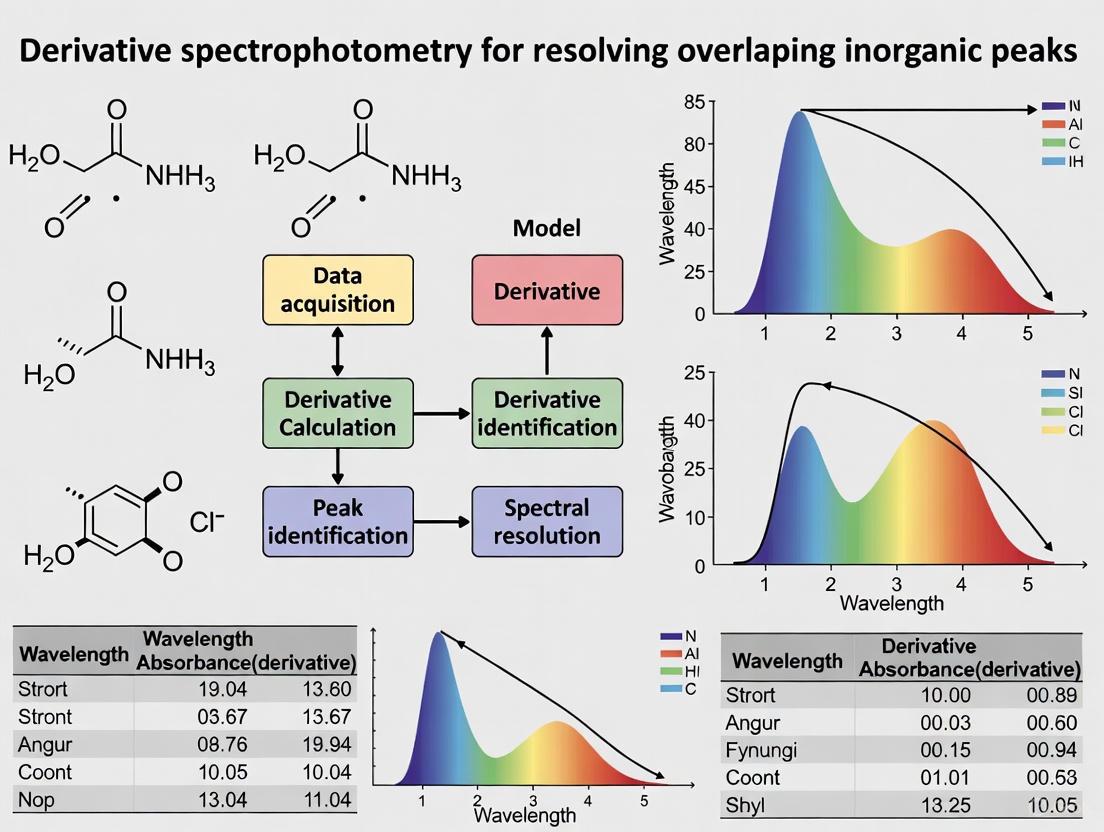
Within the context of derivative spectrophotometry for resolving overlapping inorganic peaks, the strategic selection of derivative order is a critical determinant of analytical success. This application note delineates the fundamental relationship between derivative order and spectral feature enhancement, providing a structured framework for researchers and drug development professionals to optimize their methodologies. Higher-order derivatives selectively amplify sharper spectral features and suppress broad, interfering backgrounds, making them indispensable for detecting weak analyte signals obscured by stronger, broader bands. By presenting quantitative data, detailed protocols, and decision-support tools, this document empowers scientists to systematically match derivative order to their specific analytical challenges, thereby improving the resolution and accuracy of their spectral analyses.
Derivative spectrophotometry operates on the mathematical principle of calculating the rate of change of a spectral curve (absorbance versus wavelength). The first derivative of an absorption spectrum represents its slope, the second derivative represents its curvature, and so on for higher orders [19]. This transformation of the spectral data confers several analytical advantages, including the amplification of sharp spectral features, the suppression of broad-band background interference, and the facilitation of precise peak location.
A cornerstone property of derivatives is their differential effect on peaks based on width. The amplitude of the nth derivative of a peak is inversely proportional to the nth power of its width for signals of the same shape and amplitude [19]. Consequently, narrow peaks are preserved and even enhanced in higher-order derivatives, while broad peaks are drastically attenuated. This property is the primary mechanism by which overlapping peaks can be resolved; a weak, narrow analyte peak sitting on the shoulder of a strong, broad interferent can be made analytically accessible through appropriate derivative order selection. Furthermore, derivatives enable precise identification of peak maxima and inflection points. The maximum of a symmetrical peak corresponds to a zero-crossing in its first derivative, and an inflection point in a sigmoidal curve corresponds to a zero-crossing in its second derivative [19].
Quantitative Relationship: Derivative Order and Bandwidth
The effect of derivative order on peaks of varying bandwidth can be quantitatively described, providing a predictive framework for method development. The following data, synthesized from empirical studies, allows researchers to anticipate the signal behavior of their target analytes.
Table 1: Impact of Derivative Order on Peak Amplitude Relative to Peak Width
| Derivative Order | Effect on Narrow Peaks | Effect on Broad Peaks | Approximate Amplitude Relationship |
|---|---|---|---|
| 0 (Original) | Baseline amplitude | Baseline amplitude | Amplitude ∝ H |
| 1st | Well-preserved | Significantly reduced | Amplitude ∝ H / W |
| 2nd | Enhanced | Greatly suppressed | Amplitude ∝ H / W² |
| 3rd & Higher | Further enhanced | Nearly eliminated | Amplitude ∝ H / Wⁿ |
Note: H = Peak Height, W = Peak Width (e.g., Full Width at Half Maximum, FWHM), n = Derivative Order.
Table 2: Guideline for Derivative Order Selection Based on Analytical Goal
| Analytical Goal | Recommended Order | Rationale and Application Example |
|---|---|---|
| Locate Peak Maxima | 1st | Zero-crossing of the first derivative precisely indicates the peak maximum of a symmetrical band [19]. |
| Locate Inflection Points (e.g., Titration Endpoints) | 2nd | Zero-crossing of the second derivative indicates the inflection point of a sigmoidal curve [19]. |
| Enhance Sharp Features / Detect Shoulders | 2nd | Amplifies sharp peaks and shoulders, helping to distinguish them from broader underlying bands. |
| Suppress Broad Background Interference | 2nd or 3rd | The strong inverse relationship with width (1/Wⁿ) effectively removes broad baseline drift or background signals [20]. |
| Resolve Closely Spaced Narrow Peaks | 2nd to 4th | Higher orders provide increased feature resolution, creating distinct, measurable derivative features for each narrow peak. |
Experimental Protocols
Protocol 1: Baseline Correction using the airPLS Algorithm
Prior to derivative computation, effective baseline correction is essential to remove low-frequency fluorescence or instrumental drift that can distort derivatives [20].
- Data Input: Load the spectral imaging data set as a matrix X of dimensions m by n, where m is the number of spectra and n is the number of data points per spectrum.
- Algorithm Initialization: For each spectrum (row vector x), initialize the weight vector w⁰ = 1. Set the smoothness parameter λ (a typical starting value is 10⁷) and the maximum iteration count (e.g., 20) [20].
- Iterative Fitting:
- Compute the fitted baseline z at iteration t by solving the weighted penalized least-squares problem: z = (W + λDᵀD)⁻¹ W x, where D is a derivative matrix and W is the diagonal weight matrix [20].
- Update the weight vector for the next iteration. For all points where the signal x is greater than the candidate baseline z, set their weight to zero. For points below the baseline, keep the weight as 1.
- Termination Check: The iteration stops when the termination criterion is met (the sum of absolute differences between the original signal and the fitted baseline falls below a threshold) or the maximum iteration count is reached.
- Baseline Subtraction: Obtain the corrected spectrum x* by subtracting the final fitted baseline z from the original spectrum x.
Protocol 2: Derivative Computation and Smoothing via the Savitzky-Golay Method
The Savitzky-Golay method is a robust approach that combines smoothing and derivative computation in a single step, helping to manage high-frequency noise amplified by differentiation [21] [19].
- Parameter Selection:
- Window Size (Polynomial Filter Length): Choose an odd number of data points. A larger window provides more smoothing but may obscure fine details.
- Polynomial Order: Select the order of the polynomial to be fitted to the data within the moving window (e.g., 2 or 3).
- Derivative Order: Specify the desired derivative order (e.g., 1 for first derivative, 2 for second derivative).
- Convolution: For each point in the spectrum, a Savitzky-Golay convolution filter with coefficients corresponding to the selected parameters is applied to the data within the moving window.
- Output: The algorithm outputs the smoothed derivative spectrum directly. The result is a balance between noise reduction and preservation of the true spectral shape.
The Scientist's Toolkit: Research Reagent Solutions
Table 3: Essential Materials and Algorithms for Derivative Spectrophotometry
| Item / Reagent | Function / Application | Notes |
|---|---|---|
| Savitzky-Golay Algorithm | Simultaneous smoothing and derivative calculation. | Critical for controlling noise amplification. Optimal polynomial order and window size must be determined empirically [21] [19]. |
| airPLS Algorithm | Automated baseline correction for Raman and other spectral data. | Removes fluorescent background without requiring user intervention, improving reproducibility [20]. |
| Central-Difference Method | A simple numerical method for derivative calculation. | Computes the derivative as the average slope between adjacent points (e.g., Y'ⱼ = (Yⱼ₊₁ - Yⱼ₋₁) / (Xⱼ₊₁ - Xⱼ₋₁)) without an x-axis shift [19]. |
| PCA-Despiking Algorithm | Identification and removal of cosmic ray spikes from spectral data. | Uses principal component analysis to distinguish spikes from genuine spectral features, preventing analytical distortion [20]. |
Implementation Workflow and Decision Pathways
The following diagram outlines the logical process for selecting and applying the appropriate derivative order to resolve overlapping spectral bands.
Diagram 1: Derivative Order Selection Workflow.
Advanced Applications and Considerations
Signal-to-Noise Ratio Management
A critical consideration in derivative spectroscopy is the inherent amplification of high-frequency noise. Each successive derivative operation typically degrades the signal-to-noise ratio (SNR) [19]. Therefore, the choice of derivative order is always a trade-off between the desired degree of feature resolution and the acceptable level of noise. This necessitates the integration of effective smoothing protocols, such as the Savitzky-Golay filter, directly into the derivative computation process [21]. The optimization of smoothing parameters (e.g., window size, polynomial order) must be conducted judiciously to avoid excessive distortion of the genuine spectral data.
Asymmetry Detection
Derivative analysis provides a sensitive method for detecting subtle asymmetries in otherwise symmetrical peaks. For instance, a pure Gaussian peak exhibits specific, symmetrical patterns in its derivatives. The presence of exponential broadening or other distorting factors can introduce asymmetry, which becomes readily apparent in the derivative spectra. Specifically, the second derivative of a slightly asymmetrical peak may show unequal positive peaks where a symmetrical peak would have equal ones [19]. This capability is invaluable for diagnosing peak purity and identifying the presence of unresolved secondary components.
The strategic selection of derivative order, guided by the fundamental principle of inverse dependence on peak width, is a powerful tool for enhancing the resolution of overlapping inorganic peaks in spectrophotometry. By applying lower orders (1st) for pinpointing maxima and inflection points, and higher orders (2nd to 4th) for suppressing broad backgrounds and resolving narrow, closely spaced peaks, researchers can extract critical analytical information that remains hidden in the original absorption spectrum. Successful implementation requires a holistic approach that integrates robust baseline correction, controlled smoothing, and an understanding of the signal-to-noise trade-offs. When executed within this structured framework, derivative spectrophotometry significantly advances the capabilities of quantitative and qualitative analysis in research and drug development.
Fundamental Advantages over Zero-Order Spectrophotometry in Inorganic Analysis
Derivative spectrophotometry is a powerful analytical technique that involves converting a normal zero-order absorption spectrum into its first or higher-order derivative, thereby transforming overlapping spectral features into distinct, resolvable signals [22]. This method provides a compelling solution for the analysis of complex inorganic mixtures, where overlapping absorption bands of analytes and matrix interferences often make the extraction of reliable qualitative and quantitative data difficult [22]. In inorganic analysis, this technique leverages mathematical differentiation to enhance spectral resolution, eliminate background interference, and facilitate the direct determination of metal ions even in complex matrices [22]. This application note details the specific advantages and provides a standardized protocol for exploiting derivative spectrophotometry in inorganic analysis, framed within broader research on resolving overlapping inorganic peaks.
Fundamental Advantages in Inorganic Analysis
The transition from zero-order to derivative spectrophotometry offers several distinct advantages for inorganic analysis, summarized in the table below.
Table 1: Core Advantages of Derivative over Zero-Order Spectrophotometry in Inorganic Analysis
| Feature | Zero-Order Spectrophotometry | Derivative Spectrophotometry | Impact on Inorganic Analysis |
|---|---|---|---|
| Spectral Resolution | Limited; overlapping peaks appear as a single, broad band [22] | Enhanced; resolves closely adjacent and unresolved peaks [22] [10] | Enables simultaneous determination of multiple metal ions without physical separation [22] |
| Background Elimination | Susceptible to interference from sample matrix and turbidity [22] | Effective elimination of background and matrix interferences [22] | Allows direct analysis in complex matrices (e.g., water, soils) with minimal sample pre-treatment [22] |
| Sensitivity to Subtle Features | Poor detection of weak spectral features on steep slopes [22] | Enhanced detection of minor spectral features and subtle spectral shifts [22] [10] | Improves detection of trace metals and complexes with weak or overlapping chromophores [22] |
| Signal Discrimination | Broad bands can obscure narrow analyte signals [10] | Suppresses broad band signals while enhancing sharp analyte peaks [10] | Increases selectivity for metal complexes with narrow bandwidths against a broad interfering background [22] |
| Quantification in Mixtures | Often requires prior separation or complex chemometrics [22] | Enables multicomponent analysis without prior separation using techniques like zero-crossing [22] [10] | Simplifies and speeds up the quantitative analysis of inorganic ion mixtures [22] [23] |
The theoretical basis for these advantages lies in the transformation of the spectral data. A zero-order spectrum is a plot of absorbance (A) versus wavelength (λ). The first-derivative spectrum (dA/dλ) represents the rate of change of the absorbance slope, which passes through zero at the λ_max of the original spectrum [10]. The second-derivative spectrum (d²A/dλ²) represents the curvature of the absorption spectrum and is inversely related to the original band, providing even greater resolution of sharp peaks [22] [10]. Higher-order derivatives can further enhance resolution but at the cost of a degraded signal-to-noise ratio [10].
Experimental Protocol: Determination of Metal Ions in a Mixture
The following protocol outlines a general method for the simultaneous determination of two metal ions, such as Nickel(II) and Cobalt(II), using first-derivative spectrophotometry based on complex formation with specific ligands [22].
Research Reagent Solutions
Table 2: Essential Materials and Reagents
| Item | Function/Description |
|---|---|
| UV-Vis Spectrophotometer | Double-beam instrument capable of recording derivative spectra, equipped with 1 cm quartz cells [23]. |
| Analytical Software | Software for derivative processing (e.g., 1st-4th order) and data analysis [10] [23]. |
| 2-Acetylpyridine-4-methyl-3-thiosemicarbazone (APMT) | Ligand for forming complexes with Ni(II) and Co(II) for sensitive detection [22]. |
| Methanol or Ethanol | HPLC/UV-grade solvent for preparing stock and standard solutions [23]. |
| Buffer Solution (e.g., pH 9.2) | To maintain optimal pH for complex formation [22]. |
| Nickel & Cobalt Standards | High-purity salts for preparing stock standard solutions [22]. |
Step-by-Step Workflow
The following diagram illustrates the complete experimental workflow from sample preparation to quantitative analysis.
Diagram 1: Experimental Workflow for Derivative Analysis of Metal Ions.
Detailed Methodology
Preparation of Standard Solutions:
- Prepare individual stock solutions (100 mg/L) of Ni(II) and Co(II) in high-purity water or a suitable solvent [23].
- Prepare a stock solution of the complexing ligand (APMT) in methanol or ethanol [22].
- Prepare a buffer solution to maintain the pH at the optimal value for complex formation (e.g., pH 9.2 for APMT complexes) [22].
Formation of Metal Complexes:
- Pipette appropriate aliquots of each metal stock solution into a series of 10 mL volumetric flasks.
- Add 1.0 mL of the ligand solution and 2.0 mL of the buffer solution to each flask.
- Dilute to the mark with the solvent and mix thoroughly. Allow the reaction mixture to stand for 10 minutes to ensure complete color development [22].
Spectral Acquisition and Derivatization:
- Using a double-beam spectrophotometer, record the zero-order absorption spectra of the solutions against a reagent blank over the wavelength range of 220 to 400 nm [23].
- Transfer the spectral data to the instrument's software or a dedicated data processing tool.
- Convert the zero-order spectra into first-derivative spectra (dA/dλ). The software typically uses algorithms like Savitzky-Golay for this numerical differentiation [10].
Quantitative Measurement via Zero-Crossing:
- In the first-derivative spectrum, identify the wavelength where the derivative signal for one component (e.g., Co-APMT complex) crosses the zero line (zero-crossing point) [10] [23].
- At this specific wavelength, measure the absolute value of the derivative signal (peak-to-trough) for the other component (e.g., Ni-APMT complex). This signal is directly proportional to the concentration of the second component, free from interference from the first [22] [23].
- Repeat this process at the zero-crossing point of the second component to quantify the first.
Calibration and Analysis:
- Construct first-derivative calibration curves for each metal ion at the selected zero-crossing wavelengths using a series of standard solutions [23].
- Determine the concentration of Ni(II) and Co(II) in unknown samples by interpolating their derivative signals into the respective calibration curves.
Derivative spectrophotometry provides a significant analytical advantage over zero-order methods for inorganic analysis by fundamentally enhancing the information content of UV-Vis spectra. Its ability to resolve overlapping peaks, suppress matrix interferences, and facilitate the simultaneous quantification of multiple metal ions without costly separation steps makes it an invaluable, cost-effective tool for researchers and analysts. The provided protocol offers a reliable foundation for applying this technique to the determination of metal ions in complex mixtures, contributing to more efficient and resolved analytical outcomes.
Practical Methods and Applications in Metal Ion and Inorganic Analysis
Zero-Crossing Technique for Binary and Ternary Mixture Analysis
Derivative spectrophotometry provides a powerful tool for resolving overlapping spectral bands in multicomponent mixtures without preliminary separation. The zero-crossing technique represents a specific mathematical approach within derivative spectroscopy that enables quantitative determination of individual components in binary and ternary mixtures by selecting wavelengths where derivative values of interfering compounds equal zero. This method is particularly valuable in pharmaceutical analysis for simultaneous drug quantification and in inorganic material characterization where spectral overlapping complicates direct measurement.
The fundamental principle relies on computing the first or second derivative of absorption spectra, which transforms broad, overlapping peaks into sharper, more distinct features. At specific wavelengths where the derivative spectrum of an interfering component crosses zero, the amplitude of the target compound's derivative spectrum becomes directly proportional to its concentration, enabling selective quantification amid spectral interference. This technique offers significant advantages in cost efficiency, simplicity, and environmental friendliness compared to chromatographic methods, while maintaining adequate precision and accuracy for quality control applications [24].
Theoretical Foundations
Principles of Derivative Spectrophotometry
Derivative spectrophotometry involves mathematical transformation of zero-order absorption spectra into their first, second, or higher-order derivatives. This transformation provides two primary benefits: enhanced spectral resolution and background suppression. The technique amplifies subtle spectral features while minimizing the impact of baseline shifts or tilts that often complicate direct absorbance measurements [25].
The zero-crossing method specifically utilizes the property that at wavelengths where the derivative spectrum of an interfering compound equals zero, the measured derivative signal depends solely on the concentration of the target analyte. For binary mixtures, this enables selective quantification of each component by measuring derivative amplitudes at carefully selected zero-crossing points of the other component [24].
Mathematical Formalism
The foundational mathematics for derivative spectrophotometry begins with the Beer-Lambert law. For a mixture of n components, the total absorbance at wavelength λ can be expressed as:
A(λ) = Σ εi(λ)cil + B(λ)
Where εi(λ) is the molar absorptivity of component i, ci is its concentration, l is the path length, and B(λ) represents baseline contributions.
The n^th-order derivative spectrum is then expressed as:
d^nA(λ)/dλ^n = Σ cil d^nεi(λ)/dλ^n + d^nB(λ)/dλ^n
For the zero-crossing technique, the critical condition occurs when:
d^nεj(λzc)/dλ^n = 0
At this specific wavelength (λ_zc), the derivative signal becomes dependent only on the target analyte (component i), allowing its concentration to be determined without interference from component j [24] [25].
Experimental Protocols
Protocol 1: Zero-Crossing Method for Binary Mixture Analysis
This protocol details the application of first-derivative spectrophotometry with zero-crossing points for resolving two-component mixtures, adapted from validated pharmaceutical analysis methods [25].
Materials and Reagents
Table 1: Essential Research Reagent Solutions
| Reagent/Material | Specification | Primary Function |
|---|---|---|
| UV-grade methanol | Analytical grade | Solvent for standard and sample solutions |
| Famotidine (FAM) | Reference standard (≥99% purity) | Target analyte 1 |
| Metronidazole (MET) | Reference standard (≥98% purity) | Target analyte 2 |
| Volumetric flasks | Class A, 10-100 mL capacity | Precise solution preparation |
| Quartz cuvettes | 1 cm path length | UV sample containment |
Instrumentation and Parameters
- UV/VIS Spectrophotometer capable of derivative spectrum acquisition
- Spectral bandwidth: 1 nm
- Scanning speed: Medium
- Wavelength range: 200-400 nm
- Derivative parameters: First derivative, Δλ = 10 nm, scaling factor = 10
Procedure
Standard Solution Preparation
- Prepare individual stock solutions of FAM (1000 µg/mL) and MET (2000 µg/mL) in methanol [24].
- Dilute appropriate aliquots to prepare working standards in the concentration ranges of 3-20 µg/mL for FAM and 4-20 µg/mL for MET.
Spectral Acquisition
- Scan the zero-order absorption spectra of all standard solutions against a methanol blank.
- Compute the first-derivative spectra using the established parameters.
Zero-Crossing Point Determination
- Examine the first-derivative spectrum of pure FAM to identify wavelengths where it crosses zero.
- Similarly, identify zero-crossing points in the first-derivative spectrum of pure MET.
- Confirm that at these wavelengths, only one component contributes to the derivative signal.
Calibration Curve Construction
- For FAM quantification: Measure the derivative amplitudes of standard solutions at the zero-crossing wavelength of MET.
- For MET quantification: Measure the derivative amplitudes of standard solutions at the zero-crossing wavelength of FAM.
- Plot derivative amplitudes versus concentrations to establish linear calibration curves.
Sample Analysis
- Process unknown samples identically to standard solutions.
- Determine component concentrations using the established calibration equations.
Figure 1: Binary mixture analysis workflow using zero-crossing technique
Protocol 2: Advanced Techniques for Ternary Mixture Resolution
This protocol extends the zero-crossing principle to ternary systems using derivative double divisor ratio spectra (D/DDRS) and Fourier function transformation, suitable for analyzing complex three-component mixtures with significant spectral overlap [24].
Materials and Reagents (Additional)
Table 2: Additional Reagents for Ternary Mixture Analysis
| Reagent/Material | Specification | Primary Function |
|---|---|---|
| Amoxicillin (AMX) | Reference standard (≥99% purity) | Third analyte for ternary system |
| Simulated gastric fluid | pH ~1.2 | Biological matrix simulation |
| Fourier transform processing | Software capability | Advanced spectral resolution |
Procedure: Derivative Double Divisor Ratio Spectra Method
Standard Solution Preparation
- Prepare stock solutions of all three components: FAM (1000 µg/mL), MET (2000 µg/mL), AMX (2000 µg/mL) in methanol.
- Prepare working standards in the ranges: 3-20 µg/mL (FAM), 4-20 µg/mL (MET), 12-40 µg/mL (AMX) [24].
Double Divisor Preparation
- Create a standard mixture solution containing two of the three components at fixed concentrations (e.g., MET and AMX as divisors for FAM quantification).
Ratio Spectrum Generation
- Divide the absorption spectrum of the ternary mixture by the spectrum of the double divisor solution to obtain the ratio spectrum.
Derivative Calculation
- Compute the first derivative of the ratio spectrum.
- Select appropriate wavelengths for each component where the derivative values of interferents are zero.
Calibration and Quantification
- Measure derivative amplitudes at selected wavelengths for standard solutions.
- Construct calibration curves and determine concentrations in unknown samples.
Procedure: Fourier Function Transformation Method
Spectral Acquisition
- Record zero-order absorption spectra of standard and sample solutions as previously described.
Fourier Transformation
- Apply Fourier transform to the absorbance spectra to convert them from wavelength domain to frequency domain.
- This process helps eliminate various types of interferences and resolves overlapping peaks [24].
Component Resolution
- Process the transformed spectra using appropriate algorithms to isolate individual component signals.
- Quantify each component based on their unique spectral features in the transformed domain.
Applications and Data Analysis
Pharmaceutical Analysis Case Study
The zero-crossing technique has been successfully applied to the simultaneous determination of famotidine, metronidazole, and amoxicillin in combined tablet formulations and simulated gastric fluid [24]. This triple therapy combination is commonly used for Helicobacter pylori eradication, requiring precise quality control methods.
Table 3: Quantitative Parameters for Ternary Mixture Analysis
| Parameter | Famotidine (FAM) | Metronidazole (MET) | Amoxicillin (AMX) |
|---|---|---|---|
| Linearity Range (µg/mL) | 3-20 | 4-20 | 12-40 |
| Correlation Coefficient (r) | 0.9999 | 0.9999 | 0.9999 |
| Zero-Crossing Wavelengths (nm) | 320 (1st derivative) | 285 (1st derivative) | 308 (2nd derivative) |
| Limit of Detection (LOD) | Not specified | Not specified | Not specified |
| Limit of Quantification (LOQ) | Not specified | Not specified | Not specified |
Analytical Performance
The validated methods demonstrate excellent precision and accuracy for quality control applications. The greenness assessment using NEMI, AGREE, GAPI, and CALIFICAMET-HEXAGON tools confirmed the environmental advantages of these spectrophotometric methods compared to HPLC or HPTLC techniques [24]. The main benefits include:
- Reduced analysis time and operational complexity
- Lower solvent consumption and waste generation
- Cost-effectiveness for routine analysis
- Adequate accuracy and precision for quality control purposes
Figure 2: Method selection for ternary mixture resolution
Troubleshooting and Optimization
Common Technical Issues
- Insufficient resolution: Optimize derivative parameters (Δλ, scaling factor) or consider alternative techniques like double divisor ratio spectra or Fourier transformation for highly overlapping mixtures [24].
- Baseline drift: Ensure proper solvent matching between samples and blanks. Use higher derivative orders to minimize baseline effects.
- Poor reproducibility: Standardize sample preparation procedures and instrument calibration. Verify the stability of standard solutions.
Method Optimization Guidelines
- Selection of Δλ value: Larger Δλ values provide better noise reduction but decrease spectral resolution. Optimal Δλ typically ranges between 5-15 nm.
- Derivative order: First derivative is most common for zero-crossing applications. Second derivative provides enhanced resolution but with increased noise.
- Wavelength selection: Verify zero-crossing points carefully using pure standards. Ensure the selected wavelength provides adequate sensitivity for the target analyte.
The zero-crossing technique in derivative spectrophotometry provides a robust, cost-effective approach for resolving binary and ternary mixtures in pharmaceutical and inorganic material analysis. The method's strength lies in its mathematical simplicity, experimental efficiency, and alignment with green analytical chemistry principles. While chromatographic methods may offer higher specificity for complex mixtures, the zero-crossing technique delivers adequate performance for quality control applications while minimizing operational costs and environmental impact. The continued development of complementary approaches like derivative double divisor ratio spectra and Fourier function transformation extends the applicability of these methods to increasingly challenging analytical problems.
Quantitative Determination of Metal Ions via Complex Formation with Diverse Ligands
The quantitative determination of metal ions through complex formation represents a cornerstone of analytical chemistry, with critical applications spanning environmental monitoring, pharmaceutical development, and clinical diagnostics. When metal ions coordinate with organic ligands, they typically form complexes that exhibit distinct absorption spectra in the ultraviolet-visible (UV-Vis) region, enabling their detection and quantification even in complex matrices [22]. The fundamental principle underpinning this methodology is the linear correlation between the concentration of the metal-ligand complex in solution and the absorbance measured at a characteristic wavelength, as described by the Beer-Lambert law [26].
Traditional direct absorption spectrophotometry, however, often encounters limitations when analyzing mixtures where spectral bands significantly overlap, potentially obscuring the target analyte's signal. Derivative spectrophotometry effectively addresses this challenge by mathematically transforming the zero-order absorption spectrum into its first or higher-order derivatives [22] [10]. This transformation confers significant advantages, including enhanced spectral resolution, the ability to identify subtle spectral features, and effective elimination of background interference from other sample components [22]. By converting inflections in the original spectrum into distinct maxima and minima, derivative techniques facilitate the discrimination and quantification of metal ions in multi-component systems without requiring prior physical separation [22] [10].
Key Spectrophotometric Methods for Complex Analysis
Several established experimental methods enable researchers to determine both the stoichiometry and formation constants of metal-ligand complexes, which are crucial parameters for developing robust quantitative assays.
Method of Continuous Variations (Job's Method)
The method of continuous variations is used to determine the stoichiometry of a metal-ligand complex. In this approach, a series of solutions is prepared such that the total moles of metal and ligand remains constant across all solutions, but their relative mole fractions vary systematically [26].
- Procedure: The absorbance of each solution is measured at a wavelength where the complex absorbs strongly. A graph of absorbance versus the mole fraction of the ligand typically displays two linear branches that intersect at a point corresponding to the stoichiometric ratio of the complex [26].
- Data Interpretation: The mole fraction of ligand at the intersection point is used to determine the stoichiometry of the complex. For a complex of formula ML
y, the value ofyis calculated asy = (X_L / (1 - X_L)), whereX_Lis the mole fraction of the ligand at the intersection [26].
Mole-Ratio Method
The mole-ratio method provides an alternative for determining metal-ligand stoichiometry. In this procedure, the amount of one reactant, typically the metal, is held constant, while the amount of the other reactant (the ligand) is varied [26].
- Procedure: The absorbance of each solution is monitored at an appropriate wavelength. A plot of absorbance against the ratio of ligand to metal concentration will show a distinct break point or change in slope once all metal ions have been complexed, indicating the stoichiometric ratio [26].
- Data Interpretation: The ligand-to-metal ratio at which the absorbance plot changes slope reveals the complex's composition. This method is particularly effective for complexes that exhibit strong and exclusive absorption at the monitored wavelength [26].
Slope-Ratio Method
The slope-ratio method is especially useful for studying weak complexes and is applicable only to systems where a single complex species is formed. The underlying assumption is that the complex formation reaction can be driven to completion by a significant excess of either the metal ion or the ligand [26].
- Procedure:
- A series of solutions is prepared with a large excess of ligand, and absorbance is measured as a function of varying metal concentration.
- A second series is prepared with a large excess of metal, and absorbance is measured as a function of varying ligand concentration.
- Data Interpretation: The slopes of the resulting linear plots of absorbance versus concentration are determined. The ratio of these slopes (slope from ligand variation / slope from metal variation) yields the ligand-to-metal combining ratio in the complex [26].
The following workflow diagram illustrates the logical process of method selection and analysis for determining metal-ligand complex stoichiometry:
Derivative Spectrophotometry for Resolution of Overlapping Peaks
Derivative spectrophotometry serves as a powerful enhancement to conventional absorption measurements, particularly for resolving overlapping inorganic peaks within a thesis research context. This technique functions by calculating the first or higher-order derivatives of the absorbance spectrum with respect to wavelength [10].
The process of derivative transformation enhances the visibility of subtle spectral features and narrow bands that might be obscured in the zero-order spectrum. The fundamental equations governing derivative spectra are [10]:
- First Derivative (dA/dλ): Represents the rate of change of absorbance with wavelength. Its spectrum passes through zero at the λ_max of the original absorption band.
- Second Derivative (d²A/dλ²): Represents the curvature of the absorption spectrum. It is directly proportional to the analyte concentration and is particularly effective for suppressing broad, featureless background interference.
The following diagram illustrates the spectral transformation process from zero-order to higher-order derivatives, showing how overlapping peaks can be resolved:
Higher-order derivatives (third and fourth) can provide even greater resolution for complex mixtures, though they often come with a decreased signal-to-noise ratio [10]. The zero-crossing technique is a commonly employed measurement method in derivative spectrophotometry, where the amplitude is measured at a wavelength where the derivative of the interfering component crosses zero, effectively eliminating its contribution [10].
Experimental Protocols and Data Analysis
Protocol for Determination of Nickel(II) Using Derivative Spectrophotometry
This protocol details the determination of Ni(II) using a complexing agent based on adaptations from published methodologies [22].
- Principle: Ni(II) ions form a colored complex with 2-acetylpyridine-4-methyl-3-thiosemicarbazone (APMT), which can be quantified using first or second-order derivative spectrophotometry to minimize matrix interference.
- Reagents:
- Stock Ni(II) solution (1000 mg/L in deionized water).
- APMT solution (0.5% w/v in methanol).
- Buffer solution (ammonium acetate-acetic acid, pH 5.5).
- Procedure:
- Calibration Curve: Transfer aliquots of standard Ni(II) solution (0.5-5.0 µg/mL) into a series of 10 mL volumetric flasks.
- Add 2.0 mL of buffer solution and 1.5 mL of APMT reagent to each flask.
- Dilute to the mark with deionized water and mix thoroughly. Allow 15 minutes for full color development.
- Sample Preparation: Treat the unknown sample solution identically to the calibration standards.
- Spectral Measurement: Record the absorption spectra of both standards and samples between 350-450 nm. Convert the zero-order spectra to second-order derivative spectra (typically using Δλ = 4-6 nm).
- Quantification: Measure the amplitude of the second-derivative peak (or trough) for each standard and construct a calibration curve. Determine the Ni(II) concentration in the unknown sample by interpolating from this curve.
Protocol for Simultaneous Determination of Copper(II) and Iron(II)
This protocol outlines an approach for the simultaneous determination of Cu(II) and Fe(II) in a mixture using first-order derivative spectrophotometry [22].
- Principle: Both metal ions form complexes with specific ligands, exhibiting overlapping absorption spectra. Derivative spectrophotometry allows for the resolution of these overlapping bands.
- Reagents:
- Stock Cu(II) and Fe(II) solutions.
- Appropriate complexing agent (e.g., ammonia buffer system or specific chelating agents).
- Procedure:
- Calibration: Prepare separate series of standard solutions for Cu(II) and Fe(II) across the expected concentration range.
- Form the complexes and record the zero-order absorption spectra (e.g., 500-800 nm).
- Transform the spectra into first-derivative spectra.
- Wavelength Selection: Identify two wavelengths: λ₁ where the derivative value for Fe(II) is zero (for Cu determination), and λ₂ where the derivative value for Cu(II) is zero (for Fe determination).
- Measurement: For the mixture, measure the first-derivative absorbance values at λ₁ and λ₂.
- Calculation: Using the calibration curves prepared from pure standards, calculate the concentrations of Cu(II) and Fe(II) in the mixture by solving simultaneous equations derived from the derivative absorbance measurements at the two selected wavelengths.
Table 1: Quantitative Data for Metal Ion Determination via Complex Formation
| Metal Ion | Complexing Ligand | λ_max (nm) | Linear Range (µg/mL) | Remarks / Derivative Order |
|---|---|---|---|---|
| Ni(II) | APMT [22] | ~390 (Zero-order) | 0.5 - 5.0 | 2nd derivative for enhanced selectivity |
| Zr(IV) | Mixed Aqueous-Organic [22] | Varies | Micro-amounts | Normal and 1st-derivative methods used |
| Co(II) | APMT [22] | ~390 (Zero-order) | Not Specified | Simultaneous determination with Ni(II) |
| Pd(II) | Not Specified [22] | Not Specified | Trace quantities | Non-extractive method reported |
| Cu(II) | Various [22] | Varies | Varies | Often used in simultaneous analysis with other metals (e.g., Zn²⁺, Cd²⁺) |
| Fe(II) | Various [22] | Varies | Varies | Simultaneous determination with Cu(II) reported |
Table 2: Comparison of Key Spectrophotometric Methods for Stoichiometry Determination
| Method | Principle | Key Advantage | Primary Application |
|---|---|---|---|
| Continuous Variations (Job's Method) | Varies mole fraction while keeping total moles constant. | Directly identifies stoichiometric ratio from a single graph. | Ideal for determining stoichiometry of strong, single complexes. |
| Mole-Ratio | Varies ligand/metal ratio while keeping metal concentration constant. | Simple interpretation; works well for strong complexes with exclusive absorption. | Suitable for systems where the complex has a unique, strong absorption band. |
| Slope-Ratio | Measures slopes of absorbance plots under conditions of large excess of metal or ligand. | Applicable to weak complexes that do not form quantitatively at equimolar ratios. | Best for systems where a single complex is formed but has moderate stability. |
The Scientist's Toolkit: Essential Reagents and Materials
Table 3: Key Research Reagent Solutions for Metal-Ligand Complexation Studies
| Reagent / Material | Function / Purpose | Example Specifications / Notes |
|---|---|---|
| Standard Metal Ion Solutions | Primary standards for calibration. | High-purity salts (e.g., NiCl₂·6H₂O, CuSO₄·5H₂O); concentration typically 1000 mg/L in 1-2% HNO₃. |
| Complexing Agents (Ligands) | Selective complex formation with target metal ions. | APMT for Ni/Co [22], DMHBIH for Zn [22], 1,10-phenanthroline for Fe. Purity >98%. |
| Buffer Solutions | Maintain constant pH for optimal complex formation. | Ammonium acetate (pH ~5.5), phosphate buffers, borate buffers. Choice depends on ligand and metal chemistry. |
| UV-Vis Spectrophotometer | Measure absorbance of metal-ligand complexes. | Capable of scanning 200-800 nm; derivative software functionality is essential. |
| Derivative Spectrophotometry Software | Generate 1st to 4th derivative spectra from zero-order data. | Built-in instrument software or external data processing packages (e.g., Savitzky-Golay algorithm [10]). |
Advanced Techniques and Complementary Methods
While UV-Vis derivative spectrophotometry is a powerful tool, comprehensive characterization of metal-ligand complexes often requires integration with other analytical techniques.
Computational Chemistry methods, particularly Density Functional Theory (DFT), provide deep theoretical insights into the charge-transfer interactions responsible for the absorption spectra of metal complexes. DFT calculations can predict ground state structures, identify the most probable bonding sites between metals and ligands, and calculate the energies of the Highest Occupied and Lowest Unoccupied Molecular Orbitals (HOMO-LUMO), which is crucial for understanding charge transfer transitions observed in UV-Vis spectra [27]. The HOMO-LUMO energy gap also serves as an indicator of the complex's chemical reactivity [27].
Other Spectroscopic Techniques provide complementary information:
- Electron Paramagnetic Resonance (EPR) Spectroscopy: A versatile tool for probing the geometric and electronic structures of paramagnetic metal centers in complexes, such as those of copper(II) or iron, providing details unavailable from UV-Vis spectroscopy alone [28].
- Nuclear Magnetic Resonance (NMR) Spectroscopy: Particularly useful for diamagnetic metal complexes, NMR (especially (^{13})C and (^{31})P NMR) can help determine the composition of the internal coordination sphere and identify specific ligand atoms involved in metal binding [29].
Calorimetric Techniques like Isothermal Titration Calorimetry (ITC) offer a direct route to determining the thermodynamic parameters of metal complexation, including binding constants (K), reaction enthalpy (ΔH), and entropy (ΔS). This information is vital for understanding the driving forces behind complex formation and for applications like drug delivery system design [30].
Solid Phase Derivative Spectrophotometry (SPDS) for Preconcentration and Determination
Solid Phase Derivative Spectrophotometry (SPDS) is a powerful hybrid analytical technique that combines the matrix cleanup and analyte preconcentration capabilities of solid-phase extraction (SPE) with the enhanced selectivity of derivative spectrophotometry for the determination of trace analytes in complex mixtures [1] [31]. This method is particularly valuable for resolving overlapping absorption spectra of inorganic compounds, enabling accurate quantification without prior separation [1] [32]. The integration of these techniques allows researchers to achieve lower detection limits, improve method selectivity, and analyze complex sample matrices that would otherwise present significant challenges for conventional spectrophotometric methods [31] [32].
Theoretical Principles
Fundamentals of Solid-Phase Extraction
Solid-phase extraction is a sample preparation technique that operates on the principle of distributing analytes between a liquid sample phase and a solid stationary phase [31]. The process involves four key steps: conditioning the sorbent, loading the sample, washing away interferents, and eluting the target analytes. This process effectively separates analytes from the sample matrix, reduces interferent concentrations, and preconcentrates the target species to enhance detection capability [31].
The historical development of SPE has evolved from early applications using animal charcoal in the 1940s to modern cartridges and disks containing advanced sorbent materials [31]. Contemporary SPE configurations include traditional cartridges, pipette-tip SPE for small sample volumes, disks for processing large sample volumes, and multi-well formats for high-throughput applications [31].
Derivative Spectrophotometry Principles
Derivative spectrophotometry generates derivative spectra (first, second, or higher order) from parent zero-order absorption spectra through mathematical transformation [1]. This signal processing technique enhances spectral resolution by revealing subtle spectral features that are obscured in conventional absorption spectra. Key advantages include:
- Separation of overlapping signals: Derivative transformation can resolve closely spaced absorption bands [1]
- Elimination of background interference: Constant background contributions are minimized in higher-order derivatives [1]
- Enhanced selectivity: The technique provides a more characteristic spectral profile with additional maxima, minima, and zero-crossing points [1]
The combination of SPE with derivative spectrophotometry creates a synergistic effect where SPE handles matrix complexity through physical separation while derivative spectrophotometry addresses spectral complexity through mathematical enhancement [1] [32].
Applications and Quantitative Data
SPDS has been successfully applied to the determination of various inorganic analytes across different sample matrices. The table below summarizes key applications and their analytical performance characteristics:
Table 1: Applications of SPDS for Inorganic Analysis
| Analyte | Sample Matrix | Solid Phase | Derivative Order | Wavelength (nm) | LOD | Linear Range | Reference |
|---|---|---|---|---|---|---|---|
| Niobium (Nb) | Environmental waters, alloy steel | Microcrystalline naphthalene | Second | 380 | 25 ppb | Not specified | [32] |
| Niobium (Nb) | Aqueous samples | Microcrystalline naphthalene | Normal and Second | 380 | 25 ppb | Not specified | [32] |
The determination of niobium demonstrates the capability of SPDS for trace metal analysis, with the molar absorptivity reported as 1.40 × 10⁴ L/(mol·cm) and sensitivity of 0.0066 µg/cm² [32]. The optimal pH range for quantitative recovery of niobium(V) was established between 7-8 [32].
Table 2: Performance Characteristics for Niobium Determination Using SPDS
| Parameter | Value | Condition |
|---|---|---|
| Molar Absorptivity | 1.40 × 10⁴ L/(mol·cm) | 380 nm |
| Sensitivity | 0.0066 µg/cm² | - |
| Optimal pH Range | 7-8 | - |
| Preconcentration Factor | Not specified | - |
| Recovery | Quantitative | Optimized conditions |
Experimental Protocol: Niobium Determination
Reagents and Solutions
- Niobium standard solution: Prepare appropriate stock solutions of Nb(V) in high-purity water
- Complexing agent: Select based on specific methodology (not specified in source)
- Buffer solution: pH 7-8 for optimal sorption conditions
- Naphthalene adsorbent: Freshly precipitated microcrystalline form
- Elution solvent: Appropriate for desorbing the Nb complex from solid phase
Apparatus and Instrumentation
- UV-Vis spectrophotometer: Capable of recording normal and derivative spectra
- Solid-phase extraction assembly: Vacuum manifold or alternative extraction system
- pH meter: For precise adjustment of sample pH
- Centrifuge: For phase separation if required
Step-by-Step Procedure
SPE Cartridge Preparation:
- Prepare freshly precipitated microcrystalline naphthalene as adsorbent
- Pack the adsorbent into appropriate SPE cartridge or column
Sample Pretreatment:
- Adjust sample pH to 7-8 using appropriate buffer solution
- Add complexing agent if required for Nb(V) complex formation
Solid-Phase Extraction:
- Condition the naphthalene sorbent with appropriate solvent
- Load the prepared sample through the SPE cartridge at controlled flow rate
- Wash with suitable solution to remove interferents
- Elute the adsorbed Nb complex with optimal elution solvent
Spectrophotometric Measurement:
- Record zero-order absorption spectrum of the eluate
- Generate second-derivative spectrum from the parent spectrum
- Measure the amplitude at appropriate wavelength (380 nm for Nb)
Quantification:
- Construct calibration curve using standard solutions processed identically
- Determine Nb concentration in unknown samples from calibration curve
Optimization Parameters
- pH effect: Systematically evaluate pH 6-9 to confirm optimal range
- Sorbent mass: Optimize amount of naphthalene for quantitative recovery
- Flow rates: Control sample loading and elution flow rates for maximum efficiency
- Eluent volume: Minimize while maintaining complete elution
- Derivative parameters: Optimize wavelength scanning speed and derivative order
SPDS Workflow
The following workflow diagram illustrates the complete SPDS process from sample preparation to quantitative analysis:
Research Reagent Solutions
Table 3: Essential Materials for SPDS Experiments
| Reagent/Material | Function | Application Example |
|---|---|---|
| Microcrystalline naphthalene | Solid phase adsorbent | Preconcentration of niobium from aqueous samples [32] |
| C18 bonded silica | Reversed-phase sorbent | Extraction of non-polar complexes |
| Ionic contrast media | Potential interferent study | Investigation of analytical interference [33] |
| pH buffer solutions | Sample pH adjustment | Optimization of sorption efficiency (pH 7-8 for Nb) [32] |
| Derivatizing agents | Analyte complex formation | Enhanced retention and detection sensitivity |
Advantages and Limitations
Advantages of SPDS
- Enhanced sensitivity: Preconcentration step improves detection limits [31] [32]
- Improved selectivity: Derivative processing resolves overlapping peaks [1]
- Matrix cleanup: SPE removes interfering components [31]
- Green chemistry aspects: Reduced solvent consumption compared to liquid-liquid extraction [31]
Limitations and Considerations
- Reproducibility challenges: Dependence on instrumental parameters and derivatization conditions [1]
- Method optimization requirements: Need for careful optimization of both SPE and derivative parameters [1] [32]
- Potential interferents: Contrast media and other compounds may cause analytical interference [33]
Solid Phase Derivative Spectrophotometry represents a robust analytical approach for the determination of trace inorganic species in complex matrices. The method effectively combines the strengths of solid-phase extraction for sample preparation with the resolution enhancement of derivative spectroscopy. The application to niobium determination demonstrates the practical utility of SPDS for environmental and industrial analysis. Further development of new sorbent materials and optimization of derivative parameters will continue to expand the application scope and performance characteristics of this valuable analytical technique.
Simultaneous Analysis of Multiple Inorganic Ions in Environmental and Biological Samples
The simultaneous analysis of multiple inorganic ions is a critical requirement in environmental monitoring, biological research, and drug development. Traditional methods such as titration and colorimetry often lack the specificity, accuracy, and throughput needed for modern analytical challenges, particularly when dealing with complex sample matrices [34]. The presence of overlapping spectral peaks further complicates the precise quantification of individual ions, a challenge that derivative spectrophotometry helps to resolve by enhancing spectral resolution.
This application note provides a comprehensive framework for the simultaneous determination of common inorganic ions, including anions (SO₄²⁻, Cl⁻, NO₃⁻) and cations (Na⁺, NH₄⁺, K⁺, Mg²⁺, Ca²⁺), as well as nutrients like phosphate and silicate [35]. We detail advanced chromatographic and spectrophotometric protocols, complete with visualization tools and essential reagent solutions, to support researchers in obtaining accurate and reproducible results.
Key Analytical Techniques for Simultaneous Ion Analysis
Various techniques are employed for the detection of inorganic ions, each with distinct advantages and limitations. The choice of method depends on factors such as the required detection limit, sample complexity, and available instrumentation.
Comparison of Traditional Detection Methods for Inorganic Ions
| Method | Typical Ions Detected | Limit of Detection | Key Advantages | Key Disadvantages |
|---|---|---|---|---|
| Ion Chromatography | Common anions (SO₄²⁻, Cl⁻, NO₃⁻) and cations (Na⁺, NH₄⁺, K⁺, Mg²⁺, Ca²⁺) [35] | Varies by ion | High sensitivity and reproducibility; Can detect multiple ions simultaneously [34] | Requires specialized equipment |
| Atomic Absorption Spectrophotometry (AAS) | Metal ions (e.g., Pb²⁺) [36] | 0.1 - 2.5 μg/L [36] | Low detection limit, high accuracy, good selectivity | Expensive equipment; Matrix interference can be significant |
| Fluorimetry | Heavy metal ions (e.g., Hg²⁺) [37] | Calculated LOD for Hg²⁺: 1.41 μM [37] | High temporal and spatial resolution; Enables real-time in situ detection and imaging [37] | Often specific to a single or limited number of analytes |
| Dithizone Colorimetric Method | Pb²⁺ [36] | 0.01 mg/L [36] | Accessible for most laboratories; Low cost | Complex procedure; Use of highly toxic reagents (KCN); Large waste production |
| Voltammetry | Pb²⁺ [36] | 0.1 μg/L [36] | Simple operation; Relatively cheap equipment; Minimal waste liquid | Interference from ions like Sn²⁺ and As³⁺ requires pretreatment |
Experimental Protocols
Protocol 1: Simultaneous Analysis of Common Ions by Advanced Ion Chromatography
This protocol is adapted from methods successfully applied to river water and wastewater, allowing for the simultaneous determination of common anions, cations, and nutrients [35].
- 1. Principle: Ions are separated based on their affinity to a polymethacrylate-based weakly acidic cation-exchange column. Differential elution is achieved using an acidic eluent, and ions are detected via conductivity and spectrophotometry [35].
- 2. Sample Preparation:
- Water Samples: Filter environmental water samples (river water, wastewater) through a 0.45 μm membrane filter to remove particulate matter.
- Biological Samples: For serum or urine, proteins may need to be precipitated (e.g., using acetonitrile) and removed via centrifugation prior to filtration.
- Dilute samples as necessary to fall within the instrument's calibration range.
- 3. Reagents and Equipment:
- Ion Chromatograph: Equipped with conductivity and UV-Vis detectors.
- Analytical Column: TSKgel Super IC-A/C (150 mm x 6.0 mm i.d.) or equivalent.
- Eluent: Prepare a mixture of 100 mmol/L ascorbic acid and 4 mmol/L 18-crown-6. Filter and degas before use.
- Standard Solutions: Prepare stock solutions of all target anions and cations from high-purity salts.
- 4. Procedure:
- System Equilibration: Pump the eluent through the system at a flow rate of 1.0 mL/min until a stable baseline is achieved.
- Calibration: Inject a series of mixed standard solutions at known concentrations to generate a calibration curve for each ion.
- Sample Injection: Inject the prepared sample using the same chromatographic conditions.
- Data Analysis: Identify ions based on their retention times and quantify them by comparing peak areas to the calibration curves.
- 5. Application Note: This method effectively reveals differences in ion concentration between untreated and treated wastewater, providing insights into the variation of ionic species during biological treatment processes [35].
Protocol 2: Heavy Metal Ion Detection via Near-Infrared Fluorescent Probe
This protocol details the use of a hemicyanine-based fluorescent probe (HCYS) for the detection of mercury ions (Hg²⁺), a method that can be adapted for imaging in living systems [37].
- 1. Principle: The probe HCYS, decorated with thiomorpholine as a recognition group, coordinates with Hg²⁺. This interaction causes significant fluorescence quenching at 674 nm, which can be reversibly recovered by the addition of EDTA, confirming the specificity of the interaction [37].
- 2. Sample Preparation:
- Prepare probe stock solution in an appropriate solvent (e.g., DMSO).
- Dilute the probe in PBS (10 mM, pH = 7.40) to the desired working concentration.
- For environmental samples, filter water to remove suspended solids.
- 3. Reagents and Equipment:
- Fluorescence Spectrophotometer: e.g., HITACHI F-4700.
- Fluorescent Microscope: For imaging applications (e.g., Olympus FluoView).
- Probe HCYS: Synthesized as described in literature [37].
- Buffer: Phosphate Buffered Saline (PBS), 10 mM, pH 7.40.
- EDTA Solution: 0.5 M, pH 8.0.
- 4. Procedure:
- Spectral Acquisition: Place the diluted probe solution in a cuvette. Set the excitation wavelength to 615 nm and record the emission spectrum from 630 to 750 nm.
- Analyte Addition: Add escalating concentrations of Hg²⁺ (0–100 µM) to the probe solution and record the fluorescence emission at 674 nm after each addition.
- Reversibility Test: Add an excess of EDTA to the probe-Hg²⁺ complex and observe the recovery of fluorescence intensity.
- Selectivity Test: Test the probe's response to other potential interfering ions to confirm selectivity for Hg²⁺.
- Living System Imaging: Incubate living cells, zebrafish, or plant sprouts (e.g., pea sprouts) with the probe, then expose to Hg²⁺. Perform fluorescence imaging to visualize mercury distribution [37].
- 5. Application Note: The probe HCYS exhibits fast response, high selectivity, and low cytotoxicity, making it suitable for monitoring mercury ions in aqueous solutions and visualizing mercury uptake and distribution in living cells and organisms [37].
Workflow and Signaling Pathway Visualizations
Experimental Workflow for Simultaneous Ion Analysis
The following diagram outlines the core procedural pathway for the simultaneous analysis of inorganic ions, integrating sample preparation, separation, detection, and data interpretation.
Signaling Pathway of a Fluorescent Probe for Metal Ion Detection
This diagram illustrates the molecular-level mechanism of a fluorescent probe like HCYS for detecting a target metal ion such as Hg²⁺, leading to a measurable optical signal.
The Scientist's Toolkit: Essential Research Reagents and Materials
Key Research Reagent Solutions for Inorganic Ion Analysis
| Reagent/Material | Function/Application |
|---|---|
| TSKgel Super IC-A/C Column | A polymethacrylate-based weakly acidic cation-exchange column for the simultaneous separation of common anions and cations in ion chromatography [35]. |
| Ascorbic Acid / 18-crown-6 Eluent | An acidic eluent mixture used in ion chromatography to achieve differential elution and separation of ionic species [35]. |
| Hemicyanine-based Fluorescent Probe (e.g., HCYS) | A near-infrared fluorescent probe used for the selective detection and imaging of specific metal ions (e.g., Hg²⁺) in environmental and biological samples via coordination and fluorescence quenching [37]. |
| Dithizone | A classic colorimetric reagent for the detection of lead ions (Pb²⁺); forms a colored complex with lead that can be extracted and measured [36]. |
| Chelating Resin | Used for solid-phase extraction (SPE) to pre-concentrate trace metal ions from large volume water samples prior to analysis by techniques like AAS [36]. |
| Dispersive Liquid-Liquid Microextraction (DLLME) Solvents | A green pre-concentration technique using solvents like octanol (extraction agent) and methanol (disperser) to enrich trace lead ions from samples for highly sensitive detection [36]. |
The quantitative analysis of inorganic ion mixtures, such as those containing Zr(IV), Co(II), Ni(II), Cu(II), Zn(II), Cd(II), and Pd(II), is frequently complicated by significant spectral overlap in their ultraviolet-visible (UV-Vis) absorption spectra. This overlap challenges the accurate identification and concentration determination of individual species in a mixture using zero-order (conventional) spectrophotometry. Derivative spectrophotometry provides an elegant solution to this problem by enhancing spectral resolution and selectivity [38]. This technique is based on the differentiation of the absorption spectrum with respect to wavelength, which transforms broad, overlapping bands into sharper, more distinct features, allowing for the quantitative resolution of complex mixtures [39].
Framed within broader thesis research on advanced spectrophotometric methods, this application note provides detailed protocols and case studies for applying derivative spectrophotometry to resolve overlapping peaks of the specified metal ions. It is designed to support researchers, scientists, and drug development professionals who encounter metal ion analysis in catalyst characterization, impurity profiling, or environmental monitoring.
Theoretical Background of Derivative Spectrophotometry
Fundamental Principles
Derivative spectrophotometry utilizes the first or higher-order derivatives (dnA/dλn) of the zero-order absorption spectrum (A) for analytical purposes [39]. The process amplifies subtle spectral features and suppresses a constant or linearly sloping background, such as interference from excipients or sample turbidity [38].
The relationship between derivative value and analyte concentration is derived from the differentiation of the Lambert-Beer law: dnA/dλn = (dnε/dλn) * c * l Where:
- A is absorbance
- ε is the molar absorptivity
- c is the analyte concentration
- l is the path length
For a multi-component mixture, the additivity of derivative signals is a key property, allowing the total derivative spectrum at any wavelength to be expressed as the sum of the contributions from each individual component [39].
Advantages for Inorganic Analysis
The primary advantages of derivative spectrophotometry in inorganic analysis include:
- Increased Selectivity: Differentiation reveals spectral details (e.g., shoulder peaks) that are obscured in the zero-order spectrum, enabling the determination of an analyte in the presence of a spectrally overlapping interferent [38] [39].
- Enhanced Signal-to-Noise for Narrow Peaks: The intensity of a derivative signal is inversely related to the half-width of the corresponding zero-order band. Narrow peaks are amplified relative to broad ones, which is particularly useful for resolving sharp transition metal peaks [39].
- Elimination of Matrix Interference: The technique can effectively cancel out background absorption from a sample matrix, provided the background spectrum has a different curvature compared to the analyte signal [38].
Experimental Protocols
General Workflow for Method Development
The following workflow outlines the key steps in developing a derivative spectrophotometric method for resolving metal ion mixtures. This process ensures optimal signal resolution and quantitative accuracy.
Detailed Procedural Steps
Step 1: Instrument Preparation and Standard Solution Preparation
- Ensure a double-beam UV-Visible spectrophotometer or a high-performance instrument coupled with advanced software for derivative processing is used.
- Prepare stock standard solutions (e.g., 1000 mg/L) of the target metal ions from high-purity salts (e.g., chlorides, nitrates) in high-purity water or an appropriate acidic matrix to prevent hydrolysis.
- Prepare a series of working standard solutions for each metal ion by diluting the stock solution. The concentration range should be relevant to the expected levels in the samples.
Step 2: Acquisition of Zero-Order Spectra
- Using a matched pair of quartz cuvettes (e.g., 1 cm path length), record the zero-order absorption spectra of all standard solutions and the sample mixture across a suitable wavelength range (e.g., 200-800 nm).
- Use the solvent or acid matrix as the blank for background correction.
- Critical parameters to set include: a slow scan speed, a narrow spectral bandwidth (e.g., 1 nm), and a small data interval (e.g., 0.1-0.5 nm) to ensure a high-fidelity spectrum capable of withstanding differentiation without significant noise amplification [40].
Step 3: Generation and Processing of Derivative Spectra
- Using the instrument's software, process the saved zero-order spectra to generate first, second, third, and/or fourth derivative spectra.
- Apply the Savitzky-Golay algorithm for smoothing and derivative calculation. Typical initial parameters are a 3rd-order polynomial and a filter width of 15-25 data points [39].
- Optimize the smoothing factor to achieve a balance between noise reduction and signal preservation.
Step 4: Selection of Analytical Wavelength
- Examine the derivative spectra of individual components and the mixture.
- Identify wavelengths where the signal for the analyte of interest is significant and stable, while the contribution from interfering species is at a minimum or zero (e.g., at a zero-crossing point of the interferent).
- The second derivative is often most effective for resolving overlapping peaks of inorganic ions as it produces a sharp, negative peak corresponding to the absorption maximum [38].
Step 5: Calibration and Quantification
- Measure the derivative amplitude (peak-to-zero, peak-to-trough, or tangent) at the selected analytical wavelength for each standard solution.
- Construct a calibration curve by plotting the derivative amplitude against the corresponding concentration for each metal ion.
- The curve should be linear over the working range. Determine the correlation coefficient, slope, and intercept.
- Measure the derivative amplitude of the sample at the same wavelength and use the calibration curve to determine the unknown concentration.
The Scientist's Toolkit: Essential Research Reagents and Materials
Table 1: Key reagents, materials, and instruments required for derivative spectrophotometric analysis of metal ions.
| Item | Specification/Function |
|---|---|
| Spectrophotometer | Double-beam UV-Vis instrument with derivative software and variable spectral bandwidth. |
| Cuvettes | Matched quartz cuvettes (e.g., 1 cm path length) for UV-Vis range. |
| Metal Salts | High-purity chlorides/nitrates of Zr, Co, Ni, Cu, Zn, Cd, Pd for standard preparation. |
| Complexing Agents | Reagents like dimethylglyoxime, PAN, or 4-(2-pyridylazo)resorcinol (PAR) to form colored complexes if direct metal ion absorption is weak [41]. |
| Acids & Buffers | High-purity HNO₃, HCl for sample dissolution and pH buffers to control complexation. |
| Data Processing Software | Software capable of applying Savitzky-Golay smoothing and derivative transformations (e.g., Origin, Matlab) [39]. |
Anticipated Data and Results
While the search results do not provide specific derivative data for the exact mixture of Zr(IV), Co(II), Ni(II), Cu(II), Zn(II), Cd(II), and Pd(II), the principles are universally applicable. The following table and diagram summarize the expected outcomes and the logical decision-making process for a typical analysis.
Table 2: Theoretical framework for derivative analysis of target metal ions. Parameters like optimal derivative order and wavelength must be determined experimentally for a specific sample matrix.
| Metal Ion | Common Chromogenic System | Expected Optimal Derivative Order | Key Advantage of Derivative Approach |
|---|---|---|---|
| Co(II) | Aqua ion / Chloro complex | 1st or 2nd | Resolves overlap with Ni(II) in the 500-550 nm region. |
| Ni(II) | Aqua ion / DMG complex | 2nd | Enhances resolution from Co(II) and Cu(II) bands. |
| Cu(II) | Aqua ion / Ammonia complex | 1st or 2nd | Eliminates background scattering in environmental samples. |
| Zn(II) | Dithizone / Zincon complex | 2nd | Selective determination in the presence of Cd(II) and Pb(II). |
| Cd(II) | Dithizone / Iodide complex | 2nd | Resolves spectral overlap with Zn(II) and Pb(II) complexes. |
| Pd(II) | PAR / Stannous chloride | 1st or 2nd | Provides high selectivity in catalyst impurity profiling. |
| Zr(IV) | Xylenol Orange / Arsenazo III | 2nd | Minimizes interference from other tetravalent cations. |
Decision Pathway for Peak Resolution
The following diagram illustrates the logical process for selecting the appropriate strategy when facing overlapping peaks, leading to a definitive analytical outcome.
Discussion
Derivative spectrophotometry has proven to be a powerful technique for the analysis of multi-component systems, as evidenced by its extensive applications in pharmaceutical analysis for resolving drugs in the presence of degradation products or co-formulated compounds [38]. This utility directly translates to the field of inorganic analysis. The successful application of this method to metal ion mixtures hinges on several factors:
- Instrumental Parameters: The spectral resolution of the spectrometer itself is a critical foundation. Factors such as slit width, diffraction grating density, and detector pixel array density determine the instrument's innate ability to distinguish closely spaced wavelengths [40] [42]. A balance must be struck between high resolution (narrow slit) and sufficient signal-to-noise (wider slit).
- Optimization is Key: The selection of derivative order, wavelength, and smoothing parameters is not merely procedural but requires systematic optimization to maximize the signal-to-noise ratio and resolution for each specific analytical problem [39].
- Complementary Techniques: For exceptionally complex mixtures or cases where derivatization remains insufficient, hyphenated techniques such as anion exchange chromatography coupled with UV detection can provide an additional dimension of separation prior to spectrophotometric analysis [43] [44].
This application note outlines a robust and detailed protocol for employing derivative spectrophotometry to resolve the challenging spectral overlap encountered in mixtures of Zr(IV), Co(II), Ni(II), Cu(II), Zn(II), Cd(II), and Pd(II). By following the structured workflow and optimization strategies described, researchers can develop highly selective and sensitive analytical methods. The derivative approach enhances the information extracted from UV-Vis spectra, facilitating accurate quantitative analysis without the need for extensive physical separation, thereby saving time and resources in research and quality control environments.
Overcoming Challenges and Optimizing Analytical Parameters
Reproducibility is a fundamental requirement in analytical science, yet it remains a significant challenge in derivative spectrophotometry, particularly when applied to resolving overlapping peaks in inorganic analysis. The enhanced sensitivity and selectivity of derivative techniques come with a heightened susceptibility to variations in instrumental parameters [45]. Without stringent control of these parameters, spectral artifacts can be introduced, leading to inconsistent derivative spectra that compromise both qualitative identification and quantitative measurements [46]. This application note establishes detailed protocols for controlling instrumental parameters to ensure reproducible derivative analysis of inorganic materials, framed within broader research on methodological reliability.
The Impact of Instrumental Parameters on Derivative Spectra
Derivative spectrophotometry functions by converting a zero-order absorption spectrum into its first or higher-order derivatives, which enhances the resolution of overlapping spectral bands [45]. This mathematical transformation amplifies subtle spectral features but simultaneously magnifies high-frequency noise and instrumental artifacts [46]. The reproducibility challenge stems from the fact that different instruments, or even the same instrument under varying conditions, can produce quantitatively different derivative spectra from identical samples.
The relationship between instrumental performance and derivative spectral quality is particularly critical for inorganic analysis, where subtle peak shifts and shoulder resolutions often provide the primary analytical information [47]. Table 1 summarizes the key instrumental parameters requiring control and their specific impacts on derivative data quality.
Table 1: Key Instrumental Parameters Affecting Reproducibility in Derivative Spectrophotometry
| Parameter | Impact on Derivative Spectra | Consequence for Inorganic Analysis |
|---|---|---|
| Wavelength Precision | Alters peak positions in derivative spectra; causes shifts in zero-crossing points [48] | Misidentification of inorganic species; inaccurate quantification |
| Spectral Bandwidth (SBW) | Affects resolution of overlapping peaks; modifies derivative amplitude [48] | Reduced ability to resolve overlapping inorganic peaks (e.g., metal complexes) |
| Signal-to-Noise Ratio (S/N) | Noise is amplified by derivative transformation; higher derivatives worsen S/N [45] | Decreased detection limits; poor precision in trace metal analysis |
| Scan Speed | Can cause spectral distortions with fast scanning on some instruments [48] | Altered peak shapes and heights in time-dependent phenomena |
| Detector Linearity | Non-linearity causes incorrect derivative amplitudes and shapes [48] | Inaccurate quantitative results across concentration ranges |
Pre-Experimental Instrument Evaluation Protocol
Before undertaking any derivative analysis, a comprehensive instrument evaluation is essential. This protocol, adapted from spectrofluorometry practices with applicability to UV-Vis spectrophotometry, ensures the instrument is capable of producing reproducible derivative spectra [48].
Wavelength Precision Verification
Objective: To verify and correct the wavelength accuracy of both the excitation (source) and emission (detector) monochromators.
Materials:
- Holmium oxide or didymium glass filters (for UV-Vis)
- Atomic emission lamps (e.g., mercury, neon) when applicable [48]
Procedure:
- Emission Monochromator Calibration:
- Place the appropriate wavelength standard in the sample compartment.
- Set the instrument to scan the emission monochromator across its usable range with a narrow slit width (e.g., 0.5 nm SBW).
- Record the apparent peak positions of known reference lines (e.g., holmium oxide peaks at 279.4, 287.5, 333.7, 360.9, 418.5, 453.2, 460.0, 536.2, and 637.5 nm).
- Calculate the wavelength correction function by comparing measured versus certified peak positions [48].
- Excitation Monochromator Calibration (Indirect Method):
- Place a scattering solution (e.g., Ludox or a saturated glycogen solution) in a cuvette in the sample compartment.
- Set the emission monochromator to a fixed, previously calibrated wavelength (e.g., 400 nm).
- Scan the excitation monochromator through a range that includes the set emission wavelength and its harmonics.
- Record the excitation spectrum. Sharp peaks will appear at the set emission wavelength and at half that wavelength (second harmonic) if those wavelengths are produced by the source.
- Use these peaks to correct the excitation wavelength axis [48].
Acceptance Criteria: Wavelength errors should be ≤ ±0.2 nm for the excitation monochromator and ≤ ±0.1 nm for the emission monochromator for reproducible derivative work [48].
Spectral Bandwidth (SBW) Precision and Centering
Objective: To ensure the slit mechanism functions correctly and the specified SBW is accurate across the operational range.
Procedure:
- Emission SBW Verification:
- Using an atomic line source (e.g., low-pressure mercury lamp) or a suitable solid standard with very sharp peaks, scan an isolated, sharp emission line with the emission monochromator set to its minimum SBW.
- Measure the Full Width at Half Maximum (FWHM) of the recorded peak. This measured FWHM represents the actual SBW of the emission monochromator.
- Repeat at different SBW settings (e.g., 1, 2, 5 nm) to verify linearity [48].
- Excitation SBW Verification:
- With a scattering solution in the sample compartment, set the emission monochromator to its minimum SBW at a wavelength present in the source spectrum.
- Scan the excitation monochromator across this wavelength and measure the FWHM of the resulting peak to estimate the excitation SBW [48].
Troubleshooting: If the measured SBW significantly deviates from the set value, or if the error varies with the set SBW, the mechanical slits may require servicing. A wavelength error that depends on SBW also indicates a slit centering issue [48].
Detector Linearity Assessment
Objective: To confirm the detector's response is linear over the intended absorbance range, critical for accurate derivative amplitudes.
Procedure:
- Prepare a series of calibrated neutral density filters or stable standard solutions with known absorbances spanning the expected range (e.g., 0.1 to 2.0 AU).
- Measure the absorbance of each standard using the instrument.
- Plot measured absorbance versus expected absorbance (or transmittance) and perform linear regression.
- The coefficient of determination (R²) should be ≥ 0.999 for quantitative derivative work.
Standardized Protocol for Reproducible Derivative Spectra Acquisition
Once the instrument is verified, the following standardized acquisition protocol ensures reproducible derivative spectra for inorganic analysis.
Sample and Solvent Considerations
- Solvent Selection: Use high-purity solvents with appropriate UV-Vis cutoff. The polarity of the solvent can cause shifts in peaks and troughs in the derivative spectrum; therefore, the same solvent must be used consistently throughout a study [45].
- Cuvette Selection and Handling: Use matched quartz cuvettes. Avoid cuvettes that auto-fluoresce, especially in the UV region. Ensure cuvettes are scrupulously clean and free of scratches. Cuvette positioning must be consistent as the point of intersection of the excitation and detection paths can affect the measured intensity [48].
Instrument Parameter Settings for Derivative Acquisition
The workflow for establishing robust instrumental settings is outlined below.
Figure 1: Workflow for establishing reproducible derivative spectrophotometry methods.
Data Acquisition Parameters:
- Spectral Bandwidth (SBW): Select an SBW that balances resolution and signal-to-noise. A good starting point is 1-2 nm. Keep this constant for all measurements within a study [48].
- Scan Speed: Use a slow to moderate scan speed to minimize temporal distortions. For critical work, ensure the product of scan speed (nm/s) and response time (s) is less than the SBW (nm) [48].
- Data Interval (Δλ): Set the data sampling interval to be significantly smaller than the SBW (e.g., Δλ ≤ SBW/4) to properly define spectral features [46].
- Smoothing: Apply consistent smoothing prior to derivative calculation to improve S/N. Savitzky-Golay smoothing is most common. The polynomial degree and window size must be kept identical for all samples and standards [45]. Over-smoothing will distort the derivative spectrum.
Derivative Calculation and Measurement
Objective: To generate derivative spectra consistently for quantitative analysis.
Procedure:
- Acquire Zero-Order Spectra: Record the absorbance spectrum of the sample and blank over a wavelength range that fully encompasses the features of interest.
- Apply Derivative Transformation: Use the instrument's built-in software or validated external software to calculate the derivative spectrum. The Savitzky-Golay algorithm is the most robust numerical method [45].
- Select Derivative Order: The optimal order is application-dependent. Higher derivatives offer better resolution of sharp, closely spaced bands but severely degrade S/N [45].
- Quantitative Measurement:
- Peak-to-Trough Method: For a given analyte, measure the vertical distance between a maximum and adjacent minimum in the derivative spectrum [45].
- Zero-Crossing Method: Measure the amplitude of the derivative signal at a wavelength where the spectrum of an interfering compound crosses zero [49] [45].
- Area Under the Curve (AUC): In some cases, integrating the area of a derivative peak provides better reproducibility.
Critical Control Point: All derivative parameters (order, Δλ, smoothing window, polynomial degree for Savitzky-Golay) must be documented and kept constant.
The Scientist's Toolkit: Essential Reagents and Materials
Table 2: Key Research Reagent Solutions for Reproducible Derivative Spectrophotometry
| Item | Function & Importance |
|---|---|
| Holmium Oxide Filter | Certified wavelength standard for verifying and calibrating the wavelength scale of the spectrophotometer, critical for peak assignment [48]. |
| Stable Inorganic Standards | High-purity salts or complexes (e.g., K₂CrO₄, CuSO₄) for preparing calibration standards and verifying method performance over time. |
| High-Purity Solvents | Spectroscopic-grade solvents minimize interfering background absorption, especially in the UV range where many inorganics absorb. |
| Matched Quartz Cuvettes | Provide identical pathlength and optical properties; essential for obtaining accurate, comparable absorbance values. |
| Scattering Suspensions | (e.g., Ludox) Used for the indirect calibration of the excitation monochromator and for checking instrument alignment [48]. |
| Neutral Density Filters | Calibrated filters for assessing the linearity range of the spectrophotometer's detector, crucial for quantitative accuracy [48]. |
Data Presentation and Quantitative Comparison
Reproducible data must be presented with all critical acquisition and processing parameters. Table 3 provides a template for documenting these conditions, enabling direct comparison between experiments and laboratories.
Table 3: Quantitative Data and Parameter Summary for Derivative Analysis of a Model Inorganic Mixture
| Parameter Category | Setting/Value | Notes |
|---|---|---|
| Sample | Model Mixture: 10 µM K₂CrO₄ & 20 µM KMnO₄ in H₂O | - |
| Instrument Model | Agilent Cary 3500 | - |
| Wavelength Range | 300 - 600 nm | - |
| Data Interval (Δλ) | 0.5 nm | - |
| Spectral Bandwidth (SBW) | 1.0 nm | Fixed |
| Scan Speed | 120 nm/min | - |
| Smoothing Algorithm | Savitzky-Golay | Applied before derivation |
| Smoothing Points | 11 | - |
| Polynomial Degree | 2 | - |
| Derivative Order | 2 | - |
| Measurement Method | Peak-to-Trough (CrO₄²⁻) | 445-465 nm |
| Calibration R² | 0.9992 | For K₂CrO₄ (2nd derivative) |
| LOD (K₂CrO₄) | 0.15 µM | Based on 3σ of blank |
| LOQ (K₂CrO₄) | 0.50 µM | Based on 10σ of blank |
| Intermediate Precision (%RSD) | 1.8% (n=6) | Across 3 days, same operator |
The enhanced resolution provided by derivative spectrophotometry for inorganic analysis is fully dependent on the rigorous control of instrumental parameters. The protocols detailed herein for wavelength verification, SBW confirmation, and standardized acquisition provide a concrete framework for overcoming reproducibility challenges. By adopting a standardized approach that includes comprehensive instrument evaluation prior to analysis and meticulous documentation of all derivative processing parameters, researchers can generate reliable, comparable data that validates the power of derivative techniques within inorganic chemistry.
Derivative spectrophotometry is a powerful analytical technique that enhances the resolution of overlapping spectral bands and eliminates interference from background matrices, making it invaluable for the quantitative analysis of complex mixtures without prior separation [1]. The core principle involves calculating the derivative of a zero-order absorption spectrum with respect to wavelength, which transforms broad spectral features into sharper, more distinct profiles with characteristic maxima, minima, and zero-crossing points [1] [50]. This transformation reveals spectral details obscured in conventional spectra, allowing analysts to extract qualitative information and perform precise quantification even for compounds with significantly overlapping absorption profiles [50].
The analytical performance of derivative methods depends critically on the proper optimization of instrumental parameters. Scan speed, slit width, and smoothing parameters directly influence spectral resolution, signal-to-noise ratio, and the reproducibility of derivative spectra [1]. Unfortunately, the literature indicates a lack of homogeneous optimization protocols, which contributes to the technique's perceived reproducibility challenges [1]. This application note provides structured guidance and experimental protocols for systematically optimizing these critical variables, with specific application to resolving overlapping peaks in inorganic and pharmaceutical analysis.
Theoretical Background and Parameter Influence
The process of generating a derivative spectrum mathematically amplifies sharper spectral features while suppressing broad, background interference. The first derivative of an absorption spectrum (dA/dλ) corresponds to the slope of the tangent to the zero-order curve, passing through zero at the wavelength of maximum absorption (λmax) [1]. Higher-order derivatives (second, third, fourth) offer progressively enhanced resolution of overlapping bands but may also amplify high-frequency noise [50] [51].
Key instrumental parameters interact to determine the final quality of the derivative signal:
- Slit Width directly controls the spectral bandwidth of light entering the monochromator. A narrower slit provides better spectral resolution, crucial for distinguishing closely spaced peaks, but reduces energy throughput, potentially degrading the signal-to-noise ratio [1].
- Scan Speed affects the data density (number of data points per nanometer) and the signal integration time. Excessively high scan speeds can lead to undersampling, distorting spectral features and reducing the accuracy of derivative calculations [1].
- Smoothing Parameters (e.g., Savitzky-Golay filters) and the differentiation width (Δλ) are mathematical processing parameters. Δλ defines the wavelength interval over which the derivative is calculated. Larger Δλ values provide effective noise suppression but at the cost of reduced spectral resolution, potentially broadening or distorting derivative peaks [51].
Parameter Optimization Guidelines
Optimizing derivative spectrophotometric methods requires a systematic approach where parameters are adjusted sequentially to achieve the best compromise between signal-to-noise enhancement and spectral resolution. The following guidelines provide a structured framework.
Quantitative Parameter Recommendations
Table 1: Optimization Guidelines for Critical Derivative Spectrophotometry Parameters
| Parameter | Influence on Spectrum | Optimization Goal | Recommended Starting Points & Ranges | Trade-offs & Considerations |
|---|---|---|---|---|
| Scan Speed | Data density, signal-to-noise ratio, measurement fidelity [1] | Maximize signal-to-noise without distorting spectral contours [1] | Slow to medium speeds (e.g., 100-200 nm/min); Data pitch ≤1 nm [50] [52] | High speed causes spectral distortion and amplitude reduction; Low speed increases analysis time and potential for instrumental drift [1] |
| Slit Width | Spectral bandwidth and radiant power reaching the detector [1] | Balance between resolution and signal-to-noise [1] | Narrow slit (e.g., 1-2 nm) for most applications [52] [51] | Narrow slit improves resolution but decreases energy, increasing noise; Wide slit improves signal-to-noise but degrades resolution [1] |
| Smoothing & Δλ | Resolution of overlapping bands and high-frequency noise suppression [51] | Effective noise reduction with minimal loss of spectral information [51] | Δλ = 2-8 nm [51]; Scaling factor = 10 [51]; Savitzky-Golay smoothing (e.g., 5-11 points) [51] | Larger Δλ and aggressive smoothing reduce noise but decrease amplitude and broaden peaks; Smaller Δλ preserves resolution but retains more noise [51] |
Workflow for Systematic Optimization
The following diagram illustrates the logical sequence for optimizing parameters, from initial setup to final method validation.
Experimental Protocols
Protocol 1: Resolving a Binary Mixture Using Zero-Crossing Method
This protocol is adapted from methods used for the simultaneous determination of drugs like sulphamethoxazole and trimethoprim [52] or remogliflozin and vildagliptin [51].
4.1.1 Research Reagent Solutions
Table 2: Essential Materials for Derivative Spectrophotometry
| Item | Specification | Function/Purpose |
|---|---|---|
| Double-Beam UV-Vis Spectrophotometer | Software capable of derivative processing (e.g., Shimadzu UV-1800) [50] [23] | Generates and processes absorption spectra. |
| Quartz Cuvettes | 1 cm path length, matched pair [52] [51] | Holds sample and reference solutions. |
| Standard Compounds | High purity (e.g., >98%) analytical standards [51] [23] | Provides reference spectra for method development. |
| Dilution Solvent | Appropriate for analytes (e.g., water, 70% ethanol, pH-buffered solutions) [52] [51] | Dissolves and dilutes samples without interfering spectrally. |
| Volumetric Flasks | Class A, various volumes (e.g., 10 mL, 50 mL, 100 mL) [52] | Precise preparation of standard and sample solutions. |
| pH Meter | Calibrated with standard buffers [52] | Ensures consistent pH for ionizable analytes. |
4.1.2 Step-by-Step Procedure
- Solution Preparation: Prepare stock solutions of individual analytes (A and B) at a concentration of 100 µg/mL in a suitable solvent (e.g., aqueous ethanol or buffer at pH 4.5) [52]. Dilute to create a series of standard solutions for each analyte covering the expected concentration range.
- Zero-Order Spectra Acquisition: Scan the standard solutions of each pure analyte and their binary mixtures over the relevant wavelength range (e.g., 200-300 nm) using initial instrument settings: slit width = 2 nm, scan speed = 135 nm/min, data pitch = 0.2-1 nm [50] [52].
- First Derivative Generation: Using the instrument software, generate the first derivative (D¹) spectra of the pure analytes. A Δλ of 4 nm and a scaling factor of 10 are effective starting points [51].
- Zero-Crossing Point Identification: Examine the D¹ spectrum of analyte A to find a wavelength where its amplitude is zero. Confirm that analyte B has a significant non-zero amplitude (a peak or trough) at this same wavelength, and vice versa [52] [23]. For example, in the case of saquinavir and piperine, a zero-crossing for piperine was found at 245 nm, allowing quantification of saquinavir [23].
- Calibration Curves: Measure the D¹ amplitudes at the selected zero-crossing wavelengths for the series of standard solutions of each analyte. Construct calibration curves by plotting the absolute D¹ amplitude versus concentration.
- Sample Analysis: Process sample solutions containing the binary mixture following the same derivative procedure and quantify each component using the established calibration curves.
Protocol 2: Parameter Optimization for Inorganic System Analysis
This protocol provides a systematic approach for adapting derivative methods to inorganic analyses, such as characterizing lignin or metal complexes.
4.2.1 Step-by-Step Procedure
- Establish a Baseline: Select a model inorganic compound or mixture with known, slightly overlapping spectral features. Acquire a zero-order spectrum under default instrument conditions.
- Iterative Parameter Adjustment:
- Slit Width Optimization: Keeping other parameters constant, record spectra while sequentially narrowing the slit width (e.g., from 5 nm to 1 nm). Select the narrowest slit that maintains an acceptable signal-to-noise ratio for the feature of interest [1].
- Scan Speed & Data Density Optimization: With the optimized slit width, acquire spectra at different scan speeds. Ensure the data pitch is ≤1 nm. Select the fastest speed that does not cause a reduction in the amplitude or a shift in the peak position of the derivative signal [1] [50].
- Δλ and Smoothing Optimization: Generate derivative spectra (typically first or second order) using different Δλ values (e.g., 2, 4, 8 nm). Evaluate the trade-off between noise reduction and the sharpness of critical derivative features (maxima/minima). Subsequently, apply smoothing functions and note the minimal settings required to achieve a stable, reproducible signal [51].
- Method Validation: Using the optimized parameter set, validate the method by assessing its linearity, precision, accuracy, and limit of detection for the target inorganic analytes. The method's ability to study reaction equilibria or determine physico-chemical constants can then be exploited [1].
The successful application of derivative spectrophotometry for resolving complex mixtures hinges on the deliberate and systematic optimization of critical instrumental variables. By following the structured workflows and protocols outlined in this document, researchers can develop robust, reproducible, and highly selective analytical methods. Mastering the interplay of scan speed, slit width, and smoothing parameters transforms derivative spectrophotometry from a simple data processing technique into a powerful tool for advancing research in drug development, inorganic chemistry, and material science.
In the analysis of complex mixtures via derivative spectrophotometry, the composition and pH of the sample solution are not mere solvents but active, controllable variables that directly dictate the success of the analytical method. The strategic optimization of these solution chemistry parameters is critical for enhancing spectral resolution, minimizing matrix effects, and achieving accurate quantitative results, particularly when resolving overlapping peaks of inorganic or pharmaceutical analytes [22] [53]. This document provides detailed application notes and protocols, framed within advanced research on derivative spectrophotometry, to equip researchers with practical strategies for leveraging solution chemistry to its fullest potential.
Theoretical Foundations and Key Concepts
Derivative Spectrophotometry for Spectral Resolution
Derivative spectrophotometry functions by applying mathematical transformations to the zero-order absorption spectrum, converting the original spectral curve into its first or higher-order derivatives [22]. This process offers several compelling advantages for resolving overlapping bands:
- Enhanced Resolution of Overlapping Peaks: Inflections in the original spectrum are converted into distinct maxima or minima, allowing for the identification and differentiation of closely adjacent peaks [22].
- Elimination of Background Interference: Broadband background absorption from the matrix can be effectively discriminated against, as its contribution to the higher-order derivative signal is minimal [22].
- Multi-Component Analysis: It enables the simultaneous quantification of two or more analytes in a mixture without preliminary physical separation, making it a powerful tool for analyzing complex samples [22] [23].
The technique has proven invaluable in diverse fields, from quantifying metal ions in environmental samples to analyzing active pharmaceutical ingredients in combined dosage forms [22] [23] [54].
The Critical Role of Solution Chemistry
The efficacy of derivative spectrophotometry is profoundly influenced by the chemical environment of the analyte. Two aspects of solution chemistry are paramount:
- pH Optimization: The pH of a solution can directly alter the electronic structure of an analyte, leading to shifts in its absorption spectrum. For ionizable compounds, the protonated and deprotonated forms often exhibit distinct spectral profiles. Manipulating pH can thus be used to maximize spectral differences between overlapping compounds or to enhance the molar absorptivity of a target analyte [22].
- Managing Matrix Effects: Matrix effects (ME) refer to the combined influence of all sample components other than the analyte on the measurement of the quantity [53]. In spectrophotometry, interfering substances can cause spectral overlap or alter the analyte's apparent absorptivity, leading to inaccurate quantification. These effects are particularly pronounced in complex matrices such as biological fluids, environmental samples, and pharmaceutical formulations [53].
The following diagram illustrates the decision-making workflow for developing an analytical method that accounts for solution chemistry from the outset.
Experimental Protocols
Protocol 1: Systematic pH Optimization for Spectral Resolution
Objective: To identify the optimal pH that maximizes spectral separation between two or more overlapping analytes for subsequent derivative analysis.
Materials:
- UV-Vis spectrophotometer with derivative processing capability (e.g., Shimadzu UV-1800)
- 1 cm quartz cells
- Stock standard solutions of each pure analyte (e.g., 100 µg/mL in suitable solvent)
- Buffer solutions covering a relevant pH range (e.g., pH 2.0 - 12.0)
- pH meter
Procedure:
- Preparation of pH-Buffered Analytic Solutions:
- Pipette 1.0 mL of each analyte stock solution into separate 10 mL volumetric flasks.
- Fill each flask to the mark with a different, appropriately pH-buffered solution. Ensure the final analyte concentration is within the linear range.
- Prepare a blank for each buffer solution identically but without the analyte.
Acquisition of Zero-Order Spectra:
- Using the spectrophotometer, scan the zero-order absorption spectrum of each pH-buffered analyte solution against its respective blank over a suitable wavelength range (e.g., 200-400 nm).
- Save all spectra digitally.
Generation and Analysis of Derivative Spectra:
- Using the instrument software (e.g., UV-Probe) or external data processing tools, generate the first-order derivative spectra (¹D) of all recorded scans.
- Identify the wavelengths at which the derivative signals (peaks or troughs) for each analyte are most distinct and pronounced. The optimal pH condition is the one that provides the greatest amplitude difference and resolution at the chosen measurement wavelength for each analyte in the mixture [23] [55].
Protocol 2: Quantitative Assessment of Matrix Effects
Objective: To quantitatively evaluate the extent of ionization suppression or enhancement caused by the sample matrix using the post-extraction spike method [53].
Materials:
- Blank matrix (e.g., drug-free plasma, filtered environmental water)
- Pure analyte standard
- Appropriate solvents and equipment for sample preparation
Procedure:
- Prepare Two Sets of Samples:
- Set A (Neat Standard): Prepare analyte standard solutions in a pure, simple solvent (e.g., methanol/water) at low, medium, and high concentrations within the calibration range (e.g., 5, 50, 100 µg/mL).
- Set B (Matrix-Spiked Standard): Process the blank matrix through the entire sample preparation/extraction procedure. After the final extract is obtained, spike it with the same volumes of analyte standard used in Set A to achieve identical nominal concentrations.
Acquire and Measure Analytical Signals:
- Analyze all samples in Set A and Set B using the developed derivative spectrophotometric method.
- Record the derivative amplitude (e.g., peak-to-zero) at the predetermined wavelength for each sample.
Calculate the Matrix Effect (ME):
- For each concentration level, calculate the ME percentage using the formula: ME (%) = (Signal of Matrix-Spiked Standard / Signal of Neat Standard) × 100% [53]
- An ME of 100% indicates no matrix effect. Values >100% signify ionization enhancement, and values <100% indicate ionization suppression.
Table 1: Interpretation of Matrix Effect (ME) Results
| ME Value Range (%) | Interpretation | Impact on Analysis |
|---|---|---|
| 85 - 115 | Negligible Matrix Effect | Method is robust for the intended matrix [53]. |
| < 85 | Signal Suppression | May lead to reduced sensitivity and under-reporting of analyte concentration. |
| > 115 | Signal Enhancement | May lead to inflated sensitivity and over-reporting of analyte concentration. |
Applications and Data Presentation
The following case studies demonstrate the practical application and effectiveness of these protocols.
Case Study 1: Quantification of Saquinavir in a Eutectic Mixture A first-order derivative method was developed to quantify Saquinavir (SQV) in the presence of its bioenhancer, Piperine (PIP), whose spectra significantly overlap. The derivative transformation revealed a zero-crossing point for PIP at 245 nm, where its derivative absorbance is zero. This allowed for the direct quantification of SQV at this wavelength without interference from PIP [23]. The method was validated showing excellent linearity and sensitivity, proving that derivative spectrophotometry can resolve complex mixtures where traditional zero-order methods fail.
Case Study 2: Simultaneous Determination of Antihypertensive Drugs Research on the mixture of Amlodipine besylate (AMLB) and Telmisartan (TEL) explored multiple spectrophotometric techniques, including the first-derivative method, to resolve their overlapped spectra. The study emphasized the importance of green solvent selection, ultimately choosing propylene glycol based on a green solvent selection tool. This approach highlights that effective resolution and sustainable chemistry are not mutually exclusive goals [55].
Table 2: Summary of Analytical Performance from Case Studies
| Analytical Method | Analyte(s) | Linear Range (µg/mL) | Limit of Detection (LOD, µg/mL) | Key Solution Chemistry Factor |
|---|---|---|---|---|
| 1st Derivative [23] | Saquinavir | 0.5 - 100.0 | 0.331 | Identification of isosbestic/zero-crossing points in 70% ethanol. |
| 1st Derivative [54] | Paracetamol | 2.5 - 30 | Not Specified | Use of methanol as solvent; measurement at zero-crossing point (262 nm). |
| 1st Derivative [54] | Meloxicam | 3 - 30 | Not Specified | Use of methanol as solvent; direct measurement at 361 nm possible. |
| Ratio Difference [54] | Domperidone | 2.5 - 15 | Not Specified | Use of methanol as solvent; divisor concentration (50 µg/mL PAR) critical. |
The Scientist's Toolkit: Essential Research Reagents and Materials
The following reagents are critical for developing and applying derivative spectrophotometric methods focused on solution chemistry.
Table 3: Key Research Reagents and Materials for Method Development
| Reagent/Material | Function in Solution Chemistry | Application Example |
|---|---|---|
| Buffer Solutions (various pH) | Controls the ionization state of analytes, inducing spectral shifts to enhance resolution [22]. | pH optimization for the analysis of ionizable pharmaceuticals like amlodipine and telmisartan [55]. |
| High-Purity Solvents (e.g., Methanol, Ethanol) | Dissolves analytes and forms the medium for spectral measurement; choice can affect solubility and spectral fine structure [23] [54]. | Used as a solvent for paracetamol and meloxicam in zero-order and derivative analysis [54]. |
| Green Solvents (e.g., Propylene Glycol) | Reduces environmental impact of analysis while maintaining effective solubilization and spectral properties [55]. | Primary solvent for dissolving and analyzing amlodipine and telmisartan in a green-chemistry context [55]. |
| Hydrotropic Agents (e.g., Sodium Acetate, Urea) | Increases the aqueous solubility of poorly soluble compounds, avoiding the use of toxic organic solvents [55]. | Investigated for dissolving amlodipine besylate before final solvent selection [55]. |
| Internal Standards (e.g., Isotope-Labeled) | Compensates for matrix effects and variability in sample preparation by normalizing the analyte signal [53]. | Used in mass spectrometry to correct for ME; concept can be adapted for complex spectrophotometric analyses. |
The following diagram maps the strategic approach to dealing with matrix effects once they have been identified, based on the required sensitivity of the assay and resource availability.
Managing Signal-to-Noise Ratio in Higher-Order Derivative Spectra
Derivative spectrophotometry is a powerful technique for the resolution of overlapping spectral bands, a common challenge in the analysis of inorganic ion mixtures [22]. By converting a normal zero-order absorption spectrum into its first or higher-order derivatives, this method enhances minor spectral features and suppresses broad-band background interference, facilitating multicomponent analysis without preliminary separation [22] [10]. However, a significant challenge emerges with higher-order derivatives: a progressive deterioration of the signal-to-noise ratio (SNR) [10]. This application note details the theoretical foundations of this phenomenon and provides validated protocols for managing SNR to obtain reliable quantitative data in inorganic analysis, supporting broader research on derivative spectrophotometry for resolving overlapping inorganic peaks.
Theoretical Background: SNR in Derivative Spectrophotometry
The Nature of the Noise Problem
The process of numerical differentiation inherently amplifies high-frequency noise [56]. As the derivative order increases, the signal becomes weaker and more structured, while high-frequency noise components are disproportionately enhanced. This results in a degraded SNR, making higher-order derivatives (e.g., third and fourth) particularly susceptible to noise interference [10]. The standard deviation of the noise in an nth-order derivative spectrum (σn) can be calculated from the standard deviation of the normal spectrum of the blank (σ0), formally illustrating this amplification [10].
Key Signal-to-Noise Ratio Definitions
For numerical differentiation, the traditional SNR calculation based on the root-mean-squared (RMS) value of the entire signal can be ineffective. A more relevant metric for derivative applications is the ratio of the RMS of the derivative of the signal to the RMS of the derivative of the sensor noise [56]. The following table summarizes SNR thresholds as defined by the ICH Q2(R1) guideline and typical real-world values observed in chromatographic and spectroscopic methods [57].
Table 1: SNR Thresholds for Analytical Method Performance
| Performance Level | ICH Guideline SNR | Typical Real-World SNR | Approximate %RSD |
|---|---|---|---|
| Limit of Detection (LOD) | 3:1 | 3:1 to 10:1 | ≈ 15% |
| Limit of Quantification (LOQ) | 10:1 | 10:1 to 20:1 | ≈ 5% |
Experimental Protocols for SNR Management
This protocol outlines the steps for acquiring and processing UV-Vis spectra to generate higher-order derivative data with an optimized signal-to-noise ratio, specifically for resolving overlapping peaks of metal ions.
Spectral Acquisition and Derivative Generation
Materials:
- Spectrophotometer with derivative processing capability
- Sample cells (e.g., quartz cuvettes)
- High-purity solvents and reagents
- Standard solutions of target analytes (e.g., metal ions)
Procedure:
- Instrument Calibration: Ensure the spectrophotometer is calibrated according to manufacturer specifications. Use high-purity solvents to establish a stable baseline.
- Initial Parameter Setting: Set the instrument to scan the wavelength region of interest. Key parameters to optimize include:
- Scanning Speed: Use a slower scan speed to improve the fidelity of the original (zero-order) spectrum [22].
- Slit Width: A narrower slit width provides better spectral resolution but reduces light throughput, potentially affecting SNR. A wider slit can improve SNR but may decrease resolution; find an optimal balance [22].
- Data Acquisition Rate: Set the data system sampling rate to acquire 10-20 data points across the narrowest peak of interest [58].
- Zero-Order Spectrum Collection: Record the absorption spectrum of the sample and blank. Replicate scans and average them to improve the initial SNR prior to derivatization [10].
- Derivative Generation: Apply the derivative algorithm (e.g., Savitzky-Golay) within the instrument software to generate the desired higher-order (1st to 4th) spectra from the averaged zero-order scan.
Signal Processing and Smoothing
Smoothing is a critical preprocessing step to reduce high-frequency noise without significant loss of the analytical signal [10].
- Apply Smoothing Algorithm: Use the Savitzky-Golay (SG) smoothing filter, which fits a polynomial to a moving window of data points. This method is superior to simple averaging as it preserves the underlying spectral features [57].
- Optimize Smoothing Parameters: The two key parameters for SG smoothing are:
- Window Size (Number of Points): A larger window provides more smoothing but can overly broaden sharp peaks. A smaller window preserves features but provides less noise reduction.
- Polynomial Degree: A low polynomial degree (e.g., 2 or 3) is typically sufficient for spectral data. An inappropriate polynomial degree can distort the derivative spectrum [10].
- Iterative Assessment: Visually inspect the smoothed derivative spectrum to ensure noise is reduced without significant distortion of the analytical signal. Over-smoothing will flatten smaller peaks, rendering them undetectable [57].
Optimization Strategies for SNR Enhancement
A systematic approach to improving SNR involves both increasing the analyte signal and reducing the system noise.
Table 2: Strategies for Optimizing Signal-to-Noise Ratio
| Category | Strategy | Mechanism of Action | Considerations |
|---|---|---|---|
| Noise Reduction | Signal Averaging | Averages multiple scans of the zero-order spectrum to reduce random noise. | Increases analysis time. Most effective when applied to the raw zero-order data [10]. |
| Smoothing Filters | Applies digital filters (e.g., Savitzky-Golay) to the spectrum to attenuate high-frequency noise. | Must be optimized to avoid signal distortion and loss of smaller peaks [57]. | |
| Environmental Control | Stabilizing column and detector temperature reduces baseline drift and noise. | Essential for achieving reproducible, low-noise baselines [58]. | |
| Sample Clean-up | Removes extraneous materials that can contribute to background noise. | Reduces column contamination and background interference [58]. | |
| Signal Enhancement | Wavelength Selection | Operating at the analyte's maximum absorbance wavelength maximizes the signal. | Lower wavelengths often give stronger signals but may increase background [58]. |
| Increased Sample Mass | Injecting more analyte increases the signal. | Can be achieved via on-column concentration using a weak injection solvent [58]. | |
| Alternative Detection | Using a more selective detector (e.g., fluorescence, MS) for specific analytes. | Can provide huge increases in signal without a proportional increase in noise [58]. |
The Scientist's Toolkit: Essential Reagents and Materials
Table 3: Key Research Reagent Solutions for Derivative Spectrophotometry
| Item | Function / Rationale |
|---|---|
| HPLC-Grade Solvents | Provides the lowest background signal in UV-Vis detection, minimizing baseline noise [58]. |
| High-Purity Complexing Ligands | Forms specific, high-absorbance complexes with target metal ions (e.g., Zn(II), Ni(II)), enhancing sensitivity and selectivity [22]. |
| Potassium Bromide (KBr) | Used for preparing transparent pellets for transmission FTIR spectroscopy, an alternative technique [59]. |
| ATR Crystals (e.g., Diamond, ZnSe) | Enables Attenuated Total Reflectance (ATR) sampling for FTIR, allowing direct analysis of solids and liquids with minimal preparation [59]. |
| Savitzky-Golay Smoothing Algorithm | The standard numerical method for generating derivative spectra and smoothing data, minimizing noise amplification [10]. |
Workflow and Decision Pathway
The following diagram illustrates the logical workflow for managing SNR in a higher-order derivative analysis, from initial setup to data interpretation.
Effectively managing the signal-to-noise ratio is paramount for exploiting the full potential of higher-order derivative spectrophotometry. The degradation of SNR with increasing derivative order is an inherent challenge, but it can be successfully mitigated through careful optimization of instrumental parameters, strategic application of smoothing algorithms, and adherence to robust sample preparation techniques. By implementing the protocols and strategies outlined in this document, researchers can reliably use higher-order derivatives to resolve complex overlapping bands, such as those found in inorganic ion mixtures, thereby extracting precise and accurate qualitative and quantitative information.
Strategies for Minimizing Interference from Co-existing Ions and Organic Matter
The accurate quantification of target analytes in complex matrices is a fundamental challenge in analytical chemistry. The presence of co-existing ions and organic matter frequently compromises result accuracy by causing spectral interference or altering the analytical signal. In the specific context of a broader thesis on derivative spectrophotometry for resolving overlapping inorganic peaks, these interferents can severely limit the applicability of direct ultraviolet-visible (UV-Vis) spectrophotometric methods.
This document provides detailed application notes and protocols for mitigating these pervasive challenges. The strategies outlined herein are designed to be integrated into robust analytical methods, ensuring data reliability for researchers, scientists, and drug development professionals. We place particular emphasis on the power of derivative spectrophotometry and complementary sample preparation techniques to achieve superior analytical specificity.
Core Principles and Key Strategies
Interference from matrix components manifests primarily as overlapping spectral peaks or as physical/chemical alterations of the analyte's environment, affecting its spectroscopic properties. The following table summarizes the primary sources of interference and the corresponding strategic approach for its minimization.
Table 1: Common Interferents and Strategic Countermeasures
| Interferent Category | Nature of Interference | Primary Mitigation Strategy |
|---|---|---|
| Inorganic Ions (e.g., Cl⁻, Br⁻) | Direct absorbance in the low-UV range, overlapping with analytes like nitrate [60]. | Derivative Spectrophotometry & Absorbance Correction Algorithms [60]. |
| Dissolved Organic Matter | Broad-band absorption, causing a sloping baseline that obscures analyte peaks [60]. | Second Derivative Spectrophotometry to eliminate baseline offsets [60]. |
| Particulate Matter | Light scattering, leading to signal instability and inflated absorbance readings. | Sample Filtration (e.g., 0.45 µm membrane filter) [60]. |
| Co-eluting Compounds (in LC-MS) | Ion suppression/enhancement in the mass spectrometer source [61] [53]. | Improved Chromatographic Separation & Selective Sample Clean-up [61] [62]. |
The Role of Derivative Spectrophotometry
Derivative spectrophotometry is a powerful mathematical processing technique that enhances the discrimination of overlapping spectral bands. While the fundamental principle involves computing the first or higher-order derivatives of the absorbance spectrum with respect to wavelength, the practical implementation for minimizing interference is two-fold:
- Elimination of Baseline Effects: Broadband spectral contributions from organic matter or turbidity result in a slowly varying baseline in the zero-order spectrum. The process of differentiation minimizes these constant or slowly changing offsets, effectively flattening the baseline in the resulting derivative spectrum [60].
- Resolution of Overlapping Peaks: The derivative transformation sharpens spectral features and can resolve overlapping peaks that are indistinct in the zero-order spectrum. This is particularly valuable for quantifying analytes like nitrate in the presence of chloride and organic matter, where the second derivative peak for nitrate at ≈225 nm is unique and free from these interferences [60].
Experimental Protocols
Protocol 1: Determination of Nitrate in Freshwaters Using Second Derivative UV Spectrophotometry
This protocol, adapted from established flow-analysis methods, is designed for the direct measurement of nitrate in freshwaters, effectively negating interference from organic matter and common ions without the need for toxic reagents [60].
Research Reagent Solutions
Table 2: Essential Reagents and Materials
| Item | Specification/Function |
|---|---|
| Potassium Nitrate (KNO₃) | Primary standard for calibration. |
| Sodium Hydroxide (NaOH) | To prepare alkaline persulfate digestion reagent for Total Nitrogen. |
| Potassium Persulfate (K₂S₂O₈) | Oxidizing agent for total nitrogen digestion. |
| Ultrapure Water (18.2 MΩ·cm) | Preparation of all solutions to minimize blank contamination. |
| Membrane Filter (0.45 µm) | For removal of particulate matter from samples. |
Sample Pre-Treatment
- Collect water samples in clean polyethylene or glass bottles.
- Filter samples through a 0.45 µm membrane filter immediately upon collection to remove particulate matter.
- Acidify filtrate to pH <2 with sulfuric acid if total nitrogen is to be measured, otherwise, analyze filtered samples directly for nitrate.
Instrumental Procedure and Data Acquisition
- Instrument Setup: Use a UV-Vis spectrophotometer capable of recording second derivative spectra. A flow-through cell (e.g., 10 mm path length) is recommended for automated analysis.
- Spectral Acquisition: Acquire absorbance spectra of standards and samples between 200 nm and 250 nm with a 1 nm interval.
- Data Transformation: Apply the second derivative algorithm to the absorbance spectra (d²A/dλ²).
- Calibration: Prepare a series of nitrate standards (e.g., 0.1 - 5.0 mg N L⁻¹) from a stock KNO₃ solution. Measure the peak-to-peak amplitude of the second derivative signal at ≈225 nm and construct a calibration curve.
The workflow for this method, from sample preparation to quantitative analysis, is outlined below.
Protocol 2: Resolving Spectral Overlap in Pharmaceutical Mixtures using Green Spectrophotometric Methods
This protocol exemplifies the use of simple spectrophotometric methods to resolve a ternary mixture of drugs in an eye drop formulation, successfully addressing the challenge of spectral interference from a preservative [63].
Research Reagent Solutions
Table 3: Essential Reagents and Materials for Pharmaceutical Analysis
| Item | Specification/Function |
|---|---|
| Alcaftadine (ALF) & Ketorolac (KTC) | Pharmaceutical analyte standards. |
| Benzalkonium Chloride (BZC) | Preservative and potential interferent. |
| Ultra-purified Water | Green solvent for dissolution and dilution [63]. |
| Volumetric Flasks (Class A) | For precise preparation of standard and sample solutions. |
| UV-Transparent Cuvettes (1 cm) | For spectrophotometric measurement. |
Laboratory-Prepared Mixture Preparation
- Prepare individual stock standard solutions (1.0 mg/mL) of ALF, KTC, and the interferent BZC in ultra-purified water.
- Prepare a series of laboratory-prepared mixtures in 10 mL volumetric flasks to simulate the marketed formulation and test specificity. For example:
- Mixture 1: 1.0 mL ALF, 1.6 mL KTC, and 1.0 mL BZC stock solutions (final concentrations: 10.0, 16.0, and 10.0 µg/mL, respectively).
- Mixture 2: 1.0 mL of each stock solution (10.0 µg/mL each) [63].
- Dilute all mixtures to the mark with distilled water and mix well.
Method of Analysis and Quantification
- Direct Spectrophotometric Method: Record the zero-order absorption spectra of the mixtures and individual components from 200-400 nm. Exploit wavelength regions where one component absorbs exclusively or where the interferent (BZC) has minimal contribution.
- Absorbance Resolution Method: Utilize the unique property where the spectrum of one component (e.g., KTC) extends beyond that of another (ALF). Measure the absorbance of the mixture at a wavelength where only KTC contributes and use this value to resolve its concentration, followed by the determination of ALF [63].
- Factorized Zero-Order Method: Apply mathematical factorizations to the zero-order spectra of the pure components to resolve the overlapping profiles in the mixture.
Data Presentation and Analysis
The efficacy of the described strategies is quantitatively demonstrated through method validation parameters and comparative analysis.
Table 4: Quantitative Performance of Described Methodologies
| Method / Analyte | Linear Range | Limit of Detection (LOD) | Key Achievement (Interference Minimized) |
|---|---|---|---|
| Pharmaceutical Analysis (ALF/KTC) [63] | ALF: 1.0–14.0 µg/mLKTC: 3.0–30.0 µg/mL | Not Specified | Accurate quantification of ALF and KTC in the presence of 0.006% BZC preservative without separation. |
| Nitrate in Freshwater(2nd Derivative UV) [60] | Up to 5.0 mg N L⁻¹ | Not Specified | Effective elimination of interference from dissolved organic matter and chloride ions. |
| Iodide in Seawater(IC-UV) [64] | Not Specified | 0.12 nM | Precise quantification in complex saline matrix (coastal seawater) with high reproducibility (<2%). |
The strategic minimization of interference is paramount for generating reliable analytical data. The protocols detailed herein provide a clear framework for tackling these challenges. Derivative spectrophotometry emerges as a particularly powerful and reagent-less technique for resolving spectral overlaps caused by inorganic ions and organic matter, aligning perfectly with the principles of Green Analytical Chemistry by reducing or eliminating toxic reagents [63] [60]. For more complex scenarios, a judicious combination of selective sample preparation and advanced instrumental techniques like LC-MS/MS remains the gold standard. By adopting these strategies, researchers can significantly enhance the accuracy, precision, and environmental friendliness of their analytical methods.
Technique Validation, Green Assessment, and Comparative Analysis
This document outlines the application of the International Council for Harmonisation (ICH) Q2(R2) guideline for the validation of analytical procedures, with a specific focus on parameters critical to derivative spectrophotometry. This technique is particularly valuable in research for resolving overlapping peaks in the analysis of inorganic compounds, enabling accurate quantification where traditional spectrophotometric methods fail [22]. The following sections provide detailed protocols and data presentation formats essential for researchers, scientists, and drug development professionals.
Core Validation Parameters: Protocols and Data Analysis
For quantitative assays, such as those determining the potency of a drug substance or product, ICH Q2(R2) requires the validation of specific parameters to prove the method is suitable for its intended use [65]. The following parameters are typically assessed.
Accuracy
Accuracy expresses the closeness of agreement between the accepted reference value and the value found.
Protocol:
- Prepare a minimum of 9 determinations across the specified range of the assay (e.g., 3 concentrations / 3 replicates each).
- Analyze the samples using the validated method.
- Compare the measured value to the known reference value (e.g., from a spiked placebo or a certified reference material).
- Calculate the percent recovery for each measurement and report the overall mean recovery.
Data Presentation: Accuracy is typically reported as percent recovery.
| Level | Concentration (µg/mL) | Mean % Recovery | Acceptance Criteria |
|---|---|---|---|
| 1 (Low) | 50 | 98.5 | 98.0–102.0% |
| 2 (Medium) | 100 | 100.2 | 98.0–102.0% |
| 3 (High) | 150 | 99.8 | 98.0–102.0% |
Precision
Precision expresses the closeness of agreement between a series of measurements from multiple sampling of the same homogeneous sample.
Protocol:
- Repeatability: Inject a minimum of 6 determinations at 100% of the test concentration and calculate the % Relative Standard Deviation (%RSD).
- Intermediate Precision: Perform the repeatability experiment on a different day, with a different analyst, and using different equipment. The combined standard deviation from both experiments is calculated.
Data Presentation:
| Precision Type | Analyzed Component | %RSD | Acceptance Criteria |
|---|---|---|---|
| Repeatability | Active Ingredient A | 0.5 | ≤ 1.0% |
| Intermediate Precision | Active Ingredient A | 0.7 | ≤ 2.0% |
Linearity
Linearity is the ability of the method to obtain test results that are directly proportional to the concentration of the analyte.
Protocol:
- Prepare a minimum of 5 concentrations spanning the declared range of the method (e.g., 50%, 75%, 100%, 125%, 150% of the target concentration).
- Analyze each concentration in duplicate or triplicate.
- Plot the analytical response versus the analyte concentration.
- Perform linear regression analysis to calculate the correlation coefficient (r), slope, and y-intercept.
Data Presentation:
| Parameter | Result | Acceptance Criteria |
|---|---|---|
| Correlation Coefficient (r) | 0.9998 | r ≥ 0.999 |
| Slope | 10589 | - |
| Y-Intercept | 125 | - |
| Residual Sum of Squares | < 1.0% | - |
Limit of Detection (LOD) and Limit of Quantitation (LOQ)
The LOD is the lowest amount of analyte that can be detected, but not necessarily quantified. The LOQ is the lowest amount that can be quantified with acceptable precision and accuracy [65].
Protocol (Based on Standard Deviation of the Response and Slope): This method is robust and commonly used for chromatographic and spectrophotometric data [66].
- Generate a Calibration Curve: Prepare a linearity series in the low-concentration range of the analyte.
- Perform Regression Analysis: Use software (e.g., Excel's linear regression tool) to obtain the slope (S) of the calibration curve and the standard error of the regression (σ), which is used as the standard deviation of the response [66].
- Calculate LOD and LOQ: Apply the ICH formulas:
- Experimental Verification: The calculated values must be verified experimentally by preparing and analyzing samples at the LOD and LOQ concentrations. For LOQ, this includes demonstrating acceptable precision (e.g., %RSD ≤ 5%) and accuracy [66].
Example Calculation from Calibration Data:
| Parameter | Value |
|---|---|
| Standard Error of Regression (σ) | 0.4328 |
| Slope of Calibration Curve (S) | 1.9303 |
| LOD | 3.3 × 0.4328 / 1.9303 = 0.74 ng/mL |
| LOQ | 10 × 0.4328 / 1.9303 = 2.2 ng/mL |
Application to Derivative Spectrophotometry for Inorganic Analysis
Derivative spectrophotometry is a powerful technique for resolving overlapping absorption bands of inorganic analytes in mixtures, thereby facilitating their quantitative analysis without physical separation [22].
Workflow for Method Validation
The following diagram illustrates the logical workflow for developing and validating a derivative spectrophotometric method according to ICH guidelines.
Resolving Overlapping Peaks
The primary advantage of derivative spectrophotometry in inorganic analysis is its ability to resolve overlapping spectra from multiple metal ions or complexes, eliminating background interference and enhancing minor spectral features [22]. The diagram below conceptualizes this process.
The Scientist's Toolkit: Essential Reagents and Materials
The following table lists key materials required for developing and validating a derivative spectrophotometric method for inorganic analysis.
| Item | Function/Application |
|---|---|
| High-Purity Metal Salts | Used as primary standards for preparing calibration curves of target inorganic analytes (e.g., Ni, Co, Zn) [22]. |
| Complexing Ligands | React with metal ions to form colored complexes suitable for UV-Vis detection (e.g., isonicotinoyl hydrazone derivatives) [22]. |
| Spectrophotometer with Derivative Software | Instrument capable of acquiring zero-order spectra and performing mathematical transformations to generate 1st- or higher-order derivative spectra [22]. |
| pH Buffers | Critical for controlling the analytical reaction, as the formation and stability of metal-ligand complexes are often pH-dependent [22]. |
| Reference Material | Certified reference material of a known inorganic mixture for verifying method accuracy during validation. |
Within research on derivative spectrophotometry for resolving overlapping inorganic peaks, the imperative to validate new analytical methods against established standards is paramount. High-performance liquid chromatography (HPLC) is widely recognized as a gold-standard technique for compound separation and quantification, often serving as the reference method in comparative studies [68]. A rigorous comparison between a newly developed derivative spectrophotometric method and HPLC necessitates a robust statistical framework to demonstrate equivalence, superiority, or non-inferiority. T-tests and F-tests provide fundamental statistical tools for this purpose, enabling researchers to make data-driven decisions regarding method accuracy and precision [69].
This Application Note delineates protocols for the experimental and statistical evaluation of derivative spectrophotometry against HPLC, contextualized within inorganic analysis. We provide detailed methodologies for data collection, step-by-step procedures for statistical hypothesis testing, and guidance for interpreting results to validate new analytical methods.
Theoretical Foundations and Key Comparisons
Derivative spectrophotometry functions by converting a normal zero-order absorption spectrum into its first or higher-order derivatives. This mathematical transformation enhances spectral resolution, allowing for the detection of subtle spectral features and the effective discrimination of overlapping peaks from multiple analytes—a common challenge in inorganic analysis [22]. The technique offers advantages in simplicity, speed, and cost-effectiveness compared to chromatographic methods, making it an attractive alternative for routine analysis [22] [8].
In contrast, HPLC is a separation-based technique prized for its high specificity and reliability, particularly in complex matrices. It is frequently used as a reference method for validation purposes [68] [8]. A comparative analysis framework is outlined in the following diagram, illustrating the workflow from experimental data collection to final statistical interpretation.
Table 1: Key Characteristics of Analytical Techniques
| Feature | Derivative Spectrophotometry | HPLC |
|---|---|---|
| Principle | Spectral differentiation and resolution enhancement [22] | Physical separation of compounds followed by detection [68] |
| Key Strength | Resolution of overlapping peaks without prior separation; cost-effective [22] [8] | High specificity and reliability in complex matrices [68] [70] |
| Typical Analysis Time | Minutes | Tens of minutes |
| Cost | Lower operational cost | Higher instrumental and consumable cost [8] |
| Role in Comparison | New method under validation | Reference method |
Experimental Protocols
Materials and Reagents
Table 2: Essential Research Reagents and Materials
| Reagent/Material | Function | Example in Protocol |
|---|---|---|
| Analytical Reference Standards | Serves as pure benchmark for quantifying accuracy and precision [8] [71] | Lamivudine and Tenofovir disoproxil fumarate standards for drug assay [8] |
| HPLC-Grade Solvents | Ensures minimal background interference and consistent chromatographic performance [8] [71] | Acetonitrile, methanol, and buffer salts for mobile phase preparation [8] [71] |
| Inorganic Ligands/Complexing Agents | Forms UV-absorbing complexes with metal ions for spectrophotometric analysis [22] | Diverse ligands for derivative spectrophotometric determination of metal ions [22] |
| Buffer Solutions | Controls pH of the mobile phase or sample solution to ensure reproducibility and stability [8] [71] | Potassium dihydrogen phosphate buffer (10 mM, pH ~3.5) [8] [71] |
Protocol for Method Comparison Using HPLC and Derivative Spectrophotometry
Sample Preparation
- Standard Solutions: Prepare a series of standard solutions encompassing the expected working concentration range for the target analyte(s) (e.g., metal ions or inorganic complexes) [22] [8].
- Sample Matrix: For method validation in real-world scenarios, prepare samples in the appropriate matrix (e.g., water, plasma, or formulated product) to evaluate the impact of potential interferences [72].
Instrumental Analysis
HPLC Analysis:
- Chromatographic Conditions: Utilize a reversed-phase C18 column (e.g., 150 mm x 4.6 mm, 5 µm). The mobile phase should be optimized for the separation; a common example is a mixture of methanol and an aqueous buffer (e.g., 70:30 v/v methanol: 10 mM KH₂PO₄) [8]. Adjust the pH if necessary [71].
- Detection: Typically uses a UV detector set at the λmax of the analyte.
- Procedure: Inject each prepared standard and sample solution in triplicate. Record the peak areas and retention times [8] [71].
Derivative Spectrophotometry Analysis:
- Spectral Acquisition: Scan the standard and sample solutions over a suitable UV-Vis range (e.g., 200-350 nm) to obtain the zero-order absorption spectra [8].
- Derivative Transformation: Using the instrument's software, calculate the first or higher-order derivative spectra (e.g., third or fourth derivative). The choice of order (Δλ) and scaling factor should be optimized to achieve maximum resolution of the overlapping peaks [22] [8].
- Measurement: For quantification, measure the amplitude (peak-to-trough) at a wavelength where the derivative signal for the analyte is distinct and the contribution from interferents is zero [8] [71].
Data Collection for Statistical Analysis
- Analyze a minimum of n = 5-6 independent samples for each method to ensure statistical power [69].
- For each sample, record the quantified value (e.g., concentration) obtained from both the HPLC and the derivative spectrophotometry methods.
Statistical Evaluation Protocol
Step-by-Step Statistical Analysis
Precision Comparison: F-Test
The F-test is used to compare the precisions (variances) of the two methods [69].
- Calculate Variances: Compute the variance (s²) for the results obtained from each method.
- ( s1^2 ): Variance of the derivative spectrophotometry method.
- ( s2^2 ): Variance of the HPLC method (reference).
- Compute F-statistic:
- ( F{calc} = \frac{s1^2}{s_2^2} ) (Place the larger variance in the numerator)
- Determine Critical F-value: Obtain ( F{crit} ) from F-distribution tables with degrees of freedom ( df1 = n1 - 1 ) and ( df2 = n_2 - 1 ) at a significance level of ( α = 0.05 ).
- Hypothesis Testing:
- Null Hypothesis (H₀): ( s1^2 = s2^2 ) (The precisions of the two methods are not significantly different).
- Alternative Hypothesis (H₁): ( s1^2 \ne s2^2 )
- Decision: If ( F{calc} < F{crit} ), fail to reject H₀. There is no significant difference in precision. If ( F{calc} > F{crit} ), reject H₀, indicating a significant difference in precision [69].
Accuracy Comparison: Paired t-Test
The paired t-test determines if a significant difference exists between the mean values obtained by the two methods, thus evaluating accuracy [69].
- Calculate Differences: For each of the n paired samples, calculate the difference ( di = y{i(spectro)} - x_{i(HPLC)} ).
- Compute Mean and Standard Deviation of Differences: Calculate the mean difference ( \bar{d} ) and the standard deviation of the differences ( s_d ).
- Calculate t-statistic:
- ( t{calc} = \frac{\bar{d}}{sd / \sqrt{n}} )
- Determine Critical t-value: Obtain ( t_{crit} ) from t-distribution tables with degrees of freedom ( df = n - 1 ) and ( α = 0.05 ) (two-tailed).
- Hypothesis Testing:
- Null Hypothesis (H₀): ( \mud = 0 ) (There is no significant difference between the mean values of the two methods).
- Alternative Hypothesis (H₁): ( \mud \ne 0 )
- Decision: If ( |t{calc}| < t{crit} ), fail to reject H₀, concluding no significant bias. If ( |t{calc}| > t{crit} ), reject H₀, indicating a statistically significant difference between the methods [69].
Workflow and Data Interpretation
The following diagram illustrates the logical decision process for interpreting the results of the F-test and t-test in sequence, leading to a final conclusion about the acceptability of the new derivative spectrophotometric method.
Application Example & Data Analysis
A published study compared a third-order derivative (D³) spectrophotometric method with an HPLC-UV method for the simultaneous determination of lamivudine (LAM) and tenofovir disoproxil fumarate (TDF) in fixed-dose combinations [8]. This provides a robust model for the application of the described statistical protocols.
Table 3: Exemplary Statistical Data from Method Comparison [8]
| Parameter | Derivative (D³) Spectrophotometry (LAM) | HPLC (LAM) | Derivative (D³) Spectrophotometry (TDF) | HPLC (TDF) |
|---|---|---|---|---|
| Linearity Range (µg/mL) | 2 - 10 | 0.25 - 5.0 | 8 - 24 | 0.25 - 5.0 |
| Determined Amount in Formulation (%) | ≥ 99.59 ± 1.19 | ≥ 99.86 ± 0.50 | ≥ 99.39 ± 0.63 | ≥ 99.87 ± 0.32 |
| Student's t-test (Paired) Result | Not significant (p > 0.05) | Not significant (p > 0.05) | ||
| F-test Result | Not significant (p > 0.05) | Not significant (p > 0.05) |
Interpretation of Exemplary Data
- Precision (F-test): The non-significant F-test results (p > 0.05) indicate that the variances of the derivative method and the HPLC method are statistically indistinguishable. The precision of the derivative method is therefore equivalent to the reference HPLC method for both analytes [8].
- Accuracy (t-test): The non-significant paired t-test results (p > 0.05) demonstrate that there is no statistically significant difference between the mean values obtained by the two methods. This confirms that the derivative method provides accurate results comparable to the HPLC method [8].
- Overall Conclusion: The combination of these statistical tests allowed the researchers to conclude that the proposed derivative spectrophotometric method was precise, accurate, and suitable for its intended purpose, providing a cheaper and simpler alternative to HPLC [8].
This protocol has detailed the application of t-tests and F-tests for the rigorous statistical evaluation of derivative spectrophotometry against HPLC. The structured approach—encompassing experimental design, data collection, and sequential hypothesis testing—provides a clear framework for demonstrating the validity of a new analytical method. As shown in the exemplary data, the successful application of these statistical tools can conclusively establish that a well-developed derivative spectrophotometric method exhibits accuracy and precision equivalent to HPLC, offering a reliable, cost-effective, and simpler alternative for resolving overlapping inorganic peaks and other analytical challenges.
The growing emphasis on environmental sustainability has made Green Analytical Chemistry (GAC) a fundamental approach for reducing the ecological impact of analytical procedures. GAC aims to mitigate the adverse effects of analytical activities on the environment, human safety, and health while maintaining the quality of analytical results [73]. The development and application of greenness assessment tools are crucial for evaluating and improving the environmental footprint of analytical methods, including techniques like derivative spectrophotometry used for resolving overlapping inorganic peaks [74] [73].
Among the various metrics available, the Analytical GREEnness (AGREE) calculator and the Complementary Green Analytical Procedure Index (ComplexGAPI) represent significant advances in the field. These tools help researchers quantify and visualize the environmental sustainability of their methodologies, promoting the adoption of greener practices in analytical laboratories [75] [73]. This application note details the implementation of these metric tools within the context of derivative spectrophotometry for inorganic analysis, providing structured protocols for researchers and scientists.
Table 1: Key Green Analytical Chemistry Metric Tools
| Metric Tool | Primary Characteristics | Assessment Scope | Output Format |
|---|---|---|---|
| AGREE | Assesses 12 principles of GAC, flexible, open-source software [73] | Entire analytical procedure | Pictogram with overall score (0-1) [73] |
| ComplexGAPI | Extends GAPI to include processes prior to analysis [75] | Sample collection, transport, preparation, analysis, and reagent production [75] | Multi-section pictogram with color code [75] |
| Analytical Eco-Scale | Penalty point system subtracted from base of 100 [73] | Reagents, energy, waste, hazards [73] | Numerical score [73] |
| NEMI | Simple pictogram with four criteria [73] | Chemical hazards, waste quantity, corrosivity [73] | Quadrant pictogram [73] |
Theoretical Foundations of AGREE and ComplexGAPI
The AGREE Metric System
The AGREE metric is a comprehensive assessment approach that transforms the 12 principles of GAC into a unified 0-1 scale [73]. This tool evaluates multiple criteria including energy consumption, waste generation, reagent toxicity, and operator safety. The calculation produces an easily interpretable circular pictogram with colored sections representing each principle and an overall score at the center [73]. Higher scores (closer to 1) indicate greener methods. The availability of freeware software makes AGREE accessible and straightforward to implement, enabling researchers to quickly assess their analytical procedures.
The ComplexGAPI Framework
ComplexGAPI builds upon the established Green Analytical Procedure Index (GAPI) by expanding its assessment scope to include processes performed prior to the analytical procedure itself [75]. This tool uses a pentagram-shaped pictogram with additional hexagonal fields representing different aspects of the analytical process, including the synthesis and production of reagents, solvents, and materials used in the analysis [75]. Each field is colored based on whether specific environmental requirements are met, providing an at-a-glance evaluation of the method's greenness and highlighting areas for improvement. The tool is supported by freeware software for generating ComplexGAPI pictograms [75].
Application in Derivative Spectrophotometry for Inorganic Analysis
Context of Derivative Spectrophotometry
Derivative spectrophotometry is an analytical technique of significant importance for obtaining both qualitative and quantitative information from spectra with unresolved bands [10]. This method utilizes first or higher derivatives of absorbance with respect to wavelength to enhance selectivity by eliminating spectral interferences and resolving overlapping peaks [10]. The technique is particularly valuable in inorganic analysis, where it helps resolve overlapping peaks of metal ions and other inorganic compounds in complex matrices, enabling their simultaneous determination without extensive separation procedures.
The application of greenness metrics to derivative spectrophotometry methods is essential for developing environmentally sustainable analytical approaches for inorganic analysis. When combined with green assessment tools, derivative spectrophotometry can provide both analytical superiority and environmental responsibility, aligning with the principles of white analytical chemistry that balance analytical performance with ecological impact [74].
Synergy with Green Chemistry Principles
Derivative spectrophotometry naturally aligns with several GAC principles through its potential for direct analysis without extensive sample preparation, reduced reagent consumption, and minimal waste generation. The technique can frequently eliminate the need for separation procedures that typically consume significant amounts of solvents and reagents, thereby reducing environmental impact [10]. When implementing derivative methods for inorganic analysis, researchers can further enhance their greenness by selecting environmentally friendly solvents, minimizing energy consumption, and employing proper waste management practices—all of which are evaluated comprehensively by AGREE and ComplexGAPI metrics.
Experimental Protocols
Protocol for AGREE Assessment of an Analytical Method
Purpose: To evaluate the greenness profile of an analytical method using the AGREE metric tool.
Table 2: Research Reagent Solutions for Derivative Spectrophotometry
| Reagent/Material | Function in Analysis | Green Alternatives & Considerations |
|---|---|---|
| Sample Solvents | Dissolving and diluting inorganic samples | Prefer water, ethanol, or other safer solvents over hazardous organic solvents |
| Reference Standards | Calibration and quantification | Optimize concentration to minimize waste; use stable standards to reduce repetition |
| Buffer Solutions | pH adjustment for metal complexation | Use non-toxic buffer components; minimize concentration |
| Derivatizing Agents | Enhancing spectral properties for detection | Select least toxic agents; utilize minimal effective concentrations |
Procedure:
- Software Acquisition: Download the free AGREE calculator software from the official source.
- Data Collection: Compile all relevant data about the analytical method, including:
- Sample preparation steps and consumption amounts
- Reagent types, quantities, and hazard classifications
- Energy requirements of instruments
- Waste generation quantities and disposal methods
- Throughput and analysis time
- Input Parameters: Enter the collected data into the AGREE software, addressing all 12 GAC principles:
- Principle 1: Direct analysis without sample preparation (if applicable)
- Principle 2: Minimal sample size
- Principle 3: In-line measurements
- Principle 4: Organic solvent elimination or reduction
- Principle 5: Minimized reagent consumption
- Principle 6: Energy reduction considerations
- Principle 7: Use of renewable reagents
- Principle 8: Minimized waste generation
- Principle 9: Integration of analysis steps
- Principle 10: Use of safe reagents
- Principle 11: Real-time analysis for pollution prevention
- Principle 12: Operator safety considerations [73]
- Score Calculation: Run the calculation to generate the AGREE pictogram.
- Interpretation: Analyze the results:
- Review the overall score (0-1); higher scores indicate greener methods.
- Examine individual sections to identify weaknesses in greenness.
- Compare with alternative methods to select the greenest approach.
- Optimization: Use the results to refine the method for improved greenness and reassess.
AGREE Assessment Workflow
This diagram illustrates the iterative process for assessing analytical methods using the AGREE metric, from initial data collection through optimization and final implementation.
Protocol for ComplexGAPI Assessment
Purpose: To conduct a comprehensive greenness evaluation of an analytical method using ComplexGAPI, including processes prior to analysis.
Procedure:
- Software Setup: Obtain the ComplexGAPI freeware software.
- Lifecycle Inventory: Document all stages of the analytical procedure:
- Pre-analysis processes: Synthesis and production of reagents, solvents, sorbents, and other materials [75]
- Sample collection, transport, preservation, and storage [75]
- Sample preparation steps: Extraction, purification, derivatization
- Final analysis: Instrumentation and detection
- Waste management: Treatment and disposal of all wastes
- Data Input: Enter the inventory data into the software, specifying:
- Quantities and hazards of all chemicals used
- Energy consumption at each stage
- Waste generation and management practices
- Safety considerations for operators
- Efficiency of resource utilization
- Pictogram Generation: Generate the ComplexGAPI pictogram, which includes:
- A central pentagram representing core analytical steps
- Additional hexagonal fields for pre-analysis processes [75]
- Color-coded assessment (green, yellow, red) for each section
- Analysis: Interpret the pictogram to:
- Identify environmental hotspots throughout the method's lifecycle
- Compare the comprehensive greenness with alternative methods
- Pinpoint specific areas for greenness improvement
- Method Refinement: Modify the method to address identified weaknesses, focusing particularly on pre-analysis processes and reagent selection, then regenerate the assessment.
ComplexGAPI Assessment Process
This diagram shows the comprehensive assessment process using ComplexGAPI, which expands beyond the analytical procedure itself to include pre-analysis processes such as reagent production and synthesis.
Implementation Example: Derivative Spectrophotometry for Overlapping Inorganic Peaks
Background: Development of a green derivative spectrophotometry method for resolving and quantifying overlapping peaks of metal ions (e.g., Fe³⁺ and Cu²⁺) in aqueous samples.
Method Details:
- Sample Preparation: Minimal preparation; direct analysis of filtered water samples
- Derivatization: Second-order derivative spectra to resolve overlapping peaks [10]
- Instrumentation: UV-Vis spectrophotometer with derivative capability
- Solvents: Water as primary solvent; minimal chelating agents
- Analysis Time: 5 minutes per sample
AGREE Assessment Results:
- Overall Score: 0.82
- Strengths: Minimal sample preparation, low energy consumption, aqueous-based method
- Weaknesses: Moderate reagent use for calibration, limited throughput
ComplexGAPI Assessment Results:
- Comprehensive pictogram showing mostly green fields for analysis phase
- Yellow fields for reagent production and waste management
- Overall assessment: Good greenness profile with specific improvement areas
The AGREE and ComplexGAPI metric tools provide robust, standardized approaches for assessing the environmental sustainability of analytical methods, including derivative spectrophotometry applications for inorganic analysis. By implementing these assessment protocols, researchers and drug development professionals can quantitatively evaluate their methods, identify areas for improvement, and develop genuinely greener analytical procedures. The iterative application of these tools enables continuous improvement in analytical greenness, contributing to broader sustainability goals in chemical research and development while maintaining the high analytical performance required for resolving challenging analytical problems such as overlapping inorganic peaks.
In the field of pharmaceutical analysis and research, the determination of compounds with overlapping spectral profiles presents a significant challenge. Derivative spectrophotometry has emerged as a powerful technique to resolve such complex mixtures, offering a compelling alternative to more established chromatographic methods. This application note provides a detailed comparison of these techniques, focusing on the critical parameters of cost, speed, and simplicity, framed within ongoing research on resolving overlapping inorganic peaks. We present structured quantitative data, detailed experimental protocols, and analytical workflows to guide researchers and drug development professionals in selecting the appropriate methodology for their specific applications, particularly in resource-conscious environments.
The following tables summarize the key advantages and performance metrics of derivative spectrophotometry and chromatographic methods based on current literature.
Table 1: Direct Comparison of Cost, Speed, and Simplicity
| Feature | Derivative Spectrophotometry | High-Performance Liquid Chromatography (HPLC) |
|---|---|---|
| Instrument Cost | Low [8] | High [8] |
| Operational Cost | Low (minimal solvents, often water) [63] [76] | High (consumable columns, expensive organic solvents) [77] [8] |
| Analysis Time | Fast (minutes, minimal preparation) [76] [8] | Longer (includes column equilibration, longer run times) [77] |
| Sample Preparation | Simple, often no prior separation needed [76] [8] | Can be complex, requiring extraction or filtration [76] |
| Skill Requirement | Lower technical expertise [76] | Requires specialized training [8] |
| Environmental Impact | Greener (uses water or ethanol as solvent) [63] [77] | Lower greenness (hazardous organic solvents) [77] |
Table 2: Analytical Performance Metrics for Derivative Spectrophotometry
| Application | Analytes | Linear Range (μg/mL) | Limit of Detection (LOD) | Reference |
|---|---|---|---|---|
| Ophthalmic Solution | Ciprofloxacin HCl | 50 - 100 | Not Specified | [78] |
| Fixed-Dose Combination | Lamivudine & Tenofovir | 2-10 & 8-24 | ≤ 0.46 μg/mL & ≤ 2.61 μg/mL | [8] |
| Liposomal Formulation | Gadodiamide | 25 - 285 μmol/mL | 12.42 μmol/mL | [79] |
| Binary Mixture (Urine) | Ciprofloxacin & Phenazopyridine | 1.0 - 16.0 | Not Specified | [76] |
Experimental Protocols
Protocol 1: First-Derivative Spectrophotometry for Ciprofloxacin HCl in Ophthalmic Solution
This protocol is adapted from a validated method for determining ciprofloxacin hydrochloride by forming a colored complex with Fe (III) and measuring the first-derivative spectrum [78].
3.1.1. Research Reagent Solutions
| Item | Function/Brief Explanation |
|---|---|
| Ciprofloxacin HCl Reference Standard | Primary standard for accurate quantification. |
| Ferric (III) Chloride (1.0% aqueous solution) | Complexing agent that reacts with ciprofloxacin to form a yellow-colored product. |
| Milli-Q Water | Solvent; ensures purity and minimizes interference. |
| Volumetric Flasks (10 mL) | For precise preparation and dilution of standard and sample solutions. |
| UV-Vis Spectrophotometer | Instrument for measuring the absorption spectrum and calculating its derivative. |
3.1.2. Procedure
- Stock Solution: Prepare a stock standard solution of 1000 μg/mL of ciprofloxacin HCl in water.
- Working Standards: Piper aliquots of the stock solution (0.5 to 1.0 mL) into separate 10 mL volumetric flasks.
- Complex Formation: To each flask, add 1.0 mL of 1.0% ferric chloride aqueous solution.
- Dilution: Dilute the solutions to the mark with water and mix well.
- Sample Preparation: Transfer a 200 μL aliquot of the ophthalmic solution (claimed 3.5 mg/mL) into a 10 mL volumetric flask. Add 1 mL of ferric chloride solution and dilute to volume with water.
- Spectral Acquisition: Record the absorption spectra of the solutions against a reagent blank in the 360-500 nm range.
- Derivative Measurement: Obtain the first-derivative spectra (dA/dλ) using a delta lambda (Δλ) of 8 nm. Measure the amplitude of the negative peak at 386.4 nm.
- Quantification: Construct a calibration curve by plotting the derivative amplitude versus the concentration of the working standards. Determine the concentration of the unknown sample from the calibration curve.
Protocol 2: Third-Order Derivative Spectrophotometry for Binary Drug Mixtures
This protocol is based on methods used to resolve overlapping spectra of drugs in fixed-dose combinations, such as lamivudine and tenofovir [8] or gadodiamide in liposomes [79].
3.2.1. Research Reagent Solutions
| Item | Function/Brief Explanation |
|---|---|
| Lamivudine & Tenofovir Standards | Primary standards for calibration. |
| Hydrochloric Acid (0.1 M) | Solvent medium for dissolving and diluting drug standards. |
| Methanol | Used for initial dissolution of standard compounds. |
| UV-Vis Spectrophotometer with Software | Instrument capable of higher-order derivative calculations (e.g., 3rd derivative). |
3.2.2. Procedure
- Stock Solutions: Prepare individual stock solutions of 1000 μg/mL for each drug (e.g., Lamivudine and Tenofovir) in 0.1 M HCl.
- Mixed Standard Solutions: Prepare mixtures of the two drugs at different concentration ratios within the linear range using 0.1 M HCl as solvent.
- Sample Solution: Dilute an accurately weighed portion of the pharmaceutical formulation with 0.1 M HCl to a suitable concentration.
- Zero-Order Scanning: Scan the individual drug solutions and the mixture in the UV range (e.g., 200-350 nm) to obtain the zero-order absorption spectra.
- Derivative Processing: Calculate the third-order derivative (D³, d³A/dλ³) of the overlapped spectra using the instrument's software.
- Wavelength Selection: Identify the wavelengths where the derivative amplitude for one drug is zero (a "zero-crossing" point) while the other shows a measurable signal. For example, in one study, Tenofovir and Lamivudine were determined at 240 nm and 262.5 nm, respectively, in their third-order derivative spectra [8].
- Calibration and Analysis: Construct calibration curves for each drug at the selected wavelengths using the derivative amplitudes of the standard solutions. Calculate the concentration of each component in the sample mixture.
Workflow and Decision Pathway
The following diagram illustrates the logical workflow for choosing between derivative spectrophotometry and chromatographic methods based on project goals and constraints.
Method Selection Workflow
Critical Discussion
Advantages of Derivative Spectrophotometry
The primary advantages of derivative spectrophotometry are its cost-effectiveness, speed, and operational simplicity, making it exceptionally suitable for routine quality control in resource-limited settings [76] [8]. The technique eliminates the need for expensive columns and high-purity organic solvents, significantly reducing operational costs. Furthermore, the ability to analyze samples with minimal preparation directly in water or eco-friendly hydro-ethanolic mixtures aligns with the principles of Green Analytical Chemistry (GAC), providing a sustainable alternative [63] [77]. From a technical perspective, the derivation process enhances spectral resolution by amplifying subtle spectral features and eliminating baseline shifts caused by irrelevant absorption or formulation matrix effects, which is crucial for resolving overlapping inorganic or drug peaks [8] [79].
Limitations and When to Choose Chromatography
Despite its advantages, derivative spectrophotometry has inherent limitations. Its application is generally confined to binary or simple ternary mixtures [8]. For complex mixtures with more than three components, the derivative spectra can become too convoluted for reliable quantification. Moreover, the technique lacks the definitive identification power of chromatographic techniques. While it can quantify known substances, it cannot confirm their identity or purity with the same level of confidence as HPLC coupled with a mass spectrometer (LC-MS). Chromatographic methods are indispensable for analyzing complex biomolecules like mRNA and adeno-associated viruses (AAVs) in next-generation therapeutics, where separation based on size and affinity is required [80]. Therefore, for applications demanding high sensitivity, analysis of highly complex samples, or definitive peak identification, chromatographic techniques remain the superior and often the only viable choice.
Derivative spectrophotometry is a powerful analytical technique that enhances the resolution of overlapping spectral bands, a common challenge in the analysis of multi-component mixtures in pharmaceutical and environmental samples. By converting normal absorption spectra into their first, second, or higher-order derivatives, this technique improves spectral resolution, suppresses background interference, and enables precise quantification of individual components without prior separation [11]. Within quality control laboratories, these attributes are particularly valuable for analyzing complex matrices where conventional zero-order spectroscopy proves inadequate.
The fundamental principle of derivative spectroscopy relies on calculating the rate of change of absorbance with respect to wavelength. This mathematical transformation amplifies subtle spectral features while minimizing the effect of baseline shifts or broad background absorption [81]. For pharmaceutical analysis, this enables the simultaneous determination of drugs in combination formulations, while in environmental monitoring, it facilitates the detection of trace contaminants amidst complex sample matrices. This application note details standardized protocols and practical applications of derivative spectrophotometry for resolving overlapping peaks in both pharmaceutical and environmental quality control.
Experimental Protocols
Key Research Reagent Solutions
Table 1: Essential materials and reagents for derivative spectrophotometric analysis
| Reagent/Material | Function/Specification | Application Examples |
|---|---|---|
| Double-beam UV-Vis Spectrophotometer | Must possess derivative software capabilities; spectral bandwidth ≤1 nm; wavelength range 190-400 nm | All pharmaceutical and environmental analyses [82] [83] |
| Quartz Cuvettes | 1 cm pathlength; UV-transparent | All UV spectral measurements [11] |
| Methanol, Acetonitrile (HPLC Grade) | Solvent for standard and sample preparation | Dissolving drug compounds for pharmaceutical analysis [82] |
| Distilled Water | Dilution solvent for aqueous solutions | Preparing working standards and samples [11] [83] |
| Primary Reference Standards | Certified purity (>99%) for quantification | Calibration curve construction [11] [82] |
Protocol 1: Resolution of Binary Pharmaceutical Mixtures Using Zero-Crossing Method
Principle: The zero-crossing method utilizes wavelengths where the derivative spectrum of one component crosses zero (shows no amplitude), thereby allowing direct measurement of the other component without interference [84].
Procedure:
- Standard Solution Preparation: Prepare individual stock solutions (100-1000 µg/mL) of each drug in appropriate solvent (e.g., methanol, water). Dilute to obtain working standard solutions (10-100 µg/mL) [11].
- Spectral Acquisition: Scan the zero-order absorption spectra (200-400 nm) of pure individual components and the mixture using a double-beam UV-Vis spectrophotometer.
- Derivative Transformation: Generate first-order derivative spectra (Δλ = 1-4 nm) using instrument software. Identify zero-crossing points for each component where the derivative amplitude equals zero [84].
- Calibration: Measure the derivative amplitudes of standard solutions at the zero-crossing point of the interfering compound. Construct calibration curves by plotting amplitude versus concentration.
- Sample Analysis: Apply the same derivative transformation to sample solutions and quantify components using the established calibration curves.
Application Example: Simultaneous determination of ascorbic acid and folic acid in multivitamin tablets using first-derivative zero-crossing method. Ascorbic acid was measured at 249.6 and 281 nm (zero-crossing points of folic acid), while folic acid was determined at 265.6 nm (zero-crossing point of ascorbic acid) [84].
Protocol 2: Ratio Spectrum Derivative Method for Complex Formulations
Principle: This approach involves dividing the absorption spectrum of a mixture by the spectrum of a standard solution of one component, then generating the derivative of the resulting ratio spectrum to eliminate interference [82].
Procedure:
- Divisor Selection: Scan the zero-order spectra of all pure components and the mixture. Select an appropriate divisor concentration (typically one component at mid-range concentration).
- Ratio Spectrum Generation: Divide the absorption spectrum of the mixture and standard solutions by the spectrum of the divisor using spectrophotometer software.
- Derivative Transformation: Obtain the first derivative of the ratio spectra (scaling factor = 10, Δλ = 8-10 nm) [82].
- Calibration: Measure the derivative amplitudes of standard solutions at selected wavelengths where the component of interest shows significant amplitude while the interferent shows minimal contribution. Construct calibration curves.
- Quantification: Analyze samples following the same procedure and calculate concentrations using derived regression equations.
Application Example: Analysis of Terbinafine HCl and Ketoconazole in combined tablet formulation using first derivative of ratio spectra. Terbinafine spectra were divided by a Ketoconazole divisor (3.0 µg/mL), and amplitudes were measured at 214.3 nm [82].
Protocol 3: Advanced Absorbance Subtraction for Environmental Samples
Principle: This method utilizes absorbance differences at two wavelengths where one component shows equal absorbance (isoabsorptive point) while the other shows significant difference, enabling cancellation of the first component's contribution [83].
Procedure:
- Wavelength Selection: Identify an isoabsorptive point (where both components have equal absorptivity) and a second wavelength where the component of interest shows significant absorbance difference.
- Calibration: Prepare standard solutions of the interfering compound across the expected concentration range. Confirm that the absorbance difference between the two wavelengths is zero for this component.
- Sample Analysis: Measure sample absorbance at both selected wavelengths. The difference in absorbance corresponds solely to the component of interest, as the interfering compound's contribution is cancelled.
- Quantification: Calculate concentration using a regression equation derived from standard solutions of the target compound.
Application Example: Determination of pesticide mixtures (iprodione, procymidone, chlorothalonil) in groundwater samples. The technique enabled resolution of overlapping chromatographic peaks in diode-array HPLC, maintaining sensitivity in complex environmental matrices [85].
Data Presentation and Analysis
Comparative Method Performance
Table 2: Performance characteristics of derivative spectrophotometric methods for pharmaceutical analysis
| Method | Drug Combination | Linear Range (µg/mL) | LOD (µg/mL) | Accuracy (% Recovery) | Reference |
|---|---|---|---|---|---|
| Zero-Crossing (1st Derivative) | Ascorbic Acid / Folic Acid | 0.17-12.0 (AA); 0.26-15.0 (FA) | 0.057-0.121 | 98.5-101.2% | [84] |
| Ratio Difference | Terbinafine / Ketoconazole | 0.6-12.0 (TF); 1.0-10.0 (KT) | 0.11-0.15 | 99.2-100.8% | [82] |
| Advanced Absorbance Subtraction | Ciprofloxacin / Metronidazole | 1-17 (CIP); 5-37.5 (MET) | 0.24-0.38 | 98.8-101.5% | [83] |
| Dual Wavelength | Hydroxychloroquine / Paracetamol | 3-25 (HCQ); 2-35 (PAR) | 0.09-0.18 | 99.0-101.0% | [11] |
Table 3: Greenness assessment of derivative spectrophotometric methods using various metrics
| Method | Analytical Eco-scale | GAPI | AGREE | BAGI | Environmental Advantages |
|---|---|---|---|---|---|
| Ratio Difference Spectrophotometry | >75 (Excellent) | Low risk | 0.78 | High score | Minimal organic solvent; no prior separation [82] |
| Zero-Crossing Derivative | >80 (Excellent) | Low risk | 0.82 | High score | Water-based diluent; energy efficient [84] |
| Conventional HPLC | <50 (Acceptable) | High risk | 0.45 | Moderate | Large solvent volume; waste generation [82] |
Workflow Visualization
Figure 1: Decision pathway for selecting appropriate derivative spectrophotometric methods in quality control analysis.
Derivative spectrophotometry provides quality control laboratories with robust, cost-effective, and environmentally friendly alternatives to chromatographic methods for resolving overlapping peaks in pharmaceutical and environmental samples. The techniques outlined in this application note enable precise quantification of complex mixtures without extensive sample preparation or sophisticated instrumentation. With demonstrated applications spanning combination drugs, pesticide residues, and environmental contaminants, these methods offer validated approaches for routine analysis while aligning with green chemistry principles through reduced solvent consumption and minimal waste generation. Implementation of these protocols can significantly enhance analytical capabilities in resource-limited settings while maintaining compliance with international regulatory standards.
Conclusion
Derivative spectrophotometry stands as a robust, accessible, and increasingly sophisticated technique for resolving complex inorganic mixtures. By transforming overlapped zero-order spectra into well-resolved derivative profiles, it enables the simultaneous quantification of multiple metal ions without costly separation steps. While challenges in reproducibility and parameter optimization persist, they are surmountable with careful method development. The technique's validation against established methods like HPLC, coupled with its high greenness scores when using solvents like water, solidifies its role as a sustainable and reliable tool. Future directions will likely see deeper integration with chemometrics and automated systems, further expanding its applications in biomedical research, environmental monitoring, and industrial quality control, especially in laboratories where advanced instrumentation is not readily available.
References
- 1. Derivative spectrophotometry—recent applications and ... [https://www.sciencedirect.com/science/article/abs/pii/S0039914004002152]
- 2. First-order derivative spectrophotometric estimation of ... [https://pmc.ncbi.nlm.nih.gov/articles/PMC3658079/]
- 3. Innovative derivative/zero ratio spectrophotometric method ... [https://www.sciencedirect.com/science/article/abs/pii/S1386142521012002]
- 4. Zero Order Spectrophotometric Method for Estimation of ... [https://pmc.ncbi.nlm.nih.gov/articles/PMC3019386/]
- 5. Development of a first order derivative spectrophotometry ... [https://www.sciencedirect.com/science/article/pii/S0308814619304637]
- 6. Second derivative UV–visible spectroscopy characterizing ... [https://enveurope.springeropen.com/articles/10.1186/s12302-022-00609-z]
- 7. Development of the second-order derivative UV ... [https://pubmed.ncbi.nlm.nih.gov/14520684/]
- 8. Comparative application of derivative spectrophotometric and ... [https://fjps.springeropen.com/articles/10.1186/s43094-023-00471-7]
- 9. Derivatization for success: a tutorial review of liquid ... [https://www.sciencedirect.com/science/article/abs/pii/S0003267025007482]
- 10. A REVIEW ON DERIVATIVE UV-SPECTROPHOTOMETRY ... [https://www.scielo.cl/article_plus.php?pid=S0717-97072018000304126&tlng=en&lng=en]
- 11. Various techniques for resolving overlapping ultraviolet ... [https://pmc.ncbi.nlm.nih.gov/articles/PMC11134631/]
- 12. Derivatization - Chemistry LibreTexts [https://chem.libretexts.org/Bookshelves/Analytical_Chemistry/Supplemental_Modules_(Analytical_Chemistry)/Analytical_Sciences_Digital_Library/Contextual_Modules/Sample_Preparation/06_Derivatization]
- 13. Derivatization in Mass Spectrometry [https://www.spectroscopyonline.com/view/derivatization-mass-spectrometry]
- 14. Methods for Changing Peak Resolution in HPLC [https://www.chromatographyonline.com/view/methods-changing-peak-resolution-hplc-advantages-and-limitations]
- 15. A simple background elimination method for Raman spectra [https://www.sciencedirect.com/science/article/abs/pii/S016974390900094X]
- 16. A Statistical Approach of Background Removal and ... [https://www.nature.com/articles/s41598-020-58061-z]
- 17. Derivative Spectrophotometric Method for Estimation of ... [https://pmc.ncbi.nlm.nih.gov/articles/PMC3846014/]
- 18. Real Solutions to Improve Your HPLC Peak Resolution [https://www.thermofisher.com/blog/analyteguru/real-solutions-to-improve-your-hplc-peak-resolution/]
- 19. Intro. to Signal Processing:Differentiation [https://terpconnect.umd.edu/~toh/spectrum/Differentiation.html]
- 20. Method for Removing Spectral Contaminants to Improve ... [https://www.nature.com/articles/srep39891]
- 21. Derivative Analysis of Hyperspectral Data [https://www.sciencedirect.com/science/article/pii/S0034425798000327]
- 22. Derivative UV-vis absorption spectra as an invigorated ... [https://www.sciencedirect.com/science/article/abs/pii/S0165993615301357]
- 23. A First-Order Derivative Spectrophotometric Method for the ... [https://www.mdpi.com/2673-4532/5/4/42]
- 24. Green Chemometric—assisted UV spectrophotometric ... [https://pmc.ncbi.nlm.nih.gov/articles/PMC11841278/]
- 25. Smart green spectrophotometric assay of the ternary mixture ... [https://bmcchem.biomedcentral.com/articles/10.1186/s13065-023-01097-9]
- 26. 9.7: Spectrophotometric Studies of Complex Ions [https://chem.libretexts.org/Courses/Providence_College/CHM_331_Advanced_Analytical_Chemistry_1/09%3A_Applications_of_Ultraviolet-Visable_Molecular_Absorption_Spectrometry/9.07%3A_Spectrophotometric_Studies_of_Complex_Ions]
- 27. Spectroscopic Investigation of Charge Transfer Interaction ... [https://www.spectroscopyonline.com/view/spectroscopic-investigation-of-charge-transfer-interaction-between-five-antibiotics-depending-on-density-functional-theory]
- 28. EPR spectroscopy: A versatile tool for exploring transition ... [https://www.semanticscholar.org/paper/EPR-spectroscopy%3A-A-versatile-tool-for-exploring-in-Ju-Kim/61fb6485ce314e30f1a4778ec624308b8bd333b7]
- 29. Potentiometric and spectroscopic characterization of ... [https://www.nature.com/articles/s41598-025-17498-w]
- 30. Quantitative Analysis of the Interactions of Metal Complexes ... [https://pmc.ncbi.nlm.nih.gov/articles/PMC8946196/]
- 31. A review of the modern principles and applications of solid ... [https://pmc.ncbi.nlm.nih.gov/articles/PMC9659506/]
- 32. Normal and second derivative spectrophotometric ... [https://link.springer.com/article/10.1134/S1061934815090075]
- 33. Interference of medical contrast media on laboratory testing [https://pmc.ncbi.nlm.nih.gov/articles/PMC3936969/]
- 34. The Role of Ion Chromatography in Environmental Sciences [https://www.labmanager.com/the-role-of-ion-chromatography-in-environmental-sciences-28955]
- 35. [Application of simultaneous determination of inorganic ionic species by advanced ion chromatography for water quality monitoring of river water and wastewater] - PubMed [https://pubmed.ncbi.nlm.nih.gov/22799192/]
- 36. Detection Techniques for Lead Ions in Water: A Review - PMC [https://pmc.ncbi.nlm.nih.gov/articles/PMC10143718/]
- 37. Insight into mercury ion detection in environmental ... [https://www.sciencedirect.com/science/article/abs/pii/S0925400524004970]
- 38. Derivative Spectrophotometry - an overview [https://www.sciencedirect.com/topics/biochemistry-genetics-and-molecular-biology/derivative-spectrophotometry]
- 39. 2.5. Derivative Spectroscopy [https://bio-protocol.org/exchange/minidetail?id=9256727&type=30]
- 40. Spectral Resolution of Spectrometer [https://www.ossila.com/pages/spectrometer-resolution]
- 41. Colorimetric detection of heavy metal ions with various ... [https://www.sciencedirect.com/science/article/abs/pii/S030438942201682X]
- 42. Understanding Spectrometer Resolution Specifications [https://avantesusa.com/understanding-spectrometer-resolution-specifications/]
- 43. Anion exchange chromatography coupled to mass ... [https://www.sciencedirect.com/science/article/pii/S1570023225000364]
- 44. Methods for the Anion Exchange Chromatographic ... [https://www.waters.com/nextgen/us/en/library/application-notes/2021/methods-for-the-anion-exchange-chromatographic-analysis-of-mrnas.html?srsltid=AfmBOoqJUqPnJKWbe3bHztqe4nCS-IMPA1rEvYyDdTEM6Z5fcTgzufZf]
- 45. A REVIEW ON DERIVATIVE UV-SPECTROPHOTOMETRY ... [https://www.scielo.cl/scielo.php?script=sci_arttext&pid=S0717-97072018000304126]
- 46. Recent developments in derivative ultraviolet/visible ... [https://www.sciencedirect.com/science/article/abs/pii/S0003267004006476]
- 47. Fourier transform infrared spectroscopic technique for analysis ... [https://pubs.rsc.org/en/content/articlehtml/2025/na/d5na00522a]
- 48. Challenges of Spectrofluorometry, Part 2: Instrument- ... [https://www.spectroscopyonline.com/view/challenges-of-spectrofluorometry-part-2-instrument-specific-concerns]
- 49. Spectrophotometric and Multivariate Calibration ... [https://pmc.ncbi.nlm.nih.gov/articles/PMC7275968/]
- 50. UV spectrophotometric methods for quantitative ... [https://www.sciencedirect.com/science/article/abs/pii/S1386142520311549]
- 51. Development of Ecofriendly Derivative Spectrophotometric ... [https://pmc.ncbi.nlm.nih.gov/articles/PMC8537597/]
- 52. Spectrophotometric determination of sulphamethoxazole ... [https://pmc.ncbi.nlm.nih.gov/articles/PMC2141573/]
- 53. Compensate for or Minimize Matrix Effects? Strategies ... [https://pmc.ncbi.nlm.nih.gov/articles/PMC7412464/]
- 54. Simple spectrophotometric methods for the quantitative ... [https://pmc.ncbi.nlm.nih.gov/articles/PMC12560461/]
- 55. Spectrophotometric approach for deconvolving overlapped ... [https://bmcchem.biomedcentral.com/articles/10.1186/s13065-025-01565-4]
- 56. On the Signal-to-Noise Ratio for Numerical Differentiation [https://arxiv.org/html/2501.14906v1]
- 57. Why Signal-to-Noise Ratio Determines Limit of Detection [https://www.thermofisher.com/blog/analyteguru/hplc-troubleshooting-tips-why-signal-to-noise-ratio-determines/]
- 58. The Role of the Signal-to-Noise Ratio in Precision ... [https://www.chromatographyonline.com/view/role-signal-noise-ratio-precision-and-accuracy-0]
- 59. Optimization of convenient infrared spectroscopy detection ... [https://www.sciencedirect.com/science/article/abs/pii/S0889157525010105]
- 60. Flow analysis methods for the direct ultra-violet ... [https://www.sciencedirect.com/science/article/abs/pii/S0003267011011111]
- 61. Ion Suppression: A Major Concern in Mass Spectrometry [https://www.chromatographyonline.com/view/ion-suppression-major-concern-mass-spectrometry]
- 62. Interference Testing and Mitigation in LC-MS/MS Assays [https://myadlm.org/cln/articles/2017/august/interference-testing-and-mitigation-in-liquid-chromatography-tandem-mass-spectrometry-assays]
- 63. Eco‐Friendly Spectrophotometric Methods for Resolving ... [https://pmc.ncbi.nlm.nih.gov/articles/PMC12282673/]
- 64. Environmental iodine speciation quantification in seawater ... [https://www.sciencedirect.com/science/article/pii/S0003267022012715]
- 65. ICH Q2(R2) Validation of analytical procedures [https://www.ema.europa.eu/en/ich-q2r2-validation-analytical-procedures-scientific-guideline]
- 66. Determining LOD and LOQ Based on the Calibration Curve [https://www.sepscience.com/hplc-solutions-126-chromatographic-measurements-part-5-determining-lod-and-loq-based-on-the-calibration-curve-6959]
- 67. Method Validation Essentials, Limit of Blank ... [https://www.biopharminternational.com/view/method-validation-essentials-limit-blank-limit-detection-and-limit-quantitation]
- 68. Comparative analysis of HPLC methods for measuring ... [https://www.sciencedirect.com/science/article/pii/S0304420325000313]
- 69. How to use F-test, T-test for lab data analysis [https://www.linkedin.com/posts/esamdin_hypothesis-testing-f-test-t-test-laboratory-activity-7385695049461878784-uPM6]
- 70. Advanced high-resolution chromatographic strategies for ... [https://pmc.ncbi.nlm.nih.gov/articles/PMC11576662/]
- 71. High Performance Liquid Chromatography, TLC ... [https://www.longdom.org/open-access/high-performance-liquid-chromatography-tlc-densitometry-firstderivative-and-firstderivative-ratio-spectrophotometry-for--49047.html]
- 72. Comparison of approaches for assessing detection and ... [https://www.nature.com/articles/s41598-024-83474-5]
- 73. Green analytical chemistry metrics for evaluating the ... [https://pmc.ncbi.nlm.nih.gov/articles/PMC11697060/]
- 74. Green metrics and green analytical applications [https://www.sciencedirect.com/science/article/pii/S2772577424000685]
- 75. Complementary green analytical procedure index ... [https://pubs.rsc.org/en/content/articlehtml/2021/gc/d1gc02318g]
- 76. Quotient Derivative and an Eco-Friendly Micellar... Spectrophotometry [https://pmc.ncbi.nlm.nih.gov/articles/PMC4890453/]
- 77. A dual methodology employing ion-pair chromatography and built-in... [https://bmcchem.biomedcentral.com/articles/10.1186/s13065-024-01242-y]
- 78. A First-Derivative Spectrophotometric Method for the ... [http://article.sapub.org/10.5923.j.pc.20120206.06.html]
- 79. Development and validation of high performance liquid... [https://pubs.rsc.org/en/content/articlelanding/2015/ay/c5ay01709b]
- 80. Selected new approaches and future perspectives in liquid ... [https://www.sciencedirect.com/science/article/pii/S0928098725001009]
- 81. 10: Spectroscopic Methods [https://chem.libretexts.org/Bookshelves/Analytical_Chemistry/Analytical_Chemistry_2.1_(Harvey)/10%3A_Spectroscopic_Methods]
- 82. Innovative spectrophotometric methods for the analysis of ... [https://www.nature.com/articles/s41598-025-25067-4]
- 83. Different methods for resolving overlapping UV spectra of ... [https://bmcchem.biomedcentral.com/articles/10.1186/s13065-023-01007-z]
- 84. Derivative spectrophotometric method for simultaneous ... [https://www.sciencedirect.com/science/article/pii/S2772577424000661]
- 85. Cross-sections of spectrochromatograms for the resolution ... [https://www.sciencedirect.com/science/article/abs/pii/S0039914097004232]