Bridging Theory and Experiment: A Comprehensive Guide to DFT Phonon Calculations and Raman Spectroscopy
This article provides a comprehensive guide for researchers and scientists on the synergistic relationship between Density Functional Theory (DFT) phonon calculations and Raman spectroscopy measurements.
Bridging Theory and Experiment: A Comprehensive Guide to DFT Phonon Calculations and Raman Spectroscopy
Abstract
This article provides a comprehensive guide for researchers and scientists on the synergistic relationship between Density Functional Theory (DFT) phonon calculations and Raman spectroscopy measurements. It covers the foundational principles of atomic vibrations and Raman scattering, explores methodological advances including high-throughput workflows and machine learning interatomic potentials, addresses common challenges in reconciling computational and experimental data, and establishes robust validation frameworks. By comparing these powerful characterization tools, this review aims to enhance materials characterization in biomedical and clinical research, from drug polymorph identification to the analysis of biological tissues and diagnostic applications.
Phonons and Raman Scattering: Fundamental Theory and Measurement Principles
The investigation of atomic vibrations through computational methods like density functional theory (DFT) phonon calculations and experimental techniques such as Raman spectroscopy provides a powerful, complementary framework for understanding material properties across diverse fields, from drug development to catalyst design. DFT phonon calculations yield fundamental insights into vibrational energies and symmetries from first principles, serving as idealized references free from instrumental or environmental artifacts [1] [2]. Conversely, Raman spectroscopy experimentally probes these vibrations, offering a real-world fingerprint of molecular structure and chemical environment [3] [4]. However, direct comparison is often challenged by instrumental effects in measurements and computational approximations in simulations. This guide objectively compares the performance, capabilities, and limitations of these intertwined methodologies, providing researchers with a framework for selecting and integrating these tools effectively.
Performance Comparison: DFT Phonon Calculations vs. Raman Spectroscopy
The table below summarizes the core performance characteristics and typical use cases for DFT phonon calculations and experimental Raman spectroscopy.
Table 1: Performance Comparison of DFT Phonon Calculations and Raman Spectroscopy
| Aspect | DFT Phonon Calculations | Experimental Raman Spectroscopy |
|---|---|---|
| Fundamental Basis | Quantum mechanical first principles [1] | Inelastic scattering of light [1] |
| Primary Output | Phonon frequencies, eigenvectors, Raman tensors [1] | Empirical Raman spectrum (Intensity vs. Raman shift) [3] |
| Key Strength | Provides ideal, phase-specific references; reveals unstable modes; free from instrumental effects [1] [2] | Direct, non-destructive probe of actual sample under real-world conditions (e.g., in liquid, gel, solid) [4] |
| Main Limitation | Computationally demanding; limited by system size/accuracy trade-offs; often ignores temperature, solvation [2] [5] | Signal susceptible to fluorescence interference; requires robust calibration; spectra influenced by sample properties [3] [6] [4] |
| Throughput & Speed | High-throughput workflows emerging (e.g., 5099 materials [1]); accelerated by machine learning potentials [5] [7] | Rapid acquisition (e.g., seconds per test [4]); suitable for real-time quality control [6] |
| Quantitative Accuracy | Good for frequency trends (<10 cm⁻¹ typical); intensity accuracy varies [8] | High for identification; quantitative concentration analysis possible with advanced data processing [6] [4] |
| Ideal Application | Phase identification, mode assignment, predicting properties of new materials, interpreting complex spectra [2] [4] | Quality control, non-destructive testing, in-situ monitoring, biological sample analysis [3] [6] [4] |
Experimental and Computational Protocols
A direct comparison between theory and experiment requires rigorous and well-understood protocols on both sides.
Protocol for High-Throughput Computational Raman Spectroscopy
The workflow for calculating Raman spectra from first principles, as used to create databases of thousands of materials, involves several defined stages [1]:
- Structure Optimization: The crystal structure is first relaxed to its ground-state geometry.
- Phonon Calculation: The force constant matrix is constructed, typically using the finite-displacement method where each atom in the unit cell is displaced in three Cartesian directions, and the resulting forces are computed using DFT. This data is used to build the dynamical matrix and compute phonon frequencies and eigenvectors [1] [5].
- Raman Tensor Calculation: The derivative of the electronic susceptibility (χ) with respect to each normal mode coordinate (ξν) is computed. This is often done via a finite-difference approach, where the atoms are displaced along the mass-scaled eigenvector and the change in the dielectric tensor is calculated [1].
- Spectra Simulation: The Raman intensity for each mode is determined from the Raman tensor and the phonon frequencies, finally yielding a simulated spectrum [1].
Protocol for Experimental Raman Spectroscopy and Validation
For reliable experimental data, particularly in regulated fields like pharmaceuticals, a robust measurement and validation protocol is essential [3] [4]:
- Instrument Calibration: Regular calibration of the Raman spectrometer using standard references (e.g., silicon, cyclohexane, polystyrene) is critical to ensure accuracy in both wavenumber and intensity [3].
- Sample Preparation & Measurement: Samples are measured with minimal preparation. Solids may be pressed into pellets, powders placed in holders, and liquids analyzed in cuvettes [3] [4]. Integration times are kept short (e.g., 1-4 seconds) to enable high-throughput screening [3] [4].
- Spectral Preprocessing: Acquired spectra undergo a standard pipeline including:
- Despiking: Removal of cosmic rays.
- Baseline Correction: Algorithms like adaptive iteratively reweighted Penalized Least Squares (airPLS) are used to subtract fluorescence background [4].
- Normalization: Procedures like Standard Normal Variate (SNV) or vector normalization are applied to correct for intensity variations [3].
- Data Validation: For quantitative models, performance is assessed using metrics like Root Mean Square Error (RMSE) and residual bias, often on test sets with varying physical properties to ensure model robustness [6].
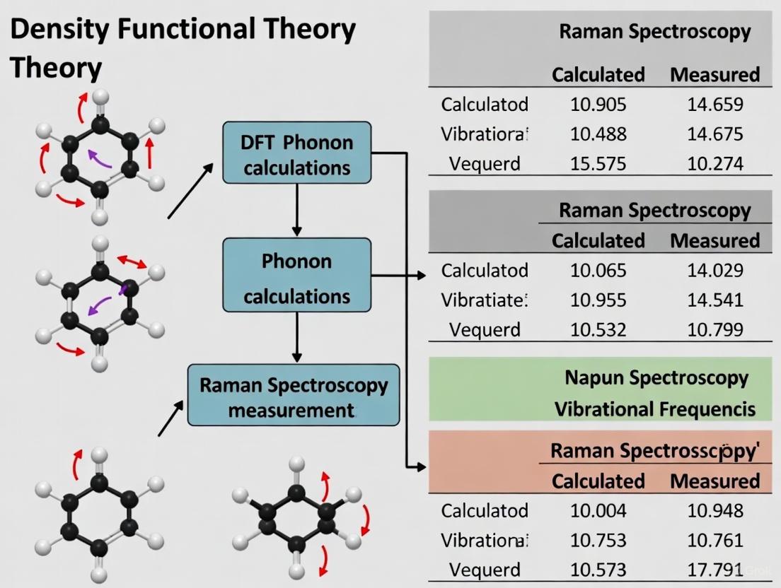
Workflow Visualization: Integrating Computation and Experiment
The following diagram illustrates the typical integrated workflow for comparing DFT phonon calculations with Raman spectroscopy measurements, highlighting their complementary roles in material characterization.
Integrated DFT-Experimental Workflow
The Scientist's Toolkit: Essential Reagents and Materials
Successful vibrational analysis, particularly for method validation and calibration, relies on a set of well-characterized standard materials.
Table 2: Essential Research Reagent Solutions for Raman Spectroscopy
| Reagent/Material | Function/Purpose | Example Uses |
|---|---|---|
| Silicon Wafer | Wavenumber and intensity calibration standard | Single sharp peak at ~520 cm⁻¹ for precise instrument calibration [3] |
| Polystyrene | Complex fingerprint standard for validation | Provides multiple well-defined peaks across a range of wavenumbers for comprehensive calibration checks [3] |
| Cyclohexane | Liquid standard for wavenumber calibration | Used for calibrating the wavenumber axis, especially in solvent or liquid measurements [3] |
| Paracetamol (Acetaminophen) | Solid pharmaceutical reference material | Serves as a stable, well-characterized solid standard for benchmarking in pharmaceutical applications [3] [4] |
| Squalene / Squalane | Lipid and biological matrix analogs | Used as reference substances whose spectra resemble biological samples (cells, tissues) for method development [3] |
Advanced Integration and Machine Learning Acceleration
The integration of DFT and Raman spectroscopy is being transformed by machine learning (ML), which overcomes traditional computational bottlenecks. ML interatomic potentials (MLIPs), such as those based on the MACE architecture, are trained on DFT data to predict energies and forces with near-DFT accuracy but at a fraction of the computational cost [5] [9] [7]. This enables high-throughput phonon and Raman spectra calculations for large and complex systems like metal-organic frameworks (MOFs) that were previously intractable [7]. Furthermore, foundation models can be fine-tuned for specific defects using surprisingly small datasets—sometimes just the data from a single atomic relaxation—to achieve highly accurate optical spectra [9]. The diagram below outlines this accelerating workflow.
ML-Accelerated Workflow
DFT phonon calculations and experimental Raman spectroscopy are not competing techniques but rather complementary pillars of modern vibrational analysis. DFT provides the foundational, interpretative framework, while Raman spectroscopy offers direct, empirical validation under realistic conditions. The ongoing integration of machine learning is breaking down historical barriers of computational cost, enabling the creation of vast databases and the accurate simulation of complex materials. For researchers in drug development and materials science, this synergistic combination provides an increasingly powerful toolkit for non-destructive characterization, quality-by-design implementation, and the rational discovery of new functional materials.
Raman spectroscopy is a powerful analytical technique used to observe vibrational, rotational, and other low-frequency modes in a molecular system, providing a unique molecular fingerprint for identifying substances and studying chemical bonding [10]. The technique is based on the Raman effect, first identified by Indian physicist Chandrasekhara Venkata Raman in 1928, which involves the inelastic scattering of monochromatic light, typically from a laser source in the visible, near-infrared, or near-ultraviolet range [11] [10]. When light interacts with matter, most photons are elastically scattered (Rayleigh scattering) at the same wavelength as the incident light. However, a tiny fraction (approximately one in a million photons) undergoes inelastic scattering at different wavelengths due to interactions with molecular vibrations—this is the Raman effect [11].
The fundamental principle governing Raman activity is a change in polarizability of the molecule during vibration. Unlike infrared spectroscopy, which depends on changes in the dipole moment, Raman scattering occurs when the incident light induces a temporary dipole moment in the molecule through distortion of its electron cloud [11] [12]. The resulting Raman spectrum presents a characteristic pattern of shifted frequencies from the incident light, providing detailed information about molecular structure, chemical composition, crystallinity, polymorphism, phase, intrinsic stress/strain, and even protein folding and hydrogen bonding [13].
Theoretical Foundations of the Raman Effect
The Semi-Classical Theory of Raman Scattering
The interaction between light and matter can be understood through a semi-classical model where the electric field of the incident light induces a polarization in the material. This relationship is represented by:
P = ε₀χE
Where P is the induced polarization vector, ε₀ is the vacuum permittivity, χ is the dielectric susceptibility tensor of the material, and E is the electric field vector of the incident light [12]. The oscillatory electric field of the incident light (angular frequency ωI, wave vector kI) interacts with the material. When the dielectric susceptibility χ also contains an oscillatory component due to modulation by a phonon or molecular vibration (angular frequency ωph, wave vector q), the product on the right-hand side of the equation generates sidebands at sum and difference frequencies ωI ± ω_ph, which constitute the Raman-scattered light [12].
To understand this more deeply, we can expand the susceptibility χ in a Taylor series with respect to the atomic displacement u:
χjk(kI,ωI) ≈ χjk + (∂χjk/∂ul)u_l + ...
The first-order term leads to the expression for the Raman-scattered component of the polarization, which oscillates at the characteristic Stokes (ωI - ωph) and anti-Stokes (ωI + ωph) frequencies [12]. The prefactor in this expression is the Raman tensor, R, which determines the amplitude of the scattered waves for a given vibrational mode and is crucial for understanding polarization dependence and selection rules [12].
The Quantum Mechanical Picture
In the quantum mechanical description, the Raman process involves the excitation of the electronic system by the photon to a higher-energy state (which may be real or virtual), interaction with phonons through electron-phonon coupling, and subsequent recombination [12]. This process can be represented by Feynman diagrams and understood through time-dependent perturbation theory. The intensity of Raman scattering is significantly enhanced when the incident photon energy approaches electronic transitions in the material (resonance Raman effect), as described by the expression:
I(ωS,ωI) ∝ |Σ ⟨i|HER(ωs)|m⟩⟨m|HEL|n⟩⟨n|HER(ωI)|i⟩ / [(ℏωI - En + iΓn)(ℏωs - Em + Γm)]|² × δ(ℏωI - ℏωS - ℏωph)
This equation shows that the intensity increases dramatically when either the incident (ωI) or scattered (ωS) photon energy matches electronic transitions (En or Em) in the system [12].
Raman Spectroscopy Compared with Other Analytical Techniques
Raman vs. IR Spectroscopy
Though both are forms of vibrational spectroscopy, Raman and IR spectroscopy differ in fundamental aspects and provide complementary information [11]. The table below summarizes their key differences:
Table 1: Comparison between Raman and Infrared Spectroscopy
| Feature | Raman Spectroscopy | Infrared Spectroscopy |
|---|---|---|
| Physical Basis | Change in molecular polarizability [11] | Change in dipole moment [11] |
| Radiation Process | Scattering of incident light [11] | Absorption of light energy [11] |
| Water Sensitivity | Low (weak Raman scatterer) [11] | High (strong IR absorber) [11] |
| Sample Preparation | Minimal to none [13] | Often requires preparation (KBr pellets, etc.) [13] |
| Spectral Bands | Homo-nuclear bonds (C-C, C=C, S-S) [11] | Polar bonds (C=O, O-H, N-H) [11] |
| Typical Excitation | Visible to NIR lasers [10] | IR source (global, etc.) [11] |
A comparison of spectra for polyethylene terephthalate (PET) demonstrates these differences: in IR measurements, spectral intensity depends on the size of the dipole moment for vibration modes (especially C=O and O-H bonds), while in Raman spectroscopy, intensity depends on the degree of polarizability for vibration modes (especially S-S, C-C, and C≡N bonds) [11].
Raman vs. Other Analytical Methods
Table 2: Raman Spectroscopy Compared with Other Analytical Techniques
| Technique | Key Advantages of Raman | Common Applications |
|---|---|---|
| FTIR Microscopy | Avoids solvent interference; better selectivity; detects IR-inactive modes [13] | Complementary molecular analysis [13] |
| Electron Microscopy | No vacuum needed; minimal sample preparation; faster analysis; ambient conditions [13] | Materials science, biology [13] |
| XRD (X-ray Diffraction) | Measures crystalline and amorphous materials; single particle analysis; no vacuum [13] | Crystalline structure analysis [13] |
Computational Approaches to Raman Spectroscopy
Traditional DFT Phonon Calculations
First-principles theoretical calculations of Raman spectra have traditionally been performed using the canonical harmonic approximation within density functional theory (DFT) [14]. This approach involves calculating the eigenvectors and dispersion relations of phonons using electronic-structure methods such as DFT, then proceeding with density functional perturbation theory (DFPT) to determine the change in polarizability for each phonon mode [14]. Specifically, these calculations determine the polarizability derivatives with respect to the phonon modes, ∂αμν/∂Qp, where Qp is the normal coordinate of phonon mode p, and αμν are the components of the polarizability tensor [14]. With these polarizability derivatives, one can compute the Raman spectrum in the harmonic approximation.
This method is well-established but faces conceptual limitations for systems with significant anharmonic vibrations. When vibrational anharmonicity is strong, atoms sample higher-order regions of the potential energy surface, and the harmonic modes may poorly approximate actual atomic motions at finite temperature [14]. This limitation is strikingly evident in cubic halide perovskites, which should be Raman-silent based on their average crystal symmetry but actually show significant Raman intensity due to strongly anharmonic vibrations [14].
Molecular Dynamics Raman (MD-Raman) Approach
The MD-Raman method provides an alternative framework rooted in statistical mechanics that treats anharmonic vibrations exactly [14]. This approach uses molecular dynamics simulations to generate a trajectory of atomic positions over time, then computes a polarizability timeseries α(t) using DFPT calculations along this trajectory [14]. The Raman spectrum is obtained from the Fourier transform of the polarizability autocorrelation function:
I(ω) ∝ ∫⟨α(0)α(t)⟩e^(-iωt)dt
This method fully incorporates vibrational anharmonicity and thermal effects but has traditionally been computationally prohibitive for practical materials computations [14]. Recent advances in machine learning have dramatically accelerated these computations by enabling rapid predictions of α(t) fluctuations with ab-initio accuracy at relatively low computational cost [14].
Table 3: Comparison of Computational Methods for Raman Spectroscopy
| Computational Method | Theoretical Basis | Advantages | Limitations |
|---|---|---|---|
| DFT Phonon (Harmonic) | Density Functional Theory + Density Functional Perturbation Theory [14] | Well-established; computationally efficient; good for perfect crystals [14] | Cannot capture anharmonic effects; fails for disordered systems [14] |
| MD-Raman | Molecular Dynamics + Polarizability Timeseries Analysis [14] | Includes anharmonic vibrations; captures thermal effects; principle exact [14] | Computationally expensive; requires long simulation times [14] |
| Machine Learning Accelerated MD-Raman | ML potentials + ML polarizability predictions [14] | Near ab-initio accuracy; significantly faster; practical for complex materials [14] | Requires training data; model validation essential [14] |
High-Throughput Computational Workflows
Recent advances have enabled high-throughput computation of ab initio Raman spectra for large material databases. One such workflow has successfully calculated Raman spectra for 3504 different two-dimensional materials using all-electron density functional perturbation theory [15]. The automated process involves: (1) obtaining material structure data from databases; (2) structural optimization; (3) harmonic Raman spectrum calculation using finite differences and DFPT; and (4) data summarization and storage in accessible databases [15]. This approach provides a valuable reference for experimentalists and represents a step toward autonomous characterization of materials.
Experimental Methodologies and Protocols
Standard Raman Spectroscopy Experimental Protocol
Instrumentation: A basic Raman spectrometer consists of a laser source (typically with wavelengths of 532 nm, 785 nm, or 1064 nm to avoid fluorescence), a sample illumination system, a spectrometer to disperse the scattered light, and a detector (typically CCD) [11] [16].
Sample Preparation: One significant advantage of Raman spectroscopy is that it typically requires no sample preparation. Analyses can be performed on 'as received' samples whether they are solid, liquid, powder, slurry, or gas [13].
Data Acquisition: The laser is focused onto the sample, and the scattered light is collected. Rayleigh scattering is filtered out using notch or edge filters, while the Raman-shifted light is directed into the spectrometer [11]. Acquisition times typically range from seconds to minutes, depending on the sample and signal strength [13].
Data Processing: The raw spectral data is processed to remove cosmic rays, correct for background fluorescence, and calibrate using known standards such as silicon [16].
Advanced Raman Techniques
Surface-Enhanced Raman Spectroscopy (SERS): Utilizes nanostructured metallic surfaces to amplify Raman signals by factors up to 10⁸-10¹¹, enabling single-molecule detection [17] [16]. SERS is particularly valuable in biomedical applications and trace analysis.
Tip-Enhanced Raman Spectroscopy (TERS): Combines scanning probe microscopy with Raman spectroscopy to achieve nanoscale spatial resolution (below the diffraction limit) [17]. TERS is essential for nanomaterials characterization and biological imaging.
Confocal Raman Microscopy: Enables high spatial resolution (sub-micron) analysis of discrete sample volumes, particularly useful for layered materials, inclusions, and chemical imaging [13].
Visualization of Raman Spectroscopy Principles
Raman Scattering Energy Level Diagram
Computational Raman Spectroscopy Workflow
Essential Research Reagent Solutions
Table 4: Essential Research Reagents and Materials for Raman Spectroscopy
| Reagent/Material | Function/Application | Key Characteristics |
|---|---|---|
| Silicon Wafer | Raman shift calibration standard [15] | Characteristic peak at 520 cm⁻¹; NIST-traceable [15] |
| SERS Substrates (Gold/Silver nanoparticles) | Signal enhancement for trace analysis [17] [16] | Nanostructured surfaces; tunable plasmon resonance [17] |
| NIST-Traceable Calibration Standards | Instrument calibration and validation [16] | Certified reference materials; guaranteed accuracy [16] |
| Specialized Lasers (532 nm, 785 nm, 1064 nm) | Excitation sources for different applications [10] | Varying penetration depth and fluorescence avoidance [10] |
| Raman Spectral Databases | Material identification and verification [13] | Extensive reference libraries; fast unknown identification [13] |
Applications in Research and Industry
Raman spectroscopy has found diverse applications across multiple scientific and industrial domains:
Pharmaceuticals and Biotechnology: Used for drug development, quality control, formulation development, and cellular analysis [17] [16]. The life sciences segment represents the fastest-growing application area, driven by advancements in diagnostics and personalized medicine [17].
Materials Science: Essential for characterizing two-dimensional materials, carbon nanomaterials (graphene, nanotubes), semiconductors, polymers, and composites [17] [15]. Raman spectroscopy can probe layer thickness, strain, doping, and crystallinity [15].
Forensic Science and Security: Employed for identification of unknown substances, trace evidence analysis, and security screening [17] [18]. The non-destructive nature preserves evidence for additional testing [13].
Environmental Monitoring: Deployed for analyzing chemical compositions in air and water, particularly with the development of portable field instruments [17].
Market Outlook and Future Perspectives
The Raman spectroscopy market is experiencing significant growth, with the global market projected to reach $472 million by 2032, exhibiting a compound annual growth rate (CAGR) of 7.0% [10]. Other estimates project an even larger market reaching $2.88 billion by 2034 [16]. This growth is driven by several key trends:
Technological Innovations: Miniaturization and portability are expanding applications into field analysis [17] [18]. Integration with artificial intelligence and machine learning is enhancing data analysis accuracy and enabling real-time decision-making [17].
Computational Advances: Machine learning-accelerated computations are making high-level theoretical predictions more accessible [14]. High-throughput computational workflows are generating comprehensive databases for material identification [15].
Emerging Applications: Growing adoption in biomedical diagnostics, environmental monitoring, and food safety testing continues to expand the technology's reach [17] [16]. The development of techniques like TERS and SERS is pushing detection limits to the single-molecule level [17].
The synergy between computational predictions and experimental measurements continues to strengthen, with MD-Raman and related methods emerging as versatile tools for theoretical prediction and characterization of molecules and materials [14]. As both experimental and computational capabilities advance, Raman spectroscopy remains an indispensable technique in the researcher's toolkit, providing unique insights into molecular structure and interactions through the fundamental principle of inelastic light scattering mediated by polarizability changes.
Density Functional Theory (DFT) provides the fundamental computational framework for modeling potential energy surfaces (PES) and interatomic force constants, which are essential for predicting and interpreting vibrational properties like Raman spectroscopy. The accuracy of these predictions heavily depends on how faithfully the computational method captures the underlying physics of atomic interactions. The PES describes the energy of a system as a function of its nuclear coordinates, while force constants—the second derivatives of energy with respect to atomic displacements—determine vibrational frequencies and phonon dispersion relations in crystalline materials [19].
While the canonical harmonic approximation has been widely successful for calculating Raman spectra, it faces significant limitations for systems with substantial anharmonic vibrations. In such cases, the harmonic approximation cannot capture temperature-dependent changes in Raman response, leading to inaccurate predictions [14]. This is particularly problematic for materials like cubic halide perovskites, which exhibit significant Raman activity despite being theoretically "Raman-silent" based on their average crystal symmetry—a phenomenon attributed to strongly anharmonic vibrations [14]. These limitations have motivated the development of more sophisticated computational approaches that go beyond harmonic approximations to better reconcile theoretical calculations with experimental Raman measurements.
Computational Methodologies: From Harmonic Approximations to Anharmonic Treatments
Density Functional Perturbation Theory (DFPT) for Harmonic Phonons
Density Functional Perturbation Theory (DFPT) has emerged as a powerful method for computing Raman spectra within the harmonic approximation. This approach calculates the linear response of electrons to perturbations caused by atomic displacements and electric fields, providing direct access to polarizability derivatives (∂αμν/∂Qp) with respect to phonon normal modes (Qp) [14]. The workflow for high-throughput Raman spectra calculations typically involves several stages: initial structure optimization, DFPT cycles for polarizability tensor calculation, and finally Raman intensity computation [15].
The harmonic DFPT approach has been successfully deployed for large-scale computational studies, such as the high-throughput calculation of Raman spectra for 3,504 two-dimensional materials [15]. However, this method inherently assumes a parabolic potential energy surface around equilibrium positions, neglecting temperature effects and anharmonic vibrations that can significantly alter Raman spectra in many functional materials [14] [15].
Molecular Dynamics-Based Raman (MD-Raman) for Anharmonic Systems
The MD-Raman approach offers a more comprehensive framework that naturally incorporates anharmonicity and temperature effects. Instead of relying on harmonic phonons, this method combines molecular dynamics simulations with subsequent calculations of polarizability tensors along the trajectory [14]. The resulting polarizability time series, α(t), is used to compute Raman spectra through correlation function analysis, effectively capturing the full anharmonic vibrational response [14].
The fundamental equation for MD-Raman derives from statistical mechanics, where the spectral density S(ω) is obtained from the Fourier transform of the polarizability autocorrelation function [14]:
S(ω) = limT→∞ (1/2T) |A(ω)|²
where A(ω) is the Fourier transform of the polarizability time series. This approach directly links atomic dynamics to spectroscopic observables without harmonic constraints, making it particularly valuable for studying systems with significant anharmonic vibrations, such as the aforementioned cubic halide perovskites that exhibit a characteristic "Raman central peak" [14].
Table: Comparison of Harmonic DFPT and Anharmonic MD-Raman Approaches for Raman Spectrum Calculations
| Feature | Harmonic DFPT Approach | Anharmonic MD-Raman Approach |
|---|---|---|
| Theoretical Basis | Density functional perturbation theory | Molecular dynamics + correlation function analysis |
| Anharmonicity Treatment | Neglects anharmonic effects | Fully includes anharmonic vibrations |
| Temperature Effects | Limited capture of thermal effects | Naturally incorporates temperature dependence |
| Computational Cost | Moderate | High (historically prohibitive) |
| System Size Limitations | Suitable for medium-sized systems | Limited by MD and polarizability calculations |
| Accuracy for Anharmonic Materials | Poor for strongly anharmonic systems | Excellent for capturing anharmonic effects |
| Key Applications | High-throughput screening of 2D materials [15] | Complex systems like halide perovskites [14] |
Machine Learning Accelerations and Cross-Functional Transferability
Machine Learning Interatomic Potentials (MLIPs)
The tremendous computational expense of traditional MD-Raman calculations has historically limited their practical application. Recent advances in machine learning interatomic potentials (MLIPs) now enable dramatic accelerations without sacrificing accuracy [14]. MLIPs learn the relationship between atomic configurations and energies/forces from DFT data, achieving near-DFT accuracy with several orders of magnitude reduction in computational cost [20].
Foundation potentials (FPs) represent a particularly promising development—these MLIPs trained on millions of DFT calculations demonstrate remarkable transferability across diverse chemical spaces [20] [21]. For Raman computations, MLIPs can accelerate both the molecular dynamics sampling and the subsequent polarizability calculations, making anharmonic Raman spectrum predictions feasible for complex materials [14].
Transfer Learning Across DFT Functionals
A significant challenge in advancing MLIPs involves transitioning from generalized gradient approximation (GGA) functionals to higher-fidelity meta-GGA functionals like r2SCAN. While r2SCAN provides improved descriptions of interatomic bonding [21], the computational cost of generating large training datasets is substantial. Transfer learning approaches, where models pre-trained on GGA data are fine-tuned on smaller r2SCAN datasets, offer a promising solution [20].
However, studies have revealed challenges in cross-functional transferability due to significant energy scale shifts and poor correlations between different DFT functionals [20]. Proper treatment of elemental energy referencing has been identified as crucial for successful transfer learning between functional types [20]. The recently introduced MatPES dataset, which includes both PBE and r2SCAN calculations, provides a valuable resource for developing more accurate MLIPs with improved transferability across functionals [21].
Diagram Title: Computational Raman Spectroscopy Workflows
Experimental Validation and Benchmarking Protocols
Validation Against Experimental Raman Spectra
Robust validation against experimental measurements is crucial for assessing the accuracy of computational methodologies. Large-scale benchmarking studies have compared calculated Raman spectra with experimental data for various material systems. For example, high-throughput DFPT calculations for 2D materials demonstrated generally good agreement with experimental spectra, though discrepancies were noted due to limitations in accounting for anharmonic effects, substrate influences, and excitonic effects [15].
For molecular systems, comprehensive benchmark studies have evaluated the performance of numerous DFT functionals for resonance Raman spectroscopy. One extensive study compared 42 different functionals for calculating resonance Raman spectra of lumiflavin, identifying HCTH, OLYP, and TPSSh as the most accurate through systematic evaluation of 0-0 transition energies, correlation with experimental spectra, and reproduction of singlet-triplet peak shifts [22].
Table: Key Research Reagent Solutions for Computational Raman Spectroscopy
| Tool Category | Specific Examples | Function/Role |
|---|---|---|
| DFT Software Packages | FHI-aims [15], CASTEP [19], Gaussian [22] | Provide electronic structure calculations and vibrational analysis capabilities |
| Machine Learning Potentials | CHGNet [20], M3GNet [21], SevenNet-MF-0 [20] | Accelerate molecular dynamics simulations while maintaining DFT-level accuracy |
| Benchmark Datasets | MatPES [21], 2DMatPedia [15], MPRelax [21] | Provide reference data for training and validation of computational models |
| DFT Functionals | PBE [19], r2SCAN [21], B3LYP [19], HCTH/OLYP/TPSSh [22] | Define exchange-correlation energy approximations with different accuracy/speed tradeoffs |
| Computational Approaches | DFPT [15], MD-Raman [14], Time-Dependent Theory [22] | Methodological frameworks for calculating Raman spectra from first principles |
Quantitative Performance Metrics
Systematic benchmarking requires well-defined quantitative metrics to evaluate computational methodologies. Key performance indicators include:
- Phonon frequency accuracy: Mean absolute error between calculated and experimental vibrational frequencies [15]
- Raman intensity correlation: Quantitative comparison of relative peak intensities in calculated versus experimental spectra [22]
- Anharmonicity capture: Ability to reproduce temperature-dependent spectral features and broadened line shapes [14]
- Computational efficiency: Time-to-solution and scalability with system size [20]
For MD-Raman approaches specifically, validation often focuses on reproducing characteristic anharmonic features observed experimentally, such as the Raman central peak in cubic halide perovskites, which harmonic calculations completely fail to predict [14].
The computational landscape for calculating potential energy surfaces and interatomic force constants is rapidly evolving, with machine learning approaches dramatically accelerating anharmonic Raman spectrum predictions while maintaining first-principles accuracy. The traditional trade-off between computational cost and anharmonic treatment is being disrupted by foundation potentials and transfer learning approaches [14] [20].
Future advancements will likely focus on improving cross-functional transferability, enabling seamless transitions between different levels of theory, and developing multi-fidelity learning frameworks that combine the efficiency of GGA calculations with the accuracy of meta-GGA functionals [20] [21]. As these computational methodologies mature, they will increasingly serve as versatile tools for theoretical prediction and characterization of molecules and materials, providing deeper insights into the relationship between atomic structure, dynamics, and spectroscopic observables.
For computational spectroscopists, the expanding toolkit offers unprecedented opportunities to bridge the gap between microscopic properties derived from calculations and macroscopic observables measured in experiments, ultimately enhancing our ability to interpret and predict material behavior across diverse chemical and materials systems [19].
The harmonic approximation stands as a cornerstone in computational materials science and chemistry, providing the foundational framework for calculating atomic vibrations and their spectroscopic signatures. Within density functional theory (DFT), this approach simplifies the complex potential energy surface of atomic interactions by considering only second-order terms in the Taylor expansion around equilibrium positions, effectively treating atomic vibrations as simple harmonic oscillators. This methodology has become indispensable for predicting various material properties, including vibrational spectra, thermodynamic behavior, and structural stability. The approximation's true power emerges when its computational predictions are compared against experimental measurements, particularly Raman spectroscopy, which serves as a critical benchmark for assessing theoretical models.
The synergy between computational harmonic analysis and experimental Raman spectroscopy has enabled researchers to interpret complex spectroscopic data, identify unknown compounds, and validate molecular structures across diverse scientific domains. However, as research progresses toward more complex materials systems and higher precision requirements, the limitations of the harmonic approximation have become increasingly apparent. This review systematically examines both the utility and constraints of harmonic approximation in phonon calculations, focusing specifically on its performance against experimental Raman spectroscopy measurements, while also exploring advanced methodologies that transcend harmonic limitations.
Theoretical Foundation and Computational Methodologies
Fundamental Principles of Harmonic Phonon Calculations
The harmonic approximation in phonon calculations operates on the fundamental principle that the potential energy surface of atomic interactions can be accurately described using a quadratic expansion around the equilibrium geometry. This approach yields several important mathematical simplifications. The interatomic force constants (IFCs), which are the second derivatives of the Born-Oppenheimer potential energy surface with respect to atomic displacements, become the central quantities defining the lattice dynamics within this framework. These second-order IFCs directly determine the phonon dispersion relations and vibrational frequencies of the system.
In practical implementation, two primary computational approaches exist for evaluating these second-order IFCs from first principles. The real-space small displacement method (often called the frozen-phonon approach) systematically displaces atoms from their equilibrium positions in finite supercells and calculates the resulting forces [23]. This method, implemented in codes like Phonopy and Phon, requires approximately 3N separate self-consistent DFT calculations for a supercell containing N atoms. Alternatively, the reciprocal-space linear response method within density functional perturbation theory (DFPT) calculates phonon properties directly in reciprocal space, offering potential computational advantages for certain systems [23].
The resulting phonon frequencies (ω) and their corresponding Raman activities are then used to simulate spectroscopic outputs. For a Raman spectrum, the intensity is typically computed from the derivative of the system's polarizability (α) with respect to the normal coordinate of each phonon mode (Qp), following the relationship: ∂αμν/∂Qp, where μ and ν are tensor components [14]. This forms the theoretical basis for connecting computational harmonic results with experimental Raman measurements.
Standard Computational Protocols for Harmonic Raman Spectra
The standard workflow for computing harmonic Raman spectra involves a sequential computational protocol that has been refined over decades of research. The process typically begins with geometry optimization, where the atomic positions and lattice parameters (for crystalline systems) are relaxed until the residual forces on atoms fall below a predetermined threshold (often 1-10 meV/Å) to ensure a valid equilibrium structure [24]. This optimized structure then serves as the reference point for subsequent vibrational analysis.
The second critical step involves vibrational frequency calculations using the harmonic approximation. For molecular systems, this is commonly performed using quantum chemistry packages like Gaussian [25], while solid-state systems typically employ DFT codes coupled with phonon packages. These calculations yield the harmonic vibrational frequencies, infrared intensities, and Raman activities. For meaningful comparison with experimental data, researchers often apply frequency scaling factors (typically ranging from 0.96 to 0.98) to mitigate systematic errors arising from the harmonic approximation and incomplete treatment of electron correlation [22].
For the specific case of comparing with resonance Raman spectroscopy, more advanced implementations of time-dependent DFT (TD-DFT) incorporate resonance effects through Franck-Condon analysis within the adiabatic Hessian formalism, enhancing the agreement with experimental measurements obtained under resonant conditions [22]. Throughout these calculations, solvation effects are frequently incorporated using implicit solvation models like the Polarizable Continuum Model (PCM), while appropriate basis sets (such as cc-pVDZ or 6-31G(d)) are selected to balance computational cost and accuracy [22] [26].
Table 1: Standard Computational Parameters for Harmonic Raman Calculations
| Computational Parameter | Typical Settings | Purpose/Rationale |
|---|---|---|
| Geometry Optimization | Force convergence: 1-10 meV/Å | Ensures valid equilibrium structure for vibrational analysis |
| Frequency Scaling Factor | 0.96-0.98 (functional-dependent) | Corrects systematic errors from harmonic approximation |
| Basis Set | cc-pVDZ, 6-31G(d), or similar | Balances computational cost and accuracy |
| Solvation Model | PCM (for solutions) | Accounts for solvent effects on vibrational frequencies |
| Dispersion Correction | D3, D3BJ, or similar | Improves treatment of van der Waals interactions |
Performance Assessment: Harmonic Approximation vs. Experimental Raman Spectroscopy
Successes and Strengths in Various Material Systems
The harmonic approximation has demonstrated remarkable success across multiple domains when compared against experimental Raman measurements. In molecular systems, particularly organic compounds and drug-like molecules, harmonic calculations show excellent agreement with experimental spectra, correctly predicting peak positions with typical errors of 10-30 cm⁻¹ after applying standard scaling factors. For instance, DFT calculations at the B3LYP/6-31G(d) level achieved "perfect agreement" with experimental Raman spectra for ponceau 4R, successfully reproducing the vibrational fingerprint of this complex organic molecule [26]. Similar success has been documented for flavin-based systems, where certain functionals like HCTH, OLYP, and TPSSh provided accurate predictions of vibrational patterns when benchmarked against experimental resonance Raman data [22].
The strength of the harmonic approach is particularly evident in its computational efficiency for high-throughput screening applications. A recent large-scale dataset comprising 220,000 molecules with computed harmonic Raman and IR spectra highlights how this approach enables the training of machine learning models for rapid spectral prediction and analysis [25]. For standard systems with minimal anharmonicity at room temperature, the harmonic approximation provides a reliable and computationally affordable method for spectral interpretation and assignment, with the entire vibrational spectrum calculable in a single step without the need for extensive configuration sampling.
Quantitative Limitations and Systematic Errors
Despite its widespread success, the harmonic approximation introduces systematic errors that become particularly evident when computational results are compared against precise Raman measurements. A comprehensive benchmark study on lumiflavin utilizing 42 different DFT functionals revealed significant variations in predicting key vibrational peaks, with even the best functionals requiring careful frequency scaling and resonance enhancements to achieve adequate agreement with experimental data [22].
Table 2: Quantitative Performance of Harmonic Approximation in Benchmark Studies
| System/Study | Key Metric | Harmonic Performance | Experimental Reference |
|---|---|---|---|
| Lumiflavin (42 functionals) [22] | 0-0 transition error | Significant variation across functionals | FMN EAS spectra |
| Strongest peak intensity at ~1498 cm⁻¹ | Required resonance correction for accuracy | ||
| Ponceau 4R [26] | Peak position agreement | Excellent with scaling factor | Solid-state Raman |
| Spectral fingerprint | Correctly reproduced pattern | ||
| Foundation MLIPs [24] | Huang-Rhys factors | ~12% deviation from DFT | Defect phonon calculations |
The most significant limitation emerges in systems with substantial anharmonicity, where the harmonic approximation fundamentally fails to capture the true physical behavior. In cubic halide perovskites, for example, harmonic calculations incorrectly predict these materials to be "Raman silent" based on their average crystal symmetry, while experimental measurements show a significant "Raman central peak" originating from strongly anharmonic atomic motions [14]. Similarly, in hydrogen-rich materials and systems with floppy modes or large-amplitude vibrations, the harmonic approximation significantly misrepresents the actual vibrational density of states, leading to inaccurate predictions of Raman band positions and shapes.
Advanced Methodologies Beyond the Harmonic Approximation
Theoretical Frameworks Addressing Anharmonicity
To overcome the limitations of the harmonic approximation, several advanced computational frameworks have been developed that explicitly account for anharmonic effects:
The Stochastic Self-Consistent Harmonic Approximation (SSCHA) provides a non-perturbative approach to anharmonicity by combining self-consistent harmonic calculations with stochastic sampling of the potential energy surface. This method effectively incorporates quantum nuclear effects and strong anharmonicity, making it particularly valuable for systems like hydrides and high-temperature phases where harmonic treatments fail [27].
Molecular Dynamics Raman (MD-Raman) approaches abandon the normal mode concept entirely in favor of a time-domain correlation function formalism. This method computes the polarizability tensor (α) along molecular dynamics trajectories, then calculates the Raman spectrum from the Fourier transform of the polarizability autocorrelation function. This approach naturally includes anharmonicity, temperature effects, and dynamical disorder [14].
Machine Learning Interatomic Potentials (MLIPs) trained on DFT data enable the efficient calculation of anharmonic properties at a fraction of the computational cost of direct ab initio methods. These potentials can capture the full anharmonic potential energy surface while maintaining near-DFT accuracy, making them particularly valuable for large systems and extensive configuration sampling [24] [27].
Comparative Performance of Anharmonic Methods
The performance advantages of these beyond-harmonic methods become particularly evident when their predictions are compared against experimental Raman measurements for challenging systems:
Table 3: Comparison of Methods for Anharmonic Raman Calculations
| Computational Method | Key Principle | Advantages over Harmonic | Computational Cost |
|---|---|---|---|
| SSCHA [27] | Self-consistent harmonic with stochastic sampling | Non-perturbative treatment of quantum fluctuations | High (~96% cost reduction with MLIP) |
| MD-Raman [14] | Time-domain correlation functions | Naturally includes temperature and dynamics | Very high (accelerated with MLIP) |
| MLIP + Phonons [24] | Machine-learned potential energy surface | DFT accuracy with force-field speed | Moderate (after training) |
| Compressive Sensing Lattice Dynamics [23] | Sarse representation of high-order IFCs | Efficient extraction of anharmonic force constants | Moderate to high |
For the specific case of PdCuH₂, a material hypothesized to exhibit high-temperature superconductivity, standard harmonic calculations predicted imaginary phonon modes suggesting dynamical instability. However, SSCHA calculations incorporating quantum nuclear effects revealed that this phase becomes dynamically stable when anharmonicity is properly accounted for, resolving the discrepancy between experimental observations and theoretical predictions [27]. Similarly, MD-Raman approaches have successfully reproduced the controversial "Raman central peak" in cubic halide perovskites, which harmonic methods completely fail to predict [14].
The "one defect, one potential" strategy represents another significant advancement, where machine learning potentials are specifically trained for individual defect systems. This approach has demonstrated remarkable accuracy in predicting defect phonons, Huang-Rhys factors, and photoluminescence spectra, achieving DFT-level accuracy while reducing computational costs by orders of magnitude [24].
Software and Code Ecosystem
The computational spectroscopy community relies on a diverse ecosystem of software packages implementing various aspects of phonon calculations and Raman spectrum simulation:
- Phonopy [23] [24]: A widely adopted package for harmonic phonon calculations using the finite displacement method, supporting both molecules and periodic systems.
- Phon [23]: Another implementation of the frozen-phonon approach for phonon band structure calculations.
- Pheasy [23]: A recently developed Python package that incorporates advanced features for handling anharmonic effects, including compressive sensing lattice dynamics and high-order interatomic force constant extraction.
- Quantum ESPRESSO [23]: A comprehensive suite for electronic structure calculations that includes DFPT capabilities for harmonic phonon calculations.
- ShengBTE [23] [27], Phono3py [23] [27]: Specialized packages for calculating lattice thermal conductivity, often used in conjunction with harmonic phonon calculations as a starting point.
- ALAMODE [23]: A package designed for anharmonic lattice dynamics, implementing compressive sensing techniques and other beyond-harmonic approaches.
- Gaussian [22] [25]: A leading quantum chemistry package extensively used for harmonic vibrational analysis of molecular systems, with capabilities for Raman intensity calculations.
Experimental-Computational Workflow Integration
The integration of computational and experimental approaches for Raman spectroscopy follows a structured workflow that maximizes synergistic benefits. The diagram below illustrates this integrated approach, highlighting key decision points where harmonic approximation may suffice versus situations requiring advanced anharmonic treatments:
The harmonic approximation remains an indispensable tool in the computational spectroscopy toolkit, providing a reasonable balance between accuracy and computational cost for many systems with minimal anharmonic character. Its success in predicting Raman spectra for a wide range of organic molecules and standard semiconductors underscores its continued relevance, particularly for high-throughput screening and initial structural characterization.
However, the comparison between computational harmonic results and experimental Raman measurements has clearly delineated the boundaries of this approximation. For systems with significant anharmonicity—including hydrogen-rich compounds, high-temperature phases, materials with floppy modes, and systems exhibiting dynamical disorder—the harmonic approximation fails to capture essential physics, necessitating more sophisticated computational approaches.
The emerging paradigm combines machine learning potentials with advanced anharmonic formulations like SSCHA and MD-Raman, offering a path forward that maintains accuracy while managing computational costs. As these methods mature and become more accessible, they promise to extend the scope of predictive computational spectroscopy to increasingly complex and functional materials, ultimately strengthening the synergy between theoretical predictions and experimental Raman measurements.
The future of computational phonon calculations lies not in abandoning the harmonic approximation, but in understanding its limitations and having robust methodologies for transitioning to beyond-harmonic treatments when necessary. This multi-scale, multi-fidelity approach will enable researchers to efficiently tackle the spectroscopic challenges presented by next-generation materials across energy, electronic, and quantum technologies.
Raman spectroscopy serves as a powerful tool for identifying materials, probing structural phase transitions, and investigating coupling phenomena in solids. The technique relies on the inelastic scattering of photons from molecular vibrations, phonons, or other excitations in a system. However, not all vibrational modes appear in Raman spectra; their activity depends fundamentally on the crystal symmetry and the resulting selection rules that govern the scattering process. For a vibration to be Raman active, it must involve a change in the molecular polarizability (α), mathematically expressed as (dα/dq)e ≠ 0, where q is the normal coordinate and e represents the equilibrium position [28] [29].
This guide explores the critical connection between crystal symmetry and spectroscopic activity, with a specific focus on comparing density functional theory (DFT) phonon calculations with experimental Raman spectroscopy measurements. Such comparisons have become indispensable in modern materials research, not only for interpreting time-resolved spectroscopy but also for aiding the design of molecules with specific properties [22]. The interplay between long-range translational symmetry in crystals versus its absence in amorphous solids creates dramatically different spectral features, enabling researchers to distinguish between structural phases and understand fundamental material properties [30].
Fundamental Raman Selection Rules
Basic Principles and Theoretical Foundation
Raman spectroscopy measures the energy shift of photons scattered inelastically by a sample. When light interacts with a molecule, the electric field of the photon induces a dipole moment (pind) in the molecule, which depends on its polarizability: pind = αE. If the polarizability changes during a vibration (dα/dq ≠ 0), the induced dipole oscillates at the vibrational frequency, resulting in scattering at shifted frequencies (Stokes and anti-Stokes scattering) in addition to the primary Rayleigh scattering [28] [29].
The spectroscopic selection rule for infrared (IR) spectroscopy differs fundamentally, requiring a change in the permanent dipole moment. This distinction makes Raman and IR spectroscopy complementary techniques. In fact, for centrosymmetric molecules (those possessing a center of inversion), the rule of mutual exclusion operates: modes that are Raman active are IR inactive, and vice versa [31] [29]. This rule emerges because IR activity requires ungerade (unsymmetric) representations under inversion, while Raman activity requires gerade (symmetric) representations [31].
Selection Rules in Crystalline Solids
In crystalline materials, vibrations are quantized as phonons—lattice vibrational waves propagating through the crystal with specific wave vectors (k), wavelengths, and frequencies [30]. The Raman activity of these phonons follows the same fundamental selection rule regarding polarizability changes [29]. However, an additional critical restriction applies to ideal crystals: only phonons near the center of the Brillouin zone (with k ≈ 0) are Raman active [30]. This restriction arises from the conservation of momentum during photon-phonon interactions in periodic structures.
The number and symmetry of Raman-active modes in a crystal depend directly on its space group symmetry. Group theory provides the mathematical framework for determining how many modes belong to each irreducible representation and which will be Raman active [30] [31]. For example, in single-crystal silicon (with a diamond cubic structure), the first-order Raman spectrum consists of one triply degenerate optical phonon at approximately 520 cm⁻¹, appearing as a very narrow band with a full width at half maximum of only 4 cm⁻¹ [30].
Table 1: Comparison of Raman Spectral Features in Crystalline and Amorphous Solids
| Feature | Crystalline Solids | Amorphous Solids |
|---|---|---|
| Spectral Bandwidth | Narrow (e.g., 4 cm⁻¹ for Si) [30] | Broad (up to several hundred cm⁻¹) [30] |
| Selection Rule | Only zone-center (k ≈ 0) phonons active [30] | All phonons across Brillouin zone become active [30] |
| Spectral Appearance | Sharp, distinct peaks [30] | Broad, featureless peaks [30] |
| Origin of Features | Discrete phonon modes [30] | Phonon density of states [30] |
| Structural Information | Long-range order, specific symmetry [30] | Short-range order, bond angle/length distributions [30] |
Computational Approaches: DFT Phonon Calculations
Methodology and Workflow
Density functional theory (DFT) calculations have become a cornerstone for interpreting and predicting Raman spectra in materials research. These first-principles computations allow researchers to investigate phonon dispersion curves, vibrational densities of states, and Raman activities by solving the electronic structure problem [32]. The typical workflow begins with determining the ground-state crystal structure and optimizing the geometry. Then, phonon frequencies and eigenvectors are calculated using density functional perturbation theory or the finite displacement method. Finally, the Raman tensors are computed for each phonon mode by determining the derivative of the polarizability with respect to atomic displacements [32].
Recent advances have extended these methods to include resonance Raman effects, which are particularly important for studying chromophore systems in solution or embedded in photoproteins [22]. Time-dependent DFT (TD-DFT) formulations can model how Raman intensities are enhanced when the excitation laser energy approaches electronic transitions in the material [22]. For the Hg₃Te₂Cl₂ crystal system, such DFT approaches have successfully calculated phonon spectra and Raman activities, providing interpretation of experimental data and revealing the contributions of different atoms to the vibrational spectra [32].
Benchmarking and Validation
The accuracy of DFT-predicted Raman spectra depends significantly on the choice of exchange-correlation functional. Extensive benchmarking studies, such as one examining 42 different DFT functionals for modeling resonance Raman spectra of lumiflavin, have helped identify optimal functionals for specific applications [22]. In this comprehensive evaluation, functionals were scored based on multiple criteria including predicted transition energies, correlation percent errors between calculated and experimental spectra, and the ability to reproduce spectral profiles [22].
Such benchmarking is essential because different functionals can yield varying results for specific material systems. For instance, in flavin-containing systems, the popular B3LYP functional has been widely used due to its accurate prediction of excitation energies, though other functionals like HCTH, OLYP, and TPSSh may provide better performance for resonance Raman calculations in certain contexts [22]. The inclusion of dispersion corrections and frequency scaling factors further refines the agreement between computational and experimental results [22].
Table 2: Key Metrics for Benchmarking DFT Calculations Against Experimental Raman Data
| Metric | Description | Application in Benchmarking |
|---|---|---|
| 0-0 Transition Energy | Energy difference between lowest vibrational states of ground and excited states | Evaluates accuracy of predicted excitation energies [22] |
| Correlation Percent Error | Quantitative measure of spectral shape agreement | Assesses how well calculated spectra match experimental peak positions and relative intensities [22] |
| Spectral Profile Compatibility | Visual inspection of spectral patterns | Determines if theoretical and experimental spectral profiles are compatible for assignment [22] |
| Peak Shift Reproduction | Ability to predict shifts between different states (e.g., singlet-triplet) | Tests performance for time-resolved studies and excited-state dynamics [22] |
| Basis Set Convergence | Stability of results with increasing basis set size | Ensures calculations are not limited by basis set choice [22] |
Experimental Protocols for Raman Spectroscopy
Standard Measurement Methodology
Modern Raman spectroscopy typically employs laser sources in the visible, near-infrared, or near-ultraviolet ranges, with the laser light interacting with molecular vibrations or phonons in the system [33]. The scattered light is collected through a lens system, with the elastically scattered Rayleigh component filtered out using notch or edge pass filters [33]. The remaining inelastically scattered light is then dispersed using a spectrograph and detected, most commonly with charge-coupled device (CCD) detectors [33].
For crystalline materials, polarization-dependent measurements can provide additional information about crystal orientation and symmetry [29]. The specific experimental setup—including laser wavelength, power, spectral resolution, and collection time—must be optimized based on the material properties. Shorter wavelength lasers generally produce stronger Raman scattering due to the ν⁴ dependence of Raman scattering cross-sections, but may cause sample degradation or fluorescence in some materials [33].
Specialized Techniques and Considerations
Several advanced Raman techniques have been developed for specific applications. Resonance Raman spectroscopy enhances sensitivity by tuning the laser frequency to match electronic transitions in the sample, resulting in signal enhancement for modes associated with those transitions [22]. For materials with weak Raman signals, surface-enhanced Raman spectroscopy (SERS) can dramatically increase sensitivity by leveraging plasmonic effects from metallic nanostructures [33].
When studying phase transitions or damage in crystals, Raman spectroscopy provides a sensitive probe of structural changes. For example, ion implantation in semiconductors can disrupt long-range translational symmetry, creating amorphous regions with characteristic broad Raman features instead of the sharp peaks of crystalline material [30]. Thermal annealing can reverse this damage, with the recovery process monitorable through the restoration of sharp crystalline peaks in the Raman spectrum [30].
Comparative Analysis: Crystalline vs. Amorphous Solids
The Raman spectra of crystalline and amorphous solids of identical chemical composition differ dramatically due to the presence or absence of spatial order and long-range translational symmetry [30]. Crystalline solids exhibit sharp, narrow Raman bands because the selection rules restrict activity to phonons at the Brillouin zone center [30]. In contrast, amorphous solids display broad spectral features because the breakdown of translational symmetry relaxes the k-vector conservation rule, making phonons across the entire Brillouin zone Raman active [30].
The Raman spectrum of an amorphous solid closely resembles the phonon density of states of the corresponding crystalline material [30]. This relationship provides a powerful connection between the vibrational properties of ordered and disordered phases. For example, fused quartz (amorphous SiO₂) shows very broad Raman peaks with widths up to several hundred wavenumbers, while crystalline quartz of the same chemical composition displays sharp, narrow bands [30]. Similarly, ion-implanted silicon exhibits broad spectral features below 550 cm⁻¹, contrasting sharply with the narrow 520 cm⁻¹ peak of single-crystal silicon [30].
Case Studies and Applications
Complex Crystal Systems: Hg₃X₂Y₂ Compounds
The Hg₃X₂Y₂ (X = S, Se, Te; Y = F, Cl, Br, I) family of compounds demonstrates the power of combining DFT calculations with experimental Raman spectroscopy. These materials crystallize in the corderoite structure (space group I2₁3) and exhibit promising properties for nonlinear optical devices [32]. DFT-guided investigations of Hg₃Te₂Cl₂ have enabled detailed analysis of phonon modes, their dispersion, symmetry properties, and Raman activities [32].
In these studies, first-principles calculations provided the theoretical foundation for assigning observed Raman peaks to specific vibrational modes and atomic contributions [32]. The calculations revealed how different atoms (Hg, Te, Cl) contribute to the vibrational spectra and enabled classification of normal mode frequencies according to irreducible representations for both infrared-active and Raman-active modes [32]. This combined computational-experimental approach offers comprehensive understanding of the relationship between crystal structure and vibrational properties in these complex materials.
Emerging Applications with Machine Learning
The integration of Raman spectroscopy with machine learning (ML) represents a cutting-edge development for materials identification and analysis. Recent research has demonstrated that ML algorithms can effectively classify and separate Raman spectra, revealing both structural similarities and subtle differences between related compounds [34]. In a study on per- and polyfluoroalkyl substances (PFAS), researchers used unsupervised ML methods like principal component analysis (PCA) and t-distributed stochastic neighbor embedding (t-SNE) to distinguish between different PFAS compounds based on their Raman spectral features [34].
DFT calculations supported this approach by modeling molecular structures and confirming experimental Raman data, thereby enhancing understanding of how molecular structures dictate spectral signatures [34]. This combined methodology—Raman spectroscopy plus DFT plus machine learning—creates a powerful framework for environmental monitoring, forensic analysis of contaminated sites, and sensor development [34].
Essential Research Reagents and Materials
Table 3: Research Reagent Solutions for Raman Spectroscopy and DFT Studies
| Reagent/Material | Function/Application | Specific Examples |
|---|---|---|
| Laser Sources | Provides monochromatic excitation for Raman scattering | Continuous wave lasers (standard Raman); pulsed lasers (time-resolved) [33] |
| DFT Software Packages | Performs quantum-mechanical calculations of structures and vibrations | ABINIT [32]; Gaussian [22] with various functionals (B3LYP, HCTH, OLYP) [22] |
| Spectrographs & Detectors | Disperses and detects Raman scattered light | CCD detectors; Fourier Transform spectrometers [33] |
| Reference Crystals | Calibration and validation of spectral measurements | Single-crystal silicon (520 cm⁻¹ peak) [30]; quartz [30] |
| Polarization Optics | Studies crystal orientation and symmetry | Polarizers, waveplates for polarization-dependent measurements [29] |
| Computational Basis Sets | Mathematical basis for electronic structure calculations | cc-pVDZ, aug-cc-pVDZ, cc-pVQZ [22] |
The connection between crystal symmetry and Raman spectroscopic activity represents a fundamental principle in materials characterization that bridges theoretical predictions and experimental observations. Through the framework of selection rules governed by group theory, researchers can understand why certain vibrational modes appear in Raman spectra while others remain silent. The integration of DFT phonon calculations with experimental Raman measurements has created a powerful synergy that enhances our ability to interpret spectral data, validate computational models, and extract detailed structural information.
As computational methods continue to advance and machine learning approaches become more sophisticated, the relationship between crystal symmetry and spectroscopic activity will undoubtedly yield new insights into material behavior. These developments will further strengthen Raman spectroscopy's position as an indispensable tool for materials research, drug development, and environmental monitoring, providing a critical bridge between atomic-scale structure and macroscopic material properties.
Computational and Experimental Workflows: From High-Throughput Screening to Biomedical Applications
The rapid advancement of high-throughput (HT) approaches has revolutionized materials science over the past decade, enabling researchers to conduct computational screening across increasingly expansive chemical spaces. [35] This paradigm shift has been particularly transformative for phonon calculations, which are essential for understanding thermal conductivity, thermodynamic stability, and spectroscopic properties of materials. Traditional first-principles phonon calculations, while accurate, remain computationally intensive, especially for large unit cells or complex materials with low symmetry. [5] The finite-displacement method, for instance, requires numerous supercell calculations using density functional theory (DFT) to capture short- to long-range interactions and achieve converged results. [5] This computational bottleneck has historically limited the availability of high-quality phonon data, creating a pressing need for automated, efficient workflows that can scale to thousands of materials.
The integration of HT phonon calculations with experimental validation through Raman spectroscopy represents a particularly powerful approach in modern materials research. Raman spectroscopy provides unique "fingerprints" of local bonding and environment through vibrational frequencies, making it indispensable for characterizing condensed materials. [36] However, interpreting Raman spectra requires robust computational references, creating a symbiotic relationship between calculation and measurement. This comparative framework enables not only materials identification but also fundamental insights into structure-property relationships that drive innovation in thermal management, energy harvesting, and electronic devices.
Comparative Analysis of Automated Workflow Solutions
Workflow Frameworks and Architectures
Table 1: Comparison of Major Workflow Automation Frameworks for Materials Computation
| Framework | Core Design Philosophy | Supported Calculators | Key Advantages | Limitations |
|---|---|---|---|---|
| AiiDA | Automated provenance tracking and reproducibility | VASP, Quantum ESPRESSO, other ab initio codes | Persistent provenance storage, error handling, modular workflows | Steeper learning curve for complex workflows |
| atomate2 | Standardization, interoperability, composability | VASP, FHI-aims, ABINIT, CP2K, MLIPs | Heterogeneous workflows, easy parameter modification, MLIP support | Relatively newer ecosystem with growing community |
| Pheasy | Specialized phonon calculations with ML-enhanced IFC extraction | Force-displacement data from various DFT codes | High-order anharmonic IFCs, advanced long-range electrostatics | Domain-specific (phonons only) |
The AiiDA framework represents a robust solution for automated high-throughput calculations, with proven capability in managing complex computational workflows. Its open-source platform enables automation of multi-step procedures with minimal user intervention while storing complete calculation provenance to ensure reproducibility. [35] Within the phononics domain, AiiDA has been successfully deployed for G0W0 calculations of excited-state properties, demonstrating its capability to handle computationally intensive post-DFT methodologies. [35]
atomate2 emerges as a comprehensive evolution of workflow frameworks, specifically designed to enhance programmability and flexibility. Its core design principles emphasize standardization of inputs and outputs, interoperability between computational methods, and composability of workflows. [37] A particularly powerful feature is atomate2's support for heterogeneous workflows, where different parts can be executed using different DFT packages or machine learning interatomic potentials (MLIPs), allowing researchers to leverage the unique strengths of each computational method. [37] This capability is crucial for phonon calculations, where initial structural relaxations might be performed efficiently with one code while subsequent phonon properties are calculated with another specialized code.
Specialized phonon codes like Pheasy complement these general frameworks by providing robust implementations for extracting interatomic force constants (IFCs) using advanced machine learning algorithms. [23] Pheasy accurately reconstructs the potential energy surface of crystalline solids via Taylor expansion of arbitrarily high order, enabling efficient extraction of IFCs from force-displacement datasets. [23] This capability is particularly valuable for high-order anharmonic lattice dynamics, which conventional approaches struggle to address due to combinatorial explosion in the number of high-order IFCs.
Computational Methods and Performance Benchmarks
Table 2: Performance Comparison of Phonon Calculation Methods
| Method | Computational Approach | Accuracy | Speed | Best Use Cases |
|---|---|---|---|---|
| Direct DFPT | Reciprocal-space linear response | High | Medium | Harmonic properties, polar materials |
| Finite Displacement | Real-space supercell calculations | High | Slow | Anharmonic properties, defect systems |
| MLIP-Accelerated | Machine learning force fields | Medium-High | Fast | High-throughput screening, large systems |
| Universal Potentials | ML trained on diverse materials | Medium | Very Fast | Initial screening, trend identification |
Traditional finite-displacement methods require numerous supercell calculations – typically 3N displacements for an N-atom system with single atom perturbations – making them computationally expensive, particularly for large unit cells. [5] Density functional perturbation theory (DFPT) provides a more efficient reciprocal-space approach for harmonic properties but becomes challenging for higher-order anharmonic properties. [23]
Machine learning interatomic potentials (MLIPs) have emerged as powerful accelerators for phonon calculations. The MACE framework, for instance, demonstrates that accurate harmonic phonon properties can be obtained with significantly reduced computational cost. [5] In one implementation, researchers trained a MACE model using only approximately six supercells per material (with all atoms randomly perturbed) rather than the traditional 3N displacements, achieving impressive accuracy with substantial computational savings. [5] This approach generated a training dataset containing 15,670 supercell structures and 8.1 million force components covering 77 elements across the periodic table. [5]
For advanced phonon scattering calculations, specialized GPU-accelerated codes like FourPhonon_GPU offer remarkable performance improvements. By leveraging OpenACC and adopting a heterogeneous CPU-GPU computing strategy, this framework achieves over 25× acceleration for scattering rate computation and over 10× total runtime speedup for four-phonon scattering calculations compared to CPU implementations. [38] This level of performance is crucial for comprehensive thermal conductivity calculations involving billions of scattering processes.
Experimental Protocols: Methodologies for Validation
Raman Spectroscopy Experimental Comparison
The validation of computational phonon spectra through experimental Raman measurements requires careful methodological considerations. In computational workflows, Raman intensities are derived from the derivative of the dielectric tensor with respect to atomic displacements, with the Raman tensor calculated as: [36]
[ \alpha{i\beta\gamma} = \frac{\sqrt{\Omega}}{4\pi} \sum{ni\gamma} \frac{\partial \varepsilon{\beta\gamma}}{\partial u{in\nu}} e{in\gamma} mn^{-\frac{1}{2}} ]
where ( \Omega ) represents the unit cell volume, ( \partial \varepsilon{\beta\gamma}/\partial u{in\nu} ) is the derivative of the dielectric tensor with respect to displacement, ( e{in\gamma} ) is the eigenvector of the dynamical matrix, and ( mn ) is the atomic mass. [36]
For polycrystalline materials commonly encountered in experimental settings, the Raman intensity must be averaged over all possible crystal orientations. This is accomplished by separating the total intensity into depolarized (( I{\perp} )) and polarized (( I{||} )) components: [36]
[ I{\perp} \sim (\omegaL - \omegai)^4 \frac{n(\omegai)+1}{30\omegai} [5G{i,1} + 3G_{i,2}] ]
[ I{||} \sim (\omegaL - \omegai)^4 \frac{n(\omegai)+1}{30\omegai} [10G{i,0} + 4G_{i,1}] ]
where ( G{i,0} ), ( G{i,1} ), and ( G_{i,2} ) are rotational invariants of the Raman tensor. [36]
The technical validation process involves matching computed vibrational modes with experimental Raman peaks using a linearly modeled cost metric: ( u = w1(v{mode} - v{peak}) + w2(I{mode} - I{peak}) ), which considers proximity in both wavenumber and normalized intensity. [36] To ensure reliable matching, computational spectra are processed by applying a signal-to-noise threshold (typically 0.356%, derived from experimental data) to remove vibrational modes with experimentally undetectable intensities. [36]
High-Throughput Computational Specifications
Standardized computational parameters are essential for generating consistent, comparable phonon data across large material datasets. Successful high-throughput implementations typically employ the following protocols:
- DFT Settings: Plane-wave energy cutoffs of 600 eV, k-point densities of 3,000 per reciprocal atom, and GGA/PBE+U exchange-correlation functionals. [36]
- Phonon Calculations: Finite displacement method with 0.01-0.05 Å atomic displacements, using the Compressive Sensing Lattice Dynamics approach for efficient force constant extraction. [5]
- MLIP Training: MACE models trained on diverse datasets covering multiple elements, with cut-off radii of 5.0 Å and 3,000 epochs of training to achieve force accuracy of ~30 meV/Å. [5]
The Materials Project database serves as a primary source for relaxed crystal structures, ensuring consistency with existing computational data. [36] For automated workflow management, error handling and restart capabilities are implemented to ensure robust execution across thousands of materials without manual intervention.
The Scientist's Toolkit: Essential Research Reagents and Computational Solutions
Table 3: Essential Computational Tools for High-Throughput Phonon Calculations
| Tool/Solution | Function | Implementation |
|---|---|---|
| VASP | DFT calculations for energies and forces | Plane-wave PAW pseudopotentials, DFPT |
| Phonopy | Finite displacement phonon calculations | Supercell generation, post-processing |
| Pheasy | High-order IFC extraction and phonon properties | Machine learning algorithms for force constants |
| ALIGNN | Direct phonon spectrum prediction | Graph neural networks with bond-angle information |
| MACE MLIP | Machine learning force fields | Message passing neural networks |
| FourPhonon_GPU | Anharmonic scattering calculations | GPU-accelerated phase space computations |
The computational tools ecosystem for high-throughput phonon calculations has diversified significantly, offering researchers multiple pathways depending on their accuracy and speed requirements. Established DFT packages like VASP provide the foundational quantum mechanical calculations, with specific implementations for dielectric tensor components and DFPT phonon calculations. [36]
Specialized phonon codes like Pheasy offer robust implementations for extracting interatomic force constants using advanced machine learning algorithms, effectively addressing the combinatorial explosion challenge in high-order anharmonic IFCs. [23] Pheasy incorporates complete sets of invariance conditions on harmonic IFCs and dimension-dependent treatments for long-range Coulomb interactions, which are crucial for recovering physical quadratic dispersions of flexural acoustic modes and correct LO-TO splitting in low dimensions. [23]
For maximum computational efficiency, graph neural network approaches like ALIGNN (Atomistic Line Graph Neural Network) enable direct prediction of phonon density of states without explicit force constant calculations. [5] These models combine crystal graph neural networks with line graphs incorporating bond connectivity and bond angle information, achieving reasonable accuracy with significantly reduced computational cost.
The evolving landscape of high-throughput DFT phonon calculations demonstrates a clear trajectory toward increasingly automated, efficient, and integrated workflows. The combination of robust workflow frameworks like AiiDA and atomate2 with specialized phonon codes and machine learning accelerators has created a powerful infrastructure for computational materials discovery. These advancements are particularly valuable when coupled with experimental validation through Raman spectroscopy, establishing a closed-loop methodology for materials characterization and design.
The most successful implementations leverage the complementary strengths of different computational approaches – using high-accuracy DFT methods for benchmark calculations while employing MLIPs for rapid screening across extensive materials spaces. As these workflows continue to mature and computational resources grow, the generation of comprehensive phonon databases for thousands of materials will become increasingly routine, providing foundational data for machine learning models and enabling predictive materials design for thermal management, energy storage, and electronic applications.
The integration of GPU acceleration for computationally intensive tasks like four-phonon scattering calculations further extends the frontiers of computational phononics, making previously intractable problems amenable to first-principles investigation. This progress, combined with standardized validation protocols against Raman spectroscopic measurements, establishes a robust framework for accelerating the discovery and development of novel materials with tailored vibrational and thermal properties.
Finite-Displacement Method vs. Density Functional Perturbation Theory for Raman Tensors
Raman spectroscopy serves as a powerful, non-destructive tool for obtaining vibrational frequencies and local chemical bonding information in condensed materials, providing a unique "fingerprint" for material characterization [36]. The interpretation of these spectra relies heavily on understanding Raman tensors—3×3 matrices that describe how a molecule's polarizability changes with its normal modes of vibration [39]. For biologically important macromolecules, Raman tensors have been determined for several hundred vibrational Raman bands, enabling advanced structural studies of proteins, nucleic acids, and even filamentous bacteriophages [39].
Computational methods play an indispensable role in determining these Raman tensors, with Density Functional Perturbation Theory (DFPT) and the Finite-Displacement Method (FDM) emerging as two predominant first-principles approaches [36] [1]. Both methods aim to calculate the Raman susceptibility tensor, which relates atomic displacements to changes in the dielectric response, but they differ fundamentally in their mathematical formulation and computational implementation. This guide provides a comprehensive comparison of these methodologies within the broader context of comparing density functional theory (DFT) phonon calculations with Raman spectroscopy measurements, enabling researchers to select the most appropriate technique for their specific applications in materials science and pharmaceutical development.
Theoretical Background of Raman Scattering
The Raman Tensor Fundamentals
The Raman effect occurs when incident laser photons interact inelastically with molecular vibrations or phonons in crystalline materials, resulting in scattered photons with frequencies shifted from the original incident light [36]. This process is characterized by the Raman tensor, a fundamental property that interrelates the electric vector of the exciting radiation (x₁, y₁, z₁) with the electric vector of the Raman scattered radiation (x₂, y₂, z₂) through a 3×3 matrix representation [39]:
[ \begin{bmatrix} x2 \ y2 \ z_2
\end{bmatrix}
\begin{bmatrix} \alpha{xx} & \alpha{xy} & \alpha{xz} \ \alpha{yx} & \alpha{yy} & \alpha{yz} \ \alpha{zx} & \alpha{zy} & \alpha{zz} \end{bmatrix} \begin{bmatrix} x1 \ y1 \ z1 \end{bmatrix} ]
Since the tensor is symmetric (αₓᵧ = αᵧₓ, αᵧ𝓏 = α𝓏ᵧ, α𝓏ₓ = αₓ𝓏), only six independent components are necessary to fully characterize it [39]. When the principal axes of the Raman tensor are selected as the coordinate system, the six non-zero components reduce to three diagonal elements: αₓₓ, αᵧᵧ, and α𝓏𝓏. Complete characterization of a Raman band therefore requires determining these three components along with three angular parameters that orient the tensor's principal axes within the molecular framework.
Raman Intensity Calculations
The intensity of Raman scattering for a specific vibrational mode depends on both the Raman tensor components and experimental conditions. For a normal mode i with polarization along β and electric field along γ, the intensity can be represented as [36] [40]:
[ I{i\beta\gamma} = \frac{2\pi h(\omegaL - \omegai)^4}{c^4\omegai} [n(\omegai) + 1] \alpha{i\beta\gamma}^2 ]
where ħ is the reduced Planck's constant, ωL is the laser frequency, ωi is the vibrational mode frequency, c is the speed of light, n(ωi) + 1 is the Bose occupation factor accounting for temperature effects, and α{iβγ} represents the Raman tensor component.
For polycrystalline samples or powdered materials commonly encountered in pharmaceutical applications, the intensity must be averaged over all possible crystal orientations. This is accomplished by separating the total intensity I = I⊥ + I∥ into depolarized and polarized components, respectively [36] [40]:
[ I{\perp} \sim (\omegaL - \omegai)^4 \frac{n(\omegai) + 1}{30\omegai} [5G{i,1} + 3G_{i,2}] ]
[ I{\parallel} \sim (\omegaL - \omegai)^4 \frac{n(\omegai) + 1}{30\omegai} [10G{i,0} + 4G_{i,1}] ]
where G{i,0}, G{i,1}, and G_{i,2} are rotation invariants derived from the Raman tensor components [36] [40].
Methodological Approaches
Finite-Displacement Method (FDM)
The Finite-Displacement Method employs a direct numerical approach to calculate Raman tensors by computing the derivative of the dielectric tensor with respect to atomic displacements. The fundamental equation implemented in FDM is [36] [40]:
[ \alpha{i\beta\gamma} = \frac{\sqrt{\Omega}}{4\pi} \sum{n i \gamma} \frac{\partial \varepsilon{\beta\gamma}}{\partial u{in\nu}} e{in\gamma} mn^{-\frac{1}{2}} ]
where Ω represents the unit cell volume, ∂εβγ/∂uᵢₙᵥ is the derivative of the dielectric tensor with respect to displacement of atom n in direction ν, eᵢₙᵧ is the eigenvector of the dynamical matrix, and mₙ is the mass of atom n.
In practice, this derivative is computed using a central difference scheme, where atoms are displaced independently by a small amount (typically 0.005 Å) in both positive and negative directions along the normal mode eigenvectors [36] [40]. The dielectric tensor is calculated for each displaced configuration, and the derivative is approximated from the finite differences. This process must be repeated for each Raman-active vibrational mode, making the computational cost scale linearly with the number of modes under investigation.
Table 1: Key Parameters in Finite-Displacement Method Implementation
| Parameter | Typical Value | Description |
|---|---|---|
| Displacement size | 0.005 Å | Atomic displacement for numerical derivative |
| K-point density | 3,000 per reciprocal atom | Brillouin zone sampling [36] [40] |
| Plane wave cutoff | 600 eV | Basis set size for expansion [36] [40] |
| Functional | GGA/PBE+U | Exchange-correlation functional [36] [40] |
Density Functional Perturbation Theory (DFPT)
Density Functional Perturbation Theory takes an analytical approach to computing Raman tensors by leveraging quantum mechanical perturbation theory. Instead of explicitly displacing atoms, DFPT calculates the third-order mixed derivative of the total energy with respect to atomic displacements and electric field components [41]:
[ \frac{\partial^3 E}{\partial u{in\nu} \partial \epsilon\beta \partial \epsilon_\gamma} ]
This derivative can be efficiently evaluated using the (2n+1) theorem in quantum mechanics, which states that if perturbed wavefunctions are known to order n, then the perturbed energy can be evaluated up to order 2n+1 [41]. In CASTEP implementations, this involves first computing the second-order derivatives with respect to the electric field, then constructing the full set of Raman tensors during phonon perturbation calculations [41].
The Raman susceptibility tensor in DFPT is defined as [1]:
[ R{\nu\beta\gamma} = \frac{Vc}{4\pi} \frac{\partial \chi{\beta\gamma}}{\partial \xi{\nu}} ]
where V_c is the unit cell volume, χβγ is the electronic susceptibility tensor, and ξν is the normal-mode coordinate along the mass-scaled eigenvector.
Comparative Analysis: FDM vs. DFPT
Computational Efficiency
The computational efficiency of FDM and DFPT exhibits a strong dependence on system size and complexity. For small systems with few atoms, FDM often proves more efficient as it avoids the overhead associated with the initialization phase required in DFPT [41]. However, as the number of atoms in the unit cell increases, DFPT becomes increasingly advantageous.
Table 2: Computational Efficiency Comparison Between FDM and DFPT
| System Size | FDM Performance | DFPT Performance | Comparative Efficiency |
|---|---|---|---|
| Small cells (e.g., BN) | Fast | ~2x slower | FDM preferred [41] |
| Medium cells (20-50 atoms) | Moderate | Comparable | System-dependent |
| Large cells (50-100 atoms) | Slow | Fast | DFPT can be 10x faster [41] |
This divergence in scaling behavior occurs because FDM requires separate calculations for each Raman-active mode, causing its computational cost to grow linearly with system size. In contrast, DFPT computes all modes simultaneously after the initial setup phase, resulting in better scaling properties for larger systems [41].
Implementation Complexity and Accuracy
Both methods can achieve the same final result—the Raman susceptibility tensor—but follow significantly different mathematical and computational pathways [41]. FDM employs a more straightforward numerical approach centered on finite differences, making it conceptually simpler to implement and understand. DFPT, however, requires more sophisticated mathematical formalism based on quantum mechanical perturbation theory.
In terms of accuracy, both methods are susceptible to convergence errors related to basis set size (energy cutoff) and k-point sampling [41]. As Raman intensities represent third-order derivatives of the total energy, they demand highly accurate calculations with stringent convergence criteria. Studies have validated both approaches against experimental databases like the RRUFF project, showing good agreement for a wide range of inorganic compounds [36] [1].
High-Throughput Applications and Database Development
The development of automated computational workflows for Raman spectra has enabled the creation of large-scale databases containing thousands of calculated spectra. Both FDM and DFPT have been employed in these high-throughput efforts, facilitating materials discovery and classification.
The Materials Project database has incorporated computational Raman spectra using workflows based primarily on FDM approaches [36]. Meanwhile, more recent efforts have leveraged DFPT to compute Raman spectra for 5,099 compounds across diverse material classes, significantly surpassing previous computational databases in size and matching the scope of experimental ones [1].
Table 3: High-Throughput Computational Raman Databases
| Database | Method | Number of Compounds | Reference |
|---|---|---|---|
| Materials Project | FDM | 55 | [36] |
| C2DB | DFPT | 733 | [1] |
| WURM | Not specified | 461 | [1] |
| Recent High-Throughput | DFPT | 5,099 | [1] |
These computational databases provide valuable reference spectra that are free from experimental limitations like instrumental contributions or sample purity issues [36]. They also enable accelerated classification of vibrational modes, discovery of structure-property correlations, and screening of materials for specific applications.
Experimental Validation and Case Studies
Technical Validation Approaches
Validating computational Raman spectra requires careful matching between calculated vibrational modes and experimental peaks. Researchers typically employ a cost metric that considers both the proximity in wavenumber (Δv) and normalized intensity (ΔI) [36] [40]:
[ u = w1(v{mode} - v{peak}) + w2(I{mode} - I{peak}) = w1\Delta v + w2\Delta I ]
This approach ensures that computed spectra are accurately benchmarked against experimental references from databases like the RRUFF project [36]. Additionally, a signal-to-noise threshold (typically 0.356%) is often applied to remove vibrational modes with experimentally undetectable intensities before the matching process [36].
Case Study: CrPS₄ Phonon Properties
A comprehensive investigation of the van der Waals layered antiferromagnetic semiconductor CrPS₄ demonstrates the practical application of these computational methods. Researchers combined experimental Raman spectroscopy with first-principles calculations based on DFPT to unravel the material's anisotropic phonon symmetry and phonon-phonon interactions [42].
The calculations employed a supercell approach with finite displacements, using a 2×2×4 supercell and 4×4×2 k-point sampling to compute phonon dispersion and phonon density of states [42]. The results revealed strong in-plane optical and electrical anisotropy, enabling the development of polarization-sensitive photodetectors. Temperature-dependent Raman studies further validated the computational predictions, showing red shifts and line width broadening of phonon peaks with increasing temperature that indicated significant lattice anharmonicity [42].
Essential Computational Toolkit
Successful implementation of either FDM or DFPT requires specific computational tools and parameters carefully chosen to balance accuracy and efficiency.
Table 4: Essential Research Reagent Solutions for Raman Tensor Calculations
| Tool/Parameter | Function | Typical Options |
|---|---|---|
| DFT Code | Electronic structure calculations | VASP [36] [42], CASTEP [41] |
| Phonon Software | Lattice dynamics | Phonopy [43] [42] |
| Exchange-Correlation Functional | Electron interactions modeling | GGA/PBE+U [36] [42] |
| Pseudopotentials | Electron-ion interactions | PAW pseudopotentials [36] [40] |
| k-point Sampling | Brillouin zone integration | 3,000 per reciprocal atom [36] |
The workflow typically begins with structure relaxation, followed by phonon calculations to determine vibrational frequencies and eigenvectors. For FDM, atoms are then displaced along these eigenvectors, and dielectric tensors are computed for each displacement. Finally, Raman tensors are derived from the numerical derivatives, and intensities are calculated using the appropriate orientation averaging [36].
Workflow Visualization
Computational Workflow for Raman Tensor Calculation
This workflow diagram illustrates the shared initial steps and method-specific processes for both FDM and DFPT approaches. The pathway diverges after phonon calculations, with FDM following an explicit displacement route while DFPT utilizes perturbation theory, ultimately converging at Raman tensor determination and spectrum generation.
The choice between Finite-Displacement Method and Density Functional Perturbation Theory for calculating Raman tensors depends primarily on the specific research requirements, system size, and available computational resources. FDM offers conceptual simplicity and implementation straightforwardness, making it ideal for smaller systems and educational purposes. DFPT provides superior computational efficiency for larger, more complex systems encountered in high-throughput materials screening and pharmaceutical applications.
Both methods have demonstrated remarkable accuracy when validated against experimental databases, establishing computational Raman spectroscopy as a reliable complement to experimental approaches. As computational resources continue to improve and algorithms become more refined, the integration of these computational methodologies with experimental Raman spectroscopy will undoubtedly expand, offering researchers powerful tools for materials characterization, drug development, and fundamental studies of vibrational phenomena in biological and synthetic systems.
The synergy between theoretical simulations and experimental measurements is a cornerstone of modern materials science. In the study of vibrational properties, density functional theory (DFT) phonon calculations are frequently compared with experimental Raman spectroscopy measurements to validate and interpret findings [14]. However, traditional ab initio molecular dynamics (AIMD) simulations, which provide the foundation for these calculations, face severe computational limitations that restrict their application to small system sizes and short timescales [44]. The emergence of machine learning interatomic potentials (MLIPs) represents a paradigm shift, offering to bridge the accuracy of electronic structure methods with the efficiency of classical force fields [45]. This review provides a comprehensive comparison of universal MLIPs and graph neural network architectures, evaluating their performance in rapid force calculations with particular emphasis on their applicability to phonon and Raman spectroscopy research.
Benchmarking Universal Machine Learning Interatomic Potentials
Performance Metrics and Computational Efficiency
Universal machine learning interatomic potentials (uMLIPs) have emerged as foundational models capable of handling diverse chemistries and crystal structures. Recent benchmarking studies have evaluated these models on their ability to predict harmonic phonon properties, which are critical for understanding vibrational and thermal behavior in materials [46]. The table below summarizes the performance of leading uMLIPs on a standardized phonon dataset:
Table 1: Performance comparison of universal machine learning interatomic potentials for phonon property prediction
| Model | Energy MAE (meV/atom) | Force MAE (meV/Å) | Phonon Frequency MAE (cm⁻¹) | Geometry Relaxation Failure Rate (%) |
|---|---|---|---|---|
| M3GNet | 12.3 | 31.5 | 12.7 | 0.15 |
| CHGNet | 24.1 | 32.8 | 13.9 | 0.09 |
| MACE-MP-0 | 9.8 | 26.3 | 10.5 | 0.16 |
| SevenNet-0 | 8.7 | 24.9 | 9.8 | 0.17 |
| MatterSim-v1 | 10.2 | 27.1 | 11.2 | 0.10 |
| ORB | 7.9 | 22.4 | 8.7 | 0.52 |
| eqV2-M | 6.5 | 19.8 | 7.3 | 0.85 |
The benchmarking data reveals several key trends. While models like eqV2-M demonstrate superior accuracy in force and phonon frequency prediction, they exhibit higher failure rates in geometry relaxation tasks [46]. This trade-off between accuracy and reliability is an important consideration for researchers selecting models for specific applications. Notably, the force MAE across all models remains within a range that makes them suitable for molecular dynamics simulations and phonon calculations, with the best performers achieving errors below 20 meV/Å.
Methodology for uMLIP Benchmarking
The assessment of uMLIP performance follows rigorous experimental protocols to ensure fair comparison across different architectures:
Dataset Construction: Benchmarking utilizes standardized datasets containing thousands of non-magnetic semiconductors covering diverse elements across the periodic table. The MDR database, comprising approximately 10,000 compounds, is frequently employed for this purpose [46].
Training Methodology: uMLIPs are typically trained on large-scale DFT databases such as the Materials Project, Open Quantum Materials Database, or Alexandria. Training incorporates not only equilibrium structures but also off-equilibrium configurations from molecular dynamics trajectories and intentionally distorted geometries to better sample the potential energy surface [46].
Architecture Variants: Different uMLIPs employ distinct architectural strategies:
- M3GNet utilizes three-body interactions and incorporates atomic positions directly [46].
- CHGNet implements a charge-equilibrium principle while maintaining a compact architecture with approximately 400,000 parameters [46].
- MACE-MP-0 employs atomic cluster expansion as a local descriptor, reducing the number of required message-passing steps [46].
- eqV2-M uses equivariant transformers to achieve higher-order equivariant representations [46].
Evaluation Metrics: Models are assessed using multiple metrics including mean absolute error (MAE) in energy, forces, stresses, and phonon frequencies. Additionally, failure rates during geometry optimization provide important practical reliability measures [46].
Graph Neural Network Architectures for Force Field Development
Architectural Comparison and Performance
Graph neural networks have emerged as a particularly powerful framework for developing machine learning force fields due to their natural alignment with atomic systems. The table below compares prominent GNN architectures for force prediction:
Table 2: Comparison of graph neural network architectures for force prediction and molecular dynamics
| Architecture | Key Features | Force Prediction Method | Reported Force MAE | Computational Efficiency |
|---|---|---|---|---|
| GNNFF | Direct force prediction; rotationally-covariant features | Direct force prediction without PES derivatives | ~80 meV/Å (Li₇P₃S₁₁) | 1.6× faster than SchNet |
| MGNN | Moment representation; Chebyshev polynomial distance encoding | Automatic differentiation of energy or direct prediction | Multiple SOTA on revised MD17 | High efficiency for universal potentials |
| SchNet | Continuous-filter convolutional layers | Derivatives of predicted potential energy surface | Benchmark on ISO17 | Moderate computational cost |
| GNS | Encoder-processor-decoder; interaction networks | Acceleration prediction from particle interactions | MSE 3.04e-9 (accelerations) | Suitable for large-scale particle systems |
GNN architectures demonstrate diverse approaches to addressing the fundamental challenges in force field development. GNNFF innovates by predicting atomic forces directly using automatically extracted structural features that are translationally invariant but rotationally covariant, bypassing the computational bottleneck of calculating potential energy surface (PES) derivatives [44]. MGNN utilizes moment representation learning and Chebyshev polynomials to encode interatomic distances, achieving state-of-the-art results on multiple benchmarks including QM9 and revised MD17 [47]. The Graph Network-based Simulator (GNS) framework employs an encoder-processor-decoder architecture with interaction networks to simulate complex physics, predicting particle accelerations which are then integrated to update positions [48].
GNN Force Field Training Methodologies
The development of accurate GNN force fields follows carefully designed experimental protocols:
Data Preparation:
- Atomic structures are represented as graphs with atoms as nodes and edges connecting atoms within a defined cutoff radius [44] [47].
- Edge features typically include relative displacement vectors and interatomic distances encoded using Gaussian filtering or Chebyshev polynomials [44] [47].
- Node features incorporate element information using one-hot encoding or embedding layers [44].
Architecture Configuration:
- Message Passing: GNNs employ iterative message passing between connected nodes, updating node and edge representations through multiple layers [47].
- Equivariance Handling: Rotationally equivariant architectures ensure forces transform correctly with molecular orientation [47].
- Output Heads: Separate network branches predict energies (scalars), forces (vectors), and other properties as needed [47].
Training Strategy:
- Models are typically trained using combined loss functions incorporating energy, force, and sometimes stress components.
- Training incorporates robustness techniques such as input corruption with random walk noise to simulate error accumulation in rollout simulations [48].
- Transfer learning approaches enable models trained on small systems to generalize to larger structures, with GNNFF demonstrating within 3% accuracy difference when predicting forces for large systems after training on smaller counterparts [44].
Applications to Raman Spectroscopy and Phonon Calculations
Machine Learning-Accelerated Raman Computations
The integration of machine learning approaches has dramatically accelerated Raman computations from molecular dynamics (MD-Raman), addressing traditional limitations in capturing anharmonic vibrational effects [14]. The conventional approach to Raman spectrum calculation relies on the harmonic approximation and density functional perturbation theory (DFPT) to determine polarizability derivatives with respect to phonon modes. However, this method fails for systems with significant anharmonicity, such as cubic halide perovskites which exhibit Raman activity despite being theoretically "Raman silent" based on their average crystal symmetry [14].
The MD-Raman method addresses these limitations through a statistical mechanics framework:
Molecular Dynamics Trajectories: DFT-based MD simulations generate trajectories sampling the potential energy surface, naturally incorporating anharmonic effects [14].
Polarizability Calculation: DFPT calculations along the MD trajectory compute a polarizability time series, α(t) [14].
Spectrum Generation: The Raman spectrum is obtained from the Fourier transform of the polarizability autocorrelation function [14].
Machine learning accelerates both components of this workflow - MLIPs replace expensive DFT calculations for generating MD trajectories, while ML models directly predict polarizability fluctuations, bypassing costly DFPT computations [14] [47].
Workflow Integration for Raman Spectroscopy
Diagram 1: ML-accelerated Raman computation workflow
The diagram illustrates the integrated workflow for machine learning-accelerated Raman computations, highlighting how ML models replace computationally intensive components of traditional approaches. This integration enables the simulation of systems with strong anharmonicity that challenge conventional harmonic approximation methods [14].
Table 3: Essential research reagents and computational resources for ML-accelerated force field development
| Resource Category | Specific Tools | Function/Purpose | Key Applications |
|---|---|---|---|
| Universal MLIP Models | M3GNet, CHGNet, MACE-MP-0, MatterSim-v1 | Foundation models for diverse materials systems | Phonon calculations, molecular dynamics, structure optimization |
| GNN Architectures | GNNFF, MGNN, SchNet, GNS | Specialized frameworks for force prediction and simulation | Complex physics simulation, force field development |
| Training Datasets | Materials Project, OQMD, MDR phonon database | Benchmark data for training and validation | Model development, transfer learning, performance assessment |
| Spectroscopy Methods | MD-Raman, Raman spectroscopy, SERS, TERS | Experimental validation and comparison | Anharmonic vibrational analysis, chemical characterization |
| Simulation Packages | DFT codes (VASP), MLIP implementations | Reference calculations and production simulations | Training data generation, large-scale molecular dynamics |
The rapid advancement of machine learning approaches for force calculations has produced remarkable capabilities in universal interatomic potentials and graph neural network force fields. Current benchmarking demonstrates that leading uMLIPs can achieve force accuracies below 20 meV/Å while maintaining computational efficiency several orders of magnitude greater than traditional DFT calculations [46]. For Raman spectroscopy research, these developments enable the incorporation of anharmonic effects through accelerated MD-Raman computations, providing more accurate theoretical comparisons for experimental measurements [14].
Despite significant progress, important challenges remain. The trade-off between accuracy and reliability in uMLIPs, particularly for systems far from equilibrium, requires continued architectural innovation and expanded training data coverage [45] [46]. The integration of ML-accelerated polarizability predictions with MLIP-driven molecular dynamics represents a promising direction for complete Raman computation workflows [14] [47]. As these technologies mature, they are poised to transform computational materials discovery and characterization, bridging the gap between accurate electronic structure methods and efficient empirical potentials while enabling previously intractable simulations of complex materials behavior.
Raman spectroscopy is a powerful analytical technique used across various scientific disciplines for characterizing molecules and materials by analyzing inelastically scattered light. For decades, first-principles theoretical calculations of Raman spectra have predominantly relied on the canonical harmonic approximation, which provides the foundational framework for interpreting Raman activity in molecular systems. While this approach has proven valuable for many applications, it possesses an inherent limitation: it cannot capture crucial thermal changes and anharmonic vibrational effects that become increasingly significant at finite temperatures [14].
The recognition of substantially anharmonic vibrations in various materials has stimulated demand for theoretical treatments that move beyond harmonic phonons. Vibrational anharmonicity—the presence of higher-order terms in the potential energy surface—manifests when thermally-activated atomic motions sample non-parabolic regions of the energy landscape. These effects play crucial roles in thermal phenomena including thermal expansion, heat conduction, phase transitions, and even optoelectronic properties like fundamental band gaps in semiconductors [14]. Molecular Dynamics Raman (MD-Raman) represents a sophisticated computational framework rooted in statistical mechanics that, in principle, treats these anharmonic vibrations exactly by combining molecular dynamics simulations with polarizability calculations [14].
This guide provides an objective comparison between traditional density functional theory (DFT) phonon calculations and the emerging MD-Raman approach, examining their performance in capturing finite-temperature and anharmonic effects critical for accurate spectroscopic predictions.
Theoretical Foundations and Computational Frameworks
Traditional DFT Phonon Calculations
Traditional DFT-based phonon calculations employ a well-established workflow beginning with geometry optimization of the equilibrium structure, followed by calculation of harmonic force constants, often using density functional perturbation theory (DFPT). The method determines polarizability derivatives with respect to phonon normal modes ((∂α{μν}/∂Qp)), which directly yield Raman intensities in the harmonic approximation [14]. This approach assumes a parabolic potential energy surface and relies on the average crystal structure, making it computationally efficient for many systems.
The strength of traditional DFT phonon calculations lies in their ability to provide symmetry-based predictions of Raman-active modes. For crystalline materials with minimal anharmonicity, this method can achieve remarkable accuracy, with reported deviations from experimental measurements of <5 cm⁻¹ (<0.6 meV) in favorable cases [49]. This precision has enabled unambiguous polymorph identification in organic semiconductor systems like 2,7-dioctyloxy[1]benzothieno[3,2-b]benzothiophene, where distinct packing motifs (parallel-stacked vs. herringbone) produce unique spectroscopic fingerprints [49].
MD-Raman Methodology
The MD-Raman approach fundamentally differs by employing a time-correlation function formalism derived from statistical mechanics. Instead of analyzing normal modes, it computes Raman spectra from the Fourier transform of the polarizability autocorrelation function derived from molecular dynamics trajectories [14] [50]. This method incorporates anharmonicity naturally because the MD simulations sample the full potential energy surface without being confined to parabolic regions near minima.
The critical computational steps in MD-Raman involve:
- Performing first-principles molecular dynamics simulations at relevant temperatures
- Calculating the polarizability tensor α(t) along the MD trajectory using DFPT or machine learning approaches
- Computing the autocorrelation function of polarizability fluctuations
- Applying Fourier transformation to obtain the Raman spectrum [14]
This framework explicitly accounts for temperature effects, including nuclear quantum effects and anharmonic mode couplings, which are particularly important for systems with large-amplitude motions or significant anharmonicities [50].
Table: Fundamental Methodological Differences Between Computational Approaches
| Feature | DFT Phonon Calculations | MD-Raman Approach |
|---|---|---|
| Theoretical Foundation | Harmonic approximation, perturbation theory | Statistical mechanics, time-correlation functions |
| Potential Energy Surface | Parabolic expansion around equilibrium | Full anharmonic surface sampling |
| Temperature Treatment | Implicit (through population factors) | Explicit (via MD simulations) |
| Polarizability Calculation | Derivatives at equilibrium structure (∂α/∂Q_p) | Time series along MD trajectory (α(t)) |
| Computational Demand | Moderate | High (historically), reduced with ML acceleration |
| Anharmonicity Handling | Limited to perturbative treatments | Naturally includes all orders of anharmonicity |
Performance Comparison: Capabilities and Limitations
Accuracy in Handling Anharmonic Systems
The most significant performance differentiator between these methods emerges in systems with substantial anharmonic vibrations. Traditional DFT phonon calculations fail dramatically for certain anharmonic materials, most notably cubic halide perovskites. According to group theory analysis of their average crystal symmetry, these materials should be Raman-silent; however, experimental measurements show significant Raman intensity, particularly a broad low-frequency feature known as the "Raman central peak" [14]. This discrepancy arises because anharmonic atomic motions, specifically octahedral tilting, break the average symmetry sampled over time. MD-Raman successfully captures this effect by explicitly modeling these temporal fluctuations [14].
For molecular crystals like anthracene and naphthalene, which exhibit complex intermolecular dynamics, MD-Raman with machine learning acceleration can probe anharmonic fingerprints in polarization-orientation Raman spectra. However, recent studies indicate that simulated polarization dependence may show only subtle deviations from quasi-harmonic predictions, suggesting that certain reported experimental anomalies might involve peak deconvolution challenges rather than profound anharmonic effects [50].
Temperature Dependence Modeling
Accurately modeling temperature-dependent spectral changes represents another critical differentiator. Traditional harmonic calculations typically incorporate temperature effects only indirectly through Bose-Einstein population factors, lacking explicit treatment of thermal expansion and temperature-induced softening of potential energy surfaces.
MD-Raman incorporates temperature explicitly through MD simulations, naturally capturing thermal expansion effects and temperature-dependent vibrational dynamics. This capability proves particularly valuable for studying systems where temperature significantly alters interatomic interactions, such as in organic semiconductors and molecular crystals [50]. For infrared spectroscopy (closely related to Raman), analytical expressions have been derived for temperature-dependent intensities in fully coupled anharmonic systems, demonstrating significant spectral changes with temperature that harmonic approaches cannot capture [51].
Computational Requirements and Scalability
Traditional DFT phonon calculations remain significantly less computationally demanding than MD-Raman approaches. A single-point DFPT calculation for polarizability derivatives requires substantially less resources than lengthy MD simulations with continuous polarizability evaluation along the trajectory.
The historical computational burden of MD-Raman involved two primary bottlenecks: the MD simulation itself and the polarizability tensor calculations using DFPT at each time step [14]. Recent advances in machine learning potentials and machine-learned polarizability models have dramatically accelerated these computations without sacrificing accuracy [14] [50]. These developments are transforming MD-Raman from a specialized method into a more versatile tool for theoretical characterization.
Table: Performance Comparison for Different Material Classes
| Material System | DFT Phonon Performance | MD-Raman Performance | Key Experimental Evidence |
|---|---|---|---|
| Harmonic Crystals (e.g., Silicon) | Excellent: High accuracy with minimal computational cost | Comparable accuracy with excessive computational demand | Peak positions match within 1-2 cm⁻¹ [14] |
| Organic Molecular Crystals (e.g., C8O-BTBT-OC8) | Good for stable polymorphs: <5 cm⁻¹ deviation | Captures thermal broadening and subtle shifts | Polymorph identification confirmed by XRD [49] |
| Anharmonic Perovskites (e.g., cubic CsPbBr₃) | Failure: Predicts Raman-silent behavior incorrectly | Success: Captures central peak and anharmonic modes | Experimental Raman shows distinct peaks [14] |
| Molecular Crystals with Large Amplitude Motions (e.g., anthracene) | Limited: Misses anharmonic coupling | Reveals subtle anharmonic polarization dependence | Discrepancies in temperature-dependent polarization [50] |
Experimental Protocols and Methodologies
Standard DFT Phonon Raman Protocol
The established protocol for computing Raman spectra via DFT phonon calculations involves sequential steps:
- Geometry Optimization: Fully optimize the crystal structure or molecular geometry using DFT with appropriate van der Waals corrections (e.g., MBD-vdW for molecular crystals) [49]
- Harmonic Frequency Calculation: Compute second derivatives of the energy with respect to atomic displacements (Hessian matrix) using DFPT or finite-difference approaches
- Polarizability Derivative Calculation: Determine the change in polarizability with respect to each normal mode coordinate ((∂α{μν}/∂Qp)) using DFPT
- Spectrum Generation: Create the simulated Raman spectrum by placing Lorentzian or Gaussian functions at each harmonic frequency with heights proportional to the computed Raman activities
This protocol typically employs packages such as VASP, Quantum ESPRESSO, or CASTEP with carefully selected exchange-correlation functionals and vdW corrections crucial for accurate vibrational properties [49].
MD-Raman Computational Workflow
The MD-Raman protocol involves distinct steps that explicitly account for temperature and anharmonicity:
- Machine Learning Potential Generation: Train ML interatomic potentials (e.g., neural network potentials or Gaussian approximation potentials) on reference DFT calculations [50]
- Polarizability Model Training: Develop machine learning models for polarizability tensors based on DFPT calculations for representative structures [50]
- Molecular Dynamics Production Run: Perform large-scale (nanosecond) MD simulations at target temperatures using ML potentials
- Polarizability Time Series Calculation: Evaluate polarizability tensors along MD trajectories using ML models
- Correlation Function Analysis: Compute the polarizability autocorrelation function (⟨α(0)α(t)⟩) and Fourier transform to obtain the Raman spectrum
This workflow captures anharmonic effects, temperature-dependent line shapes, and spectral broadening naturally through the dynamics [14] [50].
Figure 1: MD-Raman computational workflow diagram showing key steps from initialization to spectrum generation.
The Scientist's Toolkit: Essential Research Reagents and Computational Solutions
Table: Essential Computational Tools for Raman Spectroscopy Simulations
| Tool Category | Specific Examples | Function/Role | Suitability |
|---|---|---|---|
| DFT Software Packages | VASP, Quantum ESPRESSO, CASTEP | Electronic structure calculations for geometry optimization and harmonic phonons | Both approaches |
| Phonon Calculation Codes | Phonopy, DFPT implementations | Harmonic vibrational frequency and Raman activity calculation | Primarily DFT phonon approach |
| Machine Learning Potentials | ANI, SchNet, GAP | Accelerated force evaluations for MD simulations | Primarily MD-Raman approach |
| Polarizability Models | TensorNet, ML polarization frameworks | Predicting polarizability tensors for arbitrary atomic configurations | Primarily MD-Raman approach |
| Molecular Dynamics Engines | LAMMPS, i-PI, ASE | Performing MD simulations with various potentials | Primarily MD-Raman approach |
| Spectral Analysis Tools | Custom Python/R scripts, Sigma | Processing correlation functions and generating spectra | Both approaches |
The comparison between DFT phonon calculations and MD-Raman approaches reveals a complementary relationship rather than a simple superiority hierarchy. Traditional DFT phonon methods provide computational efficiency and excellent accuracy for systems with minimal anharmonicity, particularly for polymorph identification and structural characterization at low temperatures [49]. In contrast, MD-Raman offers unique capabilities for capturing anharmonic effects, finite-temperature phenomena, and dynamics-induced spectral features that harmonic methods cannot reproduce [14].
The emerging integration of machine learning acceleration is dramatically transforming the practical utility of MD-Raman computations. ML potentials and polarizability models can reduce computational costs by several orders of magnitude while maintaining ab-initio accuracy [14] [50]. This trend suggests that hybrid approaches—using traditional DFT phonons for initial screening and MD-Raman for detailed investigation of specific temperature-dependent phenomena—will become increasingly feasible and powerful.
For researchers and drug development professionals, the choice between these methods should be guided by specific research questions: DFT phonon calculations suffice for harmonic systems and structural fingerprinting, while MD-Raman becomes essential for investigating temperature-dependent spectral changes, anharmonic materials, and dynamical processes in complex molecular systems. As machine learning methodologies continue to mature, the balance between computational cost and physical accuracy will likely shift further toward MD-based approaches that comprehensively capture the rich anharmonicity inherent in molecular materials.
Raman spectroscopy has established itself as a powerful, non-destructive analytical technique in biomedical research, providing detailed molecular fingerprints based on the inelastic scattering of light from vibrational modes in a sample [10] [52]. Its label-free nature makes it particularly valuable for studying delicate biological systems without the need for staining or complex sample preparation. When combined with first-principles theoretical calculations, specifically density functional theory (DFT) phonon calculations, Raman spectroscopy transforms from a mere identification tool into a powerful method for fundamental interpretation and prediction of molecular behavior.
This guide explores the synergy between experimental Raman spectroscopy and computational DFT phonon calculations across three critical biomedical applications: protein characterization, drug polymorph identification, and tissue analysis. We objectively compare the performance of this integrated approach against alternative analytical techniques, providing experimental data and methodologies to guide researchers in selecting the optimal strategy for their biomedical investigations.
Theoretical Foundation: Integrating DFT Phonon Calculations with Raman Spectroscopy
Fundamental Principles of Raman Spectroscopy
Raman spectroscopy probes the vibrational dynamics of molecules and materials. The technique relies on the inelastic scattering of monochromatic light, typically from a laser, to observe vibrational, rotational, and other low-frequency modes in a system [10]. The resulting spectrum provides a unique molecular fingerprint invaluable for identifying substances and studying chemical bonding and molecular structure [10].
The intensity of a Raman scattering peak is governed by the Raman activity of the corresponding vibrational mode, which is defined as [52]: [ \text{Raman Activity} = a \cdot G^{\prime 2} + b \cdot G^{\prime \prime 2} ] Where ( G' ) and ( G'' ) are Raman rotational invariants expressed in terms of the electric polarizability tensor components ( \alpha_{ij} ). This relationship indicates that only vibrational modes associated with a change in the molecular polarizability yield non-zero Raman intensity [52]. This contrasts with infrared (IR) spectroscopy, which detects modes associated with a change in the dipole moment, making Raman and IR complementary techniques governed by different selection rules [52].
Density Functional Theory (DFT) for Phonon Calculations
DFT-based phonon calculations provide the theoretical framework for interpreting Raman spectra. Within the harmonic approximation, the vibrations in a molecule or solid are described by normal modes or phonons, obtained by diagonalizing the dynamical matrix derived from the second derivatives of the potential energy surface [52].
For a crystalline solid, the equation of motion for vibrational dynamics is [52]: [ ma \omega^2 ea = \sum{a'} D{a a'}(q) e{a'} ] where ( ma ) is the mass of atom ( a ), ( \omega ) is the phonon frequency, ( ea ) is the polarization vector, and ( D{a a'}(q) ) is the dynamical matrix at wavevector ( q ). Solving this eigenvalue equation yields phonon frequencies and polarization vectors [52].
The canonical harmonic approximation, while well-established, cannot capture certain thermal effects and fails significantly for systems with strong anharmonicity [14]. In such cases, molecular dynamics (MD) approaches combined with machine learning (ML) potentials have emerged as powerful alternatives, dramatically accelerating computations while maintaining accuracy [14] [53].
Figure 1: Synergy between experimental Raman spectroscopy and computational DFT phonon calculations in biomedical research.
Comparative Performance Analysis of Analytical Techniques
Protein Secondary Structure Characterization
Raman spectroscopy is particularly sensitive to protein secondary structure elements through characteristic amide bands and amino acid side chain vibrations.
Table 1: Comparison of Techniques for Protein Secondary Structure Determination
| Technique | Principle | Sample Requirements | Resolution | Key Raman Markers | Limitations |
|---|---|---|---|---|---|
| Raman Spectroscopy | Inelastic light scattering | Minimal (aqueous solutions, solids) | ~1-2 cm⁻¹ | Amide I (1640-1680 cm⁻¹), Amide III (1230-1300 cm⁻¹), S-S stretch (500-550 cm⁻¹) | Fluorescence interference, weak signal |
| FTIR Spectroscopy | Infrared absorption | Thin films, KBr pellets | ~2-4 cm⁻¹ | Amide I (1620-1700 cm⁻¹), Amide II (1480-1580 cm⁻¹) | Strong water absorption, sample preparation needed |
| Circular Dichroism | Differential absorption of polarized light | Dilute solutions | N/A | Secondary structure content (%) | No spatial resolution, solution phase only |
| X-ray Crystallography | X-ray diffraction | High-quality crystals | Atomic | Atomic coordinates | Requires crystallization, static structure |
Experimental Protocol for Protein Raman Spectroscopy:
- Sample Preparation: Prepare protein solution (≥0.5 mM) in appropriate buffer. Avoid TRIS and other highly fluorescent buffers. For solid samples, use quartz capillaries or aluminum foil substrate.
- Data Acquisition: Use 532 nm or 785 nm laser excitation to minimize fluorescence. Set laser power to 1-10 mW to prevent sample degradation. Accumulate 10-60 scans with 2-4 cm⁻¹ spectral resolution.
- Spectral Processing: Subtract buffer background, correct baseline, and normalize to internal standard (e.g., phenylalanine band at 1003 cm⁻¹).
- Secondary Structure Analysis: Deconvolute Amide I region (1600-1700 cm⁻¹) using Gaussian/Lorentzian curves: α-helix (1650-1658 cm⁻¹), β-sheet (1665-1680 cm⁻¹), random coil (1640-1648 cm⁻¹).
DFT Integration: DFT calculations of model peptides (e.g., α-helical and β-sheet structures) provide reference spectra for assigning experimental bands and quantifying secondary structure content. Anharmonic corrections from MD simulations improve accuracy for flexible regions [14].
Drug Polymorph Identification
The pharmaceutical industry relies heavily on polymorph identification, as different crystal forms can significantly alter a drug's bioavailability, stability, and processing characteristics.
Table 2: Comparison of Techniques for Drug Polymorph Identification
| Technique | Detection Limit | Quantification Ability | Sample Preparation | Throughput | Key Raman Advantages |
|---|---|---|---|---|---|
| Raman Spectroscopy | ~1-5% | Excellent with calibration | Minimal (direct analysis) | High (microscopy mapping) | Spatial mapping, in-process monitoring |
| X-ray Powder Diffraction | ~2-5% | Excellent | Moderate (grinding, packing) | Moderate | Gold standard for crystal structure |
| Differential Scanning Calorimetry | ~2-10% | Good | Sealed pans | Low | Detects amorphous content, thermal behavior |
| Near-IR Spectroscopy | ~3-7% | Good | Minimal | High | Penetration depth, process analytics |
Experimental Protocol for Polymorph Screening:
- Reference Standard Preparation: Crystallize all known polymorphs using controlled conditions. Confirm purity by XRD.
- Spectral Collection: Use Raman microscope with 785 nm laser to minimize fluorescence. Collect spectra from multiple sample spots (≥10) to assess homogeneity. Parameters: 2 cm⁻¹ resolution, 10-30s accumulation, 10-100x objective.
- Multivariate Analysis: Perform principal component analysis (PCA) on preprocessed spectra (vector normalization, Savitzky-Golay smoothing) to identify polymorph clusters.
- Quantification Model: Develop partial least squares (PLS) regression model using known mixtures of polymorphs. Validate with independent test set.
DFT Integration: DFT phonon calculations predict the Raman spectrum for each proposed crystal structure, enabling definitive assignment of experimental spectra to specific polymorphs. The approach is particularly valuable for distinguishing forms with similar XRD patterns but different molecular conformations [52].
Tissue Analysis and Disease Diagnosis
Raman spectroscopy offers unique advantages for tissue analysis, including label-free molecular specificity, minimal sample preparation, and the ability to work with fresh, frozen, or fixed tissues.
Table 3: Comparison of Techniques for Tissue Analysis in Biomedical Research
| Technique | Molecular Specificity | Spatial Resolution | Penetration Depth | Label Required | Key Raman Applications |
|---|---|---|---|---|---|
| Raman Spectroscopy/Microscopy | High (vibrational fingerprints) | ~0.5-1 μm (microscopy) | ~0.1-1 mm | No | Cancer diagnosis, lipid accumulation, drug distribution |
| Histopathology | Low (morphology-based) | ~0.2-0.5 μm | N/A | Yes (stains) | Gold standard for disease diagnosis |
| Immunofluorescence | High (antigen-specific) | ~0.2-0.5 μm | Limited by antibody penetration | Yes (antibodies) | Protein localization, cell typing |
| Mass Spectrometry Imaging | High (mass-based) | ~1-100 μm | Surface analysis | No | Metabolite distribution, drug penetration |
Experimental Protocol for Tissue Analysis:
- Sample Preparation: Use fresh frozen sections (5-10 μm thickness) on calcium fluoride or aluminum slides. Avoid formalin fixation if possible to preserve native biochemical state.
- Spectral Mapping: Set up Raman microscope with 785 nm laser, 100x objective, and motorized stage. Define mapping area with 1-5 μm step size. Integration time: 0.5-2 seconds per spectrum.
- Data Preprocessing: Remove cosmic rays, correct baseline, normalize to total intensity or internal standard (e.g., CH deformation at 1440 cm⁻¹).
- Spectral Analysis: Use multivariate methods (PCA, cluster analysis) to identify distinct tissue regions based on molecular composition. Develop classification models for disease diagnosis.
DFT Integration: While full DFT calculations of entire tissues are impractical, calculations of key biomolecules (lipids, proteins, nucleic acids) provide reference spectra for interpreting tissue Raman signatures. This is particularly valuable for identifying subtle spectral changes associated with disease states [52].
Figure 2: Integrated workflow for combining experimental Raman spectroscopy with DFT phonon calculations in biomedical research.
The Scientist's Toolkit: Essential Research Reagents and Materials
Table 4: Essential Research Reagent Solutions for Raman Spectroscopy in Biomedical Research
| Category | Specific Items | Function & Importance | Selection Criteria |
|---|---|---|---|
| Substrates | CaF₂ slides, Aluminum foil, Quartz capillaries | Minimal background interference, suitable for different sample types | Low fluorescence, chemical inertness, appropriate for laser wavelength |
| Calibration Standards | Silicon wafer (520.7 cm⁻¹), Toluene, Neon lamp | Daily instrument calibration ensuring spectral accuracy and reproducibility | Stable, sharp peaks, covering relevant spectral range |
| Reference Materials | Polystyrene beads, Acetaminophen, L-phenylalanine | Quality control, method validation, intensity normalization | Well-characterized spectra, chemical stability |
| Software Tools | Python/R packages, Commercial spectral software (WiRE, OPUS) | Spectral processing, multivariate analysis, database management | Compatibility with instrument, analysis capabilities, scripting support |
| Computational Resources | VASP, Quantum ESPRESSO, Gaussian | DFT phonon calculations, spectral prediction, mode assignment | Accuracy for molecular systems, computational efficiency, Raman capability |
The integration of Raman spectroscopy with DFT phonon calculations represents a powerful paradigm in biomedical research, combining experimental observation with theoretical interpretation. As demonstrated across protein characterization, polymorph identification, and tissue analysis, this combined approach provides deeper insights than either method alone.
Recent advancements in machine learning interatomic potentials are dramatically accelerating both MD simulations and Raman computations, making anharmonic treatments increasingly accessible for complex biological systems [14] [53]. The growing availability of user-friendly computational packages and standardized protocols further lowers the barrier for biomedical researchers to leverage these powerful tools.
For future studies, researchers should consider the expanding capabilities of portable and handheld Raman systems [10], which enable new applications in point-of-care diagnostics and real-time process monitoring. Concurrently, ongoing developments in AI-powered spectral analysis [52] promise to further enhance the speed and accuracy of extracting biological insights from Raman signatures, solidifying the technique's position as an indispensable tool in the biomedical research arsenal.
Resolving Discrepancies: Addressing Anharmonicity, Sampling, and Computational Limitations
The accurate computational modeling of atomic vibrations is fundamental to understanding the properties of soft materials, including molecular crystals and low-dimensional systems. For decades, the harmonic approximation has served as the cornerstone of first-principles phonon calculations, providing a computationally tractable framework for predicting vibrational spectra and thermodynamic properties. However, this approach assumes atomic displacements follow a perfectly parabolic potential energy surface, neglecting the significant anharmonic effects that dominate the physical behavior of many soft materials at finite temperatures. These limitations become particularly evident when comparing computational results with experimental Raman spectroscopy measurements, where harmonic calculations often fail to capture temperature-dependent peak shifts, broadening, and the appearance of symmetry-forbidden features.
The recognition of anharmonicity as a crucial factor in materials science has stimulated the development of sophisticated computational strategies that move beyond the harmonic approximation. This guide provides an objective comparison of these advanced methods, examining their theoretical foundations, computational requirements, and accuracy in predicting experimental observables. By comparing performance metrics and implementation protocols across different approaches, we aim to equip researchers with the information needed to select appropriate methodologies for their specific materials and research questions.
Theoretical Framework: From Harmonic Limitations to Anharmonic Realities
The Harmonic Approximation and Its Shortcomings
In the harmonic approximation, the potential energy surface is expanded to second order around the equilibrium atomic positions, resulting in vibrational modes (phonons) that are independent, non-interacting, and exhibit no temperature dependence. While this approach works well for many covalent crystals at low temperatures, it fails dramatically for soft materials where weak intermolecular interactions and large-amplitude atomic motions lead to significant anharmonic effects [14]. The limitations become evident in several common phenomena:
- Inaccurate temperature-dependent properties: Harmonic phonons cannot capture the thermal expansion, temperature-induced peak shifts and broadening in Raman spectra, or thermal conductivity reduction from phonon-phonon scattering [14] [54].
- Failure for dynamically disordered systems: Materials with potential energy surfaces featuring multiple minima or flatter regions, such as cubic halide perovskites, exhibit Raman activity despite being "Raman-silent" within the harmonic approximation based on their average crystal symmetry [14].
- Neglect of mode mixing: The rigid separation between inter- and intramolecular vibrations breaks down when modes of comparable energy interact, particularly in the low-frequency region crucial for understanding thermal and charge transport properties [55].
Anharmonic Manifestations in Experimental Measurements
Raman spectroscopy serves as a sensitive experimental probe for detecting anharmonic effects through various spectral signatures:
- Temperature-dependent peak shifts: Frequency changes with temperature arise from the thermal expansion of the lattice and the intrinsic anharmonicity of the potential energy surface [54].
- Linewidth broadening: The finite lifetime of phonons due to decay into other phonons (phonon-phonon scattering) or electrons (electron-phonon scattering) results in broadening of Raman peaks [56] [54].
- Appearance of symmetry-forbidden modes: Dynamic symmetry breaking due to anharmonic atomic motions can activate Raman modes that are formally forbidden in the harmonic approximation [14].
Table 1: Key Characteristics of Anharmonic Effects in Selected Materials
| Material | Anharmonic Manifestation | Impact on Material Properties |
|---|---|---|
| Cubic Halide Perovskites | Appearance of low-frequency "Raman central peak" despite centrosymmetric structure [14] | Strong coupling between anharmonic vibrations and optoelectronic properties |
| GaPS₄ | Pronounced phonon redshift and broadening; higher wavenumber peaks show stronger anharmonicity [56] | Thermal properties significantly influenced by cubic and quartic phonon scattering |
| Graphene | G-mode frequency decrease and anomalous linewidth temperature behavior [54] | Electron-phonon coupling competes with phonon-phonon interactions |
| Molecular Crystals | Mixing of rigid-body motions and intramolecular vibrations, especially in larger molecules [55] | Charge transport strongly hampered by large-amplitude molecular motion |
Computational Methodologies for Anharmonic Vibrations
Molecular Dynamics Raman (MD-Raman) Calculations
The MD-Raman approach is rooted in statistical mechanics and provides a framework that, in principle, treats anharmonic vibrations exactly. This method combines molecular dynamics simulations with calculations of polarizability evolution along the trajectory [14]:
MD-Raman Computational Workflow. Yellow boxes indicate computationally expensive steps, while green boxes show machine-learning accelerated components.
The MD-Raman method calculates Raman spectra from the Fourier transform of the polarizability autocorrelation function: $$I(\omega) \propto \int_{-\infty}^{\infty} \langle \alpha(0)\alpha(t) \rangle e^{-i\omega t} dt$$ where $\alpha(t)$ represents the polarizability tensor at time $t$, and the angle brackets denote ensemble averaging over the MD trajectory [14].
Computational Bottlenecks: Traditional implementations face two major challenges: (1) the cost of first-principles MD simulations requiring thousands of DFT calculations, and (2) the even greater expense of computing polarizability tensors via density functional perturbation theory (DFPT) at each MD step [14].
Machine Learning Acceleration: Recent advances address both bottlenecks through machine learning force fields (MLFFs) for generating accurate MD trajectories and machine learning models for predicting polarizability tensors, dramatically reducing computational cost while maintaining accuracy [14].
Machine Learning-Accelerated Approaches
Machine learning methods for anharmonic phonon calculations fall into two main categories:
Table 2: Machine Learning Approaches for Anharmonic Phonon Calculations
| Method Category | Representative Techniques | Key Features | Limitations |
|---|---|---|---|
| Direct Phonon Prediction | Atomistic Line Graph Neural Network (ALIGNN) [5], Euclidean Neural Network (E(3)NN) [5], Virtual Node Graph Neural Network (VGNN) [5] | Bypasses interatomic potentials entirely; predicts phonon properties directly from structure; fast inference | Limited by training data quantity/quality; difficult to generalize beyond training domain |
| Machine Learning Interatomic Potentials (MLIPs) | Message-Passing Neural Networks (MPNNs) [5], Materials Graph with Three-body Interactions (M3GNet) [5], Moment Tensor Potentials (MACE) [5] | Learns potential energy surface; can be combined with MD for anharmonic properties; more transferable | Requires careful training on diverse reference data; computational cost scales with system size |
The "universal potential" strategy aims to create MLIPs trained on diverse materials, enabling the model to identify underlying similarities across different structures and chemistries. This approach can significantly reduce the number of supercells required for accurate phonon calculations while maintaining predictive accuracy [5].
Reduced-Displacement and Hybrid Methods
For molecular crystals, the Minimal Molecular Displacement (MMD) method offers a specialized approach that leverages the molecular nature of these materials. By using a basis of molecular coordinates (rigid-body translations, rotations, and intramolecular vibrations) instead of atomic displacements, MMD reduces the number of required supercell calculations by up to a factor of 10 while maintaining accuracy, particularly for the important low-frequency region [55].
Experimental Protocol for MMD:
- Isolated Molecule Calculations: Compute intramolecular normal modes for isolated molecules
- Crystal Structure Optimization: Relax the crystal structure using DFT with appropriate van der Waals corrections
- Molecular Coordinate Definition: Define the basis set consisting of (a) rigid-body translations, (b) rigid-body rotations, and (c) intramolecular normal modes
- Targeted Supercell Calculations: Perform only the essential supercell calculations corresponding to the molecular displacement basis
- Dynamical Matrix Construction: Build the dynamical matrix in the molecular coordinate basis and transform to atomic coordinates
- Phonon Property Calculation: Compute phonon dispersion, density of states, and thermodynamic properties [55]
Special Considerations for Electron-Phonon Coupling
In materials with strong electron-phonon interactions, such as graphene and other 2D materials, a comprehensive treatment must account for electron-mediated anharmonicity. This involves calculating the phonon self-energy arising from electron-phonon coupling:
$${\pi}{\nu}(\mathbf{q},\omega) = \sum{\mathbf{k}nm} \left\vert g{\nu}^{nm}(\mathbf{k},\mathbf{q}) \right\vert^2 \frac{f{n\mathbf{k}+\mathbf{q}} - f{m\mathbf{k}}}{\omega + \varepsilon{n\mathbf{k}+\mathbf{q}} - \varepsilon_{m\mathbf{k}} + i\eta}$$
where $g{\nu}^{nm}(\mathbf{k},\mathbf{q})$ are electron-phonon matrix elements, $f{n\mathbf{k}}$ are Fermi-Dirac occupation factors, and $\varepsilon_{n\mathbf{k}}$ are electron energies [54].
The overall phonon self-energy must include both electron-mediated and conventional lattice anharmonicity: $$\Pi{\text{total}} = \Pi{\text{EPC}} + \Pi{\text{anh}}$$ where $\Pi{\text{EPC}}$ encompasses contributions from first-order electron-phonon coupling and electron-mediated phonon-phonon coupling, while $\Pi_{\text{anh}}$ represents the standard lattice anharmonicity [54].
Performance Comparison and Validation
Quantitative Assessment of Method Performance
Table 3: Performance Comparison of Anharmonic Computational Methods
| Method | Computational Cost | Anharmonic Treatment | Key Applications | Accuracy vs Experiment |
|---|---|---|---|---|
| MD-Raman (Full DFT) | Extremely high (months of CPU time) | Exact within classical nuclei approximation | Small systems; benchmark calculations | Excellent when feasible [14] |
| ML-Accelerated MD-Raman | Moderate to high (days to weeks) | Nearly exact with proper ML models | Medium to large systems; high-throughput screening | Excellent with well-trained models [14] [5] |
| Minimal Molecular Displacement | Low (factor of 4-10 reduction vs conventional) [55] | Approximate but tailored to molecular crystals | Molecular crystals with many atoms per cell | Excellent for low-frequency modes [55] |
| Electron-Mediated Anharmonicity | High (requires EPC calculations) | Specific to electron-phonon interactions | Graphene, doped semiconductors, superconductors | Resolves discrepancies in doped graphene [54] |
Case Study: GaPS₄ - Quantifying Anharmonic Scattering
Research on the van der Waals thiophosphate GaPS₄ demonstrates how combined computational approaches can quantify anharmonic effects. This study revealed pronounced anharmonic scattering, with both cubic and quartic phonon processes significantly influencing phonon redshift and broadening [56].
Key Findings:
- Higher wavenumber Raman peaks exhibited markedly stronger anharmonic scattering
- Quartic phonon scattering led to conspicuous nonlinear broadening
- A large fraction of cubic and quartic scattering events were Umklapp processes
- Molecular dynamics calculations confirmed extensive redshift and broadening and suggested stronger anharmonic scattering beyond the Brillouin zone center [56]
Case Study: Graphene - Electron vs Lattice Anharmonicity
The graphene G mode presents a particularly illuminating case where different anharmonic mechanisms dominate under different conditions:
Computational Protocol for Graphene G Mode:
- Standard Lattice Anharmonicity: Calculate four-phonon scattering processes using perturbation theory
- First-Order Electron-Phonon Coupling: Compute using density functional perturbation theory
- Electron-Mediated Phonon-Phonon Coupling: Include through temperature-dependent phonon-induced electron-hole pair self-energy
- Combined Treatment: Sum contributions from all mechanisms to obtain total frequency shift and linewidth [54]
Regime-Dependent Dominant Mechanisms:
- Undoped Graphene: Lattice-driven anharmonicity (three-phonon and four-phonon scattering) dominates the G phonon linewidth
- Moderately-Doped Graphene (EF = 400 meV): Both electron-phonon coupling and conventional anharmonicity contribute similarly
- Highly-Doped Graphene: Electron-mediated phonon-phonon coupling becomes the dominant relaxation channel [54]
Research Reagent Solutions: Computational Tools
Table 4: Essential Computational Tools for Anharmonic Phonon Calculations
| Tool Category | Representative Software/Methods | Primary Function | Key Considerations |
|---|---|---|---|
| First-Principles Electronic Structure | DFT, DFPT, FHI-aims [15], VASP | Provide reference calculations for forces, energies, and polarizabilities | Choice of functional (LDA [15], PBE [55], van der Waals corrections [55]) critical for soft materials |
| Machine Learning Potentials | MACE [5], M3GNet [5], Gaussian Approximation Potentials (GAP) | Accelerate molecular dynamics simulations | Training data diversity crucial for transferability; active learning recommended |
| Molecular Dynamics Engines | LAMMPS, i-PI, ASE | Perform finite-temperature simulations | Thermostat choice, simulation length, and time step affect anharmonic properties |
| Phonon Analysis Codes | Phonopy, ALMABTE, ShengBTE | Calculate phonon dispersion, thermal conductivity, and Raman spectra | Interface between different software packages often requires custom scripting |
| Specialized Raman Modules | Implementation of MD-Raman [14], DFPT Raman [15] | Compute Raman spectra from atomic trajectories | Polarizability calculation method major determinant of cost and accuracy |
The comprehensive comparison presented in this guide demonstrates that no single method universally outperforms others in modeling anharmonic vibrations across all soft material systems. The choice of computational strategy must be guided by the specific material characteristics, properties of interest, and available computational resources.
For molecular crystals, the Minimal Molecular Displacement method provides exceptional efficiency gains with minimal accuracy loss, particularly for the functionally crucial low-frequency region. In materials with significant electron-phonon coupling, approaches that explicitly include electron-mediated anharmonicity are essential for reconciling computational predictions with experimental Raman measurements. Machine learning-accelerated MD-Raman represents the most versatile future direction, combining the rigor of first-principles methods with dramatically reduced computational cost.
As these methodologies continue to mature, their integration into automated computational workflows will enable high-throughput screening of anharmonic properties across material families, accelerating the discovery and design of soft materials with tailored thermal, spectroscopic, and transport characteristics.
Perovskite materials, defined by the general ABX3 stoichiometry, represent one of the most important classes of functional materials in modern solid-state science and optoelectronics. According to fundamental symmetry principles and group theory, materials with ideal cubic perovskite structure should not exhibit first-order Raman scattering, as their high symmetry leads to a zero first-order derivative of the dielectric susceptibility with respect to atomic displacements along phonon modes at the Brillouin zone center [57]. Despite this theoretical prohibition, numerous cubic perovskites demonstrate intense, sharp Raman spectra that would typically be expected only for lower-symmetry structures, creating a significant paradox between theoretical predictions and experimental observations.
This contradiction, often termed the "central peak problem," has sparked intense debate and investigation within the scientific community. The unexpected Raman activity in these nominally centrosymmetric structures points to complex underlying physical phenomena, including dynamic disorder, anharmonic lattice vibrations, hidden local symmetries, or higher-order scattering processes [57] [58]. Understanding the origin of this phenomenon is not merely an academic exercise but has profound implications for accurately interpreting the structural, vibrational, and electronic properties of these materials, which are increasingly employed in photovoltaic devices, sensors, and other optoelectronic applications.
This review comprehensively examines case studies of Raman-active cubic perovskites that challenge conventional symmetry predictions, comparing experimental Raman spectroscopy measurements with density functional theory (DFT) phonon calculations. By systematically analyzing the discrepancies and agreements between theoretical predictions and experimental data, we aim to elucidate the common mechanisms responsible for this symmetry-breaking phenomenon across different perovskite families.
Fundamental Principles: Raman Spectroscopy and Lattice Dynamics
Theoretical Framework of Raman Scattering in Crystals
Raman spectroscopy probes the inelastic scattering of light from molecular or lattice vibrations, providing detailed information about the vibrational properties of materials. In the off-resonance Raman regime, where the frequency of incoming light (ωin) is much larger than phonon frequencies but smaller than electronic excitations, the measured Raman intensity I(ω) can be expressed as [57]:
I(ω) ∝ ∑n̂outαn̂outβLαγβδ(ω)n̂inγn̂inδ
where n̂in and n̂out represent the polarization vectors of incoming and outgoing light, and L(ω) is the Raman lineshape function obtained from the Fourier transform of the dielectric susceptibility time correlation function. For a mode to be Raman-active in first-order scattering, the derivative of the dielectric susceptibility tensor with respect to the normal coordinate of the vibration must be nonzero, a condition strictly governed by crystal symmetry.
In ideal cubic perovskites (space group Pm3̄m), factor group analysis predicts no first-order Raman-active modes, as all phonons at the Brillouin zone center belong to irreducible representations that do not generate changes in the polarizability tensor required for Raman activity. The unexpected observation of Raman spectra in these materials therefore suggests either the presence of residual disorder lowering the local symmetry, or the involvement of higher-order scattering processes that are symmetry-allowed.
Computational Approaches for Phonon Spectra
Table: Computational Methods for Phonon and Raman Spectrum Calculations
| Method | Key Features | Applications | Limitations |
|---|---|---|---|
| DFT with Harmonic Approximation | First-order expansion of dielectric susceptibility; relies on symmetry analysis [57] | Ideal for ordered structures with no symmetry breaking; identifies symmetry-forbidden modes | Fails to explain Raman activity in cubic systems; neglects anharmonicity |
| Molecular Dynamics (MD) with DFT-derived Potentials | Calculates time correlation of dielectric susceptibility; captures anharmonicity and higher-order effects [57] | Explains second-order scattering in cubic BaZrO3; models temperature effects | Computationally intensive; requires accurate susceptibility models |
| Neuroevolution Potential (NEP) | Machine learning potential trained on DFT data; efficient for MD simulations [57] | Studies pressure-induced phase transitions; models complex perovskites | Training data dependent; relatively new method |
| Hybrid and Meta-GGA Functionals | Improved exchange-correlation functionals with exact Hartree-Fock exchange [59] | Provides more accurate band gaps; better for electronic structure | Increased computational cost; parameter sensitivity |
Case Studies of Raman-Active Cubic Perovskites
Barium Zirconate (BaZrO3): The Second-Order Scattering Explanation
Barium zirconate (BaZrO3) represents a particularly intriguing case study, as it maintains a cubic structure down to 0 K but exhibits intense, sharp Raman features under ambient conditions [57] [60] [58]. Early interpretations suggested that this unexpected Raman activity might originate from nanodomains, locally distorted regions, or residual strain. However, recent combined computational and experimental investigations have demonstrated that the Raman spectrum of cubic BaZrO3 arises primarily from second-order scattering processes.
Using molecular dynamics simulations with a neuroevolution potential (NEP) model trained on DFT data, researchers have calculated the Raman spectrum of BaZrO3 through the time correlation function of the dielectric susceptibility tensor [57]. After correcting for classical statistics inherent in MD approaches, these simulations show excellent agreement with experimental Raman spectra of single-crystal BaZrO3. The analysis reveals that the relatively sharp spectral features originate from second-order scattering involving phonons throughout the Brillouin zone rather than zone-center phonons. This mechanism explains how a cubic perovskite can exhibit a rich Raman spectrum without violating symmetry principles, as second-order processes are symmetry-allowed even in centrosymmetric structures.
At elevated pressures, BaZrO3 undergoes a phase transition to a tetragonal structure, where first-order Raman scattering becomes symmetry-allowed. The emergence of clearly identifiable first-order peaks in the tetragonal phase, combined with the presence of a broad "central Raman peak" just below the phase transition pressure, provides additional insights into the lattice dynamics of perovskite systems [57].
Metal-Halide Perovskites (MHPs): Central Peak and Dynamic Disorder
The family of metal-halide perovskites (MHPs), including MAPbI3, FAPbI3, MAPbBr3, and CsPbBr3, exhibits a characteristically broad low-frequency Raman response known as the "central Raman peak," which increases in magnitude toward the elastically scattered light peak [58]. This phenomenon has been extensively documented but remains debated in terms of its physical origin. Initial proposals attributed this feature to the "liquid-like" nature of the perovskite lattice, temperature-activated A-cation rotation, octahedral tilting, or cation lone pair effects.
Systematic investigation using ultra-low frequency (ULF) Raman and terahertz time-domain spectroscopy (THz-TDS) on a wide range of metal-halide semiconductors has ruled out several potential explanations [58]. The central Raman response observed in MHPs cannot be primarily caused by extrinsic defects, octahedral tilting, or stereochemically active lone pairs, as similar broad low-frequency Raman features are observed in materials lacking these characteristics (e.g., AgI). Instead, the evidence suggests that the central Raman response results from an interplay of significant broadening of Raman-active, low-energy phonon modes that are strongly amplified by the Bose-Einstein population factor toward low frequencies.
Interestingly, the central Raman response visible in Raman spectra does not appear in IR spectra of the same materials, indicating different decay channels for Raman-active and IR-active phonons [58]. This spectral signature has implications for electron-phonon coupling and charge-carrier dynamics in photovoltaic applications, influencing hot-carrier cooling, dynamic disorder, and charge-carrier recombination processes.
Hybrid Organic-Inorganic Perovskites (HOIPs): Ferroelectricity and Phonon Signatures
Two-dimensional hybrid organic-inorganic perovskites (HOIPs) such as (BA)2(MA)n-1PbnBr3n+1 (where BA is butylamine and MA is methylamine) with n > 1 exhibit fascinating ferroelectric properties despite their nominally high symmetry [61]. These materials crystallize in the polar orthorhombic space group Cmc21 (point group mm2) at room temperature, undergoing reversible phase transitions to centrosymmetric structures (I4/mmm) at elevated temperatures. Temperature-dependent Raman spectroscopy on highly ordered ferroelectric domains in these HOIPs has identified characteristic phonon signatures associated with ferroelectric behavior.
In the ferroelectric phase of (BA)2(MA)2Pb3Br10 (n = 3), these characteristic modes exhibit a redshift compared to those in (BA)2(MA)Pb2Br7 (n = 2), reflecting a reduced energy barrier for ferroelectric switching [61]. DFT calculations correlate these modes with specific spectral signatures in Raman spectroscopy, particularly highlighting zone-boundary modes that diminish upon transitioning to the paraelectric phase. Polarized Raman mapping further reveals adjacent ferroelectric domains with orthogonal polarization orientations, directly linking phonon activity to domain configuration.
This research establishes a framework for investigating dimensionality-dependent phonons critical to ferroelectric behavior and demonstrates how Raman spectroscopy can identify characteristic modes associated with symmetry-breaking processes in perovskite structures.
Methodological Comparison: Experimental and Computational Approaches
Experimental Protocols for Raman Spectroscopy in Perovskites
Table: Key Experimental Techniques for Raman Spectroscopy of Perovskites
| Technique | Configuration | Spectral Range | Key Applications | Notable Findings |
|---|---|---|---|---|
| Ultra-Low Frequency (ULF) Raman | Continuous wave (CW) pump laser (e.g., 900 nm); below bandgap excitation [58] | >10 cm⁻¹ (~0.3 THz) | Central peak analysis; low-energy lattice vibrations | Identified broad central Raman response in MHPs; ruled out lone pair effects |
| Terahertz Time-Domain Spectroscopy (THz-TDS) | Transmission of THz pulses through samples on z-cut quartz; electro-optic detection [58] | THz frequency range | IR-active phonon modes; complementary to Raman | Revealed absence of central peak in IR spectra; different selection rules |
| Polarized Raman Mapping | Controlled linear polarization relative to crystal axes; spatial mapping [61] | Standard Raman shift range | Ferroelectric domain imaging; symmetry analysis | Visualized adjacent ferroelectric domains with orthogonal polarization |
| Temperature-Dependent Raman | Variable temperature cells; heating/cooling cycles [61] | Standard and low-frequency ranges | Phase transitions; anharmonicity studies | Tracked phonon evolution across ferroelectric-paraelectric transition |
| High-Pressure Raman | Diamond anvil cell (DAC); ruby pressure calibration [60] | Standard Raman shift range | Pressure-induced phase transitions | Revealed structural transitions in Rb₂TeBr₆ at 8.0 GPa |
Computational Methodologies for Phonon and Raman Spectrum Calculations
The accurate computation of phonon spectra and Raman activities in perovskites requires careful consideration of methodological approaches. For Hg₃Te₂Cl₂ crystals, first-principles calculations within the density functional theory framework using the ABINIT package with GGA/PBE approximation have successfully reproduced experimental Raman spectra [32]. These calculations employ norm-conserving pseudopotentials with atomic electronic configurations: Hg - [Xe] 5d¹⁰6s², Te - [Kr] 5s²5p⁴, and Cl - [Ne] 3s²3p⁵, with a plane-wave kinetic energy cutoff of 40 Ha and 4×4×4 k-point mesh for sampling the Brillouin zone.
For the lead-free (Na₀.₅Bi₀.₅)ZrO₃ system, researchers have explored the effectiveness of DFT semi-local, hybrid, and meta-GGA exchange-correlation functionals, assessing the impact of spin-orbit coupling (SOC) on electronic band gap estimations [59]. These calculations provide theoretical polarized Raman spectra, Born-effective charge tensors, IR reflectivity, and oscillator strengths that align well with experimental measurements.
In the case of BaZrO₃, molecular dynamics simulations with machine learning-derived potentials (neuroevolution potential) have proven particularly effective [57]. This approach captures anharmonic effects and higher-order scattering processes that are essential for explaining the Raman activity in the cubic phase, going beyond the limitations of harmonic approximation methods.
Comparative Analysis: Bridging Theoretical Predictions and Experimental Observations
Discrepancies and Agreements Across Perovskite Systems
Table: Comparative Analysis of Raman Activity in Cubic Perovskites
| Material | Nominal Symmetry | Experimental Raman Features | Theoretical Interpretation | Primary Mechanism |
|---|---|---|---|---|
| BaZrO₃ | Cubic (Pm3̄m) | Sharp, well-defined features at ambient conditions [57] | Second-order scattering processes [57] | Phonons throughout Brillouin zone; not zone-center |
| MHPs (MAPbI₃, CsPbBr₃, etc.) | Cubic (Pm3̄m) or slightly distorted | Broad central Raman peak; low-frequency response [58] | Broadening of low-energy phonon modes + Bose-Einstein statistics [58] | Anharmonicity; weak bonding; dynamic disorder |
| HOIPs ((BA)₂(MA)ₙ₋₁PbₙBr₃ₙ₊₁) | Polar orthorhombic (Cmc21) at RT | Characteristic ferroelectric modes; redshift with higher n [61] | Zone-boundary modes coupled to ferroelectricity [61] | Symmetry breaking from octahedral tilting and cation ordering |
| Rb₂TeBr₆ | Cubic (Fm3̄m) | Pressure-induced changes; phase transitions [60] | Pressure-induced octahedral distortions [60] | Compression-driven symmetry lowering |
| Hg₃Te₂Cl₂ | Cubic (I213) | Multiple Raman-active modes [32] | First-principles DFT calculations match experiments [32] | Non-cubic local coordination environments |
Common Mechanisms for Symmetry Breaking
Despite the diversity of perovskite materials exhibiting unexpected Raman activity, several common mechanisms emerge from the case studies:
Second-Order Scattering Processes: In truly cubic systems like BaZrO₃, second-order Raman scattering involving two phonons with equal and opposite wavevectors can produce sharp spectral features without violating symmetry selection rules [57].
Dynamic Disorder and Anharmonicity: Particularly prominent in metal-halide perovskites, the soft lattice and large atomic displacements lead to significant broadening of low-energy phonon modes and the characteristic central peak in Raman spectra [58].
Local Symmetry Breaking: Many nominally cubic perovskites exhibit local distortions or nanodomains that lower the symmetry at the scale probed by Raman spectroscopy, making normally forbidden modes weakly active [57].
Hidden Non-Centrosymmetricity: Some materials, particularly hybrid organic-inorganic perovskites, may adopt polar space groups that allow first-order Raman activity while maintaining an overall high-symmetry appearance in certain measurements [61].
The relative contribution of these mechanisms varies across different perovskite families, explaining the diverse spectral signatures observed experimentally.
The Scientist's Toolkit: Essential Research Reagents and Materials
Table: Key Research Reagents and Materials for Perovskite Raman Studies
| Reagent/Material | Function | Application Examples | Technical Considerations |
|---|---|---|---|
| ABINIT Software Package | First-principles DFT calculations [32] | Phonon and Raman spectrum calculation for Hg₃Te₂Cl₂ [32] | Plane-wave pseudopotential approach; GGA/PBE approximation |
| Diamond Anvil Cell (DAC) | High-pressure generation [60] | Pressure-dependent studies of Rb₂TeBr₆ and BaZrO₃ [60] | 300 μm culet diamonds; silicon oil pressure-transmitting medium |
| Ruby Microspheres | Pressure calibration [60] | In situ pressure measurement in DAC experiments [60] | Ruby fluorescence method; R₁ line shift (~0.365 nm/GPa) |
| Monovista Confocal Micro-Raman Spectrometer | Raman and PL measurements [60] | Pressure-dependent Raman of Rb₂TeBr₆ [60] | 532 nm excitation; 750 mm monochromator; backscattering geometry |
| Neuroevolution Potential (NEP) | Machine learning potential for MD simulations [57] | Raman spectrum calculation for BaZrO₃ [57] | Trained on DFT data; efficient for large-scale MD |
| PILATUS 3S 6M Detector | Synchrotron XRD measurements [60] | High-pressure structural studies [60] | Used at synchrotron facilities (e.g., ELETTRA) |
Research Workflow and Signaling Pathways
Research Workflow for Investigating Raman Activity in Cubic Perovskites
The workflow begins with selecting appropriate perovskite crystal systems based on their nominal symmetry and reported anomalies. Theoretical analysis using group theory and preliminary DFT calculations predicts expected phonon spectra and Raman activities. Simultaneously, experimental design establishes appropriate measurement conditions, including polarization configurations and environmental controls (temperature, pressure).
Sample preparation follows, with particular attention to single crystal growth or thin film deposition methods that minimize extrinsic defects while potentially enabling domain engineering. Raman spectroscopy experiments then capture the vibrational spectra, with special emphasis on ultra-low frequency regions and polarization dependence.
Computational modeling runs parallel to experiments, employing DFT-based phonon calculations and molecular dynamics simulations with accurate dielectric susceptibility models. The subsequent data comparison stage identifies discrepancies between theoretical predictions and experimental observations, leading to mechanism interpretation where specific physical origins (second-order scattering, dynamic disorder, local symmetry breaking) are assigned to the observed spectral features.
Finally, hypothesis validation employs additional experimental probes and refined computational models to verify the proposed mechanisms, creating an iterative research cycle that progressively enhances our understanding of Raman activity in these symmetry-defying perovskites.
The investigation of Raman-active cubic perovskites that defy symmetry predictions reveals a rich landscape of physical phenomena beyond the harmonic approximation and ideal crystal structure models. The case studies examined—including BaZrO₃, metal-halide perovskites, hybrid organic-inorganic perovskites, and related systems—demonstrate that apparent violations of Raman selection rules arise from diverse mechanisms such as second-order scattering processes, dynamic disorder, local symmetry breaking, and hidden non-centrosymmetricity.
The consistent agreement between advanced computational methods (particularly molecular dynamics simulations with machine-learning potentials) and sophisticated experimental measurements (especially ultra-low frequency Raman and temperature-dependent studies) provides powerful tools for deciphering these complex behaviors. Future research directions should focus on extending these investigations to broader classes of perovskite-inspired materials, exploring time-resolved Raman techniques to capture dynamic processes, and developing even more accurate computational models that efficiently handle anharmonic effects and electron-phonon coupling.
As the field progresses, the insights gained from studying these symmetry-defying perovskites will not only resolve fundamental questions in solid-state physics but also enable the rational design of advanced materials with tailored vibrational, electronic, and optical properties for next-generation energy and information technologies.
In the field of computational materials science, predicting the vibrational properties of materials, such as phonon spectra, is fundamental to understanding a wide range of phenomena, from thermal conductivity to phase transitions. Density Functional Theory (DFT) provides the foundation for these calculations, often coupled with Raman spectroscopy for experimental validation. However, the accuracy of these predictions is highly sensitive to the choice of several computational parameters. Incorrect settings can lead to spurious results, such as imaginary phonon frequencies, which falsely indicate structural instability, or inaccurate phonon band structures that poorly match measured Raman spectra. This guide objectively compares the performance and optimization strategies for the critical parameters governing the convergence of DFT-based phonon calculations: k-point sampling, supercell size for finite-displacement methods, and atomic displacement parameters. We present curated experimental data and methodologies to help researchers navigate the inherent trade-offs between computational cost and predictive accuracy.
Comparative Analysis of Computational Methodologies
Two primary methodological approaches exist for computing phonons within the DFT framework: the finite-displacement (frozen-phonon) method and Density Functional Perturbation Theory (DFPT). The choice between them directly influences how other parameters should be optimized.
- Density Functional Perturbation Theory (DFPT): This method directly computes the second-order derivative of the total energy with respect to atomic displacements, providing access to the dynamical matrix at a chosen wavevector
q. Its key advantage is that it works within the primitive cell, making it highly efficient for calculating phonon dispersions and properties like Infrared (IR) and Raman intensities [62]. A significant restriction is that, as implemented in some major codes like CASTEP, DFPT is not available for all Hamiltonians; it is typically limited to semi-local functionals (LDA, GGA) and norm-conserving pseudopotentials, and is not implemented for ultrasoft pseudopotentials [62]. - Finite-Displacement (Frozen-Phonon) Method: This approach is more universally applicable. It involves explicitly displacing atoms in a supercell and using DFT to calculate the resulting forces on all atoms, which are then used to construct the force constant matrix [63]. Its main drawback is the requirement for large supercells to negate interactions between periodic images of the displaced atom, leading to a much higher computational cost—typically requiring on the order of
3Nto6NDFT calculations for anN-atom supercell [63] [24]. However, it can be used with any functional and pseudopotential type, including DFT+U and hybrid functionals [62].
Table 1: Recommended Method Selection Based on Target Property and Hamiltonian
| Target Property | Preferred Method | Key Requirements & Restrictions |
|---|---|---|
IR/Raman Spectrum (q=0) |
DFPT [62] | Norm-conserving pseudopotentials; semi-local functionals (LDA, GGA) |
| Phonon Dispersion or DOS | DFPT + Fourier Interpolation [62] | Norm-conserving pseudopotentials; semi-local functionals (LDA, GGA) |
| Phonons with USP/DFT+U/Hybrid XC | Finite Displacement [62] | Requires larger supercell; no Hamiltonian restrictions |
Born Effective Charges (Z*) |
DFPT E-field [62] | Norm-conserving pseudopotentials |
| Defect Phonons in Large Supercells | Finite Displacement + MLIP [24] | Requires training a machine-learning potential; highly efficient for large cells |
Optimizing Critical Convergence Parameters
K-point Sampling for Electron Wavefunctions
The sampling of the electronic Brillouin zone with k-points is crucial for accurately describing the electronic structure, which in turn affects the calculated interatomic forces and phonon frequencies.
Convergence studies indicate that for semiconducting materials, a k-point grid density of 1000 k-points per reciprocal atom (kpra) is generally sufficient to converge phonon frequencies to within a few cm⁻¹ [64]. However, properties like the LO-TO splitting at the Γ-point are more sensitive and may require an even denser k-point grid for full convergence [64]. High-throughput analyses reveal that symmetric, Γ-centered k-point grids generally provide more systematic convergence compared to shifted grids [64].
Q-point Sampling for Phonon Properties
The q-point mesh determines the wavevectors at which the dynamical matrix is calculated, either directly via DFPT or through Fourier interpolation of force constants.
Research shows that for total energy-derived properties, a q-point grid density of ~1000 q-points per reciprocal atom (qpra) is often adequate. However, for obtaining well-converged phonon frequencies across the entire Brillouin zone, a higher density of 8000 qpra may be necessary to achieve convergence within 4 cm⁻¹ for 90% of materials studied [64]. A coarser mesh can lead to significant errors, particularly in the acoustic modes near the Γ-point.
Supercell Size and Displacement Parameters in Finite-Displacement Calculations
For the finite-displacement method, the supercell size must be large enough to ensure that the force constants between an atom and its periodic images decay to zero. In practice, this often requires supercells containing several hundred atoms [63]. The computational cost scales with the number of atoms (N), as typically 3N single-point DFT calculations are needed for the central-difference formula [24].
The displacement parameter must be chosen carefully. While a very small displacement (e.g., 0.01 Å) is often used to remain within the harmonic regime [24], some protocols for generating training data for machine learning potentials use slightly larger random displacements (e.g., 0.04 Å) to better sample the potential energy surface [24].
Table 2: Summary of Convergence Parameter Guidelines
| Parameter | Typical Converged Value | Key Metric | Pitfalls of Poor Convergence |
|---|---|---|---|
| K-point Grid | >1000 kpra [64] | Phonon frequencies, LO-TO splitting | Inaccurate LO-TO splitting, shifted phonon frequencies |
| Q-point Grid | ~8000 qpra [64] | Phonon frequencies across the Brillouin zone | Unphysical oscillations in acoustic branches |
| Supercell Size | Several hundred atoms [63] | Decay of force constants to zero | Imaginary phonon frequencies, poor phonon dispersion |
| Atomic Displacement | 0.01 - 0.04 Å [24] | Sampling of harmonic potential | Anharmonic effects (large displacement), numerical noise (tiny displacement) |
Automated Optimization and Uncertainty Quantification
Manually converging each parameter is computationally expensive and often material-specific. Recent advances propose a paradigm shift towards automated optimization and uncertainty quantification (UQ). This approach treats convergence parameters not as fixed inputs but as variables to be optimized to meet a user-defined target error for a specific property (e.g., bulk modulus, phonon frequency) [65].
The methodology involves computing the target property over a broad range of convergence parameters (e.g., energy cutoff and k-points) and volume. The resulting data is then decomposed to model both the systematic error (from a finite basis set) and statistical error (from Brillouin zone sampling) [65]. This model allows the construction of error phase diagrams, showing contour lines of constant error. An algorithm can then automatically select the computationally cheapest combination of parameters that guarantees the result is within the user-specified error tolerance, potentially reducing computational costs by an order of magnitude compared to standard high-throughput settings [65].
Experimental Protocols for Method Validation
High-Throughput DFPT Phonon Calculation Workflow
This protocol is adapted from studies focusing on high-throughput DFPT for semiconducting materials [64].
- Structure Relaxation: Fully relax the crystal structure (ionic positions, cell volume, and shape) using a tight force criterion (e.g., 1 meV/Å) and a dense k-point grid.
- K-point Convergence Test: Using the relaxed structure, perform a series of DFPT phonon calculations at the Γ-point, progressively increasing the k-point grid density. Compare the phonon frequencies, particularly the LO-TO splitting. A grid yielding a change of less than 2 cm⁻¹ for the highest frequency mode is often sufficient.
- Q-point Convergence for Dispersion: To compute a full phonon dispersion or density of states, calculate the dynamical matrix on a series of increasingly dense q-point meshes. Use Fourier interpolation to obtain the full band structure. A mesh is converged when the maximum difference in frequencies across the Brillouin zone is below a target threshold (e.g., 5 cm⁻¹).
- Validation Against Experiment: Compare the converged calculated frequencies with experimental Raman or IR data, if available. Agreement within ~10-20 cm⁻¹ for most modes is typically considered good, accounting for the known limitations of the chosen exchange-correlation functional.
Finite-Displacement Method with Machine Learning Potentials
This protocol outlines the "one defect, one potential" strategy for accurate and efficient phonon calculations in large supercells containing defects [24].
- Generate Training Data:
- Start from the fully relaxed defect supercell.
- Generate ~40-50 structurally perturbed supercells by randomly displacing each atom within a sphere of radius 0.04 Å.
- Use DFT to compute the total energy and atomic forces for each of these perturbed structures.
- Train a Machine Learning Interatomic Potential (MLIP):
- Use a data-efficient graph neural network framework (e.g., NequIP) trained on the DFT-calculated energies and forces.
- Use 85% of the data for training and 15% for validation.
- Phonon Calculation with MLIP:
- Use the finite-displacement method as implemented in packages like
Phonopy. - For each of the
3Ndisplacements, use the trained MLIP to predict the forces instead of running a DFT calculation. This step is orders of magnitude faster.
- Use the finite-displacement method as implemented in packages like
- Validation: Compare the MLIP-predicted phonon frequencies and eigenvectors against a full DFT calculation on a small test supercell to ensure accuracy.
Workflow and Pathway Diagrams
The Scientist's Toolkit: Essential Research Reagents and Computational Solutions
Table 3: Key Software Packages and Computational Tools for Phonon Calculations
| Tool Name | Type/Function | Key Features and Use Cases |
|---|---|---|
| ABINIT [64] [32] | DFT/DFPT Software | Used for high-throughput DFPT phonon calculations; supports norm-conserving pseudopotentials. |
| CASTEP [62] | DFT/DFPT Software | Efficient DFPT implementation for IR/Raman spectra with NCPs; detailed method selection guide. |
| Quantum ESPRESSO [66] | DFT/DFPT Software | Integrated suite (PWscf, Phonon) for DFPT calculations of IR/Raman spectra and phonon DOS. |
| VASP [24] | DFT Software | Widely used DFT code; often paired with finite-displacement methods for phonons. |
| Phonopy [67] [24] | Phonon Analysis Code | Open-source package for finite-displacement phonon calculations; works with many DFT codes. |
| Allegro/NequIP [24] | Machine Learning Potential | E(3)-equivariant neural network potentials for highly accurate and efficient force predictions. |
| pyiron [65] | Integrated Platform | Framework implementing automated parameter optimization and uncertainty quantification. |
High-throughput screening (HTS) represents a foundational approach across materials science and pharmaceutical development, enabling the rapid evaluation of thousands to millions of chemical compounds or materials. The central challenge in HTS lies in balancing the competing demands of computational efficiency with predictive accuracy—a trade-off that becomes particularly pronounced in computationally intensive fields such as density functional theory (DFT) phonon calculations and Raman spectroscopy. As research increasingly shifts toward novel target classes including protein-protein interactions, proximity-induced protein degradation, and RNA targeting, traditional physical HTS methods face practical limitations of cost, time, and compound availability [68].
Computational approaches have emerged as powerful alternatives to physical screening, potentially unlocking access to vast chemical spaces comprising trillions of yet-unsynthesized molecules [68]. Within this context, Raman spectroscopy serves as a critical bridge between computational prediction and experimental validation, providing unique molecular "fingerprints" that can validate theoretical models against empirical reality [4]. This comparison guide examines the current state of computational screening methodologies, with particular focus on their application to phonon property prediction and Raman spectral analysis, providing researchers with objective performance comparisons and detailed experimental protocols to inform their screening strategy selection.
Computational Methods Comparison: Performance Benchmarks
Table 1: Comparison of High-Throughput Screening Methodologies
| Screening Method | Typical Library Size | Computational Cost | Experimental Validation Success Rate | Primary Limitations |
|---|---|---|---|---|
| Physical HTS | 10^5-10^6 compounds [68] | High (physical resources) | 0.001-0.151% hit rates [68] | Requires existing compounds; False positives/negatives |
| Traditional DFT Phonon Calculations | ~10^4 materials [5] | Very High (1800+ calculations for 300-atom supercell) [24] | High agreement with experimental Raman spectra [1] | Computationally prohibitive for large systems |
| Universal MLIPs | 10^4-10^5 materials [5] | Moderate (training + prediction) | ~12% error in Huang-Rhys factors [24] | Transferability challenges across material classes |
| "One Defect, One Potential" MLIP | Limited only by training | Low (40-50 training structures) [24] | DFT-level accuracy for defect phonons [24] | Requires defect-specific training |
| AI-Based Virtual Screening | 16 billion compounds [68] | High (40,000 CPUs, 3,500 GPUs per screen) [68] | 6.7-7.6% hit rates across 318 targets [68] | Dependent on quality of structural data |
Performance Metrics Across Applications
Table 2: Accuracy Benchmarks for Phonon and Raman Prediction Methods
| Methodology | Phonon Frequency Error | Raman Spectral Agreement | Huang-Rhys Factor Error | Computational Speed-Up |
|---|---|---|---|---|
| DFT Finite-Displacement | Reference standard | 80-90% agreement with experimental references [1] | Reference standard | 1x (baseline) |
| High-Throughput DFT Workflow | Not specified | Good agreement across 5099 materials [1] | Not specified | Not specified |
| Universal MLIP Foundations | Variable across materials [5] | Reasonable lineshape reproduction [24] | ~12% deviation [24] | 10-100x for force evaluation [24] |
| Defect-Specific MLIP | DFT-level accuracy [24] | Accurate phonon sidebands in PL spectra [24] | Excellent agreement with DFT [24] | >10x for complete calculation [24] |
| ML-Accelerated Phonons (MACE) | Near-DFT accuracy for harmonic properties [5] | Not specified | Not specified | Significant reduction in required supercells [5] |
Experimental Protocols and Workflows
High-Throughput Computational Raman Spectroscopy
The simulation of Raman spectra from first principles involves a multi-step process that combines DFT with phonon calculations. In the standard workflow implemented in high-throughput studies, the first step involves calculating phonon modes through the finite-displacement method. This requires constructing a force constant matrix by displacing each atom in the unit cell in three Cartesian directions and computing the resulting forces using DFT [1]. For a system with N atoms, this typically requires 3N DFT calculations, making it computationally demanding for large systems.
The Raman tensor is then calculated using the Placzek approximation, where the derivative of the electronic susceptibility with respect to atomic displacements is computed. This is achieved through finite differences by displacing atoms along the mass-scaled eigenvector of each phonon mode and recalculating the dielectric tensor [1]. The Raman intensity for the Stokes component of the ν-th eigenmode is given by:
[ \frac{d\sigma{\nu}}{d\Omega} = \frac{\omegaS^4 V^2}{(4\pi)^2 c^4} \left| \hat{E}S \frac{\partial \chi}{\partial \xi{\nu}} \hat{E}L \right|^2 \frac{\hbar(n+1)}{2\omega{\nu}} ]
where (\hat{E}S) and (\hat{E}L) are polarization vectors of scattered and incident light, χ is the electronic susceptibility tensor, and ξ is the normal-mode coordinate [1]. This approach has been validated against experimental references across 5099 compounds, demonstrating good agreement while dramatically expanding database coverage compared to previous computational efforts [1].
Machine Learning Interatomic Potentials for Phonon Properties
Recent advances in machine learning interatomic potentials (MLIPs) have created alternative pathways for phonon calculation with significantly reduced computational cost. The "one defect, one potential" strategy exemplifies this approach, achieving DFT-level accuracy with minimal training data [24]. The protocol involves:
Training Data Generation: Starting from a relaxed defect structure, all atoms in the supercell are randomly displaced within a sphere of radius rmax = 0.04 Å centered at their equilibrium positions. Both radial and angular displacement components are sampled from uniform distributions [24].
MLIP Training: Using graph neural network frameworks such as NequLP or Allegro, the model is trained on DFT-calculated energies and forces from the perturbed structures. These models employ E(3)-equivariant operators that respect physical symmetries and demonstrate high data efficiency [24].
Phonon Calculation: The trained MLIP predicts forces for structures generated through the finite-displacement method, replacing expensive DFT self-consistent calculations. This approach reduces computational cost by more than an order of magnitude while maintaining accuracy in phonon frequencies, eigenvectors, and derived properties such as Huang-Rhys factors [24].
This method has been successfully applied to defect systems in GaN and ZnO, accurately reproducing phonon sidebands in photoluminescence spectra and nonradiative capture rates at the level of hybrid functionals [24].
Computational Workflow for ML-Augmented Raman Spectroscopy
AI-Enhanced Raman Spectral Analysis
Beyond first-principles calculation, artificial intelligence has also transformed the analysis of experimental Raman spectra. Deep learning approaches have demonstrated remarkable capabilities in spectral preprocessing, classification, and quantitative analysis [69]. Convolutional neural networks (CNNs) can process raw spectral data, potentially eliminating the need for manual preprocessing steps such as baseline correction [69]. For pharmaceutical applications, researchers have developed integrated systems combining Raman spectroscopy with advanced algorithms including airPLS for noise reduction and peak-valley interpolation with PCHIP for fluorescence correction [4]. This approach has achieved detection of active ingredients (antipyrine, paracetamol, and lidocaine) in just 4 seconds per test across liquid, solid, and gel formulations, with DFT simulations validating detection accuracy [4].
Table 3: Key Computational and Experimental Resources
| Resource Category | Specific Tools/Solutions | Primary Function | Application Context |
|---|---|---|---|
| DFT Packages | VASP [24], Elk [70] | First-principles electronic structure calculations | Force and energy reference calculations for MLIP training |
| Phonon Software | Phonopy [24], PHON [70] | Lattice dynamics calculations | Phonon spectrum and density of states calculation |
| MLIP Frameworks | MACE [5], NequLP [24], Allegro [24] | Machine learning force field training | Accelerated force prediction for structural perturbations |
| Raman Processing | airPLS Algorithm [4], PCHIP Interpolation [4] | Spectral baseline correction and noise reduction | Fluorescence interference removal in experimental Raman |
| Screening Libraries | Synthesis-on-demand Libraries [68] | Source of novel chemical scaffolds | Virtual screening of trillion-compound chemical spaces |
| Validation Databases | RRUFF Project [1], Raman Open Database [1] | Experimental reference spectra | Validation of computational Raman predictions |
Pathway to Integration: Bridging Computation and Experiment
Computational-Experimental Integration Pathway
The integration of computational and experimental approaches creates a virtuous cycle of improvement. Computational predictions guide experimental validation, which in turn provides high-quality data to refine computational models. This pathway is particularly effective in Raman spectroscopy applications, where DFT calculations provide reference spectra for material identification [1], while experimental measurements validate and improve the accuracy of computational methods [4]. For pharmaceutical applications, this integration has enabled the detection of active ingredients in complex formulations with minimal sample preparation, significantly accelerating quality control processes [4] [71].
The most successful implementations maintain flexibility in method selection, choosing the appropriate balance of computational cost and accuracy based on the specific research context. For high-precision defect phonon studies, the "one defect, one potential" approach provides DFT-level accuracy with dramatically reduced computational expense [24]. For large-scale material screening, universal MLIPs or direct property prediction using graph neural networks offer the best efficiency [5] [72]. In pharmaceutical settings, AI-enhanced Raman spectroscopy with integrated DFT validation delivers rapid, accurate component detection without sample preparation [4].
The evolving landscape of high-throughput screening presents researchers with multiple methodological choices, each with distinct advantages in the balance between computational efficiency and predictive accuracy. Traditional DFT calculations remain the gold standard for accuracy but prove prohibitively expensive for large-scale screening. Universal machine learning potentials offer impressive efficiency gains but with potentially limited transferability across diverse material classes. The emerging "one defect, one potential" paradigm represents a promising middle ground, providing DFT-level accuracy for specific systems with dramatically reduced computational cost [24].
For research teams designing high-throughput screening strategies, the optimal approach depends critically on project goals and resources. When maximum accuracy is required for well-defined systems, defect-specific MLIPs combined with targeted experimental validation deliver exceptional performance. For exploratory research across diverse chemical spaces, AI-based virtual screening of synthesis-on-demand libraries enables access to unprecedented molecular diversity with hit rates surpassing traditional HTS [68]. In pharmaceutical applications, integrated Raman spectroscopy with algorithmic processing provides rapid, non-destructive analysis suitable for manufacturing environments [4] [71].
As artificial intelligence methodologies continue to advance, the balance between computational cost and predictive accuracy will increasingly favor integrated computational-experimental approaches. Researchers who strategically implement these hybrid strategies stand to gain significant advantages in both the efficiency and effectiveness of their high-throughput screening efforts.
In the comparison of density functional theory (DFT) phonon calculations with experimental Raman spectroscopy, understanding and mitigating experimental artifacts is paramount for accurate data interpretation. This guide objectively analyzes three pervasive categories of artifacts: fluorescence interference, inherent microscope resolution limits, and sample degradation effects. Such artifacts can significantly skew the correlation between theoretical predictions and experimental observations, leading to erroneous conclusions in materials science research and drug development. We provide a structured comparison of these challenges, supported by experimental data, detailed protocols for identification and mitigation, and essential toolkits for researchers. The following sections break down each artifact category, offering a clear framework for improving the reliability of DFT-Raman comparison studies.
Fluorescence Interference
Fluorescence interference is a common artifact in Raman spectroscopy, where unwanted fluorescent signals can overwhelm the weaker Raman scattering, obscuring the vibrational fingerprint of the sample.
Origins and Impact
Fluorescence arises when molecules in a sample absorb excitation light and re-emit it at lower energies (longer wavelengths). In Raman spectroscopy, which relies on inelastic scattering of light, this broad-band fluorescence can elevate the spectral baseline, burying genuine Raman peaks and complicating or preventing accurate analysis [73]. This interference is particularly problematic when studying biological samples or organic materials, which may contain intrinsic fluorophores like riboflavins or NADH [73]. Furthermore, impurities, contaminants, or the sample matrix itself can exhibit autofluorescence, leading to false positives or negatives in high-content screening assays [73] [74].
Mitigation Strategies and Experimental Protocols
Several experimental strategies can be employed to minimize fluorescence interference:
- Photobleaching: Exposing the sample to the laser for an extended period prior to measurement can permanently reduce fluorescence intensity by breaking down fluorophores.
- Spectral Bleed-Through Control: In confocal microscopy, sequential rather than simultaneous scanning with multiple lasers can minimize bleed-through of one fluorophore's signal into the detection channel of another [75].
- Quenchers and Surface Selection: Using surface-based techniques like surface-based fluorescence intensity distribution analysis (sFIDA) can help by localizing signals and reducing bulk fluorescence contributions [74].
- Wavelength Selection: Using a longer wavelength (e.g., near-infrared, NIR) excitation laser can reduce the energy available to excite electronic transitions in many fluorophores, thereby minimizing fluorescence. However, this comes at the cost of reduced Raman scattering efficiency, which scales as ~ν⁴.
Table 1: Comparison of Fluorescence Mitigation Techniques
| Technique | Principle | Advantages | Disadvantages |
|---|---|---|---|
| Long-Wavelength Excitation | Reduces photon energy below fluorescence excitation thresholds | Highly effective for many organic fluorophores | Greatly reduced Raman signal intensity |
| Photobleaching | Permanently degrades fluorophores with prolonged laser exposure | Can be applied to existing samples | Risk of sample damage; time-consuming |
| Time-Gated Detection | Explores differences in fluorescence (ns) and Raman (fs/ps) lifetimes | Can separate signals electronically | Requires complex, expensive instrumentation |
| Spectral Bleed-Through Control | Uses sequential scanning and optimized filters | Reduces crosstalk in multi-label experiments [75] | Less effective for single-label autofluorescence |
Resolution Limits
The resolution of a microscope defines its ability to distinguish fine detail. In vibrational spectroscopy and imaging, this limit directly impacts the spatial fidelity with which a material's phonon properties can be mapped.
Fundamental Concepts and Calculations
Microscope resolution is fundamentally limited by the wave nature of light, a concept described by Abbe's diffraction limit. The lateral resolution, which is the minimum distance between two distinguishable points in the XY plane, is given by several similar equations depending on the specific criterion used [76] [77] [78].
- Abbe's Diffraction Limit: ( d = \frac{\lambda}{2 \, NA} )
- Rayleigh Criterion: ( d = \frac{1.22 \, \lambda}{NA{obj} + NA{cond}} ) (for transmitted light)
- Full Width at Half Maximum (FWHM): ( d = \frac{0.51 \, \lambda}{NA} )
Here, (d) is the resolvable distance, (\lambda) is the wavelength of light, and (NA) is the numerical aperture. The axial (Z) resolution is always worse and is given by ( d_z = \frac{2 \lambda n}{NA^2} ), where (n) is the refractive index of the imaging medium [78].
Table 2: Theoretical Resolution Limits for Common Microscope Objectives (λ = 520 nm)
| Magnification | Medium | NA | Lateral Resolution (dxy, µm) | Axial Resolution (dz, µm) |
|---|---|---|---|---|
| 10x | Air (n=1.0) | 0.4 | 0.79 | 6.50 |
| 40x | Air (n=1.0) | 0.65 | 0.48 | 2.46 |
| 40x | Water (n=1.33) | 0.8 | 0.40 | 2.16 |
| 100x | Oil (n=1.51) | 1.4 | 0.23 | 0.80 |
Implications for Raman Spectroscopy and Phonon Mapping
The diffraction limit constrains the smallest volume from which a Raman spectrum can be acquired. When correlating Raman maps with DFT-calculated phonon modes, features smaller than the resolution limit cannot be accurately spatially resolved. This can lead to apparent discrepancies if the DFT model assumes a perfect, bulk crystal while the experimental probe is sampling a heterogeneous region at a sub-diffraction scale. Furthermore, the choice of excitation wavelength (λ) directly impacts the theoretical resolution, creating a trade-off between spectral penetration, fluorescence avoidance, and spatial resolution.
Sample Degradation Effects
Sample degradation refers to physical or chemical changes in a specimen after collection or during analysis, which can alter its chemical composition and, consequently, its vibrational signature.
Mechanisms of Degradation
Degradation is a dynamic process influenced by multiple factors, which can be broadly categorized as follows [79] [80]:
- Biological Degradation: Action by microorganisms like bacteria and fungi that can metabolize or transform target analytes. This is particularly relevant for organic samples and biological tissues [79].
- Chemical Degradation: Includes hydrolysis (breakdown by water), oxidation (reaction with oxygen), and depurination (loss of DNA bases). These reactions can break covalent bonds, fundamentally altering the sample [80].
- Physical Degradation: Processes like volatilization (loss of volatile compounds) or adsorption (analyte sticking to container walls), which change the concentration of components without necessarily breaking chemical bonds [79].
- Photodamage: Exposure to the intense laser light used in Raman spectroscopy can cause local heating, break chemical bonds, or generate reactive oxygen species, leading to irreversible sample damage and distorted spectra.
Impact on DNA and Biological Samples
In forensic and biological sciences, DNA degradation is a well-studied problem. Factors like temperature, humidity, and ultraviolet radiation drive processes that break the DNA backbone and bases, compromising genetic analysis [80]. While less studied in the context of Raman spectroscopy, these same degradation mechanisms will alter the molecular composition of a sample, changing the vibrational modes that DFT calculations aim to predict. For instance, oxidation of proteins or hydrolysis of lipids will introduce new spectral features not present in the native state model.
Mitigation and Stability Assessment
To ensure sample integrity, researchers should adhere to strict protocols:
- Proper Preservation: Rapid freezing (cryopreservation) or chemical fixation can halt biological and chemical activity. Storage at stable, low temperatures is critical [79].
- Controlled Analysis Environment: Performing Raman measurements in an inert atmosphere (e.g., nitrogen glovebox) can prevent oxidation. Using lower laser power or flowing the sample can mitigate photodamage.
- Stability Monitoring: In wastewater-based epidemiology, the stability of different viral RNA targets (e.g., SARS-CoV-2) is assessed over 24 hours to determine the utility of composite samples [81]. A similar approach can be used for Raman samples, where repeated measurements over time can track spectral changes indicative of degradation.
The Scientist's Toolkit: Research Reagent Solutions
Successfully navigating experimental artifacts requires a suite of reliable reagents and materials. The following table details key items for experiments involving Raman spectroscopy and the comparison with DFT calculations.
Table 3: Essential Research Reagents and Materials
| Item | Function/Description | Application in DFT-Raman Studies |
|---|---|---|
| Anti-fading Reagents (e.g., n-propyl gallate) | Reduces photobleaching of fluorescent labels and samples | Preserves sample integrity during prolonged Raman mapping. |
| Specific Fluorophore Pairs (e.g., Alexa Fluor 488 & 633) | Probes with well-separated emission spectra to minimize bleed-through [75]. | For correlated fluorescence-Raman imaging; ensures clean signal separation. |
| Immersion Oils (n=1.51) | High-refractive-index medium between objective and sample. | Maximizes numerical aperture (NA) for optimal resolution in Raman microscopy [78]. |
| Stable Reference Materials (e.g., Silicon) | Provides a known, sharp Raman peak (e.g., Si peak at 520.7 cm⁻¹). | Essential for daily calibration of Raman spectrometer wavelength and intensity. |
| Cryogenic Preservation Media | Protects samples during storage at ultra-low temperatures (-80°C or in liquid N₂). | Prevents sample degradation (biological, chemical) for stable, reproducible Raman signals [79]. |
| Surface Passivation Agents (e.g., BSA) | Coats surfaces to minimize nonspecific binding of analytes. | Reduces background from sample adsorption in surface-based assays like sFIDA [74]. |
Experimental Workflow for Artifact Mitigation
A robust experimental workflow integrates strategies to identify, manage, and mitigate the artifacts discussed. The following diagram visualizes a logical pathway for ensuring high-quality data in a study comparing DFT phonon calculations with Raman measurements.
This workflow emphasizes iterative checking and validation at each stage. The process begins with Sample Preparation, where careful selection of substrates and controlled environments can preempt many issues. The Pre-Screening Check involves using a longer-wavelength laser to assess fluorescence levels; if fluorescence is high, the protocol loops back to apply mitigation techniques like photobleaching. During Microscopy & Spectral Acquisition, the system's resolution is verified using calibration standards (e.g., PSF beads), ensuring spatial sampling meets the Nyquist criterion. Throughout acquisition, Degradation Monitoring via time-series scans or stable internal reference peaks ensures signal integrity. Only when all checks are passed does the workflow proceed to Data Processing and final DFT Comparison, leading to a robust and reliable correlation between theoretical and experimental results.
Benchmarking and Synergy: Establishing Robust Validation Frameworks for Combined Analysis
Raman spectroscopy serves as a powerful, non-destructive analytical tool across numerous scientific disciplines, from mineralogy and materials science to pharmaceutical development. The technique provides a unique "fingerprint" of a material's vibrational modes, offering insights into its atomic structure, chemical composition, and physical properties [82] [36]. However, interpreting Raman spectra requires comparison to known references, traditionally supplied by experimental databases. The emergence of computational databases using density functional theory (DFT) now provides an alternative reference source with distinct advantages and limitations.
This guide objectively evaluates the performance of computational Raman databases against established experimental libraries—specifically the RRUFF database for minerals and KnowItAll's extensive collection. With the RRUFF Project containing over 4,000 public mineral samples and KnowItAll encompassing more than 25,000 records of various compounds, these experimental libraries have long served as the gold standard for spectral identification [82]. Meanwhile, computational approaches now offer systematic, contamination-free reference spectra for thousands of materials, though their accuracy depends critically on theoretical methodologies.
Database Landscape and Quantitative Comparison
The landscape of Raman spectral databases includes both experimental and computational resources serving different needs within the scientific community. The table below provides a quantitative comparison of major databases:
Table 1: Comparison of Major Raman Spectroscopy Databases
| Database Name | Type | Number of Spectra | Primary Content Focus | Key Features |
|---|---|---|---|---|
| RRUFF Project [82] [83] | Experimental | 4,112+ public mineral samples | Minerals | Integrated Raman, X-ray, infrared data, and chemistry |
| KnowItAll [82] | Experimental | 25,000+ records | Organic/inorganic compounds, polymers, monomers | Extensive commercial library |
| Raman Open Database (ROD) [82] | Experimental | 1,133 entries | Various compounds | Linked to Crystallographic Open Database |
| High-throughput Computational Database [82] | Computational (DFT) | 5,099 compounds | Multiple material classes | High-throughput DFT calculations across material classes |
| Computational 2D Materials Database (C2DB) [82] | Computational | 733 structures with Raman | 2D materials | Specialized in two-dimensional materials |
| WURM Project [82] | Computational | 461 minerals | Minerals | Computed Raman and infrared spectra |
Experimental databases like RRUFF provide authentic measurements from physical samples, offering immediate practical reference but potentially containing undocumented impurities or instrumental artifacts [82]. Computational databases deliver systematically generated, contamination-free data with uniform quality, though their accuracy depends on the underlying theoretical approximations [82]. The computational database described in the recent literature contains 5,099 compounds from diverse material classes, representing a significant scaling from earlier computational efforts that typically included only hundreds of structures [82].
Methodological Foundations: From Theory to Spectral Prediction
Computational Workflow for Raman Spectrum Calculation
The calculation of Raman spectra from first principles involves a multi-step process that transforms quantum mechanical calculations into predicted spectroscopic outputs. The standard workflow employed in high-throughput computational studies proceeds through several well-defined stages:
Figure 1: Computational workflow for generating Raman spectra from first principles [82] [36]
The process begins with density functional theory (DFT) calculations to determine the optimized crystal structure, which serves as the foundation for all subsequent computations [36]. The phonon calculation using density functional perturbation theory (DFPT) determines the vibrational frequencies and normal modes of the system [82]. The key step for Raman intensity prediction involves calculating the dielectric tensor derivatives with respect to atomic displacements, which provides the Raman tensor components [82]. Finally, these components are combined to generate the predicted Raman spectrum, often incorporating temperature effects through the Bose-Einstein statistical factor [82].
Experimental Reference Generation
Experimental databases employ markedly different methodologies centered on instrumental analysis. The RRUFF Project, for instance, utilizes electron microprobe analysis to determine mineral chemistry alongside spectral collection [84]. This integrated approach ensures that each Raman spectrum is linked to definitive compositional data, providing a comprehensive reference standard. Experimental measurements must account for instrument-specific parameters including laser wavelength, spectral resolution, and detection sensitivity, which collectively influence the final spectral profile [36].
Performance Benchmarking: Accuracy and Limitations
Quantitative Accuracy Assessment
The validation of computational Raman spectra against experimental measurements reveals a generally strong agreement with some systematic deviations. The table below summarizes key performance metrics:
Table 2: Accuracy Benchmarks for Computational Raman Predictions
| Performance Metric | Computational Accuracy | Experimental Reference | Notes |
|---|---|---|---|
| Peak Position Accuracy | <5 cm⁻¹ for well-behaved systems [49] | RRUFF database [36] | Varies with material class |
| Intensity Profile | Qualitative agreement [82] | KnowItAll, RRUFF [82] | Relative intensities less accurate than positions |
| Low-wavenumber Region (<150 cm⁻¹) | <5 cm⁻¹ with advanced DFT-MBD [49] | Experimental lattice phonon data [49] | Critical for polymorph discrimination |
| Computational Parameters | k-point density: 3,000/reciprocal atom [36] | N/A | Affects convergence |
| Plane wave cut-off: 600 eV [36] | N/A | Standardized in high-throughput |
For intramolecular vibrations (typically >150 cm⁻1), computational methods generally achieve good agreement with experimental peak positions, often within 10-20 cm⁻1 [49]. In the critical low-wavenumber region (<150 cm⁻1) dominated by lattice vibrations and particularly sensitive to polymorphic structures, modern DFT approaches incorporating many-body dispersion (MBD) van der Waals corrections achieve remarkable accuracy of better than 5 cm⁻1 [49]. This high level of accuracy enables unambiguous polymorph identification, as demonstrated in studies of organic semiconductor systems [49].
Case Study: Polymorph Identification in Organic Semiconductors
A compelling demonstration of computational Raman spectroscopy's capabilities comes from polymorph discrimination in the organic semiconductor 2,7-dioctyloxy[1]benzothieno[3,2-b]benzothiophene (C8O-BTBT-OC8). This system forms two polymorphs with distinct packing motifs: a parallel-stacked (PS) phase and a herringbone (HB) phase [49].
Figure 2: DFT-assisted polymorph identification workflow [49]
Researchers measured lattice phonon Raman spectra of crystals grown under different conditions, obtaining two distinct spectral patterns [49]. Through DFT calculations of both polymorph structures, they achieved exceptional agreement between computed and experimental spectra, with accuracy better than 5 cm⁻1 [49]. This precision enabled definitive assignment of the unknown spectra to specific polymorphic structures, validated by complementary X-ray diffraction measurements [49].
Research Reagent Solutions: Essential Tools for Raman Spectroscopy
Table 3: Essential Research Tools for Computational and Experimental Raman Spectroscopy
| Tool/Resource | Function/Role | Application Context |
|---|---|---|
| Vienna Ab-Initio Simulation Package (VASP) [36] | DFT and DFPT calculations | Computational workflow implementation |
| Many-Body Dispersion (MBD) van der Waals Correction [49] | Accurate description of dispersion forces | Critical for intermolecular vibrations |
| Density Functional Perturbation Theory (DFPT) [36] | Phonon frequency and eigenvector calculation | Lattice dynamics characterization |
| Electron Microprobe Analysis [84] | Chemical composition determination | Experimental sample characterization (RRUFF) |
| Materials Project Database [36] | Source of optimized crystal structures | Input structures for computational workflow |
Strategic Implementation Guidelines
Database Selection Framework
Choosing between computational and experimental Raman databases depends on specific research requirements:
Select computational databases when studying novel materials not yet synthesized in pure form, when polymorph identification is crucial, when systematic data free from instrumental artifacts is required, or when interpreting spectral assignments at the atomic level [82] [49].
Prioritize experimental databases when validating analytical instruments, when confirming material identity in practical applications, when working with complex multi-phase natural samples, or when computational resources are limited [82] [84].
Adopt a hybrid approach by using computational databases for initial screening and fundamental understanding, then validating key findings against experimental references where available [36] [49].
Future Directions and Development Trends
The field of computational Raman spectroscopy continues to evolve along several promising trajectories. Methodological refinements, particularly in the treatment of van der Waals interactions and anharmonic effects, are expected to further improve accuracy, especially for molecular crystals and weakly-bound systems [49]. Database expansion efforts will likely focus on increasing material coverage, particularly for pharmaceutical compounds and complex polymeric systems [82]. Integration with machine learning approaches promises to accelerate spectral prediction and interpretation while potentially reducing computational costs [82]. Finally, increased interoperability between computational and experimental databases through standardized data formats and application programming interfaces (APIs) will facilitate more seamless cross-validation and reference workflows [36].
For researchers, this progress suggests a future where computational databases will serve not merely as supplements to experimental references, but as integral components of a comprehensive materials characterization toolkit, enabling rapid identification and deep interpretation of Raman spectral features across increasingly diverse material systems.
Vibrational spectroscopy provides indispensable insights into the structural and dynamic properties of molecules and materials. Among the various techniques available, Inelastic Neutron Scattering (INS) and Infrared (IR) spectroscopy offer complementary information that, when integrated, provides a comprehensive picture of atomic and molecular behavior. INS directly probes nuclear motions by measuring the energy gain or loss of neutrons scattered from a sample, providing access to the complete vibrational density of states without optical selection rules [85]. In contrast, IR spectroscopy detects vibrations through the absorption of electromagnetic radiation, requiring a change in dipole moment and thus following strict selection rules that limit its observational range [86].
The integration of these techniques is particularly valuable in the context of validating Density Functional Theory (DFT) phonon calculations, which have become a cornerstone for interpreting vibrational spectra. While DFT provides powerful computational predictions of vibrational properties, each spectroscopic technique offers distinct experimental validation pathways. INS excels at detecting hydrogen-related vibrations and low-frequency modes often invisible to IR, while IR provides sensitive detection of polar bonds and their chemical environments. This guide systematically compares the capabilities, limitations, and appropriate application contexts for INS and IR spectroscopy, with particular emphasis on their roles in cross-validating computational predictions.
Fundamental Principles and Technical Comparison
Theoretical Foundations and Selection Rules
The fundamental difference between INS and IR spectroscopy lies in their underlying physical mechanisms and consequent selection rules. INS operates through neutron-nucleus interactions, with the double-differential scattering cross-section described by:
[ \frac{d^2\sigma}{d\Omega dE} \propto \sigma(\textbf{Q}\cdot\textbf{U}i)^2\exp(-Q^2U{Tot}^2) ]
where Q is the scattering vector, σ is the atom-specific cross-section, and Ui is the phonon eigenmode amplitude [85]. The significant aspect of this formalism is the absence of selection rules based on symmetry or electronic structure, allowing INS to detect all vibrational modes accessible within momentum and energy conservation constraints.
Conversely, IR spectroscopy follows selection rules derived from dipole moment changes during vibrations. A vibrational mode is IR-active only if it induces a change in the molecular dipole moment according to:
[ \left(\frac{\partial \mu}{\partial Q_i}\right) \neq 0 ]
where μ is the dipole moment and Qi is the normal coordinate of the i-th vibrational mode. This fundamental difference in selection rules makes the two techniques inherently complementary, with IR being highly sensitive to polar functional groups while INS provides universal detection regardless of symmetry or polarity [86].
Table 1: Fundamental Comparison of INS and IR Spectroscopy
| Parameter | Inelastic Neutron Scattering (INS) | Infrared (IR) Spectroscopy |
|---|---|---|
| Probe Particle | Neutrons | Photons |
| Detection Mechanism | Nuclear scattering | Photon absorption |
| Selection Rules | None | Requires dipole moment change |
| Sensitivity to Hydrogen | Excellent (high cross-section) | Moderate |
| Energy Range | 0–500 meV (0–4000 cm⁻¹) | 400–4000 cm⁻¹ (typical) |
| Sample Environment | Cryogenic to ambient temperature | Wide temperature range |
| Quantitative Interpretation | Direct density of states | Extinction coefficient dependent |
Technical Implementation and Sample Requirements
The practical implementation of INS and IR spectroscopy differs significantly in terms of instrumentation, sample requirements, and data collection methodologies. INS requires specialized neutron sources, typically large-scale facilities such as spallation sources or nuclear reactors, with instruments like the SEQUOIA and VISION spectrometers at Oak Ridge National Laboratory [87]. These instruments provide wide energy transfer ranges from sub-meV to eV, enabling comprehensive vibrational mapping. Sample sizes for INS are relatively large (grams) compared to other techniques, and the strong neutron scattering cross-section of hydrogen means that deuterated samples are often used to isolate specific molecular motions or reduce incoherent background.
IR spectroscopy implementations include Fourier-transform infrared (FTIR) spectrometers that utilize interferometers to simultaneously collect broad spectral ranges with high signal-to-noise ratios [86]. Modern FTIR instruments can accommodate diverse sample types including solids (KBr pellets), liquids, gases, and thin films with minimal material requirements (milligrams or less). Advanced techniques such as attenuated total reflectance (ATR) further simplify sample preparation for difficult-to-analyze materials. While IR provides excellent sensitivity for many functional groups, its detection limits for non-polar vibrations and hydrogen-bonding networks can be inferior to INS, particularly for complex systems with overlapping spectral features.
Experimental Protocols and Methodologies
INS Measurement Protocol
INS experiments require careful planning and execution to obtain high-quality data suitable for cross-validation with computational results. The following protocol outlines key considerations:
Sample Preparation: Due to the high neutron scattering cross-section of hydrogen, samples may require deuterium labeling to reduce incoherent background or highlight specific molecular regions. Typical sample mass ranges from 0.5–5 g, depending on scattering strength and instrument configuration. For temperature-dependent studies, samples are loaded in aluminum or quartz containers compatible with cryostat systems [87].
Instrument Selection: Choose appropriate spectrometer based on energy resolution and range requirements. The SEQUOIA spectrometer provides wide energy transfer (sub-meV to eV) suitable for complete vibrational mapping, while VISION offers higher resolution for chemical spectroscopy, particularly in the inter-molecular energy range [87].
Data Collection: Measurements typically involve scanning momentum transfer (Q) and energy transfer (E) to obtain the dynamic structure factor S(Q,E). For phonon dispersion measurements, maintain consistent Q-space coverage with ratio of momentum transfer coverage to Brillouin zone volume >20 for accurate phonon density of states determination [87].
Temperature Control: Implement precise temperature regulation, as the Debye-Waller factor becomes negligible below 30 K, significantly improving signal quality [85]. For studies of phase transitions, employ controlled heating/cooling rates (e.g., 0.5 K/min) through transition points.
Data Reduction: Process raw data using established software packages (e.g, Mantid) to correct for detector efficiency, background scattering, and sample container contributions. Convert to phonon density of states using appropriate Fourier transform methods [87].
IR Spectroscopy Measurement Protocol
IR spectroscopy protocols vary based on sample form and information requirements:
Sample Preparation: For solid samples, prepare KBr pellets containing 1–2% sample by weight to minimize scattering losses [86]. For solution studies, use sealed cells with controlled pathlength (typically 0.1–1 mm) and appropriate solvent references. Ensure samples are dry to avoid water vapor interference in critical regions.
Instrument Configuration: Employ FTIR spectrometer with sufficient resolution (typically 2–4 cm⁻¹) for most applications. Use liquid nitrogen-cooled detectors (MCT) for enhanced sensitivity in the far-IR region. Accumulate 64–256 scans to achieve adequate signal-to-noise ratio while minimizing measurement time [86].
Spectral Collection: Collect background spectrum under identical conditions without sample. Measure sample spectrum with same instrumental parameters (resolution, apodization, scanning velocity). For temperature-dependent studies, use controlled environment cells with temperature stability of ±0.5°C.
Data Processing: Subtract background spectrum from sample measurement. Apply appropriate baseline correction (e.g., linear or polynomial fitting) to remove scattering artifacts. For quantitative analysis, use integration methods (peak areas) rather than peak heights to minimize errors from band broadening effects.
Integrated Cross-Validation Approach
The synergistic application of INS and IR spectroscopy follows a systematic cross-validation workflow:
Experimental Cross-Validation Workflow
This integrated methodology was exemplified in studies of 5-nitro-N-salicylideneethylamine (NO₂SB), where INS provided critical low-frequency vibrational information complementary to IR data, enabling complete characterization of proton transfer dynamics in intramolecular hydrogen bonds [86]. The experimental workflow ensures comprehensive vibrational mapping by leveraging the unique strengths of each technique while mitigating their individual limitations through mutual validation.
Comparative Performance Analysis
Detection Capabilities and Spectral Features
Direct comparison of INS and IR performance reveals distinct advantages for specific applications. The table below summarizes key observational differences for representative molecular systems:
Table 2: Observed Spectral Features for Prototypical Molecular Systems
| Molecular System | INS Observations | IR Observations | Complementary Insights |
|---|---|---|---|
| Ammonia (NH₃) [87] | Complete vibrational DOS: Intermolecular modes (<100 meV), NH stretching (405 meV) | IR-active fundamentals: ν₂ symmetric bend (950 cm⁻¹), ν₃ asymmetric stretch (3444 cm⁻¹) | INS reveals anharmonic softening with temperature; IR shows polar bond sensitivity |
| Schiff Bases [86] | Low-frequency vibrations (<600 cm⁻¹), hydrogen bond modes, complete NH/OH vibrations | ν(NH⁺) at ~3000 cm⁻¹ (solid), ν(OH) at ~2700 cm⁻¹ (solution) | INS detects proton transfer states; IR distinguishes ionic vs. neutral forms |
| Chiral Tellurium [85] | Chiral phonon detection, phonon dispersion across Brillouin zone | Limited to zone-center phonons with dipole moment changes | INS accesses full momentum space; IR restricted by optical selection rules |
| Polymer Systems [88] | Side-chain dynamics, low-energy torsional modes, domain interface vibrations | Carbonyl stretches, CH deformation modes, group-specific vibrations | INS probes large-scale motions; IR identifies chemical functional groups |
Quantitative Performance Metrics
The analytical performance of INS and IR spectroscopy can be quantified through established figures of merit:
Table 3: Quantitative Performance Metrics for INS and IR Spectroscopy
| Performance Metric | INS | IR Spectroscopy |
|---|---|---|
| Energy Resolution | 1–2% ΔE/E (SEQUOIA) [87] | 0.5–4 cm⁻¹ (FTIR) [86] |
| Spectral Range | 0.1–500 meV (0.8–4000 cm⁻¹) [85] [87] | 400–4000 cm⁻¹ (mid-IR) |
| Detection Sensitivity | ~10 mg H (typical) | ~μg level (concentration-dependent) |
| Hydrogen Bond Detection | Direct (via proton dynamics) | Indirect (frequency shifts, broadening) |
| Temperature Range | 2–1000 K (specialized equipment) | 10–1000 K (commercial systems) |
| Data Collection Time | Hours to days (facility-dependent) | Minutes to hours |
| Quantitative Accuracy | ±5% (cross-section dependent) | ±10% (pathlength/concentration dependent) |
| Spatial Resolution | Bulk technique (mm³ sample volume) | μm (microspectroscopy) to mm |
Integration with Computational Methods
DFT Phonon Calculations and Validation
Density Functional Theory calculations of phonon spectra provide the critical link between INS and IR experimental data. Modern computational approaches employ Density Functional Perturbation Theory (DFPT) to simultaneously obtain IR intensities and phonon frequencies, creating a unified framework for cross-technique validation [15] [14]. The key steps in computational validation include:
Geometry Optimization: Initial structure relaxation using van der Waals-corrected functionals (e.g., PBE-TS) with convergence criteria of maximum force <0.01 eV/Å [15].
Phonon Calculation: DFPT computation of dynamical matrices and Born effective charges across the Brillouin zone, typically using finite-difference approaches with atomic displacements of 0.01 Å [14].
Spectrum Simulation: Generation of simulated INS spectra from phonon eigenvectors and frequencies, weighted by neutron cross-sections and Debye-Waller factors. IR intensity calculation from Born effective charges and mode eigenvectors [15].
Anharmonic Corrections: For high-accuracy prediction, especially at elevated temperatures, incorporate anharmonic effects through molecular dynamics simulations. Machine learning-accelerated approaches (NNQMD) now enable inclusion of nuclear quantum effects with ab-initio accuracy [87] [14].
The critical test for computational models emerges when they simultaneously predict both INS and IR spectral features. For example, in ammonia, conventional DFT simulations failed to reproduce the INS-measured vibrational density of states until nuclear quantum effects and anharmonicity were incorporated through path-integral molecular dynamics, demonstrating the essential role of multi-technique validation in refining computational methodologies [87].
Machine Learning Enhancements
Recent advances in machine learning (ML) have dramatically accelerated the integration of computational and experimental vibrational spectroscopy. ML approaches now enable:
- Neural-network quantum molecular dynamics (NNQMD) that captures nuclear quantum effects with near-DFT accuracy but at drastically reduced computational cost [87]
- ML-accelerated Raman computations from molecular dynamics trajectories, extending to anharmonic regimes inaccessible to harmonic DFPT [14]
- Computational Reverse-Engineering Analysis for Scattering Experiments (CREASE) that iteratively refines structural models to match experimental INS data [88]
These ML methodologies create a virtuous cycle where experimental data from multiple techniques constrains and validates computational models, which in turn generate deeper physical insights into the measured systems.
Research Reagent Solutions and Essential Materials
Successful integration of INS and IR spectroscopy requires specialized materials and computational tools. The following table catalogues essential resources for experimental and computational investigations:
Table 4: Essential Research Reagents and Computational Tools
| Category | Specific Resource | Function/Application |
|---|---|---|
| Isotopically Labeled Compounds | Deuterated analogs (e.g., ND₃) [87] | Selective highlighting of molecular regions in INS; background reduction |
| Computational Software | FHI-aims [15] | All-electron DFT with van der Waals corrections for accurate phonon calculations |
| Spectroscopy Databases | Computational Raman Database [15] | Reference spectra for 2D materials; validation of computational methodologies |
| Neutron Facilities | SEQUOIA, VISION (ORNL) [87] | High-resolution INS across broad energy ranges; specialized sample environments |
| Data Analysis Tools | APFEL++ [89] | NNLO implementation for DIS processes; parton distribution function determination |
| Molecular Dynamics Tools | Neural-network potentials [87] | Accelerated MD simulations with quantum accuracy for anharmonic systems |
| Reference Materials | 5-nitro-N-salicylideneethylamine [86] | Model system for proton transfer studies with well-characterized INS/IR spectra |
Application Case Studies
Ammonia Phase Transitions
Comprehensive INS and IR investigation of ammonia through its solid-to-liquid phase transition demonstrates the power of integrated spectroscopic analysis. INS measurements revealed strongly anharmonic behavior of intermolecular phonon dynamics in solid phase, with significant softening of acoustic and optical modes as temperature increased from 100 K toward the melting point (195 K) [87]. The INS data provided direct evidence of nuclear quantum effects (NQEs), particularly for high-energy N-H stretching modes, which were poorly described by conventional DFT simulations until path-integral molecular dynamics with neural-network quantum corrections were applied.
Complementary IR studies of ammonia focused on the N-H stretching region, where hydrogen bonding interactions create characteristic broadening and frequency shifts. The combined analysis established that standard DFT simulations are highly sensitive to the choice of van der Waals correction and fail to reproduce the experimental INS spectrum without explicit treatment of NQEs. This case study highlights how INS and IR collectively constrain computational models, forcing improvements in physical representation that would be unlikely based on either technique alone.
Chiral Phonon Detection in Tellurium
The detection of chiral phonons in right-handed tellurium exemplifies how INS provides unique capabilities beyond conventional spectroscopy. INS directly probed phonon eigenmodes carrying angular momentum, clearly distinguishing linear, elliptical, and chiral phonons through angle-resolved measurements across broad momentum-energy space [85]. This direct detection contrasts with indirect, photon-involved processes like transient IR spectroscopy, which is restricted to phonons at specific symmetry points.
While IR spectroscopy could access zone-center phonons with appropriate symmetry, INS provided the comprehensive momentum-space mapping necessary to identify chiral behavior unambiguously. The INS fingerprint of chiral phonons included characteristic intensity modulations as a function of scattering vector orientation, enabling direct determination of phonon handedness. This case demonstrates how INS expands the phenomenological scope of vibrational spectroscopy beyond conventional symmetry-based constraints, with particular relevance for emerging materials with non-trivial topological phonon properties.
Proton Transfer in Schiff Bases
Studies of 5-nitro-N-salicylideneethylamine (NO₂SB) illustrate the complementary nature of INS and IR for characterizing complex chemical dynamics. IR spectroscopy distinguished between proton-transfer (PT) and hydrogen-bonded (HB) forms through characteristic ν(NH⁺) bands at ~3000 cm⁻¹ (solid state, PT form) versus ν(OH) bands at ~2700 cm⁻¹ (solution, HB form) [86]. However, the low-frequency vibrations critical for understanding the proton transfer potential surface were inaccessible to conventional IR measurement.
INS spectra filled this informational gap by providing detailed vibrational profiles below 600 cm⁻¹, including hydrogen bond modes directly involved in the proton transfer coordinate. The combined analysis revealed that solid-state proton transfer in NO₂SB is facilitated by dimer formation with bifurcated hydrogen bonds, leading to self-polarization that enhances charge separation along the intramolecular O-H⋯N bridges. This comprehensive understanding of proton transfer mechanics emerged only through the integrated application of both spectroscopic techniques.
The integration of INS and IR spectroscopy establishes a powerful validation framework for computational phonon studies and experimental materials characterization. INS provides complete vibrational density of states without optical selection rules, excelling particularly in detection of hydrogen-related vibrations, low-frequency modes, and phonon dispersion across the Brillouin zone. IR spectroscopy offers complementary sensitivity to polar bonds and functional groups, with higher throughput and more accessible instrumentation. Together, these techniques constrain computational models more effectively than either could individually, driving improvements in physical representation such as inclusion of nuclear quantum effects and anharmonicity.
Future developments in this field will likely focus on several key areas. First, the ongoing integration of machine learning methodologies will further bridge computational and experimental domains, enabling real-time analysis and interpretation of complex spectral data. Second, the increasing availability of ultra-high-resolution neutron instruments will expand INS capabilities for studying subtle vibrational phenomena, particularly in quantum materials and complex molecular systems. Finally, methodological advances in multi-technique data fusion will create more systematic frameworks for leveraging the complementary strengths of INS and IR, potentially incorporating additional spectroscopic methods such as Raman scattering and inelastic X-ray scattering. These developments will solidify the role of integrated spectroscopic validation as an essential paradigm for connecting computational predictions with experimental observations across diverse materials systems.
This guide provides a structured framework for evaluating the performance of Density Functional Theory (DFT) phonon calculations against experimental Raman spectroscopy data. For researchers in materials science and pharmaceutical development, quantitative validation is essential for relying on computational results. The table below summarizes the core quantitative metrics and benchmarks for this comparison.
| Metric | Experimental Protocol | Computational Method | Typical Agreement Benchmark | Key Challenges |
|---|---|---|---|---|
| Peak Position Accuracy [90] | Instrument calibration with standard (e.g., silicon); wavelength referenced to known peaks. [91] [90] | DFT frequency calculation (e.g., r2SCAN-3c, B3LYP); apply uniform scaling factor (e.g., 0.98). [90] | High; often used for definitive structural validation. [90] | Anharmonic effects at finite temperature; Fermi resonance perturbations. [91] |
| Relative Intensity Correlation [91] | Measure relative band areas (e.g., I(ν1)/I(ν2)) via pseudo-Voigt fitting; correct for instrument response. [91] | Calculate Raman activities from polarizability derivatives; convert to relative intensities. [91] [1] | Moderate to low; most significant source of error. [91] [90] | Method/basis set sensitivity; requires high-level theory (e.g., HF/ large basis, PW91PW91). [91] |
| Lineshape Analysis [92] | Fit experimental bands to pseudo-Voigt or Voigt profiles to extract width (FWHM) and mixing parameter (η). [91] [92] | Harmonic approximation yields stick spectra; apply empirical broadening (Voigt profile). [90] [14] | Qualitative; reveals composition and phase data. [92] | Capturing intrinsic/anharmonic broadening and thermal effects requires advanced MD methods. [14] |
Experimental Protocols for Benchmarking DFT
To ensure a fair comparison between theory and experiment, rigorous and standardized experimental protocols are critical.
Spectral Acquisition and Calibration
- Wavelength/Shift Calibration: The spectrometer's wavelength scale must be calibrated using a strong scatterer like elemental sulfur, adjusting the laser wavelength until the Stokes and anti-Stokes Raman shifts for known peaks are equal within 0.2 cm⁻¹. [91]
- Intensity/Response Correction: The instrument's wavelength-dependent response function must be characterized using a NIST-traceable white light source. The measured spectrum is then ratioed against the intensity predicted by Planck's law for that source, and subsequent spectra are multiplied by the derived wavelength-specific scaling factor. The quality of this correction can be validated by checking that the relative intensities in the corrected Stokes and anti-Stokes spectra align with theoretical expectations. [91]
- Polarization Measurements: For relative intensity studies of specific symmetries, polarized spectra should be collected. This involves using a calcite Glan-Taylor analyzer placed after the Raman notch filter, with parallel and perpendicular orientations obtained by rotating the analyzer. The method should be verified by measuring a standard like CCl₄ with known depolarization ratios. [91]
Data Processing for Quantitative Extraction
- Baseline Correction: Fluorescence and linear baseline offsets must be removed before analysis. A simple two-point linear correction can be effective, while more complex scenarios may require algorithms like airPLS or asymmetric least squares (ALS). [4] [93]
- Peak Fitting for Intensities and Lineshapes: Relative intensities of specific bands are determined by fitting them to a lineshape function. A common approach is the pseudo-Voigt function, a mixture of Gaussian and Lorentzian profiles defined by peak area (A), full width at half maximum (w), peak center (ν₀), and the Lorentzian fraction (η). The areas under the fitted curves are then used for intensity ratios. [91] [92]
- Data Scaling: For comparative analysis, spectra are often normalized using techniques like Standard Normal Variate (SNV) or min-max normalization applied per sample to limit the effect of outliers. [93]
Workflow for DFT-Assisted Raman Spectral Validation
The following diagram illustrates the integrated workflow for acquiring, computing, and quantitatively comparing Raman spectra.
The Scientist's Toolkit: Essential Research Reagents and Solutions
Successful comparison requires both computational tools and experimental standards. The following table details key solutions used in the featured research.
| Tool / Solution | Function in Analysis | Example Use Case |
|---|---|---|
| Cyclohexane / Silicon [90] | Standard for instrumental calibration of Raman shift. | Calibrating the peak position axis of the spectrometer before measurement to ensure accuracy. [90] |
| Carbon Tetrachloride (CCl₄) [91] | Standard with known depolarization ratio. | Verifying the accuracy of polarization measurements and setup for intensity studies. [91] |
| SARA Software [90] | Software for quantitative match scoring between experimental and theoretical spectra. | Providing an objective, bias-free similarity score (0-100) to assist in structure determination. [90] |
| airPLS Algorithm [4] | Advanced algorithm for baseline correction in spectral data. | Effectively removing fluorescent backgrounds in complex samples like pharmaceutical tablets and gels. [4] |
| Pseudo-Voigt Profile [91] [92] | Mathematical function for fitting individual Raman peaks. | Extracting accurate peak areas, widths, and shapes for relative intensity and lineshape analysis. [91] [92] |
The quantitative metrics outlined provide a robust framework for assessing the performance of DFT phonon calculations. While modern DFT methods like r2SCAN-3c offer excellent accuracy for peak positions, relative intensities remain a significant challenge and require careful experimental calibration and high-level theory. Lineshape analysis offers valuable insights but often necessitates methods beyond the standard harmonic approximation to fully capture thermal and anharmonic effects. By adhering to rigorous experimental protocols and leveraging emerging software tools and algorithms, researchers can confidently use DFT as a powerful partner to Raman spectroscopy in material and drug development.
The correlation of Density Functional Theory (DFT) phonon calculations with experimental Raman spectroscopy measurements represents a powerful paradigm in modern materials characterization. This synergistic approach enables researchers to decode complex vibrational spectra, assign phonon modes with high confidence, and gain fundamental insights into structure-property relationships across diverse material classes. As computational methods have advanced in accuracy and efficiency, and experimental techniques have achieved greater sensitivity, this correlation has become increasingly vital for materials discovery and optimization. This case study examines successful implementations of DFT-Raman correlation in three distinct material categories—layered materials, electroceramics, and molecular crystals—highlighting methodological frameworks, key validation metrics, and emerging opportunities in the field.
DFT-Raman Correlation in Layered Materials
Layered materials, characterized by strong in-plane bonding and weak out-of-plane interactions, exhibit unique vibrational properties that make them ideal candidates for combined DFT-Raman investigation. The anisotropic nature of these systems creates distinct spectroscopic fingerprints that computational methods can successfully reproduce.
Orpiment (Crystalline As₂S₃)
A comprehensive experimental and theoretical characterization of crystalline As₂S₃ (mineral orpiment) demonstrates successful DFT-Raman correlation in a layered semiconducting material. The research employed a cross-correlated approach using confocal Raman microscopy and ab initio simulations with hybrid functionals [94].
Experimental Protocol: Natural orpiment samples were mechanically cleaved along the (010) plane to produce flakes ranging from nanometers to micrometers in thickness. Raman spectra were acquired using a confocal system with 532 nm and 785 nm laser sources, with power carefully controlled between 1-30 mW to prevent sample damage [94].
Computational Methodology: First-principles calculations within the DFT framework utilized hybrid functionals to accurately capture electronic exchange and correlation effects. The simulations computed phonon band structure and Raman activities, enabling direct comparison with experimental measurements [94].
Key Correlation Findings: The study successfully matched experimental Raman peaks with computed phonon modes, validating the computational approach. Orpiment exhibited semiconducting behavior with an indirect band gap of 2.44 eV, consistent with its Raman response. The research also provided the complete stiffness tensor and phonon band structure for the first time, information crucial for developing new applications of crystalline orpiment in technology and materials science [94].
Boron Nitride Polymorphs
Layered boron nitride (BN) exists in multiple polytypes distinguished by stacking sequences, each possessing distinct vibrational characteristics. A first-principles investigation of four BN polymorphs successfully correlated DFT-computed spectra with experimental observations [95].
Computational Framework: The study employed density functional perturbation theory (DFPT) with van der Waals corrections within the Quantum ESPRESSO package. Calculations used the PBE functional with Grimme-D2 dispersion correction, a plane-wave cutoff of 80 Ry, and a 16×16×16 Γ-centered k-point mesh [95].
Table 1: DFT-Calculated Raman Peaks for Boron Nitride Polymorphs
| Polytype | Stacking | Key Raman-Active Mode | Frequency (cm⁻¹) | Raman/IR Activity |
|---|---|---|---|---|
| e-BN | AA | E' | 1420.9 | Raman & IR active |
| h-BN | AA' | E₂g | 1415.5 | Raman active |
| r-BN | ABC | E | 1418.5 | Raman & IR active |
| b-BN | AB | E' | 1416.9 | Raman active |
Experimental Correlation: The computed Raman fingerprints showed excellent agreement with existing experimental data for h-BN and r-BN. For h-BN, the characteristic Raman E₂g line was calculated at 1415.5 cm⁻¹, matching experimental observations. The out-of-plane IR-active A₂u branch exhibited a TO/LO pair at 673.5/806.6 cm⁻¹, also consistent with measurements [95].
DFT-Raman Correlation in Electroceramics
Electroceramics present unique challenges for Raman characterization due to complex chemical formulas, defects, and long-range electrostatic forces that complicate spectral interpretation. Recent advances have made DFT-Raman correlation increasingly valuable in this domain.
Methodological Advances
Electroceramics often exhibit phenomena such LO/TO splitting, spectral broadening from defects and disorder, and ambiguous mode assignment that complicate Raman interpretation. DFT simulations help overcome these challenges by providing a theoretical basis for peak assignment [96].
Integration with Hyperspectral Imaging: The combination of Raman hyperspectral imaging with DFT calculations enables comprehensive mapping of defects, phases, textures, and residual stresses in electroceramic materials. This correlative approach bridges length scales from unit cell-level cation displacements to mesoscale domain behavior [96] [97].
Workflow for Complex Spectra: For electroceramics with significant anharmonicity or disorder, standard harmonic DFT calculations may prove insufficient. Machine learning-accelerated molecular dynamics approaches (MD-Raman) can capture anharmonic effects by computing polarizability time series along MD trajectories, providing more accurate spectral predictions for systems where harmonic approximations fail [14].
Experimental-Computational Synergy
A promising development in electroceramics characterization involves the real-time integration of Raman measurement software with databases of ab initio calculated spectra. This approach facilitates immediate interpretation of spectral features related to short-range order/disorder or defects under varying electric field, temperature, and pressure conditions [97].
DFT-Raman Correlation in Molecular Crystals
Molecular crystals, including liquid crystalline compounds, present distinct challenges for vibrational spectroscopy due to complex potential energy surfaces and anharmonic effects. DFT-Raman correlation has proven particularly valuable for understanding structure-property relationships in these systems.
Liquid Crystalline Compounds
A DFT investigation of thermodynamical, structural, and non-linear optical properties of 5O.m liquid crystalline compounds demonstrated successful correlation between computed and experimental vibrational spectra [98].
Computational Protocol: The study utilized DFT with the B3LYP functional and 6-31G(d,p) basis set to optimize molecular geometry and calculate vibrational frequencies. This approach has proven adequate and appropriate for various liquid crystalline compounds, providing a balance between accuracy and computational cost [98].
Spectroscopic Analysis: The research computed IR and Raman spectra, enabling comparison with experimental measurements. The analysis revealed how intra- and intermolecular interactions alter molecular properties during mesophase transitions, with vibrational spectroscopy proving highly effective for understanding molecular dynamics in these systems [98].
Electronic Properties Correlation: Beyond vibrational analysis, the study examined HOMO-LUMO energy gaps, electrostatic potential distributions, and nonlinear optical properties. These electronic characteristics influence Raman activities and provide additional validation for the computational models through comparison with experimental data [98].
Cross-Material Comparative Analysis
A comparative examination of DFT-Raman correlation across material classes reveals both universal principles and material-specific considerations.
Table 2: Methodological Comparison Across Material Classes
| Aspect | Layered Materials | Electroceramics | Molecular Crystals |
|---|---|---|---|
| Key DFT Considerations | van der Waals corrections, interlayer interactions | LO/TO splitting, defect modeling | Anharmonicity, dispersion corrections |
| Experimental Challenges | Layer-dependent signal, orientation effects | Weak scattering, surface roughness | Radiation damage, fluorescence |
| Correlation Success Metrics | Phonon mode assignment, stacking identification | Phase identification, defect characterization | Molecular conformation, phase transitions |
| Computational Cost Factors | Large unit cells for stacking polytypes | High dielectric constant materials | Flexible molecules with many degrees of freedom |
Experimental and Computational Protocols
Successful DFT-Raman correlation requires carefully designed experimental and computational protocols to ensure meaningful comparison between theoretical predictions and experimental observations.
Experimental Raman Methodology
Instrumentation Considerations: Modern Raman systems for materials characterization typically employ micro-backscattering configurations with optical microscopes, monochromators/spectrographs with gratings, and CCD detectors. Notch or edge filters are essential for suppressing the intense Rayleigh line (by >OD6) while maintaining high transmission of Raman signal (>90%) [96].
Spectral Acquisition Parameters:
- Laser wavelength selection (532 nm and 785 nm common for inorganic materials)
- Power optimization to prevent sample damage (e.g., 1-30 mW range for sensitive materials)
- Appropriate objective magnification and numerical aperture (20×-100× common)
- Integration time and averaging for sufficient signal-to-noise ratio [94] [96]
In-situ Capabilities: Advanced Raman systems enable measurements under varied conditions including temperature (4-1800 K), electric fields (up to 500 V), applied stress, and high pressure (using diamond anvil cells) [96].
Computational Raman Methodology
Workflow Implementation: High-throughput Raman calculations require optimized workflows that leverage existing phonon database information. An efficient implementation involves:
- Phonon calculation using density functional perturbation theory or finite-displacement method
- Raman tensor calculation via finite-difference approximation of dielectric constant derivatives
- Spectrum generation with appropriate broadening and temperature factors [82]
Key Equations: The Raman intensity for a given mode ν can be calculated as:
[ I_{\text{Raman}} = 45a^2 + 7\gamma^2 ]
where (a) is the isotropic polarizability derivative and (\gamma^2) is the anisotropic invariant [82].
For the Stokes component of Raman scattering, the differential cross section is:
[ \frac{d\sigma\nu}{d\Omega} = \frac{\omegaS^4 V^2}{(4\pi)^2 c^4} \left| \hat{E}S \frac{\partial \chi}{\partial \xi\nu} \hat{E}L \right|^2 \frac{\hbar (n+1)}{2\omega\nu} ]
where (\chi) is the electronic susceptibility tensor and (\xi_\nu) is the normal-mode coordinate [82].
Computational Parameters: Successful calculations typically employ:
- Plane-wave cutoffs (80 Ry in Quantum ESPRESSO)
- Appropriate exchange-correlation functionals (PBE, B3LYP)
- k-point sampling (16×16×16 Γ-centered mesh)
- van der Waals corrections for layered and molecular systems
- Phonon calculations using finite displacement or DFPT approaches [95] [82]
Figure 1: DFT-Raman Correlation Workflow Integrating Experimental and Computational Approaches
Essential Research Reagents and Computational Tools
Successful DFT-Raman correlation requires specific computational tools and analytical resources. The table below summarizes key solutions used across the cited studies.
Table 3: Research Reagent Solutions for DFT-Raman Studies
| Resource Category | Specific Tools/Platforms | Function/Role |
|---|---|---|
| DFT Software | Quantum ESPRESSO [95] [82] | First-principles calculations of electronic structure, phonons, and Raman tensors |
| Raman Instrumentation | Confocal Raman Microscopy (e.g., WITec alpha300R) [94] | High-spatial-resolution spectral acquisition with hyperspectral imaging capability |
| Spectral Databases | Raman Open Database (ROD), RRUFF Project [82] | Experimental reference spectra for validation and comparison |
| Computational Databases | C2DB, WURM, High-Throughput Raman Database [82] | Pre-computed phonon and Raman spectra for materials screening |
| Analysis Frameworks | Machine Learning-Accelerated MD-Raman [14] | Anharmonic Raman spectra calculation including temperature effects |
The correlation between DFT phonon calculations and experimental Raman spectroscopy has matured into an indispensable methodology across multiple materials classes. In layered materials like orpiment and boron nitride polytypes, this approach successfully identifies stacking-dependent vibrational fingerprints. For electroceramics, it addresses challenges posed by complex spectra, defects, and LO/TO splitting. In molecular crystals, it elucidates relationships between molecular conformation and mesophase behavior. Continuing advances in computational efficiency—including high-throughput workflows, machine learning acceleration, and enhanced treatment of anharmonicity—promise to further strengthen this synergistic relationship. The integration of real-time computational support with experimental measurements represents a particularly promising direction, potentially enabling automated, quantitative analysis of complex material systems under operational conditions. As both computational and experimental techniques continue to evolve, DFT-Raman correlation will undoubtedly play an increasingly central role in materials discovery and design.
The integration of machine learning (ML) with spectroscopic techniques is revolutionizing materials science, offering unprecedented capabilities for rapid material identification and characterization. This guide compares emerging ML methodologies that enhance the synergy between Density Functional Theory (DFT) phonon calculations and Raman spectroscopy measurements. Where DFT provides a first-principles foundation for understanding lattice vibrations, Raman spectroscopy serves as the experimental counterpart for fingerprinting materials based on their vibrational signatures. Machine learning bridges these domains by creating surrogate models that accelerate computations and enable inverse design—moving directly from spectral data to material properties. We objectively evaluate the performance, experimental protocols, and practical implementation of these ML approaches, providing researchers with a clear framework for selecting appropriate methodologies for their specific material analysis challenges.
Comparative Analysis of Machine Learning Approaches
The table below summarizes the core methodologies, performance metrics, and computational efficiencies of three dominant ML approaches in spectral interpretation for material identification.
Table 1: Performance Comparison of Machine Learning Approaches for Spectral Interpretation
| ML Approach | Primary Function | Reported Accuracy/Performance | Computational Efficiency | Key Advantages |
|---|---|---|---|---|
| Interpretable ML for Mineral Classification [99] | Classifies uranium minerals via Raman spectra based on secondary oxyanion chemistry | High accuracy for 8 oxyanion classifiers; Validated on minerals absent from training data | Rapid classification without exact pattern matching; No peak fitting or background subtraction required | Physical interpretability; Identifies novel spectral feature relationships; Applicable to poorly crystalline samples |
| ML Prediction of Phonon Scattering Rates [100] | Predicts phonon scattering rates and lattice thermal conductivity (κl) | Predicts κl with experimental and first-principles accuracy for Si, MgO, LiCoO2 | Up to 100x acceleration compared to first-principles 3ph+4ph scattering calculations | Handles highly skewed scattering rate distributions; Transfer learning between scattering orders |
| ML-Accelerated MD-Raman [14] | Computes Raman spectra from molecular dynamics, capturing anharmonic effects | Successfully predicts Raman activity in "Raman-silent" cubic halide perovskites | Dramatically reduces cost of polarizability calculations along MD trajectories | Captures anharmonic vibrations and thermal effects; Applicable to disordered systems |
Experimental Protocols and Methodologies
Interpretable ML for Mineral Classification
The experimental protocol for developing interpretable ML classifiers for mineral identification involves a structured workflow with distinct data processing and model training phases [99].
Data Collection and Preprocessing: The methodology utilizes the Compendium of Uranium Raman and Infrared Experimental Spectra (CURIES), the largest available dataset of Raman spectra for uranium minerals [99]. The raw spectral data is sectioned in the energy domain, focusing on peak intensity characteristics above background without performing peak fitting or background subtraction, thereby preserving potentially useful background information.
Classifier Definition and Training: Researchers define classifiers based on secondary oxyanion chemistry (e.g., vanadate, phosphate, silicate) and other physicochemical properties. The training employs a one-vs-all or binary classification approach rather than full-spectrum matching, which enhances model transferability to minerals not present in the reference library [99]. During training, the F1 score serves as the primary metric for evaluating and selecting classifiers.
Validation and Implementation: Models undergo rigorous validation through (1) strong correlation of high-confidence model regions with published spectroscopic assignments and (2) correct classification of minerals not included in the training data [99]. Successful classifiers are deployed within the Smart Spectral Matching (SSM) scientific framework, where they generate mineral profiles of physical and chemical properties for unknown samples based solely on Raman data.
ML Prediction of Phonon Scattering Rates
The protocol for ML-assisted prediction of phonon scattering rates addresses the computational bottleneck in first-principles lattice thermal conductivity calculations [100].
Table 2: Research Reagent Solutions for Phonon Scattering Calculations
| Research Tool | Function | Implementation Details |
|---|---|---|
| Descriptor Set | Characterizes phonon scattering processes | Uses phonon frequency (ω), wave vector (k), eigenvector (e), and group velocity (v) for each phonon involved in scattering [100] |
| Deep Neural Networks (DNNs) | Surrogate models for scattering rates | Two individual DNNs trained for three-phonon (Γλλ'λ''3ph) and four-phonon (Γλλ'λ''λ'''4ph) scattering rates [100] |
| Training Strategy | Mitigates data skewness | Random selection of scattering processes from phonon phase space; specialized sampling for rare high-scattering-rate events [100] |
| BTE Workflow Integration | Computes lattice thermal conductivity | Combines ML-predicted scattering rates with Boltzmann Transport Equation (BTE) via spectral Matthiessen's rule [100] |
Descriptor Selection and Model Architecture: For each 3ph or 4ph scattering process, descriptors include the frequency (ω), wave vector (k), eigenvector (e), and group velocity (v) of all participating phonons [100]. These descriptors sufficiently characterize each scattering process within the material's lattice dynamics. The approach employs separate Deep Neural Network (DNN) models for three-phonon and four-phonon scattering rates, with architectures designed to handle the high skewness in scattering rate distributions.
Training Strategy with Transfer Learning: The training process randomly selects a relatively small portion of scattering processes from the phonon phase space to calculate reference scattering rates, creating the training dataset [100]. To address the computational challenge of rare but important high-scattering-rate events, the methodology implements specialized sampling techniques. Additionally, transfer learning between different orders of phonon scattering further improves model performance.
BTE Workflow Integration: The ML-predicted scattering rates integrate into the established Boltzmann Transport Equation workflow through the spectral Matthiessen's rule: τλ-1 = (τλ3ph)-1 + (τλ4ph)-1, where τλ represents the total phonon relaxation time for mode λ [100]. This integration enables accurate prediction of lattice thermal conductivity (κl) while achieving up to two orders of magnitude acceleration compared to full first-principles calculations.
ML-Accelerated Molecular Dynamics for Raman Spectroscopy
The ML-accelerated MD-Raman protocol addresses the computational limitations of traditional approaches to capturing anharmonic vibrational effects in Raman spectroscopy [14].
Molecular Dynamics Simulations: The process begins with first-principles molecular dynamics simulations using Density Functional Theory to generate realistic atomic trajectories at finite temperatures, capturing anharmonic vibrations that deviate from the harmonic approximation [14].
Polarizability Calculations: Along the MD trajectories, researchers compute the polarizability tensor α(t) using Density Functional Perturbation Theory, which represents the most computationally expensive step in traditional MD-Raman approaches [14].
Machine Learning Acceleration: ML models dramatically accelerate the polarizability calculations by learning the relationship between atomic configurations and their corresponding polarizability tensors [14]. These surrogate models predict α(t) fluctuations with ab-initio accuracy at significantly reduced computational cost, enabling practical application of MD-Raman to complex materials systems.
Spectrum Generation: The Raman spectrum is computed from the polarizability autocorrelation function, incorporating anharmonic vibrations exactly through the MD trajectories rather than relying on harmonic normal modes [14]. This approach successfully predicts Raman activity in materials that would be "Raman-silent" under the harmonic approximation, such as cubic halide perovskites that exhibit a characteristic "Raman central peak" due to strongly anharmonic octahedral tilting motions.
Critical Implementation Considerations
Data Quality and Physics-Informed Training
The performance of ML models for spectral interpretation critically depends on training data quality and physical representativeness. Studies demonstrate that physics-informed datasets constructed using phonon displacements consistently outperform models trained on randomly generated atomic configurations, despite using fewer data points [101]. Explainability analyses reveal that high-performing models assign greater weight to chemically meaningful bonds that control property variations, underscoring the importance of physically guided data generation rather than simply maximizing dataset size.
Experimental Validation and Standardization
Robust evaluation of Raman spectroscopy systems requires biological standard samples that emulate tissue spectral properties. Dairy milk serves as an effective biological standard due to its homogeneity and spectral similarity to tissues, eliminating probe orientation dependencies [102]. A model-based correction step removes photobleaching artifacts and enables accurate signal-to-noise ratio (SNR) estimation, providing more reliable system performance assessment than traditional methods using Raman-silent regions for noise estimation [102].
Multi-Spectral Data Integration
For comprehensive functional group identification, integrated models training on multiple spectroscopic techniques (FT-IR, ¹H NMR, ¹³C NMR) significantly outperform single-technique approaches, achieving a macro-average F1 score of 0.93 compared to 0.88 for FT-IR alone [103]. This multi-spectral approach mirrors expert analysis practices and improves identification of functional groups with weak spectroscopic signatures, such as nitriles, alkyl halides, and ethers.
Machine learning methodologies for spectral interpretation demonstrate transformative potential in bridging DFT phonon calculations with experimental Raman spectroscopy. Interpretable ML classifiers offer physically meaningful mineral identification beyond traditional pattern matching, while ML-accelerated phonon scattering calculations enable accurate thermal conductivity predictions with significant computational savings. For capturing anharmonic effects, ML-accelerated MD-Raman approaches provide a powerful framework for modeling temperature-dependent spectral features. The optimal approach depends on the specific application: interpretable classification for compositional analysis, scattering rate prediction for thermal properties, and MD-Raman for strongly anharmonic systems. As these methodologies mature, they promise to accelerate materials discovery and characterization across diverse scientific and technological domains.
Conclusion
The synergy between DFT phonon calculations and Raman spectroscopy provides a powerful paradigm for materials characterization, particularly in biomedical and clinical research. The foundational principles establish a common language, while advanced methodologies like high-throughput computing and machine learning are dramatically accelerating discovery. Addressing anharmonicity and other discrepancies is crucial for accurate interpretation, especially in complex biological systems. Robust validation against established databases ensures reliability. Future directions point toward the widespread integration of ML-accelerated MD-Raman for capturing full anharmonicity, the development of specialized biomedical spectral databases, and the application of these combined techniques for real-time diagnostic spectroscopy and rational drug design. This convergence of computation and experiment will continue to deepen our understanding of the structure-dynamics-property relationships that underpin advanced therapeutic and diagnostic technologies.
References
- 1. High-throughput computation of Raman spectra from first principles | Scientific Data [https://www.nature.com/articles/s41597-023-01988-5]
- 2. DFT-Guided Operando Raman Characterization of Ni-Based Phases Relevant to Electrochemical Systems [https://arxiv.org/html/2507.04755]
- 3. Long-term device stability for Raman spectroscopy [https://pubs.rsc.org/en/content/articlehtml/2025/an/d5an00255a]
- 4. New Raman Spectroscopy Method Boosts Drug ... [https://www.spectroscopyonline.com/view/new-raman-spectroscopy-method-boosts-drug-component-detection-accuracy-and-speed]
- 5. Accelerating high-throughput phonon calculations via ... [https://www.sciencedirect.com/science/article/abs/pii/S2542529325000446]
- 6. New Correction Technique Enhances Accuracy of ... [https://www.spectroscopyonline.com/view/new-correction-technique-enhances-accuracy-of-transmission-raman-spectroscopy-in-pharmaceutical-quality-testing]
- 7. Machine learned potential for high-throughput phonon ... [https://www.nature.com/articles/s41524-025-01611-8]
- 8. Simulating Raman spectra by combining first-principles and empirical potential approaches with application to defective MoS2 | npj Computational Materials [https://www.nature.com/articles/s41524-020-0320-y]
- 9. Machine Learning Phonon Spectra for Fast and Accurate ... [https://arxiv.org/html/2508.09113v1]
- 10. Raman Spectroscopy Market Outlook 2025-2032 [https://www.intelmarketresearch.com/raman-spectroscopy-market-13663]
- 11. Principles of Raman spectroscopy (1) What is ... [https://www.jasco-global.com/principle/1-what-is-raman-spectroscopy/]
- 12. Raman Scattering - an overview [https://www.sciencedirect.com/topics/medicine-and-dentistry/raman-scattering]
- 13. Raman Imaging & Spectroscopy Comparison [https://www.horiba.com/int/scientific/technologies/raman-imaging-and-spectroscopy/comparison-with-other-techniques/]
- 14. Machine Learning Accelerates Raman Computations from ... [https://arxiv.org/html/2506.19595v1]
- 15. High-Throughput Computation of ab initio Raman Spectra ... [https://www.nature.com/articles/s41597-025-04593-w]
- 16. Best of the Week: New Raman Spectrometer Product Line ... [https://www.spectroscopyonline.com/view/best-of-the-week-new-raman-spectrometer-product-line-best-of-spectroscopy-interviews-from-june-2025]
- 17. Global Raman Spectroscopy Technology Market Study (2025 [https://www.linkedin.com/pulse/global-raman-spectroscopy-technology-market-study-2025-muifc]
- 18. Research-grade Raman Spectrometer 2025 Trends and ... [https://www.archivemarketresearch.com/reports/research-grade-raman-spectrometer-798791]
- 19. Computational spectroscopy for crystalline materials [https://pubs.rsc.org/en/content/articlehtml/2025/ce/d5ce00342c]
- 20. Cross-functional transferability in foundation machine ... [https://www.nature.com/articles/s41524-025-01796-y]
- 21. A Foundational Potential Energy Surface Dataset for ... [https://arxiv.org/html/2503.04070v1]
- 22. Extensive TDDFT Benchmark Study of the Resonance ... [https://pmc.ncbi.nlm.nih.gov/articles/PMC12495946/]
- 23. First-principles phonon physics using the Pheasy code [https://arxiv.org/html/2508.01020v1]
- 24. “One defect, one potential” strategy for accurate machine ... [https://arxiv.org/html/2509.00498v1]
- 25. A Dataset of Raman and Infrared Spectra as an Extension ... [https://www.nature.com/articles/s41597-025-05289-x]
- 26. Theoretical calculation (DFT), Raman and surface- ... [https://www.sciencedirect.com/science/article/abs/pii/S1386142512006440]
- 27. Efficient modelling of anharmonicity and quantum effects in ... [https://www.nature.com/articles/s41524-025-01553-1]
- 28. Raman active modes [https://www.doitpoms.ac.uk/tlplib/raman/active_modes.php]
- 29. 19.5: Raman Active Modes [https://eng.libretexts.org/Bookshelves/Materials_Science/TLP_Library_II/19%3A_Raman_Spectroscopy/19.5%3A_Raman_Active_Modes]
- 30. Why Are the Raman Spectra of Crystalline and Amorphous ... [https://www.spectroscopyonline.com/view/why-are-raman-spectra-crystalline-and-amorphous-solids-different]
- 31. 1.13: Selection Rules for IR and Raman Spectroscopy [https://chem.libretexts.org/Bookshelves/Inorganic_Chemistry/Supplemental_Modules_and_Websites_(Inorganic_Chemistry)/Advanced_Inorganic_Chemistry_(Wikibook)/01%3A_Chapters/1.13%3A_Selection_Rules_for_IR_and_Raman_Spectroscopy]
- 32. First-principles calculations of phonons and Raman ... [https://www.sciencedirect.com/science/article/abs/pii/S0925838816302729]
- 33. Raman spectroscopy [https://en.wikipedia.org/wiki/Raman_spectroscopy]
- 34. Raman Spectroscopy and Machine Learning Show ... [https://www.spectroscopyonline.com/view/raman-spectroscopy-and-machine-learning-show-promise-for-pfas-detection]
- 35. Automated workflow for accurate high-throughput GW ... [https://www.nature.com/articles/s41524-025-01833-w]
- 36. High-throughput computation and evaluation of raman ... [https://www.nature.com/articles/s41597-019-0138-y]
- 37. Atomate2: modular workflows for materials science [https://pubs.rsc.org/en/content/articlehtml/2025/dd/d5dd00019j]
- 38. A GPU-accelerated framework for calculating phonon ... [https://arxiv.org/html/2510.00518v1]
- 39. Raman Tensors and their application in structural studies of ... [https://pmc.ncbi.nlm.nih.gov/articles/PMC3524303/]
- 40. High-throughput computation and evaluation of raman spectra [https://pmc.ncbi.nlm.nih.gov/articles/PMC6659686/]
- 41. Raman spectra [https://www.tcm.phy.cam.ac.uk/castep/documentation/WebHelp/content/modules/castep/thcastepraman.htm?TocPath=Theory%20in%20CASTEP%7CPhonons%7C_____3]
- 42. Investigation on the Intrinsic Phonon Properties of CrPS4 [https://pmc.ncbi.nlm.nih.gov/articles/PMC12290967/]
- 43. ladyteam/ramanpy: Raman tensor calculation using finite ... [https://github.com/ladyteam/ramanpy]
- 44. Accurate and scalable graph neural network force field and ... [https://www.nature.com/articles/s41524-021-00543-3]
- 45. Performance Assessment of Universal Machine Learning ... [https://pubmed.ncbi.nlm.nih.gov/38990833/]
- 46. Universal machine learning interatomic potentials are ... [https://www.nature.com/articles/s41524-025-01650-1]
- 47. MGNN: Moment Graph Neural Network for Universal ... [https://www.nature.com/articles/s41524-025-01541-5]
- 48. Simulating Complex Physics with Graph Networks: Step by ... [https://medium.com/stanford-cs224w/simulating-complex-physics-with-graph-networks-step-by-step-177354cb9b05]
- 49. DFT-Assisted Polymorph Identification from Lattice Raman ... [https://pmc.ncbi.nlm.nih.gov/articles/PMC5545759/]
- 50. Chasing Anharmonicities in Polarization-Orientation ... [https://arxiv.org/html/2510.04843v1]
- 51. An analytical expression for temperature-dependent ... [https://www.sciencedirect.com/science/article/abs/pii/S0009261423003962]
- 52. AI-powered exploration of molecular vibrations, phonons, and ... [https://pubs.rsc.org/en/content/articlehtml/2025/dd/d4dd00353e]
- 53. A case study based on machine-learning potentials [https://www.sciexplor.com/articles/tx.2025.0001]
- 54. Electron-mediated anharmonicity and its role in the Raman ... [https://www.nature.com/articles/s41524-025-01610-9]
- 55. Accurate and Efficient Phonon Calculations in Molecular ... [https://arxiv.org/html/2503.18231v1]
- 56. Article Unveiling anharmonic scattering in van der Waals ... [https://www.sciencedirect.com/science/article/pii/S258900422402265X]
- 57. Untangling the Raman spectrum of cubic and tetragonal \ ... [https://arxiv.org/html/2409.16161v2]
- 58. Contrasting Ultra-Low Frequency Raman and Infrared Modes ... [https://pmc.ncbi.nlm.nih.gov/articles/PMC11320646/]
- 59. Insights into polarized Raman, infrared, and dielectric ... [https://pubs.rsc.org/en/content/articlehtml/2025/cp/d5cp01885d]
- 60. Pressure-Driven Phase Evolution and Optoelectronic ... [https://arxiv.org/html/2511.02311v1]
- 61. Phonon Driven Ferroelectricity and Raman Active Modes in ... [https://pmc.ncbi.nlm.nih.gov/articles/PMC12369693/]
- 62. 2 Running phonon calculations [https://www.tcm.phy.cam.ac.uk/castep/Phonons_Guide/2-sec:examples.html]
- 63. A Practical Guide to Frozen Phonon Calculations [https://sites.google.com/site/wolvertonresearchgroup/resources/a-practical-guide-to-frozen-phonon-calculations]
- 64. Convergence and pitfalls of density functional perturbation ... [https://www.sciencedirect.com/science/article/abs/pii/S0927025617307243]
- 65. Automated optimization and uncertainty quantification of ... [https://www.nature.com/articles/s41524-024-01388-2]
- 66. Phonon Structure, Infra-Red and Raman Spectra of ... [https://pmc.ncbi.nlm.nih.gov/articles/PMC9502259/]
- 67. First principles phonon calculations in materials science [https://www.sciencedirect.com/science/article/pii/S1359646215003127]
- 68. AI is a viable alternative to high throughput screening [https://www.nature.com/articles/s41598-024-54655-z]
- 69. Advances in deep learning-based applications for Raman ... [https://www.sciencedirect.com/science/article/abs/pii/S0026265X25000463]
- 70. A noncollinear density functional theory ansatz for the ... [https://www.sciencedirect.com/science/article/pii/S0022311525005458]
- 71. Raman spectroscopy as a tool to accelerate development ... [https://www.sciencedirect.com/science/article/pii/S0168365925008028]
- 72. AI-powered exploration of molecular vibrations, phonons ... [https://www.sciencedirect.com/org/science/article/pii/S2635098X2500018X]
- 73. Interference and Artifacts in High-content Screening - NCBI - NIH [https://www.ncbi.nlm.nih.gov/books/NBK615088/]
- 74. Artifact detection in fluorescence microscopy using ... [https://www.nature.com/articles/s41598-025-18943-6]
- 75. Spectral Bleed-Through Artifacts in Confocal Microscopy [https://evidentscientific.com/en/microscope-resource/knowledge-hub/techniques/confocal/bleedthrough]
- 76. Microscope Resolution: Concepts, Factors and Calculation [https://www.leica-microsystems.com/science-lab/life-science/microscope-resolution-concepts-factors-and-calculation/]
- 77. Resolution [https://www.microscopyu.com/microscopy-basics/resolution]
- 78. Resolution Limits of Optical Microscopes and Related ... [http://asiimaging.com/docs/ultimate_resolution_of_microscopes]
- 79. How Does Sample Degradation Impact Results? → Question [https://pollution.sustainability-directory.com/question/how-does-sample-degradation-impact-results/]
- 80. An overview of DNA degradation and its implications in ... [https://ejfs.springeropen.com/articles/10.1186/s41935-024-00389-y]
- 81. Degradation rates influence the ability of composite samples ... [https://pmc.ncbi.nlm.nih.gov/articles/PMC9817413/]
- 82. High-throughput computation of Raman spectra from first ... [https://pmc.ncbi.nlm.nih.gov/articles/PMC9908888/]
- 83. Index.htm - RRUFF Database: Raman, X-ray, Infrared, and ... [https://rruff.info/index.htm]
- 84. RRUFF Project [https://www.re3data.org/repository/r3d100010766]
- 85. Inelastic Neutron Scattering for Direct Detection of Chiral ... [https://arxiv.org/html/2507.20168v1]
- 86. The infrared, Raman and inelastic neutron scattering ... [https://www.sciencedirect.com/science/article/abs/pii/S0022286000004889]
- 87. Neutron scattering and neural-network quantum molecular ... [https://www.nature.com/articles/s41467-024-48246-9]
- 88. Recent advancements in neutron scattering techniques for ... [https://www.sciencedirect.com/science/article/pii/S0079670025001200?dgcid=rss_sd_all]
- 89. Deep Inelastic Scattering 2025 [https://indico.cern.ch/event/1436959/timetable/?view=standard_numbered]
- 90. Towards routine organic structure determination using ... [https://pmc.ncbi.nlm.nih.gov/articles/PMC10763559/]
- 91. Relative Raman Intensities in C6H6, C6D6, and C6F6 [https://www.kayakero.net/friends/willsd/Relative_Raman_Intensities_ms_numer_ref.htm]
- 92. Line shape analysis of the Raman spectra from pure and ... [https://www.sciencedirect.com/science/article/pii/S0016236113005796]
- 93. Open-source Raman spectra of chemical compounds for ... [https://www.nature.com/articles/s41597-025-04848-6]
- 94. Abstract - RSC Publishing [https://pubs.rsc.org/en/content/articlehtml/2025/ce/d5ce00525f]
- 95. Raman and Infrared Signatures of Layered Boron Nitride ... [https://pmc.ncbi.nlm.nih.gov/articles/PMC12566195/]
- 96. Advantages and developments of Raman spectroscopy for ... [https://www.nature.com/articles/s43246-023-00400-4]
- 97. The Application of Raman Spectroscopy in Electroceramics [https://www.spectroscopyonline.com/view/the-application-of-raman-spectroscopy-in-electroceramics]
- 98. A DFT analysis of thermodynamical, structural and non- ... [https://www.sciencedirect.com/science/article/abs/pii/S0167732224021834]
- 99. Interpretable machine learning models classify minerals ... [https://www.nature.com/articles/s41598-025-92686-2]
- 100. Fast and accurate machine learning prediction of phonon ... [https://www.nature.com/articles/s41524-023-01020-9]
- 101. Why Physics Still Matters: Improving Machine Learning ... [https://arxiv.org/html/2511.15222v1]
- 102. Performance assessment of probe-based Raman ... [https://pmc.ncbi.nlm.nih.gov/articles/PMC10368060/]
- 103. Machine-Learning Approach to Identify Organic Functional ... [https://pmc.ncbi.nlm.nih.gov/articles/PMC11966250/]