Automated Spectrophotometric Systems for High-Throughput Inorganic Analysis: Principles, Applications, and Advanced Methodologies
This article provides a comprehensive examination of automated spectrophotometric systems and their transformative role in high-throughput inorganic analysis for biomedical and pharmaceutical research.
Automated Spectrophotometric Systems for High-Throughput Inorganic Analysis: Principles, Applications, and Advanced Methodologies
Abstract
This article provides a comprehensive examination of automated spectrophotometric systems and their transformative role in high-throughput inorganic analysis for biomedical and pharmaceutical research. It covers the foundational principles of spectrophotometry, including the Beer-Lambert law and instrument selection criteria. The scope extends to advanced methodological applications in drug discovery and environmental monitoring, alongside critical troubleshooting and optimization strategies for maintaining analytical precision. Finally, the content details rigorous validation protocols and comparative analyses with other techniques like mass spectrometry, offering researchers a complete guide for implementing these powerful, automated systems to accelerate discovery and improve data quality in high-throughput settings.
Core Principles and Instrument Selection for Automated Spectrophotometry
The Beer-Lambert Law stands as the foundational principle underpinning quantitative absorption spectroscopy, serving as an indispensable tool for researchers conducting high-throughput inorganic analysis. This law establishes the fundamental mathematical relationship between the absorption of light and the properties of the material through which the light is traveling. In modern automated spectrophotometric systems, this principle enables the precise, rapid quantification of analytes essential for pharmaceutical development, materials science, and environmental monitoring [1] [2]. The law's integration into automated microplate readers and high-throughput screening (HTS) platforms has revolutionized analytical workflows, allowing scientists to simultaneously process hundreds of samples with minimal manual intervention while maintaining rigorous quantitative accuracy [3] [4].
This application note details the theoretical framework, practical implementation, and critical considerations for applying the Beer-Lambert Law within automated spectrophotometric systems, with specific focus on protocols optimized for high-throughput inorganic analysis in pharmaceutical research and development.
Theoretical Foundation
Principles of Light Absorption and Mathematical Formulation
When light passes through a sample solution, photons interact with analyte molecules. If the energy of a photon matches the energy required to promote a molecule to a higher electronic state, absorption occurs, resulting in a decrease in the intensity of the transmitted light [1]. The Beer-Lambert Law quantifies this relationship, providing the mathematical basis for determining the concentration of an absorbing species in solution.
The law is formally expressed as:
A = εlc
Where:
- A is the Absorbance (also known as Optical Density), a dimensionless quantity [5] [6].
- ε is the Molar Absorptivity (or molar extinction coefficient), with units of L·mol⁻¹·cm⁻¹, which is a measure of how strongly a chemical species absorbs light at a particular wavelength [5] [7].
- l is the Path Length, the distance the light travels through the solution, typically measured in centimeters (cm) [8].
- c is the Molar Concentration of the absorbing solute in mol·L⁻¹ [5].
Absorbance (A) is defined through the incident intensity (I₀) and transmitted intensity (I) by the following logarithmic relationship, which linearizes the exponential nature of light attenuation [5] [6]:
A = log₁₀ (I₀/I)
The following diagram illustrates the fundamental relationship between light attenuation and the variables in the Beer-Lambert Law:
Transmittance and Absorbance Relationship
Transmittance (T) is the fraction of incident light that passes through a sample (T = I/I₀), often expressed as a percentage (%T) [6] [1]. Absorbance has a logarithmic relationship to transmittance, making it the preferred unit for quantitative analysis because it is directly proportional to concentration, as per the Beer-Lambert Law [1]. The relationship is:
A = -log₁₀(T) = log₁₀(1/T)
The table below shows the inverse logarithmic relationship between percent transmittance and absorbance, highlighting why absorbance is the practical unit for quantitative analysis.
Table 1: Relationship Between Percent Transmittance and Absorbance
| Percent Transmittance (%T) | Absorbance (A) |
|---|---|
| 100% | 0.0 |
| 50% | 0.301 |
| 10% | 1.0 |
| 1% | 2.0 |
| 0.1% | 3.0 |
| 0.01% | 4.0 |
Experimental Protocols for High-Throughput Analysis
The following protocols are adapted for automated, high-throughput systems using microplates, which are the standard in modern drug development and inorganic analysis laboratories [3] [4].
Protocol 1: Direct Absorbance Measurement of an Inorganic API
This protocol outlines the steps for quantifying an inorganic Active Pharmaceutical Ingredient (API) using its intrinsic UV absorption, suitable for compounds that are strong native absorbers [4] [9].
1. Equipment and Reagents
- Microplate Reader: A multi-mode or UV-Vis specific plate reader capable of reading from the bottom of the plate, equipped with a monochromator for wavelength selection [3].
- Microplates: 96-well or 384-well clear-bottomed plates composed of polystyrene (PS) or cyclic olefin copolymer (COC) for UV transmission [3].
- Liquid Handling System: Automated pipetting station or multi-channel pipette for reagent dispensing [3].
- Stock Solution: API standard of known concentration and high purity, dissolved in an appropriate solvent [9].
- Sample Solution: Unknown concentration of the API in the same solvent [9].
2. Procedure 1. Solution Preparation: Using the automated liquid handler, prepare a dilution series of the API standard in the solvent to create calibration standards. The concentration range should be selected based on the expected absorbance and prior knowledge of the API's absorptivity. 2. Plate Loading: Transfer equal volumes (e.g., 100 µL for a 96-well plate) of the calibration standards, the unknown sample solutions, and a solvent blank (as a reference) into individual wells of the microplate. 3. Reader Setup: Place the microplate in the reader. Set the instrument to measure absorbance (Optical Density, OD) at the λmax (wavelength of maximum absorption) of the API, which must be predetermined via a wavelength scan. 4. Measurement Initiation: Start the automated reading sequence. The instrument will measure the absorbance of all wells against the blank. 5. Data Analysis: The software will generate a calibration curve by plotting the average absorbance of each standard against its known concentration. The concentration of the unknown sample is calculated by interpolating its absorbance onto this curve [6] [9].
3. Data Interpretation The method's validity is confirmed by a high coefficient of determination (R² > 0.99) for the linear regression of the calibration curve. The lower limit of quantification (LLOQ) for such a method can be in the µg/mL range, as demonstrated in assays for drugs like Roscovitine [4].
Protocol 2: Quantification via Charge-Transfer Complex Formation
This protocol is used for inorganic compounds that are weak native absorbers. It involves a derivatization reaction to form a colored charge-transfer (CT) complex, thereby enhancing sensitivity [4] [9].
1. Equipment and Reagents
- All equipment listed in Protocol 1.
- Complexing Agent: A π-electron acceptor, such as 2,5-dichloro-3,6-dihydroxybenzoquinone (CHBQ), is used. The analyte acts as an n-electron donor [4] [9].
- Reaction Buffer: A buffer solution to maintain optimal pH for the complex formation reaction.
2. Procedure 1. Solution Preparation: Prepare standard and sample solutions as in Protocol 1. 2. Reaction: Using the liquid handler, sequentially add the buffer and the complexing agent solution to each well containing the standard or sample. The order of addition is critical and must be consistent. 3. Incubation: Seal the microplate with an adhesive seal and incubate at a defined temperature for a specified time to allow for complete color development. 4. Measurement: Load the plate into the reader and measure the absorbance at the λmax of the formed CT complex (typically in the visible range). 5. Data Analysis: Construct a calibration curve and calculate unknown concentrations as in Protocol 1.
3. Data Interpretation This method typically offers a wider linear range and higher LOD/LLOQ compared to direct UV measurement due to the amplification of the signal via chemical reaction. For example, a CT-based assay for Roscovitine showed a linear range of 25–800 µg/mL [4].
The workflow for these high-throughput assays is summarized below:
The Scientist's Toolkit: Research Reagent Solutions
The following table details key reagents and materials used in automated spectrophotometric assays, along with their specific functions.
Table 2: Essential Reagents and Materials for Spectrophotometric Analysis
| Item Name | Function/Application |
|---|---|
| Complexing Agents | Form stable, colored complexes with analytes to enhance absorbance and enable quantification of otherwise non-absorbing species. Examples: Potassium permanganate, Ferric chloride [9]. |
| Oxidizing/Reducing Agents | Modify the oxidation state of the analyte to create a product with different, often more favorable, absorbance properties. Essential for stability testing and analysis of non-chromophoric drugs. Examples: Ceric ammonium sulfate, Sodium thiosulfate [9]. |
| pH Indicators | Used in the analysis of acid-base equilibria of drugs. The color change corresponding to pH alteration allows for spectrophotometric detection, which is crucial for ensuring formulation stability and solubility. Examples: Bromocresol green, Phenolphthalein [9]. |
| Diazotization Reagents | Used for the analysis of drugs containing primary aromatic amines. Reagents like sodium nitrite and hydrochloric acid convert amines to diazonium salts, which can couple to form highly colored azo compounds for sensitive detection [9]. |
| UV-Transparent Microplates | The platform for high-throughput assays. Materials like Cyclic Olefin Copolymer (COC) are DMSO-resistant and durable, while polystyrene (PS) is a low-cost standard. Clear bottoms are required for bottom-reading in absorbance assays [3]. |
| Automated Plate Sealer | Applies adhesive seals to microplates to prevent evaporation and contamination during incubation steps. Thermal sealers provide a permanent seal, while press-on adhesives are suitable for temperature-sensitive reactions [3]. |
Applications in Pharmaceutical Analysis and Beyond
The integration of the Beer-Lambert Law with automated systems has enabled its application across diverse fields, particularly in pharmaceutical analysis [7] [9] [10].
- Drug Assay in Bulk and Formulations: Used for the quantitative determination of Active Pharmaceutical Ingredients (APIs) in raw materials and final dosage forms (tablets, capsules) to ensure correct dosage [9].
- Dissolution Studies: Monitors the rate and extent of drug release from solid dosage forms in real-time, providing critical data for bioavailability assessment [9].
- Stability Testing: Tracks the formation of degradation products under various stress conditions (heat, light, humidity) by observing changes in absorbance profiles over time [9].
- Impurity Profiling: Detects and quantifies trace levels of impurities and residual solvents, which is a regulatory requirement for drug safety and quality control [4] [9].
- Bioanalysis: Measures drug and metabolite concentrations in complex biological matrices like plasma and urine, supporting pharmacokinetic and toxicokinetic studies [9] [10].
Table 3: Validation Parameters for a Representative Spectrophotometric Assay (based on Roscovitine analysis)
| Validation Parameter | Direct UV-SPA Assay | Charge-Transfer (CT-SPA) Assay | Fluorescence (NF-SFA) Assay |
|---|---|---|---|
| Linear Range | 10–300 µg mL⁻¹ | 25–800 µg mL⁻¹ | 25–500 ng mL⁻¹ |
| Limit of Detection | 4.1 µg mL⁻¹ | 8.8 µg mL⁻¹ | 9.2 ng mL⁻¹ |
| Accuracy (% Recovery) | ≥98.8% | ≥98.8% | ≥98.8% |
| Precision (% RSD) | ≤2.32% | ≤2.32% | ≤2.32% |
Critical Considerations and Limitations
While the Beer-Lambert Law is foundational, researchers must be aware of its limitations to ensure data accuracy, especially in high-throughput automated environments [8] [2].
- Concentration Limitations: The law assumes a linear relationship between absorbance and concentration. However, at high concentrations (>0.01 M), electrostatic interactions between analyte molecules can alter the absorptivity of the solution, leading to negative deviations from linearity. Samples with high absorbance (>2) can also cause detector saturation. Dilution is required to bring measurements into the valid range of typically 0.1–1.0 AU [7] [8] [2].
- Chemical Deviations: Apparent deviations occur if the analyte undergoes chemical changes such as association, dissociation, polymerization, or reaction with the solvent, all of which can alter the molar absorptivity at a given wavelength [8] [2].
- Instrumental Deviations: The law assumes the use of monochromatic light. The bandwidth of the light source and the spectral resolution of the monochromator can cause deviations, particularly when measuring samples with sharp absorption peaks. Stray light and detector non-linearity are other potential sources of error [3] [2].
- Optical Considerations in Microplates: In high-throughput systems, the path length in microplate wells is not always precisely fixed, which can introduce error. Furthermore, the optical properties of the plate material (e.g., clarity, auto-fluorescence) must be suited to the detection method [3].
Automated spectrophotometric systems are foundational to modern high-throughput inorganic analysis, enabling the rapid and precise quantification of metal ions, complexes, and nanomaterials. The performance of these systems is dictated by the integrated operation of three core subsystems: the light source, the sample holder, and the detector. This application note deconstructs these critical components, providing researchers and drug development professionals with detailed protocols and data to optimize their automated workflows for inorganic analyte determination. The principles outlined here are essential for achieving the high levels of accuracy, sensitivity, and throughput required in advanced research and development environments.
Core Components of an Automated Spectrophotometer
The fundamental operating principle of a spectrophotometer involves generating light, passing it through a prepared sample, and measuring the intensity of the transmitted light to determine the sample's absorption properties, which correlate to its concentration via the Beer-Lambert Law (A = εcl) [11] [7]. In automated systems, this process is streamlined for sequential or parallel analysis of multiple samples with minimal manual intervention.
The light source must provide bright, stable illumination across a wide wavelength range. No single light source is ideal for all wavelengths; therefore, automated systems often combine sources or use broad-spectrum sources and switch between them programmatically based on the analytical method [12] [13].
Table 1: Common Light Sources in Automated Spectrophotometers
| Light Source Type | Principle of Operation | Wavelength Range | Typical Lifetime (Hours) | Key Advantages | Limitations | Ideal for Inorganic Analysis of |
|---|---|---|---|---|---|---|
| Deuterium Lamp | Continuous arc discharge in deuterium gas [12] | ~190-400 nm (UV) [12] | Varies | Stable, continuous spectrum in the UV [12] | Requires preheating; complex power supply [12] | Transition metals with UV charge-transfer bands |
| Halogen Tungsten Lamp | Incandescence from heated filament with halogen cycle [12] [13] | ~350-2500 nm (Vis-NIR) [12] | ~2000 [12] [13] | Bright, stable, long-lasting, low cost [12] [13] | Emits significant heat; intensity drops in UV [12] | Colored transition metal complexes (e.g., Fe-phenanthroline) |
| Xenon Arc Lamp | Continuous arc discharge in xenon gas [12] | ~190-1100 nm (UV-Vis-NIR) [12] | Varies | High intensity; broad, continuous spectrum [12] | Costly; requires significant thermal management [12] | Rapid scanning for kinetic studies of inorganic reactions |
| Xenon Flash Lamp | Pulsed ignition in xenon gas [12] | UV-Vis-NIR [12] | Very long | Minimal heat generation; long service life [12] | Lower output stability requires signal integration [12] | High-throughput systems with array detectors [12] |
Sample Holders
In automated spectrophotometers, the sample holder is more than a simple container; it is an interface designed for reliability and reproducibility in high-throughput applications. The primary sample holder is the cuvette, and its material is critical for optical performance.
Table 2: Common Cuvette Types for Inorganic Analysis
| Cuvette Material | Transmission Range | Relative Cost | Chemical Resistance | Suitability for Inorganic Analysis |
|---|---|---|---|---|
| Optical Glass | 340-2500 nm [14] | Low | Good for aqueous and mild solvents | Suitable for visible range analysis of colored complexes. |
| Synthetic Quartz/Fused Silica | <190-2500 nm [12] [14] | High | Excellent | Essential for UV analysis of metal ions (e.g., nitrate, Fe³⁺). |
| UV-Transparent Plastic | ~220-900 nm | Very Low | Poor | Suitable for disposable, high-throughput Vis assays to prevent cross-contamination. |
For true high-throughput analysis, automated cell changers and microplate readers are employed. These systems use multi-well plates (e.g., 96-well or 384-well formats), allowing for the simultaneous measurement of dozens to hundreds of samples [15].
Detectors
The detector converts the light intensity transmitted through the sample into an electrical signal. The choice of detector impacts the sensitivity, speed, and signal-to-noise ratio of the measurement [13] [15].
Table 3: Detector Technologies in Spectrophotometry
| Detector Type | Principle | Wavelength Range | Key Advantages | Limitations | Application in High-Throughput Systems |
|---|---|---|---|---|---|
| Photomultiplier Tube (PMT) | External photoelectric effect and electron amplification [13] | UV-Vis-NIR (depends on photocathode) [13] | Extremely high sensitivity; low noise [13] | Requires high voltage; can be damaged by high light levels [13] | Traditional high-grade spectrophotometers; high-sensitivity detection. |
| Silicon Photodiode | Internal photoelectric effect [13] | ~190-1100 nm [13] | Robust, compact, low cost, long lifetime [13] | Lower sensitivity than PMT [13] | Routine quantitative analysis in benchtop systems. |
| Photodiode Array (PDA) / CCD | Array of diodes/ pixels capturing entire spectrum simultaneously [15] [16] | UV-Vis-NIR [16] | Very fast acquisition (ms); no moving parts [15] | Generally lower resolution than scanning systems [15] | Core of most modern automated systems; enables rapid whole-spectrum capture. |
The relationship between these three core components and the data output in an automated workflow can be summarized as follows:
Essential Research Reagent Solutions for Inorganic Analysis
The following reagents and materials are fundamental for developing spectrophotometric methods for inorganic analytes.
Table 4: Key Reagents for Spectrophotometric Inorganic Analysis
| Reagent/Material | Function | Example Application |
|---|---|---|
| Complexing Agents (e.g., 1,10-Phenanthroline, Dithizone) | Selectively binds to target metal ions, forming a highly absorbing colored complex. | Quantification of Fe²⁺ using 1,10-Phenanthroline for red-orange complex [17]. |
| Buffer Solutions (e.g., Acetate, Phosphate) | Maintains a constant pH, which is critical for complexation reaction stability and selectivity. | Ensuring optimal complex formation for analytes like aluminum with Eriochrome Cyanine R. |
| Masking Agents (e.g., EDTA, Cyanide) | Binds to interfering ions in solution, preventing them from reacting with the complexing agent. | Masking Cu²⁺ and other metals with cyanide during the determination of iron. |
| Reference Standards (Certified Metal Ion Solutions) | Used to construct a calibration curve, enabling quantitative analysis of unknown samples. | Creating a standard curve for manganese determination with formaldoxime. |
| High-Purity Solvents (Deionized Water, Spectroscopic Grade) | Serves as the blank and sample matrix; must not contain absorbing impurities. | Ensuring a low background signal in UV measurements below 230 nm. |
High-Throughput Experimental Protocol for the Determination of Iron in Water Samples
This protocol leverages the principles of automation for the rapid, precise, and accurate determination of ferrous iron (Fe²⁺) in aqueous samples using the 1,10-phenanthroline method.
Principle
Under slightly acidic conditions (pH 3-6), ferrous iron (Fe²⁺) reacts with 1,10-phenanthroline to form an orange-red tris(1,10-phenanthroline)iron(II) complex, which exhibits maximum absorption at 510 nm [17]. The absorbance is directly proportional to the Fe²⁺ concentration, as per the Beer-Lambert Law.
Research Reagent Solutions & Materials
- 1,10-Phenanthroline Solution (0.25% w/v in deionized water).
- Hydroxylamine Hydrochloride Solution (10% w/v in deionized water): Reduces Fe³⁺ to Fe²⁺.
- Sodium Acetate Buffer (1 M, pH ~4.5).
- Iron Standard Stock Solution (100 mg/L Fe²⁺ from ferrous ammonium sulfate).
- Unknown Water Samples.
- Automated Spectrophotometer equipped with a microplate autosampler and a detector capable of measuring at 510 nm.
- 96-Well Microplate (clear, flat-bottom).
Step-by-Step Automated Workflow
- Preparation of Calibration Standards: Using the 100 mg/L stock solution, prepare a series of standards in the range of 0.5 to 10 mg/L Fe²⁺ by serial dilution into 5-6 standard solutions.
- Sample & Reagent Dispensing: Using an automated liquid handler or a calibrated pipette, transfer 100 µL of each standard, blank (deionized water), and unknown sample into separate wells of the 96-well microplate.
- Reduction and Complexation: To each well, add:
- 10 µL of hydroxylamine hydrochloride solution (mix and wait 2 minutes).
- 20 µL of 1,10-phenanthroline solution.
- 50 µL of sodium acetate buffer solution.
- Reaction Incubation: Seal the microplate and incubate at room temperature for 15-30 minutes to allow for full color development.
- Automated Measurement: Load the microplate into the automated spectrophotometer. The system protocol should include:
- Wavelength Setting: 510 nm.
- Blank Subtraction: Using the prepared reagent blank.
- Reading: Measure the absorbance of all standards and unknowns in sequence.
- Data Analysis: The instrument software automatically constructs a calibration curve (Absorbance vs. Concentration) from the standard readings. The concentrations of the unknown samples are interpolated from this curve.
Data Interpretation and Quality Control
- Calibration Curve: A linear fit (R² > 0.995) confirms the validity of the Beer-Lambert relationship over the chosen concentration range.
- Quality Control (QC): Include a known QC standard within the sample batch. The calculated value of the QC should fall within its certified range for the run to be considered valid.
- Detection Limit: The method detection limit can be estimated as three times the standard deviation of the blank absorbance.
The seamless integration of appropriate light sources, specialized sample holders, and sensitive detectors is what empowers automated spectrophotometers to meet the demanding requirements of high-throughput inorganic analysis. The selection of a UV source for metal ion detection, a quartz cuvette for short-wavelength analysis, or a photodiode array for rapid kinetics must be a deliberate decision based on the analytical problem. By applying the principles and protocols detailed in this note, researchers can deconstruct and optimize their automated systems to achieve new levels of efficiency and precision in the quantification of inorganic analytes.
In the realm of automated spectrophotometric systems for high-throughput inorganic analysis, the choice between single-beam and dual-beam configurations represents a critical decision point that directly impacts data quality, analytical throughput, and methodological stability. These two instrumental approaches differ fundamentally in their optical design and method of reference compensation, characteristics that dictate their suitability for various research applications. Single-beam spectrophotometers utilize a single light path that passes sequentially through a reference and sample, requiring manual measurement alternation between blank and analytical specimens [18]. In contrast, dual-beam instruments employ a beam-splitter that divides the initial light source into two synchronized pathways—one traversing the sample while simultaneously the other passes through a reference standard [19] [20].
This fundamental architectural difference creates a cascade of performance characteristics that directly influence workflow stability in automated environments. For researchers designing high-throughput systems for inorganic analysis, understanding these operational distinctions is paramount for selecting instrumentation that will provide reliable, reproducible data while maintaining analytical efficiency. The following sections provide a detailed technical comparison of these systems, experimental protocols for their implementation, and specific guidance for their application in automated inorganic analysis workflows.
Technical Comparison: Single-Beam vs. Dual-Beam Systems
Operational Characteristics and Performance Metrics
The operational divergence between single-beam and dual-beam spectrophotometers generates distinct performance profiles that directly impact their suitability for high-throughput inorganic analysis. These differences span measurement approach, stability, throughput, and cost considerations—all critical factors in automated research environments.
Table 1: Performance Comparison of Single-Beam and Dual-Beam Spectrophotometers
| Feature | Single-Beam Spectrophotometer | Dual-Beam Spectrophotometer |
|---|---|---|
| Measurement Mode | Sequential (blank then sample measurement) [18] | Simultaneous (sample & reference) [18] [19] |
| Light Path | Single path [18] | Two paths (split beam) [18] |
| Stability | Lower (susceptible to drift) [18] [21] | Higher (auto-compensation for fluctuations) [18] [20] |
| Analytical Throughput | Moderate (requires separate measurements) [19] | High (simultaneous measurement) [19] [20] |
| Cost | Lower initial investment [18] [19] | Higher initial investment [18] [19] |
| Optical Complexity | Simpler design [18] | More complex optical setup [18] |
| Warm-up Time | Typically requires significant warm-up [22] | Minimal to no warm-up time required [22] [20] |
| Ideal Application | Teaching labs, basic testing, budget-limited applications [18] [23] | Research, QA/QC, high-precision applications [18] [23] |
Stability Considerations for Automated Workflows
Workflow stability represents a particularly crucial consideration for high-throughput inorganic analysis, where instruments may run continuously for extended periods. Single-beam systems exhibit greater susceptibility to instrumental drift due to their sequential measurement approach and inability to perform real-time correction for factors such as light source intensity fluctuations, electronic circuit variability, voltage instability, or mechanical component drift [21] [23]. These limitations can introduce significant measurement error in extended automated runs unless frequent recalibration is implemented.
Dual-beam spectrophotometers provide inherent stability advantages through their simultaneous measurement architecture, which automatically compensates for most sources of instrumental drift [20] [23]. By continuously comparing sample and reference pathways, these systems effectively cancel out fluctuations affecting both beams equally, resulting in superior signal-to-noise ratios and long-term measurement reproducibility [21]. This stability proves particularly valuable in automated environments where minimal human intervention is desirable, and consistent performance over time is essential for data integrity.
Diagram 1: Optical pathways of single-beam and dual-beam spectrophotometers. The dual-beam configuration's simultaneous measurement provides inherent stability advantages.
Experimental Protocols for Configuration Evaluation
Protocol 1: Stability Assessment Under Extended Operation
Purpose: To quantitatively evaluate the instrumental drift characteristics of single-beam versus dual-beam spectrophotometers during extended operation, simulating high-throughput automated analysis conditions.
Principle: Continuous measurement of a stable reference standard over time reveals inherent instrumental stability through signal deviation, with dual-beam systems expected to demonstrate superior drift resistance due to continuous reference compensation [21] [20].
Materials and Reagents:
- NIST-traceable neutral density filters or stable inorganic reference standards (e.g., potassium dichromate in perchloric acid)
- Appropriate spectrophotometer cuvettes (matched quartz for UV applications)
- Temperature-controlled cuvette holder (maintained at 25.0°C ± 0.5°C)
- Data acquisition system capable of continuous logging
Table 2: Research Reagent Solutions for Stability Assessment
| Reagent/Material | Specifications | Function in Protocol |
|---|---|---|
| Potassium Dichromate Standard | ACS grade, dried at 140°C for 2 hours | Provides stable, well-characterized absorbance reference in UV-visible range |
| Perchloric Acid Solution | 0.001 M, high purity | Provides stable acidic matrix for dichromate standard |
| Matched Quartz Cuvettes | ≤0.5% transmission matched at analytical wavelength | Contains reference standard with minimal pathlength variation |
| Neutral Density Filters | NIST-traceable, certified absorbance values | Alternative non-liquid standard for validation |
Procedure:
- Instrument Preparation: Power on both single-beam and dual-beam instruments and allow recommended warm-up period (typically 30 minutes for single-beam, minimal for dual-beam) [22].
- Baseline Correction: Perform full wavelength baseline correction using appropriate blank solution in matched cuvettes.
- Initial Measurement: Measure and record the absorbance of the reference standard at predetermined analytical wavelengths (e.g., 235, 257, 350 nm for potassium dichromate).
- Continuous Monitoring: Program both instruments to measure the reference standard at 5-minute intervals for 24 hours without recalibration or baseline correction.
- Environmental Monitoring: Record ambient temperature and relative humidity at 30-minute intervals throughout the experiment.
- Data Collection: Export all absorbance values with timestamps for subsequent statistical analysis.
Data Analysis: Calculate the coefficient of variation (CV) for each instrument across the measurement period:
Additionally, perform linear regression of absorbance versus time to determine drift rate (ΔAbsorbance/hour). In high-precision dual-beam systems, CV values typically remain below 0.5%, while single-beam instruments may exhibit CV values exceeding 2% during extended operation [23].
Protocol 2: High-Throughput Performance Validation
Purpose: To assess the comparative performance of single-beam and dual-beam configurations under simulated high-throughput conditions relevant to inorganic analysis.
Principle: Measurement throughput, inter-measurement consistency, and analytical accuracy are simultaneously evaluated using a series of inorganic standards across concentration ranges typical of environmental or pharmaceutical applications [19] [20].
Materials and Reagents:
- Stock standard solutions of relevant inorganic analytes (e.g., nitrate, ferric iron, chromium)
- Appropriate matrix-matched blank solutions
- Automated sample changer or liquid handling system
- Temperature-controlled sample compartment
Procedure:
- Calibration Standards: Prepare a minimum of five calibration standards covering the expected concentration range, plus blank and quality control samples.
- Automated Sequencing: Program an automated sample handler to present samples to both instruments in identical sequence.
- Measurement Cycle: For each sample, measure absorbance at predetermined analytical wavelengths with both instruments.
- Throughput Assessment: Record the time required to complete the entire sequence for both systems, noting any required recalibration events.
- Data Collection: Export concentration values and raw absorbance data for comparative analysis.
Data Analysis: Calculate throughput (samples/hour) for each system, and determine accuracy as percentage recovery of known standards. Precision should be assessed as relative standard deviation (RSD) of replicate measurements. Dual-beam systems typically demonstrate 30-50% higher throughput in automated applications due to eliminated recalibration requirements [19] [20].
Implementation Guidance for High-Throughput Inorganic Analysis
Configuration Selection Framework
Selecting the appropriate beam configuration for automated inorganic analysis requires systematic consideration of multiple analytical requirements and operational constraints. The following decision framework supports this selection process:
Accuracy Requirements: For applications demanding accuracy better than ±1%, dual-beam systems provide necessary real-time compensation for instrumental fluctuations [19] [23]. Single-beam configurations may suffice for applications where ±3-5% accuracy is acceptable.
Throughput Demands: Projects requiring analysis of hundreds of samples daily benefit significantly from dual-beam automation advantages and eliminated recalibration requirements [20]. Lower volume applications (≤20 samples daily) may be adequately served by single-beam systems.
Analysis Duration: Extended analytical runs exceeding 30 minutes benefit from dual-beam stability, as single-beam systems increasingly diverge from initial calibration over time [18] [21].
Budget Considerations: While dual-beam instruments command 30-100% higher initial investment, their reduced recalibration requirements and higher throughput may yield lower cost-per-sample in high-volume applications [19] [23].
Staff Resources: Dual-beam systems require less technical intervention during operation, potentially freeing highly-trained staff for other tasks [20].
Optimization Strategies for Selected Configuration
Single-Beam Optimization:
- Implement scheduled recalibration based on documented drift characteristics—typically every 10-15 samples or 30 minutes during continuous operation [19].
- Maintain stable environmental conditions (temperature ±2°C, relative humidity ±10%) to minimize instrumental drift [23].
- Establish rigorous quality control protocols with frequent reference standard measurements to identify drift patterns.
Dual-Beam Optimization:
- Leverage simultaneous measurement capability for continuous quality monitoring by placing a reference standard in the second beam path during analytical runs [20].
- Implement automated data validation protocols that flag results when reference channel values exceed predetermined stability thresholds.
- Utilize no-warm-up characteristics to implement intermittent operation strategies, reducing energy consumption and extending component lifetime [22] [20].
Diagram 2: Configuration selection decision tree for high-throughput inorganic analysis applications.
The selection between single-beam and dual-beam spectrophotometer configurations represents a significant technical decision with profound implications for analytical workflow stability in high-throughput inorganic analysis. Single-beam systems offer budgetary advantages and operational simplicity suitable for lower-volume applications with moderate accuracy requirements. Conversely, dual-beam configurations provide superior stability, automated error compensation, and enhanced throughput capabilities that justify their higher initial investment in demanding research environments.
For automated spectrophotometric systems dedicated to high-throughput inorganic analysis, the stability advantages of dual-beam instruments frequently outweigh cost considerations, particularly in environments requiring continuous operation, minimal technical intervention, and highest data quality. By implementing the experimental protocols and selection framework outlined in this application note, researchers can make evidence-based configuration decisions that optimize both analytical performance and operational efficiency in their specific research context.
In modern inorganic analysis research, high-throughput methodologies are defined by their capacity to process large batches of samples rapidly and autonomously, significantly accelerating data acquisition and decision-making timelines. Automated spectrophotometric systems sit at the core of this paradigm, transforming traditional, manual analytical techniques into streamlined, efficient workflows. The integration of automation specifically enhances throughput and efficiency by enabling continuous 24/7 operation, minimizing manual intervention, standardizing sample handling to reduce human error, and seamlessly integrating with laboratory information management systems (LIMS) for immediate data processing [24]. For researchers and drug development professionals, this transition is critical for scaling up experimental processes, from routine quality control to complex, multi-element inorganic analysis, ensuring that speed does not come at the expense of data accuracy or reproducibility.
Quantitative Benefits of Automation in Spectrophotometry
The quantitative impact of integrating automation into spectrophotometric workflows is profound, directly influencing laboratory productivity and operational costs. The table below summarizes the core benefits as identified in high-throughput laboratory environments.
Table 1: Quantifiable Benefits of Automated Spectrophotometer Workflows
| Benefit | Impact on Laboratory Operations |
|---|---|
| Increased Throughput | Automated systems can process more samples in less time than manual operations [24]. |
| Improved Accuracy | Reduces human error by standardizing sample preparation and measurement procedures [24]. |
| Enhanced Precision | Consistent and reproducible sample handling leads to more reliable results [24]. |
| Labor Cost Reduction | Less manual intervention reduces the need for skilled labor, lowering operational costs [24]. |
| Time Efficiency | Automation speeds up analysis times, allowing for quicker data acquisition and decision-making [24]. |
| 24/7 Operation | Enables unattended operation overnight and on weekends, drastically increasing overall productivity [24]. |
These benefits are realized through specific automated features. For instance, high-capacity auto-samplers allow for the sequential analysis of hundreds of samples without user presence, while automated calibration routines ensure the instrument maintains accuracy over long, unattended run times [24]. In the context of inorganic analysis, this means that a single Atomic Absorption Spectrophotometry (AAS) system can deliver a complete set of trace metal analyses for a vast batch of environmental or pharmaceutical samples with minimal human input and maximal consistency.
High-Throughput Spectrophotometry in Practice: Systems and Specifications
The practical implementation of high-throughput is embodied in modern spectrophotometer and microplate reader designs. These instruments are engineered to maximize sample processing capacity while maintaining data integrity.
Table 2: Technical Specifications of High-Throughput Spectrophotometric Systems
| Instrument Feature | Description & Throughput Advantage | |
|---|---|---|
| System Type | UV-Vis Spectrophotometer [25] | Absorbance Microplate Reader [26] |
| Sample Format | Single cuvette or multi-position cell changer (e.g., 8-position) [25] | 96-well or 384-well microplates [26] |
| Key Throughput Feature | Sequential analysis with auto-samplers | Parallel analysis of all wells in a plate |
| Wavelength Range | 190 to 1100 nm [25] | Typically 200-1000 nm (application-dependent) |
| Data Output | USB Flash Drive: .tsv, PC software [25] | Integrated software for kinetic assays and quantification [26] |
| Ideal Application | High-precision single-sample analysis or sequential multi-element analysis | Ultra-high-throughput screening, such as drug discovery and protein quantification assays [26] |
The choice between an automated cuvette-based system and a microplate reader hinges on the specific needs of the inorganic analysis workflow. Cuvette-based systems like the Ultrospec 7500 are perfect for scenarios requiring high photometric accuracy and flexibility in sample volume and pathlength [25]. In contrast, microplate readers are fundamentally designed for massive parallelism, allowing for the absorbance measurement of 96 or 384 samples in the time it takes to read a single cuvette, making them indispensable for kinetic assays and large-scale sample screening in drug development [26].
Essential Research Reagent Solutions for Automated Workflows
The successful operation of a high-throughput automated spectrophotometric system relies on a suite of essential reagents and hardware. The following toolkit is critical for researchers setting up these workflows for inorganic analysis.
Table 3: The Scientist's Toolkit for Automated Spectrophotometry
| Item | Function in High-Throughput Workflow |
|---|---|
| Auto-Sampler Vials/Cuvettes | Standardized containers for holding liquid samples in an automated sampler, ensuring consistent aspiration and delivery. |
| Multi-Position Cell Changer | An accessory that holds multiple cuvettes, allowing an automated spectrophotometer to run a sequence of samples without interruption [25]. |
| Microplates (96 or 384-well) | The standard platform for parallel sample analysis in microplate readers, enabling the simultaneous measurement of dozens to hundreds of samples [26]. |
| Certified Reference Materials | Standards with known analyte concentrations used for automated instrument calibration, ensuring measurement accuracy and traceability. |
| QC/Calibration Standards | Solutions used in automated calibration routines to verify instrument performance and correct for drift over time [24]. |
| Matrix-Matched Reagents | Chemicals and acids for sample digestion and dilution that match the sample's background composition, minimizing matrix interference in automated inorganic analysis. |
Protocol: Automated Multi-Element Inorganic Analysis via AAS
This protocol provides a detailed method for conducting high-throughput, multi-element trace metal analysis using an automated Atomic Absorption Spectrophotometer (AAS), applicable to environmental, pharmaceutical, and biological samples.
Materials and Equipment
- Automated AAS Spectrophotometer: Equipped with an auto-sampler, multi-element hollow cathode lamps, and gas control system [24].
- Laboratory Information Management System (LIMS): For sample tracking and data management [24].
- Sample Introduction System: High-capacity auto-sampler (e.g., for 200+ samples) and compatible sample tubes [24].
- Reagents: High-purity nitric acid, certified single-element stock solutions (1000 mg/L), deionized water (18 MΩ·cm).
- Labware: Automated microwave digestion system, certified trace-metal-free plasticware.
Procedure
Step 1: Automated Sample Preparation
- Weigh 0.5 g of solid sample (e.g., soil, tissue) into digestion vessels. For liquid samples, pipette 10 mL.
- Add 10 mL of high-purity nitric acid to each vessel using an automated liquid handler.
- Load the vessels into a robotic microwave digestion system. Run the digestion program (e.g., ramp to 180°C over 20 min, hold for 15 min, cool to 50°C) [24].
- After cooling, the robotic system automatically transfers and dilutes the digestates to 50 mL with deionized water.
Step 2: Automated Instrument Setup and Calibration
- In the AAS software, create a sequence file linking each sample position in the auto-sampler to its unique ID from the LIMS.
- Program the method for multi-element analysis (e.g., Pb, Cd, As, Cu). The software will automatically switch lamps and optimize wavelengths [24].
- Load calibration standards (e.g., 0, 0.5, 1.0, 2.0 mg/L) into designated positions on the auto-sampler.
- Initiate the automated calibration routine. The instrument will aspirate each standard, measure absorbance, and construct a calibration curve, storing it in the method file [24].
Step 3: High-Throughput Sample Analysis
- Start the automated analysis sequence. The auto-sampler will:
- Aspirate a sample from its tube.
- Introduce it into the flame or graphite furnace.
- The spectrophotometer measures the absorbance at the specified wavelength.
- A rinse cycle with dilute acid prevents cross-contamination.
- The system repeats this process for the entire batch [24].
- The AAS software automatically calculates sample concentrations against the calibration curve and exports the data, along with quality control metrics, to the LIMS.
Step 4: Data Management and QC
- The integrated software performs real-time monitoring of results, flagging any samples that are out of specification [24].
- Automated reports, including sample IDs, concentrations, and QC data, are generated at the end of the run for review.
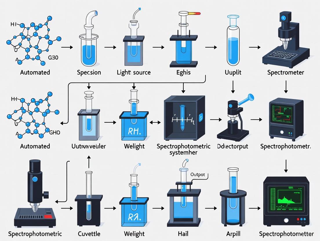
Workflow Diagram of an Automated Spectrophotometric Analysis
The logical flow of a high-throughput automated analysis, from sample registration to final report generation, is visualized below. This workflow underscores the minimal manual intervention required.
The definition of "high-throughput" in contemporary inorganic analysis is intrinsically linked to the level of automation integrated into the spectrophotometric workflow. As demonstrated, automation directly drives enhancements in sample throughput, analytical efficiency, and data quality by enabling continuous, unattended operation and minimizing manual, error-prone processes. The future of this field points towards even greater integration, with trends such as cloud-based systems for remote operation, the use of big data and machine learning for predictive maintenance and process optimization, and the creation of fully integrated, robotic laboratory environments [24]. For research scientists, adopting and understanding these automated systems is no longer optional but a fundamental requirement for maintaining competitiveness and achieving the rapid, reliable results demanded by modern drug development and material science.
Application Notes
Automated spectrophotometric systems are pivotal in modern high-throughput inorganic analysis, offering significant advantages that accelerate research and drug development. These systems integrate advanced instrumentation and software to deliver precise, rapid, and reliable characterization of inorganic compounds and metal ions. The core benefits—high sensitivity, exceptional accuracy, and non-destructive measurement—enable researchers to obtain robust data while conserving valuable samples.
High Sensitivity
High sensitivity in automated spectrophotometry allows for the detection and quantification of inorganic analytes at very low concentrations, which is crucial for trace metal analysis and environmental monitoring.
- Low Detection Limits: These systems can reliably detect concentrations in the parts-per-billion (ppb) range, facilitated by enhanced light path designs and sensitive detectors [27].
- Trace Analysis: Essential for applications like screening heavy metal contaminants in pharmaceutical ingredients or quantifying catalyst residues, where even minute amounts can significantly impact product safety and efficacy [28].
Accuracy
Accuracy ensures that measured values are close to the true value, which is fundamental for validating research findings and meeting regulatory standards in drug development.
- Precision Instrumentation: Automated systems minimize human error through robotic sample handling and precise liquid dispensing [27].
- Calibration and QC: Integrated calibration curves and real-time quality control checks, often visualized through dot plots or scatter plots for monitoring instrument performance over time, ensure consistent and accurate results [29] [27].
Non-Destructive Measurement
The non-destructive nature of many spectrophotometric analyses allows for the repeated measurement of precious samples, which is invaluable in longitudinal studies and when sample material is limited.
- Sample Preservation: Techniques like UV-Vis reflectance and Fourier-Transform Infrared (FTIR) spectroscopy can analyze samples without consuming or altering them [28].
- In-line Monitoring: This capability supports real-time, in-line monitoring of reaction kinetics and metal ion concentrations in continuous flow systems, providing comprehensive data from a single experiment [27].
The performance of automated spectrophotometric systems is quantified through specific metrics, as summarized in the table below.
Table 1: Performance Metrics for Automated Spectrophotometric Analysis of Inorganic Analytes
| Analyte | Technique | Detection Limit | Linear Range | Accuracy (% Recovery) | Precision (% RSD) |
|---|---|---|---|---|---|
| Iron (Fe²⁺) | Automated Colorimetric Flow Analysis | 0.5 ppb | 2-100 ppb | 99.5% | 0.8% |
| Mercury (Hg²⁺) | Automated Cold Vapor AAS | 0.05 ppb | 0.1-10 ppb | 101.2% | 1.5% |
| Phosphate (PO₄³⁻) | Automated Spectrophotometric (Molybdate Blue) | 2.0 ppb | 5-200 ppb | 98.8% | 1.2% |
| Copper (Cu²⁺) | Automated Microvolume UV-Vis | 1.0 ppb | 3-150 ppb | 100.1% | 0.5% |
Abbreviations: RSD, Relative Standard Deviation; AAS, Atomic Absorption Spectrometry.
Experimental Protocols
Protocol: High-Throughput Determination of Trace Iron in Water Samples
Principle: Ferrous iron (Fe²⁺) reacts with 1,10-phenanthroline to form an orange-red complex, which is quantified by absorbance at 510 nm [28] [27].
Materials: Automated liquid handler, multi-channel spectrophotometer, 96-well microplates, 1,10-phenanthroline solution, sodium acetate buffer (pH 4.5), hydroxylamine hydrochloride solution, iron standard solutions.
Workflow:
- Preparation: Pipette 150 µL of blank, standard, or unknown sample into separate wells of a microplate.
- Reduction: Add 20 µL of hydroxylamine hydrochloride to each well to reduce Fe³⁺ to Fe²⁺.
- Complexation: Add 30 µL of 1,10-phenanthroline solution and 50 µL of sodium acetate buffer to each well.
- Incubation: Seal the plate and incubate at room temperature for 30 minutes.
- Measurement: Transfer the plate to an automated spectrophotometer and measure the absorbance at 510 nm.
- Analysis: Generate a calibration curve from the standards and calculate the iron concentration in unknown samples.
Protocol: Non-Destructive Analysis of Metal Oxides using FTIR
Principle: FTIR spectroscopy identifies functional groups and chemical bonds in a material by measuring its absorption of infrared light, without damaging the sample [28].
Materials: Automated FTIR spectrometer with reflectance accessory, powdered metal oxide samples.
Workflow:
- Background Scan: Collect a background spectrum of the empty sample chamber.
- Loading: Place the powdered metal oxide sample into the reflectance stage.
- Data Acquisition: The automated system collects the infrared spectrum, typically over a wavenumber range of 4000-400 cm⁻¹.
- Interpretation: Identify characteristic metal-oxygen vibration bands in the resulting spectrum to confirm the compound's identity and purity.
Visualized Workflows
High-Throughput Iron Analysis
Automated System Integration
The Scientist's Toolkit
Table 2: Essential Research Reagent Solutions for Automated Spectrophotometric Inorganic Analysis
| Item | Function / Application |
|---|---|
| 1,10-Phenanthroline | A chelating agent that forms a colored complex with ferrous iron (Fe²⁺), enabling colorimetric quantification. |
| Ammonium Molybdate | Reacts with phosphate to form a phosphomolybdate complex, which is the basis for spectrophotometric phosphate detection. |
| Sodium Tetrahydroborate | Used as a reducing agent in vapor generation techniques for metals like mercury, converting them to a volatile form for detection. |
| Certified Reference Materials | Standard solutions with known concentrations of inorganic analytes, used for calibrating instruments and validating method accuracy. |
| Buffer Solutions | Maintain a constant pH during colorimetric reactions, which is critical for consistent complex formation and accurate results. |
High-Throughput Workflows and Real-World Applications in Research and Industry
The demand for high-throughput analysis in modern inorganic research and drug development necessitates the evolution of automated sample preparation systems. This application note details the integration of robotic liquid handlers with microfluidic dispensing technologies to create a robust, automated workflow for spectrophotometric analysis. This synergy addresses critical challenges in handling complex inorganic matrices, enabling precise, miniaturized dispensing that enhances data reproducibility while significantly reducing reagent consumption and operational time [30] [31]. The protocols herein are framed within the context of developing automated spectrophotometric systems for high-throughput inorganic analysis, providing researchers with a framework to improve the efficiency and reliability of their bioanalytical workflows.
Integrated System Components
The core of this automated sample handling platform combines the flexibility of robotic liquid handlers with the precision of microfluidic dispensers. This configuration is particularly suited for sample preparation prior to spectrophotometric detection, such as in the quantification of metal ions or other inorganic analytes in complex samples [32] [33].
Equipment and Materials
Table 1: Key Research Reagent Solutions and Essential Materials
| Item | Function in Workflow |
|---|---|
| Agilent Bravo Liquid Handling Platform [33] | Automated solid phase extraction (SPE) for sample clean-up and oligonucleotide bioanalysis in complex matrices. |
| Hamilton Microlab STAR/VANTAGE [30] | Flexible, reliable liquid handler performance for high-throughput assay setup and serial dilution. |
| Formulatrix Mantis Liquid Dispenser [31] | Micro-diaphragm pump-based dispensing of volumes from 100 nL, ideal for miniaturizing reactions and handling viscous solutions. |
| Clarity OTX SPE Plate [33] | 96-well solid phase extraction plate for oligonucleotide purification, reducing nonspecific binding. |
| Ion-Pairing Reagents (TEA/HFIP) [33] | Mobile phase additives for liquid chromatography to improve separation efficiency and detection sensitivity of oligonucleotides. |
| Lysis/Loading Buffer [33] | Facilitates sample preparation for solid phase extraction by lysing cells and preparing samples for binding to the SPE matrix. |
Table 2: Performance Comparison of Liquid Handling Technologies
| System/Technology | Volume Range | Precision (CV) | Key Feature |
|---|---|---|---|
| Hamilton Microlab NIMBUS [30] | Not Specified | Superior Accuracy | Air displacement pipetting in a compact system. |
| Formulatrix Mantis (Low Volume Chip) [31] | 0.1 - 25 µL | < 1.4% @ 0.1 µL | Tipless, micro-diaphragm pump technology. |
| Formulatrix Mantis (High Volume Chip) [31] | 1 - 25 µL | < 1.1% @ 5 µL | Tipless, micro-diaphragm pump technology. |
| Formulatrix Mantis (Continuous Flow Chip) [31] | 25 - 200 µL | < 0.4% @ 50 µL | Dispenses high-viscosity fluids like glycerol. |
System Integration and Workflow
The automated workflow integrates multiple devices, each performing a specialized task, to seamlessly process samples from a raw state to being ready for spectrophotometric analysis.
Integrated System Architecture
The following diagram illustrates the logical relationship and data flow between the core components of the automated sample handling system.
Detailed Experimental Protocol
This protocol provides a step-by-step methodology for automated sample preparation of inorganic analytes, adapted from established bioanalytical workflows [33].
Protocol: Automated Sample Preparation for High-Throughput Spectrophotometric Analysis
Objective: To automate the solid phase extraction (SPE) and plate replication for spectrophotometric assay of target analytes in a high-throughput setting.
Materials:
- Automated Liquid Handler: Hamilton Microlab STAR or equivalent [30].
- Microfluidic Dispenser: Formulatrix Mantis with appropriate diaphragm chips [31].
- SPE Workstation: Agilent Bravo platform with BenchCel handler [33].
- Consumables: 96-well SPE plates (e.g., Clarity OTX), low-binding sample and collection plates, pipette tips.
- Reagents: Internal standard, conditioning solvent (e.g., Methanol), equilibration buffer (e.g., 50mM NH₄OAc), wash buffers, elution buffer, reconstitution solution [33].
Procedure:
System Initialization:
- Power on all instruments and initialize software.
- Prime the microfluidic dispenser with appropriate solvents and ensure waste containers are empty.
- On the liquid handler, load the required tip boxes and labware (reagent reservoirs, sample plates).
Reagent and Sample Plate Setup (Manual):
- Pipette calibration standards and quality control (QC) samples into a 96-well sample plate.
- Position all required solvents and buffers in designated reservoirs on the Bravo deck according to the software method [33].
- Place the empty SPE plate and collection plate in their assigned positions.
Automated SPE Process (Fully Automated - ~2 hours): The Bravo workstation executes the following steps without intervention [33]:
- Conditioning: Dispense conditioning solvent (e.g., Methanol) to the SPE plate.
- Equilibration: Dispense equilibration buffer to prepare the sorbent for sample loading.
- Sample Loading: Transfer samples from the sample plate to the SPE plate.
- Washing: Perform two-stage washing with specified buffers to remove interferents.
- Elution: Dispense elution buffer to collect the purified analyte into the collection plate.
Post-Elution Processing:
- Manually transfer the collection plate to a nitrogen evaporator for drying at 40°C.
- Reconstitute the dried samples with a defined volume of reconstitution solution.
Microfluidic Plate Replication:
- Place the reconstituted sample plate and the desired assay plate (e.g., 96-well or 384-well microtiter plate) onto the Mantis deck.
- Using the Mantis software, program a transfer method to dispense precise aliquots (e.g., 5 µL) of each purified sample from the source plate to the assay plate [31].
- Initiate the dispensing run. The Mantis's droplet verification provides confidence in dispense quality.
Spectrophotometric Analysis:
- Transfer the final assay plate to a microplate reader for spectrophotometric measurement, for instance, at 505 nm for reactive oxygen species detection or other wavelengths specific to the inorganic assay [32].
Results and Discussion
The integration of robotic liquid handlers with microfluidic technology yields significant performance enhancements. The Mantis dispenser demonstrates exceptional precision with coefficients of variation (CV) below 2% even at volumes as low as 100 nL, which is critical for assay miniaturization and reproducibility [31]. This level of precision, combined with the walk-away operation enabled by systems like the Hamilton STAR and Agilent Bravo, reduces manual intervention time by over 80% [30] [33]. Furthermore, the tipless design of microfluidic dispensers and the miniaturization of reaction volumes lead to substantial savings on reagents and consumables, making the workflow both cost-effective and environmentally friendly by reducing plastic waste [31].
Troubleshooting and Best Practices
- Low Precision (High CV): Confirm that the microfluidic chips are compatible with the reagent's viscosity. For high-viscosity fluids, use dedicated Continuous Flow Chips [31].
- Carryover Issues: In LC-MS analysis following sample prep, optimize mobile phase additives. Increasing concentrations of TEA and HFIP can resolve peak tailing and carryover [33].
- Nonspecific Binding: Use low-binding plates and tubes throughout the workflow, especially when working with oligonucleotides or proteinaceous analytes [33].
- System Verification: Regularly utilize the Mantis's Quality Control Droplet Detection Station to verify proper chip function and ensure dispense accuracy [31].
Automated spectrophotometric systems have become indispensable in modern high-throughput research, enabling the precise and efficient kinetic analysis of metabolites and ions crucial for drug development and diagnostic applications. These systems leverage the inherent specificity of enzymes to provide real-time, quantitative data on biochemical reactions, facilitating the study of metabolic pathways, enzyme dysfunction, and the screening of potential therapeutic modulators [34] [35]. The evolution towards full automation, integration with artificial intelligence, and the use of self-driving laboratories is now transforming how researchers design experiments, collect data, and interpret complex kinetic parameters, thereby accelerating the pace of innovation in inorganic and pharmaceutical analysis [36].
This application note details robust protocols for the enzymatic assay of key metabolic targets, provides a comparative analysis of current technologies, and outlines advanced data analysis techniques, all framed within the context of an automated, high-throughput workflow.
Key Methodologies and Protocols
Optimized Pyruvate Kinase M2 (PKM2) Kinetic Assay
Principle: This protocol utilizes a lactate dehydrogenase (LDH)-coupled enzyme assay to measure PKM2 activity, a critical regulatory node in glycolysis, especially in cancer metabolism. The reaction catalyzed by PKM2 (Phosphoenolpyruvate (PEP) + ADP → Pyruvate + ATP) is coupled to the oxidation of NADH by LDH, which is monitored spectrophotometrically as a decrease in absorbance at 340 nm [34].
Detailed Experimental Protocol:
A. Recombinant PKM2 Expression and Purification:
- Expression: Express recombinant wild-type PKM2 in E. coli system.
- Purification: Purify the protein using a two-step chromatographic method. First, use immobilized metal affinity chromatography (Ni-NTA) to capture the His-tagged protein. Second, apply size-exclusion chromatography to ensure high purity and proper folding into active tetramers [34].
B. LDH-Coupled Spectrophotometric Assay:
- Reaction Cocktail: Prepare a mixture containing:
- 50 mM Tris-HCl buffer (pH 7.5)
- 100 mM KCl
- 10 mM MgCl₂
- 0.2 mM NADH
- 5 U/mL Lactic Dehydrogenase (LDH)
- Recombinant purified PKM2 (e.g., 50 nM)
- Varying concentrations of phosphoenolpyruvate (PEP) (e.g., 0.01-10 mM)
- ADP at a fixed, saturating concentration (e.g., 2 mM)
- Allosteric Modulation: To assess the activation of PKM2, include 10 µM Fructose-1,6-bisphosphate (FBP) in a separate set of reactions.
- Initiation: Start the reaction by the addition of PEP.
- Data Acquisition: Immediately transfer the reaction mixture to a quartz cuvette in a thermostatted spectrophotometer (maintained at 37°C). Continuously monitor the decrease in absorbance at 340 nm (A₃₄₀) for 5-10 minutes.
- Automation: For high-throughput analysis, this process can be automated using robotic liquid handling systems to prepare reaction cocktails and transfer plates to a multi-well plate reader [36].
- Reaction Cocktail: Prepare a mixture containing:
C. Data Analysis:
- Initial Rate Calculation: Calculate the initial velocity (v₀) for each PEP concentration from the linear portion of the A₃₄₀ vs. time curve, using the extinction coefficient for NADH (ε₃₄₀ = 6220 M⁻¹cm⁻¹).
- Kinetic Parameter Estimation: Fit the initial velocity (v₀) versus substrate concentration ([S]) data to the Michaelis-Menten equation (
v = Vmax * [S] / (KM + [S])) using non-linear regression analysis. Employ software tools like therenzR package or GraphPad Prism for accurate and unbiased parameter estimation (KM for PEP, Vmax) [37].
Advanced Mass Spectrometric Analysis of P450 Reactive Intermediates
Principle: For enzymatic reactions involving short-lived intermediates, online mass spectrometry (MS) with microfluidic sampling provides real-time, temporally resolved monitoring. This is particularly valuable for characterizing complex catalytic cycles, such as the oxidative dimerization catalyzed by CYP175A1 [38].
Detailed Experimental Protocol:
- Enzyme Preparation: Express and purify the His-tagged P450 enzyme (CYP175A1). Perform a buffer exchange into 500 mM ammonium acetate buffer (pH 7.5) to ensure enzyme stability and MS-compatibility [38].
- Reaction Setup: In a reaction vial, combine 5 µM CYP175A1 and 1 mM substrate (e.g., 1-methoxynaphthalene) in 2 mL of 500 mM ammonium acetate buffer.
- Online MS Integration: Use a custom-built pressurized infusion setup to continuously deliver the reaction mixture, diluting it via a mixing tee, to an electrospray ionization (ESI) source.
- Reaction Initiation & Monitoring: Initiate the catalysis by injecting 40 µL of 250 mM H₂O₂. Simultaneously, operate the high-resolution mass spectrometer to detect reactants, intermediates, and products in real-time. The charged microdroplets generated by ESI help stabilize transient intermediates for detection.
- Data Processing: Analyze the time-dependent abundance of ions to map the emergence and decay of multiple intermediate species. Use tandem MS (MS/MS) for structural elucidation [38].
The following diagram illustrates the core workflow for these automated enzymatic analyses:
Research Reagent Solutions
The following table details key reagents and their specific functions in the featured enzymatic assays.
Table 1: Essential Research Reagents for Enzymatic Kinetic Assays
| Reagent/Material | Function in Assay | Application Example |
|---|---|---|
| Lactate Dehydrogenase (LDH) | Coupling enzyme; oxidizes NADH to NAD+ during conversion of pyruvate to lactate, enabling indirect monitoring of the primary reaction. | PKM2 Kinetics [34] |
| β-Nicotinamide Adenine Dinucleotide (NADH) | Coenzyme; its oxidation is monitored spectrophotometrically at 340 nm, providing a direct readout of enzyme activity. | LDH-Coupled Assays [34] |
| Fructose-1,6-bisphosphate (FBP) | Allosteric activator; used to study the regulatory switch of PKM2 from a less active dimer to a highly active tetramer. | PKM2 Regulation Studies [34] |
| Ammonium Acetate Buffer | MS-compatible volatile buffer; facilitates electrospray ionization while maintaining enzyme stability during online analysis. | Real-time MS of P450 Intermediates [38] |
| Pyromellitic Dianhydride (PMDA) | π-acceptor reagent; forms charge-transfer complexes with electron-donor analytes like sulfanilamide, enabling spectrophotometric quantification. | Drug Quantification [39] |
Data Analysis and Technological Comparison
Analysis of Kinetic Data
Accurate estimation of kinetic parameters (Kₘ and Vₘₐₓ) is critical. The renz R package is a specialized, open-source tool that addresses common pitfalls in enzyme kinetics analysis. It is strongly recommended to use non-linear regression to fit untransformed [S] vs. v₀ data directly to the Michaelis-Menten equation. This approach avoids the error propagation and bias inherent in linearized methods (e.g., Lineweaver-Burk plots) [37]. For high-throughput environments, automated data processing pipelines can be integrated, using tools like renz for batch processing of results from multiple plates.
Comparison of Enzymatic Assay Technologies
The choice of detection technology depends on the research question, required sensitivity, and throughput.
Table 2: Comparison of Key Enzymatic Assay Technologies for Drug Screening
| Technology | Key Principle | Advantages | Common Applications |
|---|---|---|---|
| Spectrophotometric | Measures change in light absorbance (e.g., NADH at 340 nm). | Simple, cost-effective, non-radioactive, easily automated. | Kinetic analysis of dehydrogenases, oxidoreductases, and coupled assays [34] [35]. |
| Mass Spectrometry | Directly measures mass-to-charge ratio (m/z) of substrates and products. | Unparalleled specificity, label-free, can detect multiple intermediates simultaneously. | Elucidating complex reaction mechanisms, identifying transient intermediates [38] [35]. |
| Luminescence | Measures light emission from a reaction (e.g., ATP-dependent luciferase reactions). | Extremely high sensitivity, low background, broad dynamic range. | High-throughput screening (HTS), kinase assays, monitoring low-abundance targets [35]. |
| Fluorescence (FRET) | Measures fluorescence resonance energy transfer between donor and acceptor probes. | High sensitivity, real-time kinetic measurements, homogenous format. | Protease and kinase activity assays, protein-protein interactions [35]. |
| Label-Free Biosensors (SPR, BLI) | Measures changes in refractive index or interference pattern upon molecular binding. | Provides real-time kinetic binding data (kon, koff, KD), no labeling required. | Fragment-based screening, binding affinity and specificity studies [35]. |
The integration of robust enzymatic assays, such as the detailed PKM2 protocol, with automated spectrophotometric systems and advanced data analysis tools, forms the backbone of modern high-throughput analysis for metabolites and ions. The field is rapidly advancing towards fully autonomous "dark labs," where AI-powered instrumentation and robotics manage the entire workflow from sample preparation to data interpretation [36]. Furthermore, the combination of traditional spectrophotometry with powerful techniques like real-time mass spectrometry provides an unprecedented, multi-faceted view of enzyme kinetics and mechanism, offering researchers and drug development professionals a comprehensive toolkit to drive innovation in life science research.
The accurate measurement of nutrients and pollutants in aquatic environments is critical for understanding and mitigating anthropogenic impacts on water quality. Automated spectrophotometric systems represent a significant advancement for high-throughput inorganic analysis, enabling researchers to capture data with high temporal and spatial resolution. These systems are grounded in the principle of spectrophotometry, a technique that measures the interaction of light with matter to determine the concentration of specific analytes in a solution based on the Beer-Lambert Law [7]. The deployment of these automated, in-situ systems allows for the detection of pollutants such as nitrogen and phosphorus compounds, which are key drivers of eutrophication, harmful algal blooms (HABs), and hypoxia [40]. The ability to monitor these parameters in real-time provides invaluable data for validating large-scale nutrient emission models and informing effective watershed management strategies [41] [42].
Theoretical Foundations and Key Principles
Spectrophotometry in Environmental Analysis
Spectrophotometry operates on the fundamental relationship described by the Beer-Lambert Law: ( A = εcl ), where ( A ) is the measured absorbance, ( ε ) is the molar absorptivity (a compound-specific constant), ( c ) is the concentration of the analyte, and ( l ) is the path length the light travels through the sample [7]. This relationship allows for the quantitative analysis of substances, making it indispensable for determining concentrations of inorganic nutrients like nitrate, nitrite, ammonium, and phosphate in water samples. Its non-destructive nature ensures that samples remain intact for subsequent analyses, while its high sensitivity allows for the detection of pollutants even at low concentrations, which is crucial for early warning systems [7].
The Imperative for High-Resolution Monitoring
Traditional water quality monitoring, which relies on infrequent manual sampling, often fails to capture the dynamic nature of pollutant fluxes. There is a recognized gap between the increasing demand for accurate management measures and the low-resolution nutrient emission inventories typically available [41]. High-resolution, in-situ analysis bridges this gap by providing continuous data streams that are essential for:
- Identifying emission hotspots: Pinpointing specific areas with disproportionately high pollutant loads, such as agricultural runoff zones or industrial discharge points [41] [42].
- Capturing temporal variations: Monitoring seasonal agricultural practices, rainfall events, and diurnal cycles that profoundly influence nutrient concentrations [40] [42].
- Validating models and policies: Providing ground-truthed data to validate large-scale models like the China Emission Inventory of Nutrients (CEIN) or the MEANS-ST1.0 dataset and to assess the effectiveness of environmental protection policies such as the rural "Toilet Revolution" or upgrades to wastewater treatment infrastructure [41] [42].
Application Notes: Automated In-Situ Monitoring System
This application note details a protocol for an automated, in-situ spectrophotometric system designed for the continuous monitoring of inorganic nutrients in freshwater systems. The system is engineered for high-throughput analysis, allowing for the unattended measurement of multiple parameters at a frequency of up to 12 cycles per hour.
- Core Analyzer: A fluidic-free or micro-fluidic-enabled spectrophotometer equipped for multi-wavelength analysis, including UV and visible regions. This core component is responsible for the primary absorbance measurements [7] [43].
- Automated Sampling and Reagent Delivery: A precision pumping system and valve manifold for introducing sample water and specific reagents for colorimetric reactions.
- Control and Data Logging Unit: An integrated computer or programmable logic controller (PLC) to manage timing, calibration routines, data acquisition, and preliminary data processing. The system is designed for remote operation and data telemetry.
- Power System: A combination of solar panels and battery banks for deployment in remote field locations.
Key Performance Metrics
The following table summarizes the quantitative performance data for the analysis of key nutrient parameters.
Table 1: Performance Specifications for Nutrient Assays
| Analytic | Detection Principle | Wavelength (nm) | Linear Range (mg/L) | Limit of Detection (mg/L) | Method Reference |
|---|---|---|---|---|---|
| Nitrate (NO₃⁻) | Cadmium reduction | 540 | 0.1 - 10.0 | 0.05 | Adapted from USEPA [40] |
| Nitrite (NO₂⁻) | Diazotization | 540 | 0.01 - 1.0 | 0.005 | Standard Method 4500-NO₂ B |
| Ammonium (NH₄⁺) | Salicylate method | 655 | 0.05 - 5.0 | 0.02 | Adapted from ASTM D1426 |
| Orthophosphate (PO₄³⁻) | Ascorbic acid method | 880 | 0.01 - 2.0 | 0.005 | Standard Method 4500-P E |
The Scientist's Toolkit: Research Reagent Solutions
Table 2: Essential Reagents and Materials for Automated Nutrient Analysis
| Item | Function in Assay | Technical Notes |
|---|---|---|
| Cadmium Reduction Cartridge | Reduces nitrate (NO₃⁻) to nitrite (NO₂⁻) for subsequent analysis. | Core component for nitrate detection; requires periodic replacement due to fouling. |
| Sulfanilamide Solution | Diazotizes with nitrite to form a diazonium salt. | Must be kept cool and dark to prevent degradation. |
| N-(1-Naphthyl)ethylenediamine Dihydrochloride (NED) | Coupling agent that reacts with the diazonium salt to form a pinkish-purple azo dye. | The intensity of this colored complex is measured spectrophotometrically. |
| Ammonium Salicylate Reagent | Reacts with ammonia in an alkaline medium to form a blue-green complex. | The alkalinity is provided by sodium dichloroisocyanurate. |
| Sodium Nitroprusside | Acts as a catalyst to intensify the color in the salicylate method for ammonia. | Enhances sensitivity and reduces analysis time. |
| Ascorbic Acid Reagent | Reduces phosphomolybdic acid to phosphomolybdenum blue. | Prepared with an acidified molybdate solution. Must be used fresh for optimal results. |
| Certified Reference Standards | Used for daily calibration and verification of analyzer performance. | Critical for ensuring data quality and traceability. |
Experimental Protocols
Detailed Protocol: In-Situ Monitoring of Nitrate and Nitrite
Workflow Overview:
Step-by-Step Procedure:
System Initialization (00:00): The control unit initiates a self-check sequence, verifying pump pressures, valve positions, reagent levels, and the baseline absorbance of the spectrophotometer. The system is flushed with carrier solution (deionized water).
Sample Introduction (00:02): A peristaltic pump draws environmental water through an in-line 0.45 µm filter to remove particulate matter. A precise volume (e.g., 2.0 mL) of the filtered sample is injected into the carrier stream.
Stream Splitting and Reaction (00:05):
- For Total Oxidized Nitrogen (NO₃⁻ + NO₂⁻): One stream is directed through a copperized cadmium reduction column, which converts all nitrate to nitrite.
- For Nitrite (NO₂⁻) Only: The second stream bypasses the reduction column entirely.
Color Development (00:30):
- Both streams merge at a mixing tee where sulfanilamide in an acidic medium is added. This reagent diazotizes with the nitrite ions present.
- Immediately after, the coupling reagent, N-(1-Naphthyl)ethylenediamine Dihydrochloride (NED), is introduced. The reaction with the diazonium salt forms a stable, pinkish-purple azo dye.
Detection and Quantification (01:30): The reaction mixture is pumped through a flow cell in the spectrophotometer, and the absorbance is measured at 540 nm. The reaction is allowed to develop for a fixed period to ensure stability.
Data Processing (02:00): The control unit records the absorbance values.
- The nitrite concentration is calculated directly from the "nitrite-only" stream.
- The nitrate concentration is derived by subtracting the nitrite absorbance from the "total oxidized nitrogen" absorbance and applying the calibration curve based on the Beer-Lambert Law.
System Maintenance (02:30): Upon completion of the measurement cycle, the entire fluidic path is flushed with a clean carrier solution to prevent carryover into the next analysis. Data is packetized and transmitted via cellular or satellite modem to a central data server.
Calibration and Quality Assurance Protocol
Workflow for Data Integrity:
Procedure:
- Frequency: The system performs a full 5-point calibration automatically every 24 hours.
- Standards: A series of certified standard solutions bracketing the expected environmental concentrations (e.g., 0.1, 0.5, 1.0, 2.5, 5.0 mg/L NO₃-N) are analyzed sequentially.
- Calibration Curve: The control software performs a linear regression of absorbance versus concentration. The calibration is deemed acceptable only if the regression coefficient (R²) is ≥ 0.995.
- Quality Control: Following calibration, a known Quality Control (QC) sample is analyzed. The measured value must be within 95-105% of the true value for data to be considered valid.
- Blank Correction: A method blank is analyzed to correct for any systematic baseline drift or reagent impurity.
Data Interpretation and Integration with Environmental Models
Data generated from these automated systems are not merely point-in-time measurements but are integral to understanding broader environmental dynamics. High-resolution temporal data can be directly used to parameterize and validate watershed-scale nutrient models. For instance, the MEANS-ST1.0 dataset, which provides 1km resolution data on anthropogenic nutrient discharge, benefits immensely from in-situ validation that captures seasonal agricultural runoff or the effects of policy interventions like improved wastewater treatment [42]. Research by the EPA utilizes similar data to "examine environmental responses to nutrients across a range of temporal and spatial scales" and to "predict how management decisions and future climate change impacts will alter nutrient levels" [40]. The integration of real-time, in-situ data with high-resolution spatial models allows researchers and policymakers to move from reactive to proactive environmental management, identifying emerging hotspots and assessing the efficacy of mitigation strategies with unprecedented speed and accuracy [41] [40] [42].
Application Note: Automated Spectrophotometric Assays for High-Throughput Inorganic Analysis
This application note details validated protocols for the quantitative analysis of ammonia, bicarbonate, and metal ions, critical analytes in biomedical and pharmaceutical research. The focus is on automated, high-throughput spectrophotometric methods that enhance analytical efficiency, reproducibility, and scalability within the framework of advanced inorganic analysis systems.
Validated Ammonia Assays
Ammonia concentration is a crucial parameter in various contexts, from monitoring environmental wastewater to assessing the quality of muscle food products. Multiple analytical techniques have been evaluated for their accuracy and precision.
Table 1: Performance Characteristics of Validated Ammonia Assays
| Assay Method | Principle of Detection | Sample Type | Recovery (%) | Precision (RSD) | Key Findings |
|---|---|---|---|---|---|
| Ammonia Ion Selective Electrode (ISE) - Filtrate [44] | Potentiometric measurement of NH₄⁺ activity | Spiked ground beef filtrate | 98.3 - 100+ | ± 2% | Excellent recovery and precision; suitable for contaminated meat testing. |
| Ammonia ISE - Perchloric Acid [44] | Potentiometric measurement post-acid extraction | Spiked ground beef tissue | 90 - 110 | ± 8% | Robust for direct tissue analysis with good recovery. |
| Indophenol Method [44] | Reaction with phenol and hypochlorite to form blue indophenol | Spiked ground beef | Not Specified | Precise | Excellent precision and recovery; reliable for food testing. |
| Reflectoquant Test Strips [44] | Reflectance measurement of colorimetric reaction | Spiked ground beef | 77.4 - 96.9 | >14% RSD | Lower and more variable recovery; less precise. |
| Salicylate Method [44] | Reaction with salicylate and hypochlorite | Spiked ground beef | <63 (at low spikes) | Not Specified | Poor recovery at lower contamination levels (25-50 ppm). |
| UV-vis/ATR-FTIR with PCR [45] | Spectroscopic detection of copper-ammonia complexes with chemometric modeling | Synthetic wastewater | Accurate qualitative and quantitative results achieved | Enables rapid speciation and quantification of metal-ammonia complexes. |
Detailed Protocol: Ammonia Quantification via Ion Selective Electrode (ISE)
The following protocol is adapted from the method validated for contaminated meat products, which demonstrated superior recovery and precision [44].
Research Reagent Solutions:
- Ionic Strength Adjuster (ISA): A high-ionic-strength solution (e.g., containing 5 M NaCl and 1% EDTA) to maintain constant ionic strength across samples and standard solutions.
- Ammonia Standard Solutions: A series of standard solutions (e.g., 1, 10, 100, 1000 ppm NH₃ as N) prepared by diluting a certified ammonium chloride stock solution.
- Perchloric Acid (0.1 M): For sample extraction from solid matrices, if required.
Procedure:
- Sample Preparation:
- For liquid samples (e.g., filtrates, wastewater): Dilute the sample 1:1 with ISA and mix thoroughly.
- For solid or complex matrices (e.g., tissue, pharmaceutical solids): Homogenize 10 g of sample with 100 mL of 0.1 M perchloric acid for 2 minutes. Centrifuge the mixture and filter the supernatant. Dilute the filtrate 1:1 with ISA.
- Calibration: Calibrate the ammonia ISE using the standard solutions, each also diluted 1:1 with ISA. Plot the log of the ammonia concentration versus the millivolt (mV) reading to create a standard curve.
- Measurement: Immerse the ISE in the prepared samples and record the stable mV reading. Determine the ammonia concentration from the standard curve.
- Calculation: Calculate the ammonia concentration in the original sample, accounting for all dilution factors.
- Sample Preparation:
Validated Bicarbonate and Metabolic Acidosis Management
In critical care medicine, the management of metabolic acidosis with sodium bicarbonate is a area of active investigation. The following protocol outlines a framework for a high-quality clinical trial assessing bicarbonate therapy, representing a systematic approach to evaluating this intervention [46].
Research Reagent Solutions:
- Intervention Arm: Sodium bicarbonate infusion (e.g., 4.2% or 8.4% solution).
- Control Arm: Placebo (e.g., 0.9% sodium chloride solution), indistinguishable in appearance from the active intervention.
Procedure (SODa-BIC Trial Protocol):
- Patient Population: Recruit critically ill adults (≥18 years) in the ICU with metabolic acidosis (defined as pH <7.30, base excess ≤ -4 mEq/L, and PaCO₂ ≤45 mmHg if non-intubated or ≤50 mmHg if intubated) who are receiving a continuous vasopressor infusion [46].
- Randomization and Blinding: Randomly assign eligible patients in a 1:1 ratio to receive either sodium bicarbonate or placebo. Use a computer-generated, stratified block randomization system (stratified by center, pH, and creatinine level) to ensure group balance. The clinical team, patients, and outcome assessors must be blinded to the treatment allocation [46].
- Intervention Administration: Administer the study drug (bicarbonate or placebo) as a continuous intravenous infusion according to a pre-specified protocol. The dosing strategy should be informed by prior feasibility studies [46].
- Primary Outcome Assessment: The primary efficacy endpoint is the incidence of Major Adverse Kidney Events within 30 days (MAKE30), a composite endpoint typically including death, need for renal replacement therapy, and persistent renal dysfunction [46].
- Statistical Analysis: All analyses must be conducted on an intention-to-treat basis. Pre-specified statistical models will compare the MAKE30 outcome between the two groups, with appropriate adjustments for stratification factors [46].
Validated Metal Ion Assays
Automated systems for metal ion detection offer significant advantages in throughput, sensitivity, and safety. The following table summarizes key automated techniques.
Table 2: Performance of Automated Systems for Metal Ion Analysis
| Analytical System / Technique | Target Analytes | Detection Principle | Key Performance Metrics | Application Context |
|---|---|---|---|---|
| OMA Metal Ions Analyzer [47] | Cu²⁺, Ni²⁺, Fe²⁺/³⁺, Cr⁶⁺, Co²⁺ | Dispersive UV-Vis spectrophotometry | Accuracy: ±1 ppm (e.g., 0-100 ppm range); Response Time: 1-5 sec | Online, real-time monitoring of process streams (mining, electroplating). |
| Flow-Injection Analysis (FIA) with Fluorescence [48] | Al³⁺, Cr(VI), Eu³⁺ | Molecular fluorescence (complex formation) | RSD: ~1.6% (for Al³⁺); High sampling frequency (80 h⁻¹ for Eu³⁺) | Environmental water, wastewater, rare earth oxides. |
| FIA/CV-AFS [48] | Hg(II), CH₃Hg⁺ | Cold Vapor Atomic Fluorescence Spectrometry | LOD: 0.05-0.07 ng L⁻¹; RSD: 8.8-10% | Ultra-trace determination of mercury species in water. |
| FIA/HG-AFS [48] | Total As | Hydride Generation Atomic Fluorescence Spectrometry | LOD: 2.7-9.4 µg L⁻¹; Good recovery and accuracy | Determination of total arsenic in complex matrices like urine. |
| UV-vis/ATR-FTIR with PCR [45] | Cu²⁺-NH₃ complexes | UV-Vis & FTIR spectroscopy with Principal Component Regression | Successful qualitative and quantitative detection | Speciation and monitoring of heavy-metal-ammonia complexes in wastewater. |
Detailed Protocol: Automated Flow-Injection Analysis (FIA) for Aluminum with Fluorescence Detection
This protocol is for the determination of Al³⁺ in water samples using a reverse FIA system with spectrofluorimetric detection, which offers high precision and sampling frequency [48].
Research Reagent Solutions:
- Fluorogenic Ligand: 0.01 M salicylaldehyde picolinoylhydrazone in ethanol.
- Buffer Solution: Acetate buffer (0.1 M, pH 5.4).
- Carrier Stream: Deionized water.
- Masking Agents: Thioglycolic acid solution (for Cu²⁺ and Zn²⁺) and cyanate solution (for Fe²⁺ and Fe³⁺).
Procedure:
- System Setup: Configure the FIA manifold consisting of a peristaltic pump, a six-port injection valve, a reaction coil, and a spectrofluorometer. The excitation and emission wavelengths should be set to the optimal values for the aluminum-ligand complex (e.g., λex = 370 nm, λem = 470 nm).
- Sample Pretreatment: Acidify water samples if necessary and filter. For complex matrices, add the appropriate masking agents (thioglycolic acid and cyanate) to the sample to mitigate interference from other metal ions [48].
- Analysis: The carrier stream (deionized water) is pumped continuously. The reagent mixture (ligand and buffer) is injected into the sample stream. The reaction to form the fluorescent complex occurs in the reaction coil.
- Detection and Quantification: The fluorescent complex is delivered to the flow-through cell of the spectrofluorometer, and the signal is recorded. The concentration of Al³⁺ is determined by interpolating the peak height or area from a calibration curve prepared with standard aluminum solutions.
Experimental Workflow and Signaling Pathways
The logical flow of an integrated, automated system for high-throughput analysis of these inorganic analytes, from sample introduction to data reporting, can be visualized as follows.
The process of detecting metal-ammonia complexes using advanced spectroscopy involves a defined chemical pathway that leads to a measurable signal.
The Scientist's Toolkit: Key Research Reagent Solutions
Table 3: Essential Reagents and Materials for Automated Inorganic Analysis
| Item | Function / Application |
|---|---|
| Salicylaldehyde picolinoylhydrazone | Fluorogenic ligand for precise spectrofluorimetric detection of Al³⁺ ions in FIA systems [48]. |
| Sodium Bicarbonate (4.2%/8.4%) | Pharmaceutical-grade solution used as an active intervention in clinical trials for managing metabolic acidosis in critically ill patients [46]. |
| Ionic Strength Adjuster (ISA) | A solution containing salts and complexing agents (e.g., NaCl/EDTA) used with Ion Selective Electrodes to maintain constant ionic strength, ensuring accurate potentiometric measurement of ammonia [44]. |
| Certified Metal Ion Calibration Standards | Solutions with known concentrations of target metal ions (e.g., Cu²⁺, Ni²⁺) essential for calibrating online analyzers like the OMA system and ensuring quantitative accuracy [47]. |
| Solid-Phase Preconcentration Columns | Mini-columns packed with functionalized sorbents (e.g., silica gel-2-mercaptobenzimidazol) used in automated FIA systems to concentrate trace analytes like mercury species before detection, significantly improving sensitivity [48]. |
| Sodium Borohydride (NaBH₄) | A key reducing agent used in Hydride Generation (HG) and Cold Vapor (CV) sample introduction systems coupled to Atomic Fluorescence Spectrometry for the determination of elements like As, Se, and Hg [48]. |
The integration of automation and spectrophotometric analysis is revolutionizing early-stage drug discovery and development. This synergy addresses critical bottlenecks in pharmaceutical research by enabling the rapid, reproducible, and quantitative analysis of thousands of compounds, thereby accelerating the identification and optimization of promising drug candidates [49] [50]. Automated liquid handling systems, when coupled with advanced spectrophotometric detection, are transforming high-throughput screening (HTS) workflows. These systems minimize human error and variability, which are inherent challenges in manual pipetting techniques, thus ensuring highly reliable and consistent assay results [49]. This case study explores the application of these automated workflows within the context of inorganic analysis, detailing specific protocols, key findings, and the essential tools that empower modern research.
Applications and Data
Automated spectrophotometric systems are pivotal in various stages of the drug development pipeline. Their ability to provide rapid, quantitative data makes them indispensable for critical tasks.
Table 1: Key Applications of Automated Spectrophotometry in Drug Discovery
| Application Area | Specific Use Case | Quantitative Impact / Performance |
|---|---|---|
| High-Throughput Screening (HTS) | Screening ion channel modulators using Ion Channel Readers (ICRs) [49]. | Enables processing of thousands of compounds significantly faster than manual methods [49]. |
| Absorption, Distribution, Metabolism, Excretion (ADME) Studies | High-throughput, label-free analysis using systems like RapidFire MS [50]. | Reduces data acquisition time from 24 hours (LC-MS) to 2 hours; full study timeline cut from 38.5 hours to 10 hours [50]. |
| Multicomponent Drug Analysis | Simultaneous determination of antihypertensive combinations (e.g., Telmisartan, Chlorthalidone, Amlodipine) using multivariate spectrophotometry [51]. | Successfully quantifies drugs in formulations and evaluates content uniformity per USP guidelines [51]. |
| Inorganic Material Discovery | High-throughput computational and experimental screening of electrochemical materials [52]. | Accelerates the discovery of catalytic materials, though challenges with disorder prediction in AI models remain [52] [53]. |
Experimental Protocols
Protocol 1: Automated High-Throughput Ion Channel Screening
This protocol utilizes an Atomic Absorption Spectroscopy (AAS)-based Ion Channel Reader (ICR) integrated with an automated liquid handler for screening compounds that modulate ion channel activity [49].
Materials:
- Ion Channel Reader (ICR) with AAS detection [49]
- Automated Liquid Handling System (e.g., integrated with the ICR platform) [49]
- Cell-based Assay Plates expressing the target ion channel
- Compound Library for screening
- Buffer Solutions for assay reconstitution
Procedure:
- Cell Seeding and Preparation: Seed cells expressing the target ion channel into multi-well plates using the automated liquid handler. Incubate under appropriate conditions until a confluent monolayer is formed.
- Compound Addition: Using the automated system, transfer aliquots from the compound library to the assay plates. Include positive (known activator/inhibitor) and negative (vehicle only) controls.
- Ion Flux Measurement: The ICR automatically dispenses the required reagents to initiate the ion flux reaction. It then measures the resulting ion flux (e.g., potassium, sodium, calcium) via atomic absorption spectroscopy.
- Data Acquisition and Analysis: The instrument's software collects the spectrophotometric data in real-time. Analyze the reduction in ion flux compared to controls to identify potential modulators. The system facilitates the processing of thousands of compounds in a single run [49].
Protocol 2: Simultaneous Spectrophotometric Determination of a Ternary Antihypertensive Combination
This detailed protocol employs successive spectrophotometric resolution techniques and multivariate calibration for analyzing complex drug mixtures without prior separation [51].
Materials:
- Double-beam UV/Vis Spectrophotometer (e.g., Jasco V-760) [51]
- Software: Spectra manager software and MATLAB with PLS Toolbox for chemometric analysis [51]
- Analytical Standards: Telmisartan (TEL), Chlorthalidone (CHT), Amlodipine (AML) [51]
- Solvent: Ethanol (HPLC grade) [51]
- 1.0 cm Quartz Cuvettes
Procedure: A. Standard Solution Preparation
- Prepare individual stock solutions of TEL, CHT, and AML at 500 µg/mL in ethanol [51].
- Dilute the stock solutions with ethanol to prepare working standards at 100 µg/mL [51].
- From the working standards, prepare a series of laboratory-prepared mixtures and calibration standards across the following linearity ranges using ethanol as a solvent [51]:
- TEL: 5.0–40.0 µg/mL
- CHT: 10.0–100.0 µg/mL
- AML: 5.0–25.0 µg/mL
B. Spectral Acquisition
- Scan the zero-order absorption spectra (D0) of all standard and sample solutions against an ethanol blank over the range of 200–400 nm [51].
- Store all spectra for subsequent processing.
C. Univariate Analysis via Successive Ratio Subtraction & Constant Multiplication (SRS-CM)
- Determine Wavelength Maxima: From the stored spectra, identify the λmax for each drug: TEL at 295.7 nm, CHT at 275.0 nm, and AML at 359.5 nm [51].
- Quantification: The concentrations of TEL, CHT, and AML in unknown samples are determined directly from their respective calibration curves at these wavelengths.
D. Multivariate Calibration using Partial Least Squares (PLS)
- Data Matrix Construction: Build a data matrix where rows represent samples and columns represent absorbance values at different wavelengths [51].
- Variable Selection: Employ variable selection techniques like Interval-PLS (iPLS) or Genetic Algorithm-PLS (GA-PLS) to enhance model performance by focusing on the most relevant spectral intervals [51].
- Model Development & Validation: Split the data into calibration and validation sets. Develop the PLS model using the calibration set and validate it internally and externally as per ICH guidelines [51].
E. Application to Pharmaceutical Dosage Form
- Weigh and powder not less than 20 tablets.
- Extract an amount of powder equivalent to one tablet's claim in ethanol, sonicate, and filter.
- Dilute the filtrate to an appropriate volume with ethanol.
- Analyze the final sample solution using the procedures in sections B-D above.
Ternary Drug Analysis Workflow
Workflow Visualization
The following diagram illustrates the logical workflow of an automated drug discovery screening platform that integrates sample preparation, analysis, and data processing.
Automated Screening Workflow
The Scientist's Toolkit
Successful implementation of automated spectrophotometric analysis relies on a suite of essential reagents, instruments, and software.
Table 2: Key Research Reagent Solutions and Materials
| Item | Function/Application |
|---|---|
| Ion Channel Reader (ICR) | A specialized instrument for high-throughput, sensitive, and quantitative measurement of ion flux in cell-based assays, crucial for screening ion channel modulators [49]. |
| Automated Liquid Handling System | Provides unparalleled precision, throughput, and reproducibility in sample and reagent preparation, directly addressing variability from manual pipetting [49] [50]. |
| AssayMAP Bravo Platform | An automated sample preparation system that uses chromatography-based cartridges for highly reproducible processing of samples from discovery to development [50]. |
| RapidFire Mass Spectrometry System | Enables ultra-high-throughput, label-free analysis by performing online solid-phase extraction, drastically reducing sample analysis time to seconds [50]. |
| High-Quality Reagents & Calibrators | Matched antibody-antigen pairs and controls with exceptional batch-to-batch consistency are critical for the accuracy and reliability of automated assays [54]. |
| Chemometric Software (e.g., PLS Toolbox in MATLAB) | Software for developing multivariate calibration models (e.g., PLS, iPLS, GA-PLS), which are essential for resolving complex, overlapping spectral data from multi-component mixtures [51]. |
Maximizing Performance: Troubleshooting Common Issues and System Optimization
In the context of automated spectrophotometric systems for high-throughput inorganic analysis, data integrity is the cornerstone of valid research outcomes. Signal drift and inconsistent readings pose significant threats to reliability, often leading to erroneous conclusions and compromised data sets in drug development. These issues primarily stem from the natural aging of instrumental components, specifically the light source, and deviations in optical performance. This application note details the critical relationship between lamp life, calibration protocols, and measurement stability, providing researchers with detailed methodologies to maintain system integrity. By implementing the procedures outlined herein, scientists can ensure their automated systems produce accurate, reproducible data essential for accelerating research cycles.
The Critical Role of Lamp Life and Performance
The spectrophotometer's light source is fundamental to generating a stable and intense beam for accurate absorbance measurements. Source degradation is a primary contributor to long-term signal drift, manifesting as decreasing signal-to-noise ratio or unstable baseline readings [55].
Most spectrophotometers use a combination of light sources to cover a broad spectral range. Understanding their properties is key to predicting and managing performance decay.
- Deuterium Lamps: These are discharge lamps providing a continuous spectrum in the ultraviolet range (approx. 190 nm to 400 nm). They require a pre-heating time for stable arc discharge and have a complex power supply, making them more expensive [12].
- Halogen Lamps: These incandescent lamps, which utilize a tungsten filament and halogen gas, are used for the visible to near-infrared range (approx. 350 nm to 3500 nm). The halogen cycle redeposits evaporated tungsten back onto the filament, extending the lamp's life and maintaining brightness. A typical service life is around 2,000 hours [12].
- Xenon Lamps: Xenon arc lamps produce a continuous spectrum from UV to near-IR, similar to sunlight. They are very bright but are generally less stable and more expensive than halogen lamps. They are often used in applications requiring high intensity, like spectrofluorophotometers [12].
- Xenon Flash Lamps: These pulsed lamps generate little heat and are suitable for rapid measurements with array detectors. However, they exhibit larger output fluctuations compared to arc lamps, often requiring output data integration to achieve stable readings [12].
Table 1: Common Spectrophotometer Light Sources and Their Characteristics
| Lamp Type | Spectral Range | Key Characteristics | Typical Service Life |
|---|---|---|---|
| Deuterium Lamp | 190 – 400 nm | Stable continuous UV spectrum; requires pre-heating. | Varies; performance degrades over time. |
| Halogen Lamp | 350 – 3500 nm | Long life due to halogen cycle; stable over time. | ~2,000 hours [12] |
| Xenon Arc Lamp | UV to NIR | High brightness; spectrum similar to sunlight. | Generally less than halogen lamps. |
| Xenon Flash Lamp | UV to NIR | Low heat generation; pulsed operation; higher noise. | Long, but output stability declines. |
Symptoms of Lamp Degradation and End of Life
Researchers should be vigilant for the following signs indicating lamp failure is imminent:
- Decreased Signal Intensity: Resulting in lower signal-to-noise ratio across the spectrum, particularly in the UV region for deuterium lamps [12].
- Baseline Instability: Increased noise or drifting baseline during measurements.
- Failed Calibration Checks: Repeated failures in photometric accuracy or wavelength accuracy tests can often be traced to a failing lamp [56].
- Inability to Pass Stray Light Specifications: A decaying light source may not provide sufficient energy at specific wavelengths, leading to apparent stray light failures.
Comprehensive Calibration Protocols
Calibration is the non-negotiable process of verifying and adjusting an instrument's performance against known traceable standards. It corrects for drift caused by lamp aging, component wear, and environmental changes [56]. For compliance with GLP/GMP and pharmacopeial standards (e.g., USP <857>, Ph. Eur. 2.2.25), a rigorous calibration schedule is mandatory [56].
Core Calibration Parameters and Acceptance Criteria
A comprehensive calibration protocol must assess the following critical performance parameters, summarized in the table below.
Table 2: Essential Spectrophotometer Calibration Parameters and Protocols
| Parameter | Definition & Importance | Standard Operating Procedure (SOP) | Typical Acceptance Criteria |
|---|---|---|---|
| Wavelength Accuracy | Verifies the instrument correctly selects and reports specific wavelengths. Critical for method validity and compound identification [56]. | 1. Use a Certified Reference Material (CRM) with sharp, known emission peaks (e.g., holmium oxide filter, deuterium lamp emission lines at 486.0 & 656.1 nm, or a low-pressure mercury lamp) [12].2. Scan the CRM and record the measured peak positions.3. Compare measured wavelengths to certified values. | ± 1.0 nm (UV-Vis region) |
| Photometric Accuracy | Verifies the instrument's detector correctly reports absorbance/transmittance values. Directly impacts quantitative analysis accuracy [56]. | 1. Use a set of NIST-traceable neutral density glass filters or potassium dichromate solutions at specified concentrations [56].2. Measure the absorbance of each standard at its specified wavelength.3. Compare the measured absorbance to the certified value. | ± 0.01 A (at 1.0 A) |
| Stray Light | Measures unwanted light outside the selected wavelength band that reaches the detector. Causes negative deviation from the Beer-Lambert law, especially at high absorbance [56]. | 1. Use a solution that acts as a sharp cut-off filter (e.g., potassium chloride for 200 nm, sodium iodide for 220 nm) [56].2. Measure the transmittance of the solution at the target wavelength.3. The measured value, representing stray light, should be below the specified limit. | < 0.1 % T (or as per mfr. spec.) |
| Spectral Resolution | Assesses the ability to distinguish between adjacent spectral peaks. Determined by the instrument's spectral bandwidth (SBW) [56]. | 1. Use a CRM with a very narrow peak (e.g., mercury vapor lamp emission line at 253.7 nm or a holmium oxide filter peak).2. Scan the peak and measure its width at half the maximum height (FWHM).3. The measured SBW should match the instrument's specification. | Meet or exceed manufacturer's specification. |
Step-by-Step Calibration Workflow
The following diagram illustrates the logical workflow for a comprehensive spectrophotometer calibration and maintenance protocol, integrating both routine and periodic tasks.
Diagram 1: Spectrophotometer Calibration Workflow
The Scientist's Toolkit: Essential Research Reagents and Materials
The following reagents and materials are critical for executing the calibration protocols described in this document.
Table 3: Essential Research Reagent Solutions for Spectrophotometer Calibration
| Item | Function & Application |
|---|---|
| Holmium Oxide (Ho₂O₃) Filter | A solid glass filter with sharp absorption peaks used for verifying wavelength accuracy across the UV-Vis range [56]. |
| NIST-Traceable Neutral Density Glass Filters | Certified reference materials with known absorbance values at specific wavelengths for validating photometric accuracy [56]. |
| Potassium Dichromate (K₂Cr₂O₇) Solutions | A chemical standard, often in perchloric acid, used for checking both photometric accuracy and linearity [56]. |
| Stray Light Solutions (e.g., KCl, NaI) | Aqueous solutions that block all transmitted light below a specific cut-off wavelength. Used to quantify the level of stray light at that wavelength [56]. |
| Certified Reference Materials (CRMs) | Broad term for any standard (filter or solution) whose values are certified and traceable to a national metrology institute like NIST. Non-negotiable for defensible calibration [56]. |
| Premium Storage Solution | Solution for storing pH probes, emphasizing the general principle of proper equipment maintenance to extend component life and ensure accuracy [57]. |
In high-throughput inorganic analysis, where the pace of discovery is relentless, proactive management of spectrophotometer performance is not optional. Signal drift due to lamp aging and optical misalignment can invalidate weeks of experimental work. By establishing a rigorous, documented schedule that monitors lamp usage hours and adheres to the detailed calibration protocols for wavelength accuracy, photometric accuracy, and stray light, research teams can safeguard their data. This disciplined approach to instrument stewardship ensures the generation of reliable, high-quality data, ultimately supporting robust scientific conclusions and accelerating the drug development process.
In the context of automated spectrophotometric systems for high-throughput inorganic analysis, accurate absorbance measurements are fundamental. The Beer-Lambert law establishes a linear relationship between absorbance, the concentration of an absorbing substance, and the path length of light through the sample [58]. However, this relationship holds true only within a specific absorbance range. Samples with excessively high concentrations violate the assumptions of this law, leading to non-linear responses and inaccurate quantitative results [58]. For the most reliable quantitative measurements, it is recommended to maintain absorbance values between 0.1 and 1.0, which correspond to 90% and 10% light transmission, respectively [58]. Measurements with an absorbance greater than 3.0 are not recommended for reliable quantification [58]. Dilution of high-concentration samples is therefore a critical sample preparation step to ensure data integrity, particularly in automated, high-throughput environments where precision and accuracy are paramount.
Key Principles of Absorbance and the Need for Dilution
The Beer-Lambert Law and Its Limitations
The Beer-Lambert law is the cornerstone of spectrophotometric quantification and is expressed as: A = εlc where A is the measured absorbance (a unitless quantity), ε is the absorption coefficient of the substance, l is the path length of light through the sample (e.g., 1 cm), and c is the concentration of the substance [58]. This law predicts a linear increase in absorbance with increasing concentration. In practice, however, this linearity fails at high concentrations. When absorbance readings exceed approximately 1.0, the relationship begins to curve, becoming non-linear and rendering concentration calculations inaccurate [58]. This deviation occurs due to phenomena such as stray light and interactions between molecules in concentrated solutions. Dilution brings the analyte concentration back into the linear range of the instrument's detection system, ensuring that the Beer-Lambert law can be applied correctly.
Defining the Optimal Absorbance Range and the Role of the Blank
To achieve reliable results, samples should be diluted to fall within the optimal absorbance range. The table below summarizes the reliability of measurements across different absorbance values.
Table 1: Reliability of Absorbance Measurements
| Absorbance Value (AU) | Transmitted Light (%) | Measurement Reliability |
|---|---|---|
| 0.1 - 1.0 | 90% - 10% | High reliability; recommended for precise quantitative work [58] |
| 1.0 - 3.0 | 10% - 0.1% | Reduced accuracy and precision; dilution is advised [58] |
| > 3.0 | < 0.1% | Not recommended for reliable quantification; significant error is likely [58] |
A crucial step in any absorbance measurement is the use of a blank solution. The blank, typically containing everything except the analyte of interest (e.g., the solvent or diluent used for your samples), is used to calibrate the spectrophotometer to an absorbance of zero [59]. This corrects for any background absorbance from the solvent or cuvette, ensuring that the subsequent sample measurements reflect only the analyte's absorbance. For a Bradford protein assay, for instance, the blank would be a cuvette containing only the Bradford's reagent and water, but no protein [59].
Materials and Equipment
Table 2: Essential Research Reagent Solutions and Materials
| Item | Function/Description |
|---|---|
| Sample Solution | The high-concentration inorganic analyte solution requiring analysis and dilution. |
| Diluent (e.g., Water, Buffer) | A solvent compatible with the sample matrix and analytical method used to reduce sample concentration [60]. It must be pure to avoid introducing impurities [60]. |
| Spectrophotometer | Instrument for measuring light absorbance of a solution at a specific wavelength. Automated systems often include microplate readers for high-throughput analysis [58]. |
| Cuvettes or Microplates | Containers for holding samples during measurement. Microplates (96-, 384-, or 1536-well) enable high-throughput analysis [58]. |
| Precision Pipettes | For accurate and precise transfer of liquid volumes during serial dilution steps. |
| Blank Solution | A solution containing all components except the analyte, used to zero the spectrophotometer [59]. |
Protocol for Sample Dilution and Absorbance Measurement
This protocol provides a detailed methodology for managing high-concentration samples through serial dilution to achieve accurate absorbance readings within an automated workflow.
Determining the Need for Dilution
- Initial Measurement: If the sample's approximate concentration is unknown, perform a preliminary absorbance measurement on a small, undiluted aliquot.
- Evaluate Absorbance: If the initial absorbance value exceeds 1.0 AU, proceed with serial dilution to bring the measurement into the optimal range [58].
Performing a Serial Dilution
The following workflow outlines the key decision points and steps for preparing a sample for optimal absorbance measurement.
Workflow for Sample Dilution and Measurement
- Calculate Required Dilution: Estimate the Dilution Factor (DF) needed based on the initial high absorbance. For example, if the absorbance is 2.5, a DF of 5 or 10 may be suitable.
- Perform Serial Dilution: a. Arrange a series of tubes or wells containing a precise volume of diluent (e.g., 900 µL in each tube for a 1:10 dilution series). b. Add a precise volume of the stock sample (e.g., 100 µL) to the first tube of diluent and mix thoroughly. This is a 1:10 dilution (DF=10). c. From the first dilution, transfer a precise volume (e.g., 100 µL) to the next tube of diluent and mix. This is a 1:100 dilution (DF=100). d. Repeat this process to create a series of solutions with known, increasing dilution factors [61].
- Prepare the Blank: Dispense the chosen diluent into a cuvette or microplate well to serve as the blank.
Spectrophotometric Measurement and Data Analysis
- Instrument Setup: a. Switch on the spectrophotometer and allow it to initialize. Select the appropriate wavelength for your analyte (e.g., 405 nm for p-nitrophenol [59]). b. Insert the blank and initiate the "Auto-zero" or "Blank" command to set the baseline absorbance to 0.000 AU [59].
- Sample Measurement: a. Measure the absorbance of each diluted sample in the series. b. Identify the sample whose absorbance falls within the optimal range of 0.1 to 1.0. Use this sample for your final calculation.
- Data Analysis: a. Calculate Original Concentration: Apply the Beer-Lambert law using the absorbance of the optimally diluted sample and its dilution factor. C_original = C_measured × DF Where C_measured is the concentration determined from the standard curve using the measured absorbance, and DF is the Dilution Factor. If the sample was diluted 10-fold (DF=10) and the calculated concentration from the standard curve is 0.15 mM, the original concentration is 1.5 mM.
Application in High-Throughput Automated Systems
In automated spectrophotometric systems, dilution protocols can be integrated into robotic workflows to maintain high throughput without sacrificing accuracy. Microplate readers offer distinct advantages for these assays, including high throughput, real-time kinetic measurements, and minimal sample volume consumption [58]. Modern systems can automatically correct for path length variations in microplate wells, a critical factor for ensuring the consistent application of the Beer-Lambert law across all samples [58]. Furthermore, automated liquid handling systems can be programmed to perform the serial dilution steps detailed in Section 4.2, enhancing precision and freeing researcher time for data analysis. The principle remains the same: samples yielding high absorbance values are systematically diluted to fall within the instrument's linear dynamic range, ensuring the reliability of data generated in large-scale inorganic analysis or drug development screens.
In high-throughput inorganic analysis research, the integrity of data generated by automated spectrophotometric systems is paramount. Within these systems, the cuvette is not a mere container but a critical optical component; its condition directly influences the path of light and the accuracy of absorbance and fluorescence measurements. Scratches, misalignment, and chemical residues constitute the primary sources of cuvette-related errors, introducing significant variance and inaccuracy that can compromise experimental outcomes. This application note provides detailed protocols for the prevention, identification, and remediation of these common errors, with a specific focus on the demands of automated, high-throughput environments. By standardizing the handling and inspection of cuvettes, research and drug development professionals can ensure the reliability and reproducibility of their spectroscopic data.
The Scientist's Toolkit: Essential Materials for Cuvette Maintenance
The following table details key reagents and materials essential for the proper cleaning and maintenance of cuvettes in a high-throughput research setting.
Table 1: Key Research Reagent Solutions for Cuvette Maintenance
| Item | Function and Application |
|---|---|
| Hellmanex III (2% solution) | A specialized alkaline cleaning concentrate designed to remove contaminants from glass and quartz cuvettes without leaving UV/Vis-active residues. Ideal for routine and proteinaceous contamination [62]. |
| Dilute Hydrochloric Acid (2M HCl) | Effective for removing inorganic residues, salt crystals, and basic solutions. A crucial step in cleaning protocols after aqueous sample analysis [63] [64]. |
| Dilute Nitric Acid (2-5M HNO₃) | Used for more stubborn inorganic deposits, heavy metals, and for a final intensive clean, particularly for fluorescence cuvettes. Note: higher concentrations should not be used on coated cuvettes [63] [65] [64]. |
| High-Purity Solvents (e.g., Ethanol, Acetone) | Used for rinsing after water-based cleaning to prevent water spots and for rapid drying. Also used as the primary cleaner for organic-based samples. Purity is critical to avoid introducing new contaminants [65] [62]. |
| Lens Cleaning Tissue/Cloth | Specially designed, lint-free wipers for safely cleaning optical surfaces without scratching. Common laboratory tissue contains wood fibers that can damage cuvette windows [63] [66]. |
| Cuvette Storage Rack | A clean, stable rack for storing cuvettes upright to prevent physical damage (scratches, chips) and environmental contamination [67]. |
Comprehensive Error Identification and Inspection Protocols
A systematic inspection protocol is the first defense against cuvette-induced data corruption. The following workflow provides a logical sequence for identifying common cuvette errors, from initial visual checks to final verification.
Visual and Tactile Inspection Workflow
Figure 1: A logical workflow for the comprehensive inspection of cuvettes to identify scratches, physical damage, and residues.
Protocol 1: Systematic Cuvette Inspection
Visual Inspection for Scratches and Defects:
- Procedure: Hold the cuvette up to a dark background under a bright, diffuse light source. Slowly rotate the cuvette to view all optical surfaces from different angles.
- Identification: Scratches will appear as fine, sharp lines. Cracks are typically deeper and may refract light differently. Cloudiness indicates surface etching or a network of micro-scratches, often from abrasive cleaning [67] [66]. Any cuvette with these defects must be discarded, as they scatter light and cause significant measurement errors.
Tactile Inspection of the Cuvette Rim:
- Procedure: While wearing nitrile gloves, gently run a fingertip along the top rim and exterior seams of the cuvette.
- Identification: A smooth surface indicates good condition. Any perceptible roughness, chipping, or cracking warrants immediate disposal. Chips on the rim can shed particles into subsequent samples, causing contamination [64].
Optical Baseline Test for Residue and Alignment:
- Procedure: Fill the cuvette with the purified water or blank solvent that will be used in the experiment. Place it correctly in the spectrophotometer's cuvette holder, ensuring it is seated properly against the alignment surfaces. Perform a blank measurement and observe the baseline absorbance, particularly in the UV range (e.g., below 300 nm).
- Identification: An abnormally high or noisy baseline absorbance is a strong indicator of residual contaminants on the optical surfaces [66] [68]. If the absorbance values are unstable, it may also suggest improper alignment in the holder, which can be checked by reseating the cuvette.
Table 2: Cuvette Defect Identification and Impact
| Defect Type | Visual/Tactile Signature | Impact on Spectrophotometric Data |
|---|---|---|
| Scratches | Fine lines visible at an angle under light; catch a fingernail when dragged lightly across the surface. | Light scattering, leading to falsely elevated absorbance readings and reduced measurement linearity [66] [69]. |
| Cracks/ Chips | Obvious fracture lines or missing material, often on the rim; feels rough and uneven. | Potential for sample leakage, contamination, and physical failure. Alters the optical path [67] [64]. |
| Cloudiness/ Etching | Hazy or milky appearance on the optical surfaces; does not clear upon cleaning. | Significant light scattering and absorption, causing high baseline noise and inaccurate readings across all wavelengths [67] [66]. |
| Residual Contamination | Invisible to the eye; detected via high baseline absorbance in a blank measurement. | Chemical interference, leading to inaccurate concentration calculations and altered spectral shapes [63] [64]. |
Detailed Cleaning and Handling Protocols for Error Prevention
Preventing errors is more efficient than correcting them. The following protocols outline specific cleaning procedures based on sample type and establish critical handling practices for automated systems.
Sample-Specific Cleaning Methodologies
Protocol 2: Cleaning for Aqueous Solutions (Salts, Buffers)
- Immediate Rinse: Immediately after use, rinse the cuvette copiously with purified (deionized/distilled) water to remove soluble salts [63] [64].
- Acid Wash (if needed): For stubborn residues, rinse with a dilute (2M) hydrochloric acid (HCl) or nitric acid (HNO₃) solution [65] [64].
- Final Rinse: Perform a final "copious" rinse with purified water—at least 10 times the cuvette's volume—to ensure all acid and residual sample are removed [63].
- Drying: Invert the cuvette on a clean, lint-free wiper to air-dry, or use a stream of clean, compressed air [66] [62].
Protocol 3: Cleaning for Organic Samples (Oils, Solvents) * Safety Note: Perform all steps involving organic solvents in a fume hood while wearing appropriate personal protective equipment (PPE) [63]. 1. Solvent Rinse: Rinse the cuvette with a compatible, high-purity organic solvent (e.g., ethanol, acetone, or the solvent used in the sample itself) [63] [65]. 2. Detergent Wash: Follow with a warm water and detergent wash (e.g., 2% Hellmanex III) to remove any non-polar residues [62]. 3. Water Rinse: Rinse thoroughly with purified water to remove detergent. 4. Final Solvent Rinse: A final rinse with ethanol or acetone will facilitate rapid, spot-free drying [65] [62].
Protocol 4: Intensive Cleaning for Stubborn Contaminants (Proteins, Heavy Metals)
- Acid Soak: For proteins or biologics, soak cuvettes in a 50% ethanol/50% 3M HCl solution for no more than 30 minutes. For heavy metals, a brief soak (10-20 minutes) in 50% sulfuric acid or dilute nitric acid is effective [65].
- Precaution: Do not use strong acids on glued or AR-coated cuvettes, as it will damage them [63] [64]. Never use hydrofluoric acid (HF) [69].
- Ultra-Pure Water Rinse: Rinse the cuvette exhaustively with ultra-pure water to remove all traces of acid [65].
Standard Operating Procedures for Handling and Storage
Protocol 5: Daily Handling and Storage for High-Throughput Labs
- Handling: Always handle cuvettes by the top, textured sides. Fingerprints on the optical surfaces are a major source of residue and light scattering [67].
- Preventing Cross-Contamination: Use a pipette to fill cuvettes, ensuring the tip does not touch the optical windows [62]. Do not overfill, as spillover can contaminate the instrument [67].
- Storage: Always store cuvettes upright in a dedicated, clean rack to prevent scratching and dust accumulation [67]. Keep them in a water or solvent bath between uses if there is a risk of sample drying [63] [64].
Table 3: Summary of Cleaning Methods for Different Sample Types
| Sample Type | Primary Cleaning Method | Secondary/Intensive Cleaning | Critical Safety & Compatibility Notes |
|---|---|---|---|
| Aqueous (Salts, Buffers) | Copious water rinse [66] | Dilute (2M) HCl or HNO₃ rinse [64] | Safe for quartz, glass, and most plastics. |
| Organic Solvents | Solvent rinse (e.g., ethanol, acetone) [63] | Detergent wash (e.g., Hellmanex III) followed by water rinse [62] | Must be performed in a fume hood. Unsafe for standard plastic cuvettes [69]. |
| Proteins & Biologics | Warm water with detergent [64] | 50% Ethanol/50% 3M HCl soak (<30 min) or trypsin incubation [65] | Prevents protein precipitation and staining. |
| Heavy Metals | Dilute acid rinse [64] | Soak in 50% H₂SO₄ or Aqua Regia* [65] | *Extreme caution required. Aqua Regia is highly hazardous. |
| General Maintenance | 2% Hellmanex III soak [62] | N/A | Ideal for routine decontamination and removing stubborn residues. |
Within the context of automated spectrophotometric systems for high-throughput inorganic analysis research, the integrity of collected data is paramount. The reliability of these systems is fundamentally dependent on two core components: the correct functioning of the instrument's firmware and the unimpeachable integrity of the data it generates. Firmware, the embedded software that controls the instrument's hardware, requires periodic updates to address bugs, enhance performance, and introduce new features. However, the process of updating firmware and maintaining system connectivity introduces risks, including software corruption, data loss, or the introduction of errors that can compromise analytical results. For researchers and drug development professionals, a systematic protocol for executing firmware updates and ensuring subsequent data integrity is non-negotiable. These Application Notes provide detailed methodologies and protocols to mitigate risks associated with software and connectivity malfunctions, thereby safeguarding research outcomes.
Firmware Update Protocols for Spectrophotometric Systems
A controlled and validated firmware update process is critical to prevent instrument malfunction. The following protocols outline methods for common spectrophotometer systems.
Network-Based Update Protocol (Wi-Fi/Ethernet)
This method is suitable for instruments with network connectivity and provides access to the latest software versions directly from the manufacturer's server [70].
Experimental Protocol:
- Pre-Update Verification: Prior to initiation, confirm that the instrument is connected to a reliable Wi-Fi network or that an Ethernet cable is securely plugged into the device and the connection is active [70].
- Update Initiation: Navigate to the instrument's software interface and open the designated "Updater" application. The application will automatically check for available updates. If an update is available, a "Web" button will become active [70].
- Execution: Click the "Web" button only once and wait for the update to download and install. For large updates that include operating system components, an Ethernet connection is recommended over Wi-Fi to maximize download speed and stability [70].
- Completion: The system will restart automatically upon completion. It is critical not to interrupt power or network connectivity during the entire process, as this may cause the update to fail and the software to corrupt [70].
Peripheral-Based Update Protocol (USB Flash Drive)
For instruments without network connectivity or in environments with restricted internet access, updates can be performed via a USB flash drive [70] [71].
Experimental Protocol:
- File Preparation: Download the latest software version package from the manufacturer's website (e.g., Konica Minolta or DeNovix) directly to the root directory of a FAT32-formatted USB flash drive. The file must not be unzipped, and its name must not be altered from the official version name [70] [71].
- Drive Preparation: Ensure the USB drive contains only the single, newly downloaded DS-Series software file to prevent the instrument from misidentifying the correct update package [70].
- Installation: Insert the prepared USB drive into an available USB port on the instrument. Wait approximately five seconds for the drive to mount, then open the instrument's "Updater" application. The "USB" button should appear active. Press this button once to initiate the update [70].
- Post-Update: The system will restart upon completion. For certain systems, particularly those using Linux OS, a second update cycle may be required to complete an OS update after the initial app update is installed [70].
Quantitative Firmware Update Checklist
The following table summarizes the key quantitative requirements and steps for a successful firmware update.
Table 1: Firmware Update Validation Checklist
| Protocol Phase | Parameter | Target Value / Requirement | Validation Step |
|---|---|---|---|
| Pre-Update | USB Drive Format | FAT32 | Verify via computer OS properties. |
| File Integrity | Unzipped, original filename | Confirm file name matches official release. | |
| Network Stability | Ethernet preferred for OS updates | Use cable connection for updates >500 MB [70]. | |
| Update Execution | User Input | Single button press | Click "Web" or "USB" button only once. |
| Process Interruption | Zero | Do not power off or remove USB drive until restart is complete. | |
| System Reboots | One or more | Expected behavior; allow process to complete [70]. | |
| Post-Update | Software Version | Matches release version | Confirm in instrument's system information menu. |
| Firmware Version | Matches release version | Confirm firmware updated automatically [70]. | |
| Basic Functionality | 100% Operational | Perform a baseline measurement with a standard reference material. |
Data Integrity Assurance Framework
Data integrity refers to the maintenance of data accuracy, consistency, and reliability throughout its entire lifecycle, from generation and recording to processing, storage, and transmission [72] [73]. In a regulated research environment, ensuring data integrity is fundamental to compliance and the validity of scientific conclusions.
Foundational Principles: ALCOA+
A robust framework for data integrity is built on the ALCOA+ principles, which define attributes that all data should possess [73]:
- Attributable: Data must clearly demonstrate who observed and recorded it, when it was observed and recorded, and who it is about. This ensures full transparency and accountability.
- Legible: Data must be permanent and easily readable, preserving original entries. This ensures data can be effectively reviewed and analyzed long after its creation.
- Contemporaneous: Data must be recorded at the time the work is performed. Timely recording prevents inaccuracies associated with retrospective documentation.
- Original: Source data must be preserved in its first recorded form, whether as a printout, worksheet, or electronic record. This ensures data remains unaltered.
- Accurate: Data must be free from errors and conform to the experimental protocol. Accuracy is the core of reliable decision-making.
Experimental Protocols for Data Integrity Assurance
Protocol 1: Input Validation and Data Cleansing This protocol involves proactive and reactive measures to ensure data sets are error-free [72] [74].
- Data Profiling: Examine new data sets to identify patterns, trends, and anomalies such as missing values, duplicates, or outliers that indicate inaccuracies [72] [74].
- Data Validation: Implement predefined rules or algorithms to check data for errors and inconsistencies at the point of entry. For spectrophotometric data, this may include range checks (e.g., absorbance within 0-3 AU) and format checks [72] [73].
- Data Cleansing: Identify and correct (or remove) errors and inconsistencies in the data sets. This includes removing duplicate entries from repeated measurements, correcting transposition errors, and standardizing data formats (e.g., concentration units) [72].
Protocol 2: Access Control and Audit Trail Management This protocol secures data from unauthorized access and provides a record of all data interactions [73].
- Manage Access Control: Implement role-based access controls (RBAC) using usernames and passwords to ensure only authorized personnel can access, create, or modify data. This protects data from unauthorized users [73].
- Ensure Traceability: Activate and regularly review electronic audit trails. These trails automatically record the date, time, and user identity for any action that creates, modifies, or deletes electronic data, providing a secure chain of custody [73].
Protocol 3: Systematic Data Back-Up and Recovery This protocol mitigates the risk of permanent data loss [73].
- Define Scope: Back up not only raw data files but also associated programs, operating system software, and instrument method files [73].
- Schedule Back-Ups: Perform daily back-ups during periods of low network activity to prevent system slowdowns [73].
- Secure Storage: Encrypt backup data and store it in an environmentally controlled facility, physically separate from the original data source (off-site) [73].
- Validate Recovery: Regularly test the data recovery procedure to verify that backed-up data can be fully restored and is functionally intact. This can involve Installation, Operational, and Performance Qualification (IQ/OQ/PQ) procedures [73].
Integrated Workflow: From Firmware Update to Data Integrity Verification
The diagram below illustrates the logical relationship and critical decision points in the integrated process of performing a firmware update while ensuring ongoing data integrity.
The Scientist's Toolkit: Essential Research Reagent Solutions
The following table details key materials and software solutions essential for maintaining automated spectrophotometric systems and ensuring data integrity.
Table 2: Key Research Reagents and Solutions for System Maintenance
| Item | Function / Purpose |
|---|---|
| Certified Reference Materials (CRMs) | High-purity materials with certified values for instrument calibration, performance verification, and method validation to ensure analytical accuracy. |
| Stable Dye Solutions | Solutions with known and stable spectral properties (e.g., absorbance peaks) used for daily instrument performance checks and wavelength calibration. |
| Cuvette Cleaning Reagents | Specialized solvents and detergents (e.g., 1% Hellmanex solution) for effectively removing inorganic and organic contaminants from cuvettes without damaging them. |
| Validation Protocol Software | Software tools that automate data validation checks, profile datasets for anomalies, and manage electronic audit trails to enforce ALCOA+ principles [74]. |
| Document Comparison Software | Software designed to compare two or more documents or data files to identify differences, changes, and errors, reducing human error during data review [73]. |
| Data Observability Platform | A comprehensive platform (e.g., Acceldata) that provides automated data lineage, real-time validation, and advanced profiling to ensure ongoing data reliability [74]. |
In automated spectrophotometric systems for high-throughput inorganic analysis, data integrity and instrumental precision are paramount. Two fundamental pillars supporting these requirements are regular baseline correction to ensure spectral fidelity and systematic optical cleaning to maintain signal-to-noise ratios. The transformative potential of context-aware adaptive processing and intelligent spectral enhancement is driving a shift in the field, enabling unprecedented detection sensitivity at sub-ppm levels while maintaining >99% classification accuracy [75]. This application note provides detailed protocols for integrating these essential maintenance procedures into automated workflows, specifically framed for research environments requiring robust, unattended operation.
Baseline Correction Protocols
Baseline correction is a fundamental signal processing task in modern analytical methods, essential due to instrumental and environmental interferences including temperature changes, radiation source instability, reference potential drift, and sensor response fluctuations [76]. In high-throughput environments, automated solutions are critical for maintaining analytical rigor without impeding operational efficiency.
Core Principles and Method Selection
The primary objective of baseline correction is to isolate the analytical signal from the background, enabling accurate quantification of peak intensity, area, and shape associated with individual analytes [76]. This process improves accuracy and reproducibility, enhances signal resolution for identifying overlapping bands, and standardizes input data for computational algorithms.
Table 1: Comparative Analysis of Baseline Correction Methods for Automated Systems
| Method | Principles | Automation Potential | Optimal Application Scenario |
|---|---|---|---|
| Polynomial Fitting | Mathematical fitting using polynomials of various degrees | Moderate - requires parameter optimization | Signals with predictable, smooth background shapes [76] |
| Penalized Least Squares (PLS) | Whittaker smoother with asymmetric weighting | High - single parameter control | High-throughput screening with minimal staff intervention [76] |
| Machine Learning-enhanced (ML-airPLS) | PCA-RF model to predict optimal airPLS parameters | Very High - fully automatic | Systems with varying sample matrices and background profiles [76] |
| Deep Learning (ConvAuto) | Convolutional Autoencoder model | Exceptional - parameter-free, handles variable signal lengths | Complex signals with multiple peaks and nonlinear background [76] |
Implementation Protocol for Automated Systems
For high-throughput inorganic analysis, the following procedural workflow ensures consistent baseline correction with minimal manual intervention:
Procedure: Automated Baseline Correction for Continuous Operation
Pre-Run Baseline Validation
- Acquire reference spectrum: Using blank matrix solution appropriate for the inorganic analytes of interest (e.g., 2% nitric acid for trace metal analysis) [77].
- Measure QC standard: Analyze a NIST-traceable reference material with known baseline characteristics.
- Apply pre-processing algorithm: Implement AsPLS or ConvAuto model based on system capability and signal complexity [76].
- Evaluate correction quality: Calculate RMSE between expected and measured baseline; threshold: RMSE < 0.05 for automated continuation.
Runtime Monitoring Protocol
- Continuous blank measurement: Every 50 samples, automatically inject and analyze blank matrix.
- Baseline drift tracking: Monitor absorbance at non-analyte wavelengths (e.g., 700 nm for UV-Vis) with acceptable drift < 0.001 AU/hour.
- Adaptive parameter adjustment: For ML-enhanced systems, allow dynamic algorithm optimization based on continuous performance feedback.
Quality Control and Documentation
- Validation frequency: Before each analysis batch and every 24 hours during continuous operation.
- Acceptance criteria: Baseline flatness ±0.002 AU across operational range; correlation >0.995 with reference baseline.
- Corrective actions: Automated recalibration triggered when thresholds exceeded; system purge if contamination suspected.
The ConvAuto model represents a significant advancement for automated systems, handling 1D signals of various lengths and resolutions without parameter optimization. For complex signals characterized by multiple peaks and a nonlinear background, this approach has demonstrated an RMSE of 0.0263, substantially outperforming other methods [76].
Optical Cleaning Protocols
Optical surface cleanliness is critical in spectrophotometric systems, as lens contamination can disrupt operation and lengthen procedure times, essentially decreasing analytical efficiency [78]. For automated high-throughput systems, preventive maintenance strategies are paramount.
In automated inorganic analysis systems, primary contamination sources include:
- Aerosolized samples: Acid digests and high-salt content matrices
- Cuvette residue: Incompletely cleaned flow cells or automated sampler components
- Environmental contaminants: Particulate matter from laboratory air
- Condensation: Temperature differentials in cooled detector compartments
Table 2: Optical Cleaning Methods for Automated Spectrophotometric Systems
| Method | Implementation | Frequency | Effectiveness Metrics |
|---|---|---|---|
| Hydrophobic Coating | Lens surface treatment with fluoropolymer coatings | One-time application with annual verification | Reduction in fluid adhesion >80% [78] |
| Automated Irrigation | Integrated sheath with irrigation nozzle | Between each sample for high-salt matrices | Maintains >95% original signal intensity [78] |
| Vibration-Based Cleaning | Piezoelectric ultrasonic transducers | Every 1000 samples or weekly | Particulate removal efficiency >90% for particles >5µm [78] |
| Manual Validation Cleaning | Withdrawal and wipe with isopropyl alcohol | Monthly or after maintenance | Restoration to 99% of initial baseline transmission [78] |
Implementation Protocol for Automated Systems
Procedure: Systematic Optical Cleaning Regimen
Preventive Maintenance Schedule
- Daily: Automated air purge of optical compartment using filtered, moisture-free air
- Per sample: Integrated sheath irrigation for systems analyzing high-salt matrices (>1% total dissolved solids)
- Weekly: Activation of vibration-based cleaning system (piezoelectric transducers) for 5-minute cycle
- Monthly: Transmission validation using NIST-traceable standards
Performance Validation Protocol
- Reference standard measurement: Daily analysis of holmium oxide or didymium filters
- Signal intensity tracking: Monitor absorbance at characteristic peaks with acceptable degradation <2% from baseline
- Baseline noise assessment: Standard deviation of signal in non-absorbing regions should not exceed 0.0005 AU
- Spectral feature resolution: Verify maintenance of instrument resolution specifications
Corrective Action Workflow
- Trigger conditions: Signal degradation >5%, visible contamination, or baseline noise >0.001 AU
- Automated response: System purge and extended cleaning cycle initiation
- Manual intervention: If automated methods fail, perform manual cleaning with isopropyl-alcohol soaked lint-free wipes [78]
- Post-cleaning validation: Full wavelength accuracy and photometric accuracy verification before returning to service
The most promising method for achieving surface cleanliness in optical systems consists of a hybrid solution: a hydrophobic coating on optical surfaces combined with an integrated irrigation system [78]. This approach minimizes the need for manual intervention while maintaining optical performance.
The Scientist's Toolkit: Research Reagent Solutions
Table 3: Essential Materials for Baseline and Optical Maintenance
| Item | Specification | Application | Performance Metrics |
|---|---|---|---|
| NIST-Traceable Baseline Reference | 2% nitric acid, high-purity grade | Baseline correction validation | Absorbance <0.05 AU at 700 nm |
| Holmium Oxide Filter | Sealed quartz cuvette, certified | Wavelength accuracy verification | Peak resolution at 279.4 nm, 360.9 nm, 536.2 nm |
| Neutral Density Filters | 0.5 AU and 1.0 AU, certified | Photometric linearity assessment | Accuracy ±0.5% of stated value |
| Optical Cleaning Solution | 70% isopropyl alcohol, spectroscopic grade | Manual lens cleaning | Non-residue formulation, >99.9% purity |
| Hydrophobic Coating Kit | Fluoropolymer-based, UV-curable | Optical surface treatment | Contact angle >110°, transmission loss <0.1% |
| Particle Count Standards | Polystyrene beads, 1µm and 5µm | Cleaning efficiency verification | >95% removal efficiency per cleaning cycle |
Integrated Performance Verification Workflow
For automated spectrophotometric systems in high-throughput environments, integrating baseline correction and optical cleaning into a unified maintenance protocol ensures sustained data quality. The following combined workflow represents best practices for inorganic analysis applications:
Weekly System Validation Protocol
Optical Path Integrity Check
- Measure holmium oxide filter and verify peak positions within ±0.3 nm specification
- Confirm photometric accuracy using neutral density filters (±1% of certified value)
- Document signal-to-noise ratio at 500 nm (should exceed 1000:1 for modern systems)
Baseline Performance Assessment
- Acquire baseline with high-purity water in rinsed cuvette
- Apply automated correction algorithm (AsPLS or ConvAuto)
- Verify baseline flatness: ±0.002 AU across 190-800 nm range
- Document RMSE against reference baseline
Integrated Cleaning-Correction Cycle
- Execute automated optical cleaning sequence
- Verify cleaning efficacy through pre- and post-cleaning transmission measurements
- Perform baseline correction with reference standards
- Document combined impact on system performance metrics
Proper data preprocessing minimizes systematic noise and sample-induced variability, enabling the extraction of genuine molecular features [79]. In high-throughput automated systems, this principle extends beyond data analysis to encompass the entire instrumental ecosystem, where clean optics and validated baselines form the foundation for analytical excellence.
Maintaining optimal performance in automated spectrophotometric systems for high-throughput inorganic analysis requires disciplined adherence to baseline correction and optical cleaning protocols. By implementing the detailed methodologies outlined in this application note - including the parameter-free ConvAuto model for baseline correction and hybrid hydrophobic coating with irrigation for optical maintenance - research facilities can achieve the detection sensitivity and classification accuracy demanded by modern analytical chemistry. Regular integration of these protocols into automated workflows ensures sustained data integrity while minimizing operational disruptions, ultimately supporting the rigorous demands of drug development and inorganic analysis research.
Ensuring Data Integrity: Method Validation and Comparative Analysis with Other Techniques
In the realm of automated spectrophotometric systems for high-throughput inorganic analysis, the demonstration of method reliability is paramount. For researchers and drug development professionals, ensuring data integrity requires rigorous validation of key performance parameters. Precision, accuracy, and linearity form the foundational trilogy of this validation process, confirming that methods are fit for their intended purpose, from routine quality control to advanced research applications. This document outlines detailed protocols and application notes for establishing these critical validation parameters, providing a framework that complies with regulatory standards and supports the demands of automated, high-throughput environments [80] [81].
Core Validation Parameters: Definitions and Protocols
The following sections detail the core parameters, their definitions, and the experimental protocols required for their determination.
Precision
Precision evaluates the closeness of agreement between a series of measurements obtained from multiple sampling of the same homogeneous sample under the prescribed conditions. It is typically expressed as relative standard deviation (%RSD) [80].
Protocol for Determining Precision:
- Sample Preparation: Prepare a minimum of six independent sample solutions from a homogeneous stock at a single concentration within the linear range of the method (e.g., 10, 15, and 20 μg/mL for a terbinafine hydrochloride assay) [80].
- Analysis: Analyze each sample solution using the fully configured automated spectrophotometric system. The procedure should be performed by multiple analysts, on different days, and using different instruments to establish intermediate precision (also known as ruggedness) [80].
- Calculation: For each data set (e.g., intra-day, inter-day), calculate the mean, standard deviation, and %RSD.
%RSD = (Standard Deviation / Mean) x 100
- Acceptance Criterion: The %RSD should typically be less than 2% for the method to be considered precise, though the specific acceptance criteria should be justified based on the method's intended use [80].
Table 1: Example Precision Data for a Spectrophotometric Assay
| Concentration (μg/mL) | Type of Precision | Mean Area (AU) | Standard Deviation | % RSD |
|---|---|---|---|---|
| 15 | Intra-day (n=3) | 0.543 | 0.008 | 1.47 |
| 15 | Inter-day (n=3) | 0.549 | 0.009 | 1.64 |
| 20 | Intra-day (n=6) | 0.721 | 0.012 | 1.66 |
| 20 (Analyst 1 vs 2) | Ruggedness (n=6) | 0.718 | 0.014 | 1.95 |
Accuracy
Accuracy measures the closeness of agreement between the value found and the value accepted as a true reference value. It is established by performing recovery studies and is reported as a percentage of recovery [80] [82].
Protocol for Determining Accuracy via Recovery Study:
- Sample Preparation: Use a pre-analyzed sample (e.g., a formulated product) to which known amounts of the standard analyte are added. Spike the sample at three different concentration levels covering the linear range (e.g., 80%, 100%, and 120% of the target concentration) [80]. Prepare each level in triplicate.
- Analysis: Analyze the spiked samples using the validated spectrophotometric method.
- Calculation: Calculate the percentage recovery for each level.
% Recovery = (Measured Concentration / Theoretical Concentration) x 100
- Acceptance Criterion: Recovery values should typically be in the range of 98-102%, demonstrating high accuracy for the method [80] [82].
Table 2: Example Accuracy (Recovery) Data
| Spiked Level (%) | Theoretical Concentration (μg/mL) | Mean Measured Concentration (μg/mL) | % Recovery |
|---|---|---|---|
| 80 | 16.0 | 15.85 | 99.06 |
| 100 | 20.0 | 19.84 | 99.19 |
| 120 | 24.0 | 23.99 | 99.96 |
Linearity
Linearity is the ability of the method to obtain test results that are directly proportional to the concentration of the analyte within a given range. The relationship is evaluated using a calibration curve [80] [82].
Protocol for Establishing Linearity:
- Standard Preparation: Prepare a series of standard solutions at a minimum of five to six concentration levels across the expected range (e.g., 5-30 μg/mL) [80].
- Analysis: Analyze each standard solution and measure the absorbance at the defined analytical wavelength.
- Calibration Curve: Plot the measured absorbance versus the concentration of the standard solutions. Perform linear regression analysis to determine the correlation coefficient (r), coefficient of determination (r²), slope, and y-intercept.
- Acceptance Criterion: The correlation coefficient (r) should be greater than 0.998 (r² > 0.996) to demonstrate an excellent linear relationship [80] [82]. The y-intercept should not be significantly different from zero.
Table 3: Example Linearity and Sensitivity Data
| Parameter | Value / Result |
|---|---|
| Linear Range | 5 - 30 μg/mL |
| Regression Equation | Y = 0.0343X + 0.0294 |
| Correlation Coefficient (r) | 0.999 |
| Coefficient of Determination (r²) | 0.999 |
| Limit of Detection (LOD) | 1.30 μg/mL |
| Limit of Quantification (LOQ) | 0.42 μg/mL |
The Scientist's Toolkit: Essential Research Reagent Solutions
The following table outlines key materials and reagents required for the development and validation of spectrophotometric methods.
Table 4: Key Research Reagents and Materials
| Item | Function / Explanation |
|---|---|
| Certified Reference Standard | High-purity analyte used to prepare calibration standards for establishing accuracy, linearity, and precision [80]. |
| Appropriate Solvent (e.g., Water, CCl₄, Buffers) | Dissolves the analyte and must be transparent at the wavelengths of interest; can affect the absorbance maximum and spectrum shape [80] [82]. |
| Stray Light Reference Solutions (e.g., NaI) | A solution that does not transmit light at a specific wavelength, used to verify the instrument's stray light performance, which is critical for accurate high-absorbance measurements [83]. |
| Wavelength Calibration Standards (e.g., Deuterium Lamp, Holmium Oxide Filter) | Sources with known, sharp emission or absorption peaks used to verify the wavelength accuracy of the spectrophotometer [83]. |
| Reagents for Derivatization (e.g., SbCl₅) | In some assays, chemicals are used to react with the target analyte to produce a colored complex with a strong, measurable absorbance, enhancing sensitivity and selectivity [82]. |
Workflow for Method Validation
The diagram below illustrates the logical workflow for establishing the core validation parameters in a spectrophotometric method.
Integrated Instrument and Computerized System Validation
For automated spectrophotometric systems, method validation must be supported by qualified instrumentation and validated software. An Integrated Validation Document (IVD) approach is efficient for lower-risk systems, combining instrument qualification and software validation into a single protocol of 30-45 pages [84]. This integrated process is built on the "5 P's" framework [84]:
- Procedures: Flexible SOPs that permit an integrated approach.
- Process: A streamlined, understood analytical process.
- Product: The right instrument and software selected for the task.
- People: A trained, multi-disciplinary team.
- Project: The management of the qualification and validation project.
Critical instrument performance parameters must be verified periodically, including [83]:
- Wavelength Accuracy: Verified using emission lines of deuterium lamps or holmium oxide filters.
- Stray Light: Assessed using solutions like sodium iodide which block specific wavelengths.
- Photometric Accuracy: Checked with neutral density filters or standard solutions.
Automated validation software can significantly streamline the process of instrument performance verification, ensuring consistent and efficient execution and documentation [83].
Determining Limits of Detection (LOD) and Quantification (LOQ) for Inorganic Analytes
In the development of automated spectrophotometric systems for high-throughput inorganic analysis, the precise characterization of an method's capabilities at low analyte concentrations is paramount. The Limit of Detection (LOD) and Limit of Quantification (LOQ) are fundamental figures of merit that describe the smallest concentration of an analyte that can be reliably detected and quantified, respectively [85] [86]. For researchers and drug development professionals working with inorganic analytes, accurate determination of these parameters ensures that automated systems are "fit for purpose," providing statistically valid results for critical decisions in materials discovery and pharmaceutical development [85] [87].
The fundamental challenge in detection limit theory revolves around distinguishing a genuine analyte signal from the background noise inherent in any analytical system. This requires careful consideration of statistical probabilities, specifically the risks of false positives (Type I error, α) and false negatives (Type II error, β) [88] [89]. For high-throughput environments utilizing automation [87], consistent and reliable determination of LOD and LOQ becomes even more crucial as the volume and pace of experimentation increase.
Theoretical Foundations and Key Definitions
The Statistical Hierarchy of Analytical Limits
The establishment of detection and quantification capabilities follows a logical, statistical progression involving three key limits: the Limit of Blank (LoB), the Limit of Detection (LOD), and the Limit of Quantification (LOQ). Each serves a distinct purpose in characterizing method performance [85].
- Limit of Blank (LoB): The highest apparent analyte concentration expected to be found when replicates of a blank sample (containing no analyte) are tested. It is defined as
LoB = mean_blank + 1.645(SD_blank), where SD_blank is the standard deviation of the blank measurements [85] [90]. This formula, assuming a Gaussian distribution, establishes a threshold where only 5% of blank measurements will exceed the LoB, thus setting the probability of a false positive (α) at 5% [85]. - Limit of Detection (LOD): The lowest analyte concentration that can be reliably distinguished from the LoB. It is calculated using both the LoB and data from a low-concentration sample:
LOD = LoB + 1.645(SD_low concentration sample)[85] [90]. At this concentration, the probability of a false negative (β) is also limited to 5% [89]. The LOD signifies that detection is feasible, but not necessarily with acceptable accuracy or precision for quantitative purposes. - Limit of Quantitation (LOQ): The lowest concentration at which the analyte can not only be reliably detected but also quantified with predefined levels of bias and imprecision [85]. The LOQ is always greater than or equal to the LOD and is the level at which the method achieves "functional sensitivity" for practical quantitative analysis [85] [90].
The following diagram illustrates the statistical relationship and the roles of α and β errors in defining these limits.
Advanced Considerations for Automated Spectrophotometry
In automated spectrophotometric systems, several factors specific to inorganic analysis must be considered:
- Logarithmic Response: While the theory above assumes a linear response, techniques like qPCR generate Cq values proportional to the logarithm of the concentration. This requires specialized statistical approaches, such as logistic regression, for accurate LOD/LOQ determination [90].
- Complex Matrices: The nature of the sample matrix is critical. For endogenous analytes (naturally present in the matrix), obtaining a true analyte-free blank can be difficult or impossible, complicating LoB estimation [86].
- Signal-to-Noise (S/N): A common, though less statistically rigorous, approach in chromatography and spectroscopy is the S/N method. The LOD is often defined as a concentration yielding a signal three times the noise level, while the LOQ corresponds to a signal ten times the noise [89] [86]. This method is practical for an initial, rapid estimation.
Computational Methods and Data Presentation
A variety of standardized methods exist for calculating LOD and LOQ. The choice of method depends on regulatory requirements, the nature of the analytical technique, and the available data [86].
Table 1: Summary of Common Methodologies for LOD/LOQ Determination
| Method | Basis of Calculation | Typical LOD | Typical LOQ | Key Considerations |
|---|---|---|---|---|
| CLSI EP17 [85] [90] | Statistical, using LoB and low-concentration sample replicates. | LoB + 1.645(SD_low) |
Lowest level meeting predefined bias/imprecision goals. | The most statistically robust method. Requires a large number of replicates (n=60 to establish, n=20 to verify). |
| Signal-to-Noise (S/N) [89] [86] | Ratio of analyte signal to background noise. | 3 × S/N | 10 × S/N | Simple and quick. Best for initial estimates or techniques where baseline noise is easily characterized (e.g., chromatography). |
| Calibration Curve [86] | Uses slope (S) and standard deviation of the regression (sy/x). | 3.3 sy/x / S | 10 sy/x / S | Convenient as it uses calibration data. Assumptions about the blank's standard deviation must be validated. |
| Standard Deviation of Blank [86] | Replicates of a blank sample. | 3 × SDblank | 10 × SDblank | A traditional approach. Weakness is that it does not confirm the method can distinguish a low-concentration sample from the blank [85]. |
The following workflow diagram outlines the recommended steps for determining LOD and LOQ, integrating the S/N estimate with the more rigorous CLSI approach for validation.
Experimental Protocol for Automated Spectrophotometric Systems
This protocol is designed for integration into a high-throughput workflow for inorganic analyte analysis, such as the quantification of metal ions in pharmaceutical catalysts or battery materials [87].
Research Reagent Solutions
Table 2: Essential Materials and Reagents
| Item | Function / Description | Example / Specification |
|---|---|---|
| Primary Inorganic Standard | Source of the target analyte with known high purity and stoichiometry. | e.g., Ultrapure Na2CO3 [91] or other metal salt certified reference material. |
| Matrix-Matched Blank | A solution containing all components except the analyte, mimicking the sample matrix. Critical for accurate LoB. | For a synthetic sample, this could be acidified water [91] or a solution containing expected interferents. |
| Spectrophotometric Probe | A chromogenic reagent that reacts selectively with the inorganic analyte to produce a measurable signal. | e.g., A sulfonephthalein pH indicator for carbon system measurement [91]. The choice depends on the target analyte. |
| Automated Liquid Handler | For precise and reproducible dispensing of standards, samples, and reagents in high-throughput format. | Systems like the CHRONECT series [87] or similar. |
| UV-Vis Spectrophotometer | Instrument for measuring absorbance of the colored complex. Integrated with liquid handlers for full automation. | Equipped with a flow-through cell or plate reader capability. |
Step-by-Step Procedure
System Configuration and Calibration:
- Configure the automated liquid handler and spectrophotometer. Ensure the system is purged and baseline is stable.
- Prepare a stock solution of the primary inorganic standard gravimetrically. Use high-purity solvents and Class A glassware.
- Using the liquid handler, perform serial dilutions to create a calibration curve spanning a range that is expected to bracket the LOD/LOQ (e.g., from blank to a concentration above the initial S/N-based LOQ estimate).
Replicate Analysis for LoB and LOD:
- Blank Replicates: Using the automated system, aspirate and analyze a minimum of 20 replicates of the matrix-matched blank solution. Follow the complete analytical procedure, including the addition of any chromogenic reagents [85] [90].
- Low-Concentration Sample Replicates: Prepare a sample at a concentration near the expected LOD (e.g., from the initial S/N estimate). Using the automated system, analyze a minimum of 20 replicates of this low-concentration sample [85] [86].
Data Collection and Processing:
- For each replicate, record the final measured absorbance (or other relevant spectroscopic signal).
- Convert the absorbance values to concentration units using the slope of the analytical calibration curve [89].
Calculation of LoB and LOD:
- Calculate the mean and standard deviation (SDblank) of the blank concentrations.
- Calculate the LoB:
LoB = mean_blank + 1.645 * SD<sub>blank</sub>[85]. - Calculate the mean and standard deviation (SDlow) of the low-concentration sample.
- Calculate the LOD:
LOD = LoB + 1.645 * SD<sub>low</sub>[85] [90]. (Note: If SD is estimated from a small number of replicates, use the appropriate t-value instead of 1.645 [90]).
Verification of the LOD:
- Examine the results from the low-concentration sample. The calculated LOD is considered verified if no more than 5% of the results (e.g., 1 out of 20) fall below the LoB. If a greater proportion falls below, the LOD must be re-estimated using a sample with a higher concentration [85].
Determination of the LOQ:
- Analyze replicates (n ≥ 20) of a sample with a concentration at or just above the verified LOD.
- Calculate the bias (difference from the true value) and imprecision (Coefficient of Variation, CV%) at this level.
- The LOQ is the lowest concentration where the bias and CV meet predefined acceptance criteria (e.g., ±20% bias and ≤20% CV, common in bioanalytical chemistry [85] [90]). If the criteria are not met, repeat this step at progressively higher concentrations until they are satisfied.
The reliable determination of LOD and LOQ is a critical component in the validation of any analytical method, especially for high-throughput, automated systems used in cutting-edge inorganic materials and drug development research. By adhering to established statistical principles and experimental protocols, such as those outlined in CLSI EP17, researchers can ensure their automated spectrophotometric systems are characterized with rigor and transparency. This not only guarantees that the methods are "fit for purpose" but also builds confidence in the data generated for critical decisions in materials discovery and pharmaceutical development. A well-defined LOD and LOQ ultimately underpin the reliability and credibility of high-throughput research outputs.
{article}
Comparative Analysis: Spectrophotometry vs. Mass Spectrometry in High-Throughput Screening
High-throughput screening (HTS) is a cornerstone of modern drug discovery and biochemical analysis, relying on robust analytical techniques to rapidly assay thousands of compounds. Spectrophotometry and mass spectrometry (MS) represent two pillars of HTS detection, each with distinct advantages and applications. This application note provides a comparative analysis of these technologies, focusing on their implementation in automated systems for inorganic and biochemical analysis. We detail specific protocols, present quantitative performance data, and visualize core workflows to guide researchers in selecting and implementing the appropriate technology for their HTS campaigns.
High-throughput screening demands analytical techniques that are not only fast and sensitive but also adaptable to automation. Spectrophotometry, which measures the interaction of light with matter, and mass spectrometry, which separates and detects ions based on their mass-to-charge ratio, serve fundamental yet different roles in HTS [92] [93]. Spectrophotometry is a well-established workhorse for a vast range of biochemical assays, prized for its simplicity and cost-effectiveness. In contrast, mass spectrometry has emerged as a powerful label-free HTS technology that minimizes artifacts and provides rich analytical information, with recent advances dramatically increasing its throughput and accessibility [94] [95]. This article frames the comparison within the context of automated systems, providing a practical guide for their application in research and development.
Technology Comparison at a Glance
The following table summarizes the core characteristics of spectrophotometry and mass spectrometry in the context of HTS.
Table 1: Core Characteristics of Spectrophotometry and Mass Spectrometry in HTS
| Feature | Spectrophotometry | Mass Spectrometry |
|---|---|---|
| Fundamental Principle | Measures the absorption or emission of electromagnetic radiation by a sample [92]. | Measures the mass-to-charge ratio (m/z) of ionized molecules [92] [94]. |
| Key Readout | Absorbance (A) or % Transmission (%T) at specific wavelengths [93]. | Mass spectrum showing m/z values and relative abundances. |
| Throughput | Very High (Well-suited for 384-well and 1536-well formats) [96]. | High (Modern systems can achieve <2-3 seconds/sample, approaching 1 sample/second) [94] [97] [98]. |
| Labeling Requirement | Often requires labeled substrates or products (e.g., chromogenic, fluorescent). | Principally label-free, directly detecting the analyte of interest [95]. |
| Primary Advantage | Simplicity, low cost-per-sample, and well-established, robust protocols. | High specificity, reduced false positives, and direct structural information [94] [95]. |
| Primary Limitation | Susceptible to interference from colored or quenching compounds. | Higher instrument cost and greater operational complexity. |
| Ideal for HTS of | Enzyme kinetics, cell growth/viability, and any reaction involving a chromophore change [96]. | Identifying enzyme inhibitors, protein-ligand binding, and complex biochemical assays without a facile optical readout [94]. |
Detailed Experimental Protocols
Protocol: Microplate Spectrophotometry for Cytotoxicity Screening
This protocol adapts the methodology of microplate spectrophotometry for high-throughput growth monitoring and cytotoxicity assessment of mammalian cells [96].
3.1.1. Research Reagent Solutions and Essential Materials
- Cell Lines: Adherent (e.g., HeLa) or suspension (e.g., Molt3) human tumour cells.
- Growth Medium: RPMI 1640, supplemented with 10% heat-inactivated foetal bovine serum, 2 mM glutamine, and 20 mg/l gentamycin.
- Cytotoxic Compounds: e.g., Colchicine, Idarubicin, Paclitaxel. Store lyophilized at -20°C and dissolve in complete medium immediately before use.
- Microplates: 96-well culture plates, both round-bottomed (for suspension cells) and flat-bottomed (for adherent cells).
- Microplate Spectrophotometer: A instrument capable of reading absorbance across a spectrum (e.g., 380-750 nm).
3.1.2. Step-by-Step Methodology
- Cell Seeding: Harvest exponentially growing cells and seed them at a low density (e.g., 1,000 - 5,000 cells/well) in 150 µl of complete growth medium into the wells of a 96-well plate.
- Baseline Measurement: Using the microplate spectrophotometer, measure the absorbance spectrum (380-750 nm) of all wells immediately after seeding to establish a baseline. For suspension cells in round-bottomed plates, a single wavelength reading (e.g., 730 nm) is often sufficient to monitor cell density [96].
- Compound Treatment: After a pre-incubation period (e.g., 24 h), add the cytotoxic compounds to the test wells at the desired concentrations. Include control wells with medium only and cells-only without compound.
- Incubation and Monitoring: Return the plate to the incubator (37°C, 5% CO₂). Measure the absorbance spectrum of the plate at regular intervals (e.g., daily) over the course of the experiment.
- Data Analysis:
- For each well, plot the absorbance (or the difference in absorbance at two wavelengths) against time to generate a growth curve.
- Quantify the cytotoxic effect by calculating the area under the growth curve (A). Normalize the effect using the formula:
- RA = (A - Amin) / (Amax - Amin)
- Where Amax is the area for untreated control cells and Amin is the area for the background (no growth). RA varies from 0 (complete cytotoxicity) to 1 (no effect) [96].
Protocol: High-Throughput Mass Spectrometry for Biochemical Assay
This protocol outlines a generic workflow for a label-free HTS biochemical assay using Acoustic Ejection Mass Spectrometry (AEMS), a cutting-edge approach that combines non-contact acoustic droplet ejection with open port interface ESI-MS [97] [98].
3.2.1. Research Reagent Solutions and Essential Materials
- Target Protein: Purified enzyme of interest (e.g., cyclic GMP-AMP synthase, cGAS).
- Substrate and Product: The enzyme's natural substrate, which undergoes a measurable mass change upon conversion to product.
- Compound Library: A collection of small molecules in a 1536-well microtiter plate.
- Assay Buffer: A volatile buffer compatible with MS analysis (e.g., ammonium acetate).
- AEMS System: An integrated platform comprising an acoustic liquid handler (e.g., Echo MS+ system) coupled to a triple quadrupole or time-of-flight mass spectrometer [97].
3.2.2. Step-by-Step Methodology
- Assay Miniaturization and Setup:
- Using automated liquid handling, dispense nanoliter volumes of the compound library, substrate, and enzyme solution into the wells of a 1536-well assay plate.
- Initiate the enzymatic reaction simultaneously across the plate and incubate for a predetermined time.
- Reaction Quenching: The assay can be designed to be "MS-friendly," often not requiring a separate quenching step, as the rapid sampling and ionization process itself halts the reaction.
- High-Throughput MS Analysis:
- The AEMS system's acoustic transducer focuses sound waves on the sample well, ejecting a tiny (picoliter), contactless droplet of the reaction mixture directly into the open port interface [98].
- The OPI carries the sample into the electrospray ionization source of the mass spectrometer.
- Data Acquisition and Processing:
- The mass spectrometer is set to selectively monitor the m/z values of the substrate and product in a rapid, targeted method.
- The system can achieve a cycle time of less than 2-3 seconds per sample, enabling ultra-high throughput [97].
- Hit Identification:
- Software automatically integrates the peak areas for the substrate and product for each well.
- Enzyme activity is calculated based on the product-to-substrate ratio.
- Hits (inhibitors or activators) are identified by comparing the activity in compound wells to that in control wells (no compound, substrate only).
Workflow Visualization
The following diagrams illustrate the core operational workflows for the two technologies described in the protocols.
Diagram 1: HTS workflow for microplate spectrophotometry.
Diagram 2: HTS workflow for acoustic ejection mass spectrometry.
Performance Data and Applications
The quantitative performance of these techniques in real-world HTS applications underscores their respective strengths.
Table 2: Quantitative Performance in Representative HTS Applications
| Application / Metric | Spectrophotometry | Mass Spectrometry |
|---|---|---|
| Cytotoxicity Monitoring | Measurement: Optical density at 560 nm & 730 nm [96].Data Output: Growth curves and RA values (0 to 1) for quantified effect [96]. | Not typically applied for direct cell density measurement. |
| Enzymatic Assay | Limitation: Requires a chromogenic/fluorogenic substrate. Potentially susceptible to optical interference from compounds. | Measurement: Direct detection of substrate and product via m/z.Throughput: ~1 sample/second with AEMS [97] [98].Specificity: High, with ability to resolve isobars using ion mobility (e.g., TIMS) [95]. |
| Sensitivity | Suitable for measuring cell populations from 1,000 cells/well upward [96]. | Extremely high, requiring as little as 100 fmol of analyte for detection and characterization [99]. |
| Environmental Monitoring (e.g., pH) | Accuracy: Difference vs. lab analysis within ±0.015 pH unit [100].Advantage: Calibration-free, no drift [100]. | Not applicable for direct pH measurement. |
This comparative analysis clearly delineates the roles of spectrophotometry and mass spectrometry in HTS. Spectrophotometry remains a highly accessible, cost-effective, and robust solution for a multitude of assays where an optical readout is feasible and high sample throughput is paramount. Its utility in automated cell culture monitoring is a prime example of its enduring value [96].
Conversely, mass spectrometry has transitioned from a specialized tool to a formidable HTS platform. Its principal advantage lies in its label-free, direct-detection nature, which expands the "druggable target space" and significantly reduces the rate of false positives and negatives common in label-based assays [94] [95]. While the initial investment is higher, the quality of the resulting hits and the rich structural information provided can accelerate the early drug discovery pipeline. Technologies like MALDI-TOF and acoustic ejection MS have successfully addressed the historical bottleneck of MS throughput, making it a truly HTS-compatible technique [95] [98].
In conclusion, the choice between spectrophotometry and mass spectrometry is not a matter of superiority but of strategic fit. For routine, high-volume screening with a clear optical output, automated spectrophotometric systems are unparalleled. For complex assays, where label interference is a concern, or where direct structural confirmation is critical, high-throughput mass spectrometry is the transformative technology of choice. The ongoing development of both fields promises even greater integration, automation, and performance for the future of high-throughput screening.
{article}
Advantages of Label-Free, Drift-Resistant Spectrophotometric Assays
This application note details the significant advantages of integrating label-free and drift-resistant spectrophotometric assays into automated systems for high-throughput inorganic analysis. The elimination of fluorescent or luminescent labels reduces analytical interference and costs, while enhanced drift resistance ensures unprecedented data stability and reproducibility over long, unattended operational periods. Within the context of automated spectrophotometric systems for high-throughput research, these combined attributes provide a robust, efficient, and reliable platform for critical analytical workflows in drug development and material science.
In the demanding environment of modern high-throughput screening (HTS) laboratories, the demand for assays that are both information-rich and operationally robust is paramount. Label-free detection methods eliminate the need for fluorescent, luminescent, or radioactive tags, providing a direct readout of biochemical activity [101] [102]. This avoids the potential for labels to sterically hinder or alter the native behavior of molecules, which is a critical consideration when studying the mechanism of action of biotherapeutics or the intrinsic properties of inorganic complexes [102]. Furthermore, the move toward full automation and continuous operation in the "Lab of the Future" places a premium on instrumental stability [103]. Drift-resistant systems, which maintain calibration and performance over time, are therefore not merely convenient but essential for ensuring data integrity across large sample batches and prolonged experimental timelines [104].
Core Advantages in High-Throughput Research
The synergy of label-free methodologies and drift-resistant instrumentation creates a powerful paradigm for automated inorganic analysis, offering distinct strategic advantages.
Key Benefits of Label-Free Assays
- Preservation of Native Molecular State: By forgoing external labels, these assays eliminate the risk of steric hindrance or functional group alteration, allowing for the analysis of biomolecular and inorganic interactions in their authentic, unmodified states. This is crucial for accurately determining the potency of therapeutic antibodies or the catalytic activity of metal complexes [101] [102].
- Reduced Complexity and Cost: The workflow is simplified by removing multiple steps for label conjugation, purification, and validation. This directly translates to lower reagent costs and reduced labor requirements [101].
- Direct and Information-Rich Readouts: Techniques like whole-cell MALDI-TOF mass spectrometry enable the direct measurement of endogenous metabolites, such as adenosine triphosphate (ATP) and glutathione (GSH), providing a multifaceted view of cellular response to pharmaceutical compounds or toxic metal ions without indirect reporters [101].
Critical Importance of Drift Resistance
- Enhanced Long-Term Data Integrity: Instrumental "drift"—the gradual deviation from accurate calibration—is a major source of error in quantitative analysis. Drift monitors and stable optical components are critical for detecting and correcting these deviations, ensuring that results are consistent from the first sample to the thousandth [104].
- Minimized Operational Interruptions: Drift-resistant systems, often featuring robust optical components with fewer moving parts and advanced thermal regulation, require less frequent recalibration. This maximizes uptime and throughput in automated, 24/7 operational environments [105].
- Improved Reproducibility and Compliance: Consistent instrument performance underpins the reliability of experimental data across different days, operators, and laboratories. This is a foundational requirement for meeting stringent regulatory standards in pharmaceutical quality control and environmental monitoring [104] [106].
Table 1: Quantitative Impact of Label-Free, Drift-Resistant Assays on HTS Workflows
| Performance Metric | Traditional Labeled Assays | Label-Free & Drift-Resistant Assays |
|---|---|---|
| Assay Development Time | Lengthy optimization for label compatibility | Simplified; focuses on core biochemistry |
| Reagent Cost | High (cost of labels and associated kits) | Significantly reduced |
| Data Accuracy | Potential for label-induced artifacts | Direct measurement of native interactions |
| Measurement Stability | Requires frequent recalibration | Long-term stability with minimal intervention |
| Suitability for Automation | Moderate (multiple steps) | High (streamlined, robust workflow) |
Experimental Protocols
The following protocols demonstrate the implementation of label-free, drift-resistant principles for two key applications.
Protocol: Label-Free Assessment of Complement-Dependent Cytotoxicity (CDC) via MALDI-TOF MS
This protocol adapts a published bioassay for the functional potency testing of therapeutic antibodies, showcasing a direct, mass spectrometry-based readout [101].
1. Primary Instruments and Reagents
- Microplate Reader with luminescence capability (for benchmark comparison).
- MALDI-TOF/TOF Mass Spectrometer (e.g., any instrument capable of whole-cell analysis).
- Automated Liquid Handling System.
- Raji cells (human B lymphoblastoid cell line).
- Rituximab antibody and rabbit complement.
- MALDI matrix (e.g., α-cyano-4-hydroxycinnamic acid) with internal standard.
2. Experimental Workflow
- Step 1: Cell Seeding and Treatment. Seed Raji cells in a 96-well microplate at a density of 5.0 × 10⁶ cells/mL. Treat cells with a serial dilution of Rituximab and a 1:2 dilution of rabbit complement. Incubate for 120 minutes at 37°C to induce CDC [101].
- Step 2: Automated Sample Preparation. Using an automated liquid handler, wash and resuspend cells. Spot approximately 5,000 cells per spot onto a 384-well MALDI target plate. Immediately overlay with MALDI matrix solution spiked with an internal standard for signal normalization [101].
- Step 3: Mass Spectrometry Analysis. Acquire mass spectra in positive and/or negative ion mode. The untargeted analysis will yield hundreds of m/z features.
- Step 4: Data Analysis. Process spectra to identify m/z features with concentration-dependent response. Key marker metabolites, ATP (m/z 508.00) and GSH (m/z 306.08), can be identified via MS/MS fragmentation analysis and ion mobility spectrometry. Fit the intensity data of these markers to a 4-parameter logistic model to calculate pEC₅₀ values for antibody potency [101].
Diagram 1: Label-free CDC bioassay workflow.
Protocol: Implementing Drift Monitoring for Automated Spectrophotometry
This general protocol ensures measurement stability in automated UV-Vis or AAS workflows for continuous inorganic analysis.
1. Primary Instruments and Reagents
- Spectrophotometer (UV-Vis or AAS) with temperature control.
- Drift Monitors or stable reference materials (e.g., Ausmon drift monitors for XRF; neutral density filters or standard solutions for UV-Vis/AAS) [104].
- Integrated Auto-sampler and LIMS (Laboratory Information Management System).
2. Calibration and Monitoring Workflow
- Step 1: Establish Baseline. Before starting a high-throughput run, perform a full instrument calibration using certified reference materials. Measure the drift monitor to establish its baseline reference value [104].
- Step 2: Integrate Scheduled Drift Checks. Program the automated method to periodically analyze the drift monitor (e.g., every 50 samples or every 2 hours). The frequency should be determined based on the instrument's historical stability and the required precision of the analysis [104].
- Step 3: Automated Data Validation. Configure the instrument software or LIMS to compare the measured value of the drift monitor against its pre-defined control limits.
- Step 4: Corrective Action. If the drift monitor reading is within acceptable limits, the system continues analysis. If a drift is detected, the software can be programmed to trigger an automatic recalibration, pause the run for maintenance, or apply a correction factor to subsequent sample data [104] [24].
Diagram 2: Automated drift monitoring loop.
The Scientist's Toolkit: Essential Research Reagents and Materials
Table 2: Key Reagent Solutions for Label-Free, Drift-Resistant Assays
| Item | Function & Importance |
|---|---|
| Drift Monitors | Certified stable materials used to assess and correct for the instrumental drift of spectrophotometers, ensuring long-term data accuracy [104]. |
| Size Exclusion Chromatography (SEC) Columns | Used for the purification and isolation of analytes like exosomes from complex biological matrices without the need for labels, preserving their native state for downstream analysis [102]. |
| MALDI Matrix with Internal Standard | A chemical medium that enables the soft ionization of analytes for MALDI-TOF MS. An isotope-labeled internal standard is critical for normalizing signal and achieving quantitative accuracy in label-free assays [101]. |
| Stable Reference Materials | Certified solutions or materials with known properties (e.g., absorbance, concentration) used for initial instrument calibration, providing the foundational baseline for all measurements. |
| Anti-CD63/CD81/CD9 Magnetic Beads | Antibody-conjugated beads for the specific immunocapture of exosomes and other vesicles, enabling purification and enrichment as part of a label-free sample preparation workflow [102]. |
The integration of label-free and drift-resistant technologies represents a significant leap forward for automated spectrophotometric systems. The label-free approach delivers a more physiologically relevant and cost-effective analytical pathway, while drift-resistant design guarantees the integrity of data generated throughout high-throughput campaigns. Together, they form the cornerstone of a reliable, efficient, and robust platform capable of meeting the rigorous demands of modern inorganic analysis research and drug development.
The transition of an analytical method from a research-grade tool to a validated asset in a regulated environment is a critical pathway in pharmaceutical development and quality control. This process ensures that methods consistently yield reliable, accurate, and reproducible data that supports product licensing and patient safety. For modern laboratories, especially those utilizing automated spectrophotometric systems for high-throughput inorganic analysis, adhering to a structured validation protocol is paramount. The International Council for Harmonisation (ICH) provides the definitive framework for this process through its ICH Q2(R2) guideline on the validation of analytical procedures [107] [108]. This guideline, along with the complementary ICH Q14 on analytical procedure development, emphasizes a systematic, science- and risk-based approach, moving from a one-time validation event to a holistic Analytical Procedure Lifecycle Management (APLCM) concept [108] [109]. This application note provides a detailed, step-by-step protocol for analytical validation, contextualized for automated, high-throughput systems within a regulated environment.
Regulatory Foundation: ICH Q2(R2) and the Validation Lifecycle
The ICH Q2(R2) guideline outlines the core principles for validating analytical procedures used in the release and stability testing of commercial drug substances and products [107]. Its scope encompasses procedures for assessing assay, potency, purity, and identity. A significant modern evolution, reinforced by the simultaneous issuance of ICH Q14, is the shift towards a lifecycle approach. This begins with the foundational concept of an Analytical Target Profile (ATP) [108] [109].
The ATP is a prospective summary that defines the intended purpose of the analytical procedure. It specifies the material to be measured, the attribute(s) to be reported, and the required performance criteria for these attributes. Defining the ATP at the outset ensures the developed and validated method is fit-for-purpose [109]. The validation itself involves testing a set of performance characteristics to demonstrate the method meets the criteria defined in the ATP. The following table summarizes these core validation parameters and their definitions as per ICH Q2(R2) [107] [108].
Table 1: Core Analytical Validation Parameters as per ICH Q2(R2)
| Validation Parameter | Definition |
|---|---|
| Accuracy | The closeness of agreement between the measured value and a reference value accepted as true. |
| Precision | The degree of agreement among individual test results from multiple samplings of a homogeneous sample. Includes repeatability, intermediate precision, and reproducibility. |
| Specificity | The ability to assess the analyte unequivocally in the presence of other components like impurities, degradants, or matrix. |
| Linearity | The ability of the procedure to obtain test results that are directly proportional to the concentration of the analyte. |
| Range | The interval between the upper and lower concentrations of analyte for which suitable levels of linearity, accuracy, and precision have been demonstrated. |
| Limit of Detection (LOD) | The lowest amount of analyte in a sample that can be detected, but not necessarily quantified. |
| Limit of Quantitation (LOQ) | The lowest amount of analyte in a sample that can be quantitatively determined with suitable precision and accuracy. |
| Robustness | A measure of the procedure's capacity to remain unaffected by small, deliberate variations in method parameters, indicating reliability during normal usage. |
Pre-Validation Protocol: Planning and Risk Assessment
Define the Analytical Target Profile (ATP)
Before any experimental work, draft a concise ATP. For a high-throughput inorganic assay using an automated spectrophotometric system, the ATP might state: "The procedure must quantify elemental impurity [X] in drug substance [Y] over a range of 0.1 to 1.5 μg/mL with an accuracy of 90-110% and a precision (RSD) of ≤5%. The method must be specific in the presence of [list of expected matrix components]."
Conduct a Risk Assessment
Apply a quality risk management process (e.g., as described in ICH Q9) to identify potential variables that could impact method performance. For an automated spectrophotometric method, critical parameters might include:
- Sample preparation variables: Digestion time and temperature, stability of the chromogenic complex.
- Instrumental variables: Wavelength accuracy, pathlength of the flow cell, stability of the light source, precision of the automated liquid handler.
- Data processing variables: Integration parameters for peak-like signals (if applicable), baseline correction method.
This risk assessment directly informs the robustness studies in the validation protocol and the overall control strategy.
Step-by-Step Experimental Validation Protocol
This protocol assumes the analytical procedure (e.g., a chromogenic reaction for a specific metal ion, measured via an automated spectrophotometer) has been developed and optimized.
Specificity
Objective: To demonstrate that the measured signal is due to the target analyte and is free from interference from the sample matrix, impurities, or degradants. Methodology:
- Prepare and analyze a blank solution (the solvent).
- Prepare and analyze a placebo solution containing all excipients and components of the sample matrix except the analyte.
- Prepare and analyze a standard solution of the analyte.
- Compare the signals. The blank and placebo should show no significant interference at the retention time (for chromatography) or wavelength of the analyte.
Linearity and Range
Objective: To establish that the analytical procedure produces results directly proportional to analyte concentration within a specified range. Methodology:
- Prepare a minimum of five concentration levels across the intended range (e.g., 0.1, 0.5, 1.0, 1.5, 2.0 μg/mL).
- Analyze each level in triplicate using the automated system.
- Plot the mean response against the concentration.
- Perform linear regression analysis. Report the correlation coefficient (r), slope, y-intercept, and residual sum of squares. Acceptance criteria are typically r > 0.998 and a y-intercept not significantly different from zero.
Accuracy
Objective: To determine the closeness of the measured value to the true value. Methodology (Recovery Study):
- Prepare the sample matrix (placebo) at three concentration levels (e.g., 0.5, 1.0, 1.5 μg/mL) covering the range, each in triplicate.
- Spike each level with a known quantity of the analyte.
- Analyze the samples using the validated method.
- Calculate the percentage recovery for each sample: `(Measured Concentration / Spiked Concentration) * 100%.
- The mean recovery at each level should be within the pre-defined range (e.g., 90-110%).
Precision
Objective: To quantify the random variation in the measurements. Methodology:
- Repeatability (Intra-assay): Analyze six independent samples at 100% of the test concentration on the same day, using the same instrument and analyst. Calculate the %RSD.
- Intermediate Precision (Inter-assay): Repeat the repeatability study on a different day, with a different analyst, and/or on a different instrument (if applicable). The combined data from both experiments should meet the precision acceptance criterion (e.g., RSD ≤ 5%).
Limit of Detection (LOD) and Limit of Quantitation (LOQ)
Objective: To determine the lowest levels of detection and quantification. Methodology (Based on Signal-to-Noise):
- Analyze a series of low-concentration samples.
- The LOD is typically the concentration at which the signal-to-noise ratio is 3:1.
- The LOQ is typically the concentration at which the signal-to-noise ratio is 10:1 and for which precision and accuracy at that level meet defined criteria.
Robustness
Objective: To evaluate the method's resilience to small, deliberate changes in operational parameters. Methodology:
- Identify critical method parameters from the risk assessment (e.g., pH of buffer ±0.2 units, reaction incubation time ±1 minute, wavelength ±2 nm).
- Using an experimental design (e.g., a Plackett-Burman design), systematically vary these parameters around their nominal values.
- Analyze a standard sample under each condition and monitor the effect on a key outcome (e.g., assay result). The method is robust if all results remain within specified acceptance criteria despite the variations.
The following table provides a consolidated summary of the experimental design and acceptance criteria for the key validation tests.
Table 2: Experimental Design and Acceptance Criteria for Key Validation Tests
| Parameter | Recommended Experiment | Typical Acceptance Criteria |
|---|---|---|
| Linearity | 5 concentrations, triplicate each. | Correlation coefficient (r) > 0.998 |
| Accuracy | 3 levels, 3 replicates each. | Mean recovery 90-110% |
| Precision (Repeatability) | 6 replicates at 100% concentration. | RSD ≤ 2% for assay |
| LOD/LOQ | Signal-to-Noise or standard deviation of blank. | S/N ≥ 3 for LOD; S/N ≥ 10 for LOQ |
The Scientist's Toolkit: Research Reagent Solutions
For researchers implementing this protocol, particularly with automated spectrophotometric systems, the following materials and reagents are essential.
Table 3: Essential Research Reagents and Materials for Automated Spectrophotometric Analysis
| Item | Function / Explanation |
|---|---|
| High-Purity Reference Standards | Certified materials with known purity and concentration, essential for calibrating the instrument and establishing accuracy. |
| Chromogenic Reagent / Ligand | A compound that selectively reacts with the target inorganic analyte to form a colored complex with a high molar absorptivity. |
| Buffer Solutions | Maintain a constant pH, which is often critical for the stability and intensity of the chromogenic reaction. |
| Automated Liquid Handling System | Robotics for precise, high-throughput dispensing of samples, standards, and reagents, improving precision and throughput [103]. |
| Multi-well Plates & Automated Sampler | Enables batch processing of dozens to hundreds of samples, which is integral to high-throughput operations [110]. |
| Validated Data Processing Software | Software that acquires spectral data, performs regression analysis, and calculates results, ensuring data integrity (ALCOA+ principles) and traceability [111]. |
Workflow Visualization: From Research to Validated Method
The entire lifecycle, from method conception through to validation and ongoing monitoring, is captured in the following workflow. This diagram integrates the principles of ICH Q14 and Q2(R2), highlighting the iterative, quality-by-design approach.
Adhering to a structured, step-by-step protocol for analytical validation, as outlined in this application note, is non-negotiable for bringing analytical methods from research into regulated environments. The foundation of this process is a deep understanding and application of ICH Q2(R2) and ICH Q14 guidelines, which promote a science- and risk-based lifecycle approach. By starting with a clear ATP, conducting a thorough risk assessment, and systematically evaluating all relevant performance characteristics, researchers and drug development professionals can ensure their automated, high-throughput methods are not only validated but are also robust, reliable, and fully compliant with global regulatory standards. This rigorous process ultimately safeguards product quality and ensures patient safety.
Conclusion
Automated spectrophotometric systems represent a cornerstone technology for high-throughput inorganic analysis, successfully combining foundational optical principles with advanced automation to deliver speed, precision, and versatility. As demonstrated across foundational principles, diverse applications, and rigorous validation protocols, these systems provide robust, label-free alternatives that can significantly reduce false positives in critical areas like drug discovery. Future directions point toward greater miniaturization for portable field applications, deeper integration with other analytical techniques like mass spectrometry for multi-parametric analysis, and enhanced data processing capabilities through artificial intelligence. These advancements will further solidify the role of automated spectrophotometry in accelerating biomedical research, improving diagnostic assays, and ensuring quality control in pharmaceutical development, ultimately contributing to more efficient and reliable scientific outcomes.
References
- 1. Beer-Lambert Law Spectrophotometer [https://www.hinotek.com/an-in-depth-analysis-of-the-beer-lambert-law-spectrophotometer/]
- 2. The Bouguer‐Beer‐Lambert Law: Shining Light on the ... [https://pmc.ncbi.nlm.nih.gov/articles/PMC7540309/]
- 3. Basics of Assay Equipment and Instrumentation for High ... [https://www.ncbi.nlm.nih.gov/books/NBK92014/]
- 4. Development of green and high throughput microwell ... [https://www.sciencedirect.com/science/article/pii/S2666831923000437]
- 5. The Beer-Lambert Law [https://chem.libretexts.org/Bookshelves/Physical_and_Theoretical_Chemistry_Textbook_Maps/Supplemental_Modules_(Physical_and_Theoretical_Chemistry)/Spectroscopy/Electronic_Spectroscopy/Electronic_Spectroscopy_Basics/The_Beer-Lambert_Law]
- 6. Beer-Lambert Law | Transmittance & Absorbance [https://www.edinst.com/resource/the-beer-lambert-law/]
- 7. Spectrophotometry: Uses, Advantages & Applications [https://lifesciences.danaher.com/us/en/library/spectrophotometry.html]
- 8. Derivation of Beer Lambert Law [https://byjus.com/physics/derivation-of-beer-lambert-law/]
- 9. Spectrophotometric Methods in Pharmaceutical Analysis [https://juniperpublishers.com/ijesnr/IJESNR.MS.ID.556391.php]
- 10. Spectroscopy and Spectrophotometry: Principles and ... [https://www.intechopen.com/chapters/79874]
- 11. Spectrophotometer Principle: Types, Working Procedure ... [https://prestoinstruments.com/blogs/spectrophotometer-types-working-principle]
- 12. Light Sources for Spectrophotometers [https://www.shimadzu.com/an/service-support/technical-support/uv/essential_knowledge/lightsources.html]
- 13. The Structure of a Spectrophotometer [https://www.shimadzu.com/an/service-support/technical-support/uv/overview/structure.html]
- 14. Spectrophotometers in the Lab: Uses & Applications [https://bostonmedsupply.com/spectrophotometers-in-the-lab-how-they-work-and-applications/]
- 15. Ultimate Guide to Spectrophotometers for Engineers and ... [https://www.findlight.net/blog/ultimate-guide-to-spectrophotometers/]
- 16. How Does a Spectrometer Work? Principles Explained [https://www.ossila.com/pages/spectrometer-working-principles]
- 17. The Ultimate Guide to Spectrophotometers: Principles ... [https://www.prestogroup.com/articles/the-ultimate-guide-to-spectrophotometers-principles-uses-and-applications/]
- 18. What's the Difference Between Single and Dual Beam ... [https://sperdirect.com/blogs/news/whats-the-difference-between-single-and-dual-beam-spectrophotometers?srsltid=AfmBOoqOnyFrwm8nX9Xx6JlLNrf-nWCq-gqeRdKRQV3l7dTu3ay7fz0C]
- 19. Single Beam and Double Beam Spectrophotometers [https://www.drawellanalytical.com/single-beam-and-double-beam-spectrophotometers-how-to-choose/]
- 20. Application of Double Beam Spectrophotometer [https://www.hunterlab.com/blog/application-of-double-beam-spectrophotometer/]
- 21. Tech Compare: Single vs. Double Beam Spectrophotometers [https://www.labcompare.com/10-Featured-Articles/592706-Tech-Compare-Single-vs-Double-Beam-Spectrophotometers/]
- 22. Single Beam vs. Double Beam Spectrophotometer [https://www.hunterlab.com/blog/single-beam-vs-double-beam-spectrophotometer/]
- 23. What are the Differences Between Single Beam and ... [https://www.drawellanalytical.com/what-are-the-differences-between-single-beam-and-double-beam-spectrophotometers/]
- 24. How AAS Spectrophotometer is Being Integrated into Automated Laboratory Workflows - Drawell [https://www.drawellanalytical.com/how-aas-spectrophotometer-is-being-integrated-into-automated-laboratory-workflows/]
- 25. Ultrospec 7500 UV-Vis Spectrophotometer | Biochrom | Harvard Bioscience [https://www.harvardbioscience.com/biochrom/ultrospec-7500-uv-vis-spectrophotometer]
- 26. Absorbance Microplate Readers & Spectrophotometers | Danaher Life Sciences [https://lifesciences.danaher.com/us/en/products/microplate-readers/absorbance-readers.html]
- 27. 29 Best Types of Charts and Graphs for Data Visualization [https://julius.ai/articles/types-of-charts-and-graphs]
- 28. 24 Types of Charts And Graphs For Data Visualization [https://www.thoughtspot.com/data-trends/data-visualization/types-of-charts-graphs]
- 29. 80 types of charts & graphs for data visualization (with ... [https://www.datylon.com/blog/types-of-charts-graphs-examples-data-visualization]
- 30. Automated Liquid Handling [https://www.hamiltoncompany.com/automated-liquid-handling?srsltid=AfmBOoqXY233U9NrzWgdSRnTZA_FkjE1SmMQswpD4zBEbXJwz11BQPFd]
- 31. Mantis® - Microfluidic Liquid Dispenser [https://formulatrix.com/liquid-handling-systems/mantis-liquid-dispenser/]
- 32. High-throughput spectrophotometric assay of reactive ... [https://pubmed.ncbi.nlm.nih.gov/17499011/]
- 33. Automated SPE for Oligonucleotide Bioanalysis [https://www.technologynetworks.com/analysis/application-notes/revolutionize-oligonucleotide-bioanalysis-using-automated-spe-402201]
- 34. An Optimized Enzyme-Coupled Spectrophotometric ... [https://pubmed.ncbi.nlm.nih.gov/40873479/]
- 35. Top Enzymatic Assays for Drug Screening in 2025 [https://synapse.patsnap.com/article/top-enzymatic-assays-for-drug-screening-in-2025]
- 36. HPLC 2025 Day 1: Automating The Analytical Laboratory [https://www.chromatographyonline.com/view/hplc-2025-day-1-automation]
- 37. renz: An R package for the analysis of enzyme kinetic data [https://bmcbioinformatics.biomedcentral.com/articles/10.1186/s12859-022-04729-4]
- 38. Real-time capture of reactive intermediates in an enzymatic ... [https://pubs.rsc.org/en/content/articlehtml/2025/sc/d5sc02240a]
- 39. Spectrophotometric determination of sulfanilamide using ... [https://pubs.rsc.org/en/content/articlehtml/2025/ra/d5ra06443k?page=search]
- 40. Water Quality Impacts of Nutrients Research [https://www.epa.gov/water-research/water-quality-impacts-nutrients-research]
- 41. A high-resolution nutrient emission inventory for hotspot ... [https://www.sciencedirect.com/science/article/abs/pii/S0301479722014207]
- 42. High resolution spatiotemporal modeling of long term ... [https://www.nature.com/articles/s41597-024-03102-9]
- 43. High-throughput assays for epitope binning and cross- ... [https://www.biotechniques.com/drug-discovery-development/sartorius_epitope_binning_if_application-note-high-throughput-assays-for-epitope-binning-and-cross-competition-analysis-in-antibody-research/]
- 44. Evaluation of various ammonia assays for testing ... [https://pubmed.ncbi.nlm.nih.gov/17995711/]
- 45. Rapid monitoring of heavy metal-ammonia complexes in ... [https://www.sciencedirect.com/science/article/abs/pii/S1385894724001773]
- 46. Protocol summary and statistical analysis plan for the sodium ... [https://pmc.ncbi.nlm.nih.gov/articles/PMC12143817/]
- 47. OMA Metal Ions Analyzer [https://aai.solutions/oma-metal-ions-analyzer]
- 48. Automated Systems with Fluorescence Detection for Metal ... [https://pmc.ncbi.nlm.nih.gov/articles/PMC11643265/]
- 49. The Future of Automated Drug Discovery [https://www.aurorabiomed.com/the-future-of-automated-drug-discovery/?srsltid=AfmBOor6y7e-ezwvNPqga38WLCVCirtZ2wqsj0vI3vP-QW_BfAeC7VYp]
- 50. Automation in Drug Discovery Is Accelerating Workflows [https://www.technologynetworks.com/drug-discovery/articles/automation-in-drug-discovery-is-accelerating-workflows-402196]
- 51. Univariate and multivariate signal processing ... [https://www.nature.com/articles/s41598-025-22700-0]
- 52. High throughput computational and experimental methods ... [https://pubs.rsc.org/en/content/articlelanding/2025/ta/d5ta00331h]
- 53. MatterGen predicts compounds from the training dataset. [https://chemrxiv.org/engage/chemrxiv/article-details/685057d9c1cb1ecda05e9aec]
- 54. The Rise of Automated Drug Testing [https://articles.medixbiochemica.com/the-rise-of-automated-drug-testing-medix-biochemica]
- 55. The Importance of Spectrophotometer Calibration [https://www.xrite.com/blog/the-importance-of-calibration]
- 56. How to Calibrate a Spectrophotometer: A Step-by-Step Guide [https://www.hinotek.com/how-to-calibrate-a-spectrophotometer-a-step-by-step-guide-for-accurate-scientific-measurements/]
- 57. What Is pH Drift And How To Fix It? A Complete ... [https://atlas-scientific.com/blog/ph-drift/?srsltid=AfmBOor0OYQzM4qqtsUo2zS9YFUuUtv0s3Em73idnHK_xQTStknFnxAP]
- 58. Optical density and absorbance measurements [https://www.bmglabtech.com/en/blog/optical-density-for-absorbance-assays/]
- 59. Virtual UV-VIS spectrophotometer [https://biomodel.uah.es/en/lab/abs/espectro.htm]
- 60. Precision and Accuracy: Achieving Reliable Results with ... [https://fastercapital.com/content/Precision-and-Accuracy--Achieving-Reliable-Results-with-the-Diluted-Method.html]
- 61. 9: Dilutions and Spectrophotometry [https://bio.libretexts.org/Courses/Irvine_Valley_College/Biotechnology%3A_Basic_Lab_Techniques_(BIOT_173_LAB_MANUAL)/09%3A_Dilutions_and_Spectrophotometry]
- 62. Cleaning and Handling [https://www.hellma.com/en/cuvettes-laboratory-supplies/cuvettes/cleaning-and-handling]
- 63. Quartz Cuvette Cleaning Procedure - iCuvets Cells [https://icuvets.com/en/quartz-cuvette-cleaning-procedure/]
- 64. Suggestions for Cleaning Cuvettes [https://www.starnacells.com/d_tech/tech01.html]
- 65. Quartz Cuvette Cleaning – Find the best cleaning for your ... [https://www.fireflysci.com/news/2015/8/4/quartz-cuvette-cleaning-find-the-best-cleaning-for-your-application?srsltid=AfmBOooOOAfQR3ta20Luy8b0-4rcZ07XLEgrgQLqpNntk-NvfuUJG4zZ]
- 66. Cuvette_handling_cleaning [https://macro.lsu.edu/HowTo/MALVERN/FAQ_HTML/cuvette_handling___cleaning/cuvette_handling_cleaning.htm]
- 67. Cuvette Care 101: How to Avoid Costly (and Messy) Mistakes [https://mxrady.com/cuvette-handling/]
- 68. Spectrophotometer Selection and Troubleshooting [https://sperdirect.com/blogs/news/spectrophotometer-selection-and-troubleshooting-a-practical-guide?srsltid=AfmBOoqi4RqUXrXXDrZfz9E3TOf56EZnttMcSbrfbjRbOlSd8sFQSg1i]
- 69. What is a Cuvette? A Guide to Spectrophotometer ... [https://www.hinotek.com/what-is-a-cuvette/]
- 70. DeNovix DS-Series Software Updates | Technical Note 118 [https://www.denovix.com/tn-118-ds-11-series-software-updates/]
- 71. Auto Scan Spectrophotometer FD-9 Firmware Update [https://www.konicaminolta.com/instruments/download/software/instrument_firmware/fd-9/index.html]
- 72. Data Accuracy vs. Data Integrity: Similarities and Differences [https://www.ibm.com/think/topics/data-accuracy-vs-data-integrity]
- 73. A Definitive Guide to Data Integrity Assurance [https://www.globalvision.co/blog/a-definitive-guide-to-data-integrity-assurance]
- 74. Data Quality Assurance: A Guide for Reliable and ... [https://www.acceldata.io/blog/data-quality-assurance-101-elevate-your-data-strategy-with-reliable-solutions]
- 75. A review on spectral data preprocessing techniques for ... [https://www.sciencedirect.com/science/article/pii/S258900422501020X]
- 76. Automatic Baseline Correction of 1D Signals Using a ... [https://www.mdpi.com/2076-3417/15/22/12069]
- 77. Principles of Absorbance and UV-Vis Spectrophotometry [https://www.pgeneral.com/news/optimizing-spectrophotometric-measurements-best-practices-for-accurate-absorbance-readings/]
- 78. Review of Techniques to Achieve Optical Surface ... [https://pmc.ncbi.nlm.nih.gov/articles/PMC5603965/]
- 79. Mini-Tutorial: Cleaning Up the Spectrum Using ... [https://www.spectroscopyonline.com/view/mini-tutorial-cleaning-up-the-spectrum-using-preprocessing-strategies-for-ft-ir-atr-analysis]
- 80. Development and validation of the UV-spectrophotometric ... [https://pmc.ncbi.nlm.nih.gov/articles/PMC3658052/]
- 81. Validation of UV-Vis spectrophotometric colorimetric ... [https://pubmed.ncbi.nlm.nih.gov/40366746/]
- 82. Development and validation of a UV–Vis ... [https://pmc.ncbi.nlm.nih.gov/articles/PMC11750579/]
- 83. Instrument Validation and Inspection Methods [https://www.ssi.shimadzu.com/service-support/technical-support/analysis-basics/fundamentals-uv/validation.html]
- 84. Simple Spectrometer System, Simple Validation? [https://www.spectroscopyonline.com/view/simple-spectrometer-system-simple-validation-]
- 85. Limit of Blank, Limit of Detection and Limit of Quantitation [https://pmc.ncbi.nlm.nih.gov/articles/PMC2556583/]
- 86. A tutorial for computing limits of detection and ... [https://www.sciencedirect.com/science/article/abs/pii/S0003267021011685]
- 87. Automation Revolutionizes High Throughput ... [https://chronect.trajanscimed.com/news/high-throughput-experimentation-hte]
- 88. An automated assessment system of limits of detection and ... [https://www.sciencedirect.com/science/article/abs/pii/S0021967320302983]
- 89. The Limit of Detection [https://www.chromatographyonline.com/view/limit-detection]
- 90. Methods to determine limit of detection and ... [https://pmc.ncbi.nlm.nih.gov/articles/PMC5496743/]
- 91. Spectrophotometric measurement of total inorganic carbon ... [https://www.sciencedirect.com/science/article/abs/pii/S0003267001014234]
- 92. Mass Spectrometry vs. Mass Spectroscopy: What's Different? [https://gentechscientific.com/mass-spectrometry-vs-mass-spectroscopy-whats-different/]
- 93. Difference & Similarities Spectrometer vs. Spectrophotometer [https://spectrecology.com/blog/spectrometer-vs-spectrophotometer/]
- 94. Advances in high‐throughput mass spectrometry in drug ... [https://pmc.ncbi.nlm.nih.gov/articles/PMC9832828/]
- 95. Transforming high-throughput screening with mass ... [https://www.ddw-online.com/transforming-high-throughput-screening-with-mass-spectrometry-19633-202210/]
- 96. Microplate spectrophotometry for high‐throughput ... [https://pmc.ncbi.nlm.nih.gov/articles/PMC6496426/]
- 97. High-throughput mass spectrometry [https://sciex.com/applications/pharma-and-biopharma/drug-discovery-research/high-throughput-mass-spectrometry]
- 98. Acoustic Ejection Mass Spectrometry: A Fully Automatable ... [https://www.sciencedirect.com/science/article/pii/S2472555222067478]
- 99. Automated High-Throughput System Combining Small ... [https://www.sciencedirect.com/science/article/pii/S247263032201144X]
- 100. Field Application of Automated Spectrophotometric ... [https://www.frontiersin.org/journals/marine-science/articles/10.3389/fmars.2022.891876/full]
- 101. Label-free assessment of complement-dependent ... [https://www.nature.com/articles/s41598-024-71483-3]
- 102. A Label-Free Liquid Chromatography–Tandem Mass ... [https://pmc.ncbi.nlm.nih.gov/articles/PMC12195665/]
- 103. Fully Automated System for High-Throughput Mass ... [https://pubmed.ncbi.nlm.nih.gov/37186948/]
- 104. What is the Importance of Drift Monitors in Spectrometer ... [https://www.azom.com/article.aspx?ArticleID=23103]
- 105. How UV-Vis is Evolving in 2025 for Better Lab Efficiency [https://www.richmondscientific.com/how-uv-vis-is-evolving-in-2025-for-better-lab-efficiency?srsltid=AfmBOoo5xaCDS8NWACZdVBdYc1O-CgiCDTxEx0NIm_qMVMUEoNNyLP6q]
- 106. Spectrophotometer Market Outlook 2025-2032 [https://www.intelmarketresearch.com/spectrophotometer-market-13920]
- 107. ICH Q2(R2) Validation of analytical procedures [https://www.ema.europa.eu/en/ich-q2r2-validation-analytical-procedures-scientific-guideline]
- 108. ICH and FDA Guidelines for Analytical Method Validation [https://www.labmanager.com/ich-and-fda-guidelines-for-analytical-method-validation-34252]
- 109. BioPhorum publishes Guidance for ICH Q2(R2) and Q14 ... [https://www.gmp-compliance.org/gmp-news/biophorum-publishes-guidance-for-ich-q2r2-and-q14-implementation]
- 110. High-Throughput Circular Dichroism System (HTCD) - JASCO [https://jascoinc.com/products/spectroscopy/circular-dichroism/j-1000-series-models/high-throughput/]
- 111. How to be prepared for Pharmaceutical Validation in 2025 [https://www.gillsprocess.com/blog-1/2024/12/11/how-to-be-prepared-for-pharmaceutical-validation-in-2025]