Advanced Photocatalytic Degradation of Organic Pollutants in Wastewater: Mechanisms, Materials, and Biomedical Applications
This comprehensive review explores photocatalytic degradation as a sustainable advanced oxidation process for eliminating persistent organic pollutants from wastewater, with particular relevance for pharmaceutical and biomedical research.
Advanced Photocatalytic Degradation of Organic Pollutants in Wastewater: Mechanisms, Materials, and Biomedical Applications
Abstract
This comprehensive review explores photocatalytic degradation as a sustainable advanced oxidation process for eliminating persistent organic pollutants from wastewater, with particular relevance for pharmaceutical and biomedical research. The article covers fundamental mechanisms involving reactive oxygen species generation, examines innovative catalyst designs like heterojunctions and doped semiconductors, and details methodological approaches for optimizing degradation efficiency of drugs and dyes. It further provides troubleshooting guidance for operational challenges, presents validation protocols through kinetic and toxicity analyses, and discusses the direct implications for reducing pharmaceutical contamination in water systems, thereby supporting drug development and environmental safety.
Fundamental Principles and Emerging Photocatalyst Materials for Wastewater Remediation
Photocatalysis has emerged as a promising advanced oxidation process (AOP) for addressing the critical challenge of organic pollutant removal from wastewater. This technology leverages light-activated catalysts to generate highly reactive species that can degrade recalcitrant compounds which conventional treatment methods cannot effectively remove [1]. The process is particularly valued for its environmental friendliness, sustainability, and energy efficiency, as it primarily requires only light and semiconductors to drive the degradation of pollutants with low biodegradability and high complexity [2]. Understanding the fundamental mechanism—from initial photon absorption to the generation of reactive oxygen species (ROS)—is essential for optimizing these systems for applications ranging from industrial wastewater treatment to the removal of pharmaceuticals and agricultural chemicals [3] [2]. This document details the core principles and experimental approaches for investigating this mechanism, providing a framework for researchers and scientists working in environmental remediation and related fields.
The Photocatalytic Mechanism: A Step-by-Step Analysis
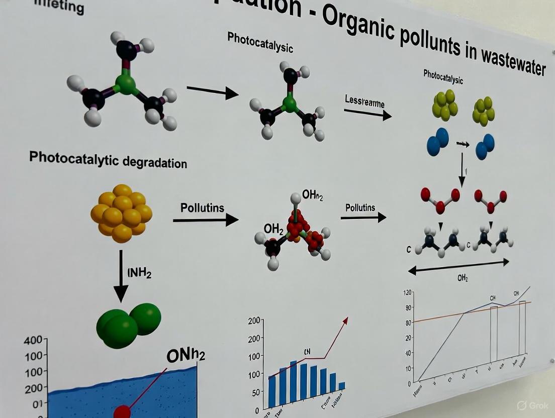
The photocatalytic degradation of organic pollutants is a complex process initiated by light absorption and culminating in the mineralization of contaminants. The mechanism can be broken down into five sequential stages, as illustrated in the following diagram and elaborated in the subsequent analysis.
Diagram 1: The sequential mechanism of photocatalysis, from light absorption to pollutant degradation.
The process begins when a semiconductor photocatalyst, such as TiOâ‚‚ or ZnO, absorbs photons with energy equal to or greater than its band gap energy (E_g). This absorption promotes an electron (eâ») from the valence band (VB) to the conduction band (CB), creating a positively charged hole (hâº) in the valence band. This results in the formation of an electron-hole (eâ»/hâº) pair [1]. A narrower band gap facilitates this electron-hole pair generation by enabling the use of lower-energy photons, such as those in the visible light spectrum [1]. For instance, while pure TiOâ‚‚ is primarily UV-active, green-synthesized ZnO nanoparticles have been reported with a band gap of 2.92 eV, allowing for more efficient utilization of visible light [2].
Charge Carrier Separation and Migration
The photogenerated electrons and holes must then separate and migrate to the surface of the photocatalyst without recombining. Recombination is a competitive process that reduces photocatalytic efficiency. Strategies to minimize recombination include doping with foreign elements, creating surface defects, forming heterojunctions with other semiconductors, and using co-catalysts [1]. For example, coupling TiOâ‚‚ with clay to form a nanocomposite not only prevents TiOâ‚‚ aggregation but also enhances charge separation, thereby improving the overall photocatalytic activity [3]. The successful migration of these charge carriers to the catalyst surface is critical for the subsequent redox reactions.
Reactive Oxygen Species (ROS) Generation
Once on the surface, the electrons and holes drive a series of redox reactions with surrounding molecules:
- Hole Reactions: The photogenerated holes (hâº) can oxidize water molecules (Hâ‚‚O) or hydroxide ions (OHâ») to produce powerful hydroxyl radicals (•OH) [3] [1].
- Electron Reactions: The excited electrons (eâ») in the conduction band can reduce molecular oxygen (Oâ‚‚) adsorbed on the catalyst surface to generate superoxide anion radicals (O₂•â»), which can further protonate to form hydroperoxyl radicals (•OOH) and eventually hydrogen peroxide (Hâ‚‚Oâ‚‚), leading to more •OH radicals [1].
Among these, hydroxyl radicals (•OH) are often the primary oxidative species due to their high reactivity, as confirmed by both radical scavenger experiments and Density Functional Theory (DFT) predictions [3].
Pollutant Adsorption and Oxidative Degradation
The final stage involves the adsorption of organic pollutant molecules onto the photocatalyst surface and their subsequent oxidation. The adsorption is influenced by the surface charge of the catalyst, which is determined by the solution pH relative to the catalyst's point of zero charge (PZC) [3] [1]. Once adsorbed, the generated ROS (e.g., •OH and O₂•â») attack the pollutant molecules, breaking them down into smaller, less harmful intermediates and ultimately mineralizing them into COâ‚‚, Hâ‚‚O, and inorganic ions [3] [1]. The degradation pathway can be complex; for the dye BR46, GC-MS analysis verified its breakdown into non-toxic intermediates [3].
Quantitative Performance Data
The efficiency of photocatalysis is quantified using several key metrics. The tables below summarize performance data from recent studies and the figures of merit used for evaluation.
Table 1: Photocatalytic Performance in Pollutant Degradation
| Photocatalyst | Target Pollutant(s) | Light Source | Optimal Catalyst Loading | Degradation Efficiency | Key Performance Metrics |
|---|---|---|---|---|---|
| TiO₂–clay nanocomposite [3] | Basic Red 46 (BR46) dye | UV-C lamp (8 W) | Immobilized bed | 98% dye removal, 92% TOC reduction | Pseudo-first-order rate constant: 0.0158 minâ»Â¹ |
| ZnO (N-gZnOw) [2] | Clomazone, Tembotrione, Ciprofloxacin, Zearalenone | Sunlight | 0.5 mg/cm³ | 98.2%, 95.8%, 96.2%, 96.6% removal, respectively | Band gap: 2.92 eV; Particle size: 14.9 nm |
Table 2: Figures of Merit (FOM) in Photocatalysis [4]
| Figure of Merit | Description | Utility & Challenges |
|---|---|---|
| Reaction Rate (r) | The speed of the photocatalytic reaction (e.g., pollutant degradation or Hâ‚‚ production). | A fundamental metric, but depends on experimental conditions like catalyst mass and light intensity. |
| Apparent Quantum Yield (AQY) | The ratio of the reaction rate to the incident photon flux. | Accounts for light absorption but does not fully consider scattering and reflection. |
| Photocatalytic Space Time Yield (PSTY) | The reaction rate per unit volume of the reactor. | Useful for evaluating reactor efficiency and scalability. |
| Turnover Number (TON) & Turnover Frequency (TOF) | TON: moles of product per mole of catalytic sites. TOF: TON per unit time. | Requires knowledge of active sites, which can be obscured in semiconductors. |
Experimental Protocols
Protocol: Photocatalytic Degradation of a Model Dye Using an Immobilized Nanocomposite
This protocol outlines the procedure for degrading organic dyes using a TiOâ‚‚-clay nanocomposite in a rotary photoreactor, based on a study that achieved 98% removal of Basic Red 46 [3].
I. Materials and Reagents
- Photocatalyst: TiOâ‚‚-P25 (Degussa).
- Support Material: Industrial clay powder.
- Immobilization Agent: Silicone adhesive.
- Model Pollutant: Basic Red 46 (BR46, Câ‚₈Hâ‚‚â‚BrN₆) or similar.
- Solvent: Distilled water.
- Substrate: Flexible plastic (talc) sheets (17 cm x 35 cm).
II. Nanocomposite Synthesis and Immobilization
- Weighing: Precisely combine 0.7 g of TiOâ‚‚ and 0.3 g of clay powder in a beaker (70:30 ratio).
- Mixing: Add 5-10 mL of distilled water and stir continuously with a magnetic stirrer for 4 hours at room temperature.
- Drying: Transfer the mixture to an oven and dry at 60°C for 6 hours.
- Grinding: Grind the dried product into a fine powder using a mortar and pestle.
- Immobilization:
- Apply a thin, uniform layer of silicone adhesive to the plastic substrate.
- Uniformly sieve the TiOâ‚‚-clay powder onto the adhesive-coated substrate.
- Allow the coated substrate to dry at ambient temperature for 24 hours.
III. Photocatalytic Reactor Setup and Operation
- Reactor Assembly: The rotary photoreactor consists of:
- A water tank (500 mL capacity).
- An electric motor to drive a PVC cylinder (17 cm length, 11 cm diameter).
- A quartz cylindrical tube placed inside the rotating cylinder to house an 8-watt UV-C lamp.
- Experimental Run:
- Prepare an aqueous solution of BR46 dye at the desired initial concentration (e.g., 20 mg/L).
- Pour the solution into the reactor tank.
- Set the rotational speed of the cylinder (e.g., 5.5 rpm) and turn on the UV lamp.
- Conduct experiments for a specified duration (e.g., 90 min), taking samples at regular intervals.
IV. Analysis and Quantification
- Dye Concentration: Monitor the degradation by measuring the absorbance of the dye solution using UV-Vis spectrophotometry.
- Mineralization Efficiency: Use a Total Organic Carbon (TOC) analyzer to quantify the reduction in organic carbon content.
- Degradation By-products: Identify intermediates using Gas Chromatography-Mass Spectrometry (GC-MS).
- Reactive Species Identification: Perform radical scavenger tests to identify the primary ROS involved (e.g., using isopropanol for •OH quenching).
Protocol: Synthesis of Green ZnO Nanoparticles for Solar-Driven Degradation
This protocol describes an eco-friendly synthesis of ZnO nanoparticles for the degradation of various organic pollutants under sunlight [2].
I. Green Synthesis of ZnO Nanoparticles
- Extract Preparation: Prepare an extract using green tea leaves and water.
- Precursor Solution: Use a zinc nitrate solution as a precursor.
- Synthesis: Mix the precursor with the plant extract in an aqueous medium to form the nanoparticles.
- Characterization: Characterize the synthesized nanoparticles (N-gZnOw) using:
- X-Ray Diffraction (XRD): For crystallinity and phase structure.
- SEM: For morphological analysis.
- UV-Vis Spectroscopy: To determine the band gap energy.
- Zeta Potential Measurements: To find the isoelectric point.
II. Photocatalytic Testing
- Pollutant Selection: Use pollutants like clomazone (herbicide), ciprofloxacin (antibiotic), or zearalenone (mycotoxin).
- Reaction Conditions:
- Use an optimal catalyst loading of 0.5 mg/cm³.
- Perform reactions under natural sunlight or a solar simulator.
- Efficiency Analysis: Use Liquid Chromatography-Electrospray Ionization-Tandem Mass Spectrometry (LC-ESI-MS/MS) to confirm pollutant degradation and identify intermediates.
The workflow for these experimental processes is visualized below.
Diagram 2: A generalized workflow for photocatalytic degradation experiments.
The Scientist's Toolkit: Key Research Reagents and Materials
Table 3: Essential Materials for Photocatalysis Research
| Material/Reagent | Function & Role in Photocatalysis | Specific Examples |
|---|---|---|
| Semiconductor Photocatalysts | The primary light-absorbing material that generates electron-hole pairs. | TiO₂-P25 [3], ZnO nanoparticles [2], NaTaO₃ [4]. |
| Support Materials & Composites | Enhance surface area, prevent aggregation, and adsorb pollutants for closer contact with ROS. | Clay in TiOâ‚‚-clay composites [3], Graphene derivatives (rGO) [3]. |
| Model Organic Pollutants | Representative compounds used to benchmark and study photocatalytic efficiency. | Basic Red 46 (azo dye) [3], Clomazone (herbicide) [2], Ciprofloxacin (antibiotic) [2]. |
| Radical Scavengers | Chemical compounds used to quench specific ROS and elucidate the degradation mechanism. | Isopropanol (for •OH), p-benzoquinone (for O₂•â»), EDTA (for hâº) [3]. |
| Immobilization Agents | Used to fix catalyst particles onto solid supports for easy separation and reusability. | Silicone adhesive [3]. |
| Characterization Tools | Instruments for analyzing the physical, chemical, and optical properties of photocatalysts. | XRD, FE-SEM, UV-Vis DRS, BET surface area analyzer [3]. |
| Benzo(b)triphenylen-10-ol | Benzo(b)triphenylen-10-ol|High Purity|C22H14O | Buy Benzo(b)triphenylen-10-ol , a research-grade PAH for biochemical studies. For Research Use Only. Not for human or veterinary use. |
| 5-Hydroxyheptan-2-one | 5-Hydroxyheptan-2-one|C7H14O2 | 5-Hydroxyheptan-2-one (C7H14O2) is a hydroxy ketone for research. This product is for laboratory research use only and is not intended for personal use. |
The photocatalytic degradation of organic pollutants presents a promising advanced oxidation process (AOP) for addressing the persistent challenge of wastewater treatment. Semiconductor-based photocatalysis utilizes light energy to generate electron-hole pairs that form highly reactive species capable of mineralizing complex organic contaminants into harmless compounds like CO₂ and H₂O [5]. This technology has gained significant research attention due to its potential to utilize solar energy, operate at ambient temperatures, and achieve complete mineralization without generating secondary pollution [6] [7]. Among various semiconductors, titanium dioxide (TiO₂), zinc oxide (ZnO), and graphitic carbon nitride (g-C₃N₄) have emerged as particularly promising materials due to their unique properties, though each presents distinct challenges that band gap engineering strategies seek to overcome.
Fundamental Properties and Band Gap Characteristics
The photocatalytic activity of semiconductors originates from their electronic structure, particularly the energy separation between the valence band (VB) and conduction band (CB) known as the band gap. When photons with energy equal to or greater than this band gap strike the semiconductor, electrons are excited from the VB to the CB, leaving holes in the VB and creating electron-hole pairs that drive redox reactions [5]. The efficiency of this process depends critically on the band gap energy, which determines the range of utilizable light, and the positions of the VB and CB, which govern the redox potential of the generated charge carriers.
Table 1: Fundamental Properties of Key Semiconductor Photocatalysts
| Semiconductor | Band Gap (eV) | Light Absorption Range | Advantages | Limitations |
|---|---|---|---|---|
| TiO₂ | 3.0–3.2 [6] | UV | Excellent photostability, non-toxicity, low cost [8] | Limited visible light utilization [6] |
| ZnO | ~3.37 [8] | UV | High photocatalytic efficiency, inexpensive [8] | Photo-corrosion, rapid charge recombination |
| g-C₃N₄ | 2.7–2.8 [6] | Visible | Visible light activity, thermal/chemical stability, metal-free [8] [9] | Fast electron-hole recombination, low surface area [9] [6] |
Band Gap Engineering Strategies
Heterojunction Construction
Creating interfaces between different semiconductors represents one of the most effective approaches to enhance charge separation and extend light absorption. The TiO₂-ZnO/g-C₃N4 nanocomposite demonstrates this principle, where the combined system shows enhanced performance for dye removal and hydrogen generation compared to individual components [8]. Similarly, TiO₂/g-C₃N₄ heterojunctions have proven highly effective for degrading persistent pollutants like monochlorophenols (MCPs), achieving removal efficiencies of 87% for 2-chlorophenol compared to less than 50% with either component alone [9]. The heterojunction between TiO₂ and g-C₃N₄ improves charge separation through transfer of photogenerated electrons from g-C₃N₄ to TiO₂, reducing recombination rates and enhancing photocatalytic activity [6].
Metal and Non-Metal Doping
Incorporating metal elements into semiconductor structures can modify their electronic properties and enhance visible light absorption. Noble metals like gold (Au) act as electron mediators when deposited on semiconductor heterostructures, promoting charge carrier transfer due to localized surface plasmon resonance (LSPR) effects [10]. In the TiO₂@Au/g-C₃N₄ system, Au nanoparticles accumulate and transfer photo-stimulated electrons from TiO₂ to g-C₃N₄, creating an efficient Z-scheme photocatalytic mechanism that preserves strong redox capabilities [10].
Nanocomposite Formation with Supporting Materials
Combining semiconductors with supporting materials like clay can enhance surface area and stability. The TiO₂-clay nanocomposite demonstrates this advantage, exhibiting an increased BET surface area of 65.35 m²/g compared to 52.12 m²/g for pure TiO₂ [3]. The clay acts as a supportive matrix that prevents TiO₂ aggregation while providing additional adsorption sites for pollutants, creating a synergistic system that achieves 98% dye removal and 92% total organic carbon reduction under optimal conditions [3].
Application Notes: Experimental Protocols
Protocol 1: Synthesis of TiO₂-ZnO/g-C₃N₄ Nanocomposite
Purpose: To create an efficient heterostructured photocatalyst for dye degradation and hydrogen generation [8].
Materials: Titanium dioxide (TiOâ‚‚), zinc oxide (ZnO), urea, methylene orange (MO), rhodamine B (RhB), methanol, ascorbic acid, citric acid, hydrochloric acid, sodium hydroxide.
Procedure:
- g-C₃N₄ Preparation: Heat urea powder in a muffle furnace at a heating rate of 10°C/min to 600°C and maintain at this temperature for 4 hours. After cooling to room temperature, collect the resulting bulk g-C₃N₄ [9].
- Nanocomposite Formation: Combine TiO₂ and ZnO precursors with the synthesized g-C₃N₄ using chemical reduction method.
- Characterization: Analyze the crystallinity and phase structure by X-ray diffraction (XRD). Examine surface morphology and elemental composition using scanning electron microscopy (SEM) and transmission electron microscopy (TEM). Determine optical properties through UV-Vis spectroscopy and photoluminescence (PL) spectroscopy [8].
Protocol 2: Microwave-Assisted Synthesis of g-C₃N₄/TiO₂ Heterostructures
Purpose: To rapidly produce visible-light-activated photocatalysts for degradation of recalcitrant azo dyes [6].
Materials: Pre-synthesized g-C₃N₄ nanosheets, titanium precursor, methyl orange (MO), urea.
Procedure:
- g-C₃N₄ Preparation: Thermal polycondensation of urea at 500-600°C for several hours [6].
- Heterostructure Formation: Mix different weight ratios of g-C₃N₄ (15, 30, and 45 wt.%) with TiO₂ precursor. Subject the mixture to microwave irradiation for 1 hour.
- Characterization: Analyze phase structure by XRD, examine morphology through SEM and STEM, determine chemical states via XPS, and evaluate optical properties using UV-Vis absorption spectroscopy [6].
Table 2: Performance Comparison of Engineered Photocatalysts
| Photocatalyst | Target Pollutant | Experimental Conditions | Removal Efficiency | Key Findings |
|---|---|---|---|---|
| TiO₂-ZnO/g-C₃N₄ [8] | Methylene Orange, Rhodamine B | Sunlight irradiation | High degradation rate | Bimetallic structure with g-C₃N₄ enhanced charge separation |
| 40TiO₂/g-C₃N₄ [9] | Monochlorophenols (2-CP, 3-CP, 4-CP) | UV-Vis light, 25 ppm, 1 g/L catalyst | 87% (2-CP), 64% (4-CP) | Superior to individual components (<50%) |
| 30% g-C₃N₄/TiO₂ [6] | Methyl Orange (MO) | Solar simulator, 4 hours | 85% | 2× and 10× more efficient than pure TiO₂ and g-C₃N₄ |
| TiO₂-clay [3] | Basic Red 46 (BR46) | UV light, 20 mg/L, 90 min | 98% | Enhanced surface area (65.35 m²/g) and stability |
Protocol 3: Evaluation of Photocatalytic Performance
Purpose: To assess the degradation efficiency of synthesized photocatalysts for organic pollutants [9] [3].
Materials: Photocatalyst, target pollutant (dye or phenolic compound), radical scavengers (e.g., ammonium oxalate, benzoquinone, isopropyl alcohol), UV-Vis spectrophotometer, TOC analyzer.
Procedure:
- Photocatalytic Testing: Prepare pollutant solution at specific concentration (e.g., 10-25 mg/L). Add photocatalyst (0.2-1 g/L) and maintain suspension under continuous stirring. Irradiate with appropriate light source (UV, visible, or solar simulator). Withdraw samples at regular intervals.
- Analysis: Measure pollutant concentration by UV-Vis spectroscopy. Evaluate mineralization efficiency through total organic carbon (TOC) analysis.
- Reactive Species Identification: Conduct radical scavenging experiments using specific quenchers—ammonium oxalate for holes, benzoquinone for superoxide radicals, and isopropyl alcohol for hydroxyl radicals [9] [3].
- Byproduct Identification: Analyze degradation intermediates using GC-MS with HP-5MS UI column (30 m × 0.25 mm × 0.25 µm) [9].
The Scientist's Toolkit: Essential Research Reagents
Table 3: Key Research Reagents for Photocatalyst Development and Evaluation
| Reagent/Chemical | Function/Application | Key Characteristics |
|---|---|---|
| Titanium Dioxide (TiOâ‚‚-P25) [3] | Benchmark photocatalyst | Mixed-phase (anatase/rutile), band gap ~3.2 eV |
| Urea [9] [6] | g-C₃N₄ precursor | Nitrogen-rich, thermal polycondensation at 500-600°C |
| Hydrofluoric Acid (HF) [10] | Morphology control for TiOâ‚‚ | Exposes high-energy (001) facets |
| Tetrabutyl Titinate [10] | TiOâ‚‚ precursor | Hydrolyzes to form anatase phase |
| Methylene Orange (MO) [8] [6] | Model azo dye pollutant | Recalcitrant, visible chromophore for degradation monitoring |
| Rhodamine B (RhB) [8] [10] | Model cationic dye | Fluorescent, used for photocatalytic activity assessment |
| Monochlorophenols (MCPs) [9] | Model persistent organic pollutants | EPA-priority contaminants, low biodegradability |
| Ammonium Oxalate [9] | Hole (hâº) scavenger | Identifies role of holes in degradation mechanism |
| Benzoquinone [9] | Superoxide (Oâ‚‚â») scavenger | Determines contribution of superoxide radicals |
| Isopropyl Alcohol [9] | Hydroxyl radical (•OH) scavenger | Evaluates role of hydroxyl radicals in degradation |
| 2-Methyl-1-phenylguanidine | 2-Methyl-1-phenylguanidine|Research Chemical | 2-Methyl-1-phenylguanidine for research. Investigating its potential as a 5-HT3 receptor ligand. This product is for Research Use Only (RUO). Not for human or veterinary use. |
| 4,4'-Dichlormethyl-bibenzyl | 4,4'-Dichlormethyl-bibenzyl|High-Purity|RUO |
Reaction Mechanisms and Pathways
The enhanced photocatalytic activity of engineered semiconductors stems from improved charge separation and tailored reaction pathways. In Z-scheme systems like TiOâ‚‚@Au/g-C₃Nâ‚„, Au nanoparticles serve as electron mediators that transfer photoexcited electrons from the TiOâ‚‚ conduction band to the g-C₃Nâ‚„ valence band, effectively separating the reduction and oxidation reactions while preserving strong redox potentials [10]. This mechanism enhances the formation of reactive oxygen species (ROS), particularly superoxide radicals (Oâ‚‚â») and hydroxyl radicals (•OH), which play pivotal roles in degrading organic pollutants.
Radical trapping experiments have demonstrated that different reactive species dominate in various systems. For g-C₃N₄/TiO₂ heterostructures, superoxide radicals were identified as the primary species responsible for methyl orange degradation, with minimal involvement of hydroxyl radicals, suggesting a type-II heterojunction mechanism [6]. In contrast, hydroxyl radicals were determined to be the main oxidative species in TiO₂-clay systems for BR46 dye degradation [3]. Density Functional Theory (DFT) calculations support these experimental findings, predicting favorable attack sites on dye molecules and confirming the degradation pathways [3].
Band gap engineering through heterojunction construction, elemental doping, and nanocomposite formation has significantly advanced the photocatalytic performance of TiO₂, ZnO, and g-C₃N₄ for wastewater treatment. The strategic combination of these materials creates synergistic systems that enhance light absorption, improve charge separation, and provide abundant active sites for pollutant degradation. The experimental protocols and application notes presented herein provide researchers with practical methodologies for developing and evaluating advanced photocatalytic systems. Future research directions should focus on targeting emerging contaminants with environmental persistence, such as perfluorinated compounds and pharmaceuticals, while improving catalyst efficiency in real water matrices with multicomponent interfering ions [5]. The development of conducting polymer-based nanocomposites also represents a promising but underexplored area for future photocatalyst design [7].
Application Notes
The photocatalytic degradation of organic pollutants in wastewater represents a promising advanced oxidation process for addressing water contamination challenges. Recent research has focused on developing next-generation photocatalytic materials that enhance visible light absorption, improve charge separation efficiency, and increase stability for practical applications. Three primary material classes have emerged as particularly effective: Z-scheme heterojunctions, doped semiconductors, and carbon-based composites.
Z-Scheme Heterojunction Photocatalysts
Z-scheme heterojunctions represent a significant advancement over traditional heterojunction systems by mimicking natural photosynthesis and enabling more efficient charge separation while maintaining strong redox potential [11]. These systems typically combine two semiconductor materials with staggered band structures, connected through an electron mediator or direct interface, to create a vectorial charge transfer pathway.
Recent studies have demonstrated the exceptional performance of 2D/2D Z-scheme WO₃/g-C₃N₄ heterojunctions for environmental applications [12]. These materials exhibit considerable photocatalytic performance for degrading various organic pollutants without requiring cocatalysts. The 40%WO₃/g-C₃N₄ composite showed exceptional degradation efficiency for multiple organic dyes, achieving nearly complete degradation of rhodamine B (RhB) within 20 minutes under visible light irradiation [12]. The same system also demonstrated applications in nitrogen fixation, simultaneously achieving photocatalytic nitrogen reduction reaction (NRR) and nitrogen oxidation reaction (NOR) to produce NH₄⺠and NO₃⻠using air as a nitrogen source [12].
Another study on g-C₃Nâ‚„/WO₃ Z-scheme heterojunctions reported a 97.9% degradation efficiency for RhB within 15 minutes and 93.3% for tetracycline hydrochloride (TC-HCl) within 180 minutes under visible light [13]. The system maintained 97.8% efficiency after four cycles, demonstrating excellent stability and reusability. Radical trapping experiments confirmed that holes (hâº) and superoxide radicals (·Oâ‚‚â») served as the primary reactive species in the degradation mechanism [13].
Table 1: Performance Metrics of Representative Z-Scheme Heterojunction Photocatalysts
| Photocatalyst System | Target Pollutant | Degradation Efficiency | Time Required | Light Source | Stability Performance |
|---|---|---|---|---|---|
| 40%WO₃/g-C₃N₄ [12] | Rhodamine B (RhB) | ~100% | 20 min | Visible light | High recyclability |
| g-C₃N₄/WO₃ [13] | RhB | 97.9% | 15 min | Visible light | 97.8% after 4 cycles |
| g-C₃N₄/WO₃ [13] | TC-HCl | 93.3% | 180 min | Visible light | Maintained high efficiency |
| 40%WO₃/g-C₃N₄ [12] | Tetracycline-HCl | Significant degradation | Not specified | Visible light | Good photocatalytic stability |
Doped Semiconductor Photocatalysts
Doping represents a strategic approach to enhance the performance of semiconductor photocatalysts by modifying their electronic properties, reducing band gaps, and minimizing electron-hole recombination [14]. Metal oxide semiconductors including TiO₂, ZnO, CeO₂, WO₃, and ZrO₂ have been extensively explored as photocatalysts due to their high stability across wide pH ranges, low toxicity, and strong oxidizing capabilities [14]. However, these materials face limitations including wide band gaps (primarily activating under UV light) and rapid recombination of photogenerated electron-hole pairs [14] [15].
Doping with rare-earth elements has proven particularly effective due to their ability to interact quickly with functional groups through 4f empty orbitals [14]. For WO₃, which has a narrower band gap (2.7-2.8 eV) than TiO₂, doping further enhances visible light absorption and charge separation efficiency [15]. Similarly, graphitic carbon nitride (g-C₃N₄), with a bandgap of approximately 2.7 eV and absorption edge at 450-470 nm, benefits from doping to reduce its inherent limitations of serious photo-induced electron-hole recombination and narrow visible light absorption range [13] [15].
Table 2: Comparison of Key Semiconductor Photocatalysts for Wastewater Treatment
| Semiconductor | Band Gap (eV) | Primary Activation Range | Advantages | Limitations |
|---|---|---|---|---|
| TiOâ‚‚ [15] | 3.0-3.2 | Ultraviolet (λ ≤ 390 nm) | High activity, well-studied, stable | Limited visible light absorption, rapid eâ»/h⺠recombination |
| WO₃ [15] | 2.7-2.8 | Visible light | Good visible light absorption, stable, non-toxic | Low conduction band position, recombination issues |
| g-C₃Nâ‚„ [15] | ~2.7 | Visible light (up to 470 nm) | Metal-free, easy synthesis, tunable | High eâ»/h⺠recombination, limited oxidation potential |
| Doped Variants [14] | Reduced | Extended visible light | Enhanced visible light response, reduced recombination | Synthesis complexity, potential cost increase |
Carbon-Based Composite Photocatalysts
Carbon-based materials have emerged as promising components in photocatalytic composites due to their exceptional electrical conductivity, tunable surface properties, structural diversity, and environmental compatibility [16]. Carbon nanostructures including graphene, carbon nanotubes, carbon dots, graphitic carbon nitride, and fullerenes offer unique advantages for enhancing photocatalytic performance through improved charge separation and transport [16].
The integration of carbon materials with metal nanoparticles creates synergistic effects that significantly boost photocatalytic activity. Gold (Au) and Silver (Ag) nanoparticles on graphene sheets have demonstrated enhanced photocatalytic performance, serving dual functions as both light absorbers and catalytic centers [16]. These carbon-metal nanocomposites facilitate rapid electron transfer during redox reactions while minimizing charge recombination, addressing critical limitations of standalone semiconductor photocatalysts [16].
Carbon-based composites have shown particular effectiveness for diverse environmental applications including degradation of organic dyes, hydrogen production through water splitting, and carbon dioxide reduction [16]. Their hydrophobic nature, chemical stability across acidic and basic conditions, thermal stability, and potential for low-cost production make them attractive for scalable wastewater treatment applications [16].
Experimental Protocols
Protocol 1: Synthesis of 2D/2D Z-Scheme WO₃/g-C₃N₄ Heterojunctions
Principle: This protocol describes the preparation of 2D/2D Z-scheme WO₃/g-C₃N₄ heterojunctions through a facile rapid calcination method, creating an efficient photocatalytic system for organic pollutant degradation and nitrogen fixation [12].
Materials:
- Sodium tungstate dihydrate (Na₂WO₄·2H₂O)
- Graphitic carbon nitride (g-C₃N₄)
- Deionized water
- Hydrochloric acid (HCl)
Procedure:
- Begin with pre-synthesized g-C₃N₄ powder, which can be prepared through thermal polymerization of nitrogen-rich precursors such as melamine at 550°C for 4 hours [13].
- Prepare a homogeneous dispersion by adding 4g of g-C₃N₄ to 25mL deionized water with ultrasonic treatment for 1 hour [13].
- Add 0.584g NaCl and 1.65g Na₂WO₄·2H₂O to the dispersion under constant stirring.
- Gradually introduce 5mL hydrochloric acid to the solution while continuing stirring for 3 hours until a yellow precipitate forms.
- Transfer the suspension to a Teflon-lined autoclave and maintain at 160°C for 12 hours.
- After cooling to room temperature, filter the product and rinse thoroughly with deionized water to remove impurities.
- Dry the obtained sample in an oven at 80°C for 8 hours.
- Finally, calcine the powder at 450°C for 3 hours with a heating rate of 5°C/min to obtain the crystalline WO₃/g-C₃N₄ composite.
- For optimization, prepare composites with different WO₃ loading percentages (e.g., 20%, 40%, 60%) by varying the precursor ratios [12].
Characterization:
- Analyze crystal structure using X-ray diffraction (XRD)
- Examine morphology and interface structure with transmission electron microscopy (TEM)
- Determine chemical states and composition via X-ray photoelectron spectroscopy (XPS)
- Assess optical properties with UV-Vis diffuse reflectance spectroscopy (DRS)
Protocol 2: Photocatalytic Degradation Assessment of Organic Pollutants
Principle: This protocol evaluates the photocatalytic performance of synthesized materials for degrading organic pollutants under visible light irradiation, quantifying degradation efficiency and identifying reactive species [13].
Materials:
- Target pollutants (RhB, TC-HCl, MB, MO, etc.)
- Photocatalyst powder
- Scavengers: Ammonium oxalate (AO, h⺠scavenger), Benzoquinone (BQ, ·O₂⻠scavenger), Isopropyl alcohol (IPA, ·OH scavenger)
- 300W Xe lamp with 400nm filter
- UV-Vis spectrophotometer
Procedure:
- Prepare pollutant solutions at specific concentrations (e.g., 20mg/L RhB, 30mg/L TC-HCl) using deionized water.
- In a standard reaction setup, disperse 0.1g photocatalyst in 100mL pollutant solution.
- Sonicate the suspension for 5 minutes to ensure uniform dispersion.
- Pre-adsorption phase: Stir the suspension magnetically in darkness for 30 minutes to establish adsorption-desorption equilibrium.
- Initiate irradiation using a 300W Xe lamp equipped with a 400nm filter to provide visible light.
- At predetermined time intervals (e.g., every 5 minutes for RhB, longer intervals for TC-HCl), withdraw 4mL aliquots.
- Immediately filter the aliquots to remove catalyst particles.
- Analyze the filtrate using UV-Vis spectrophotometry to determine residual pollutant concentration.
- Calculate degradation efficiency using the formula: Efficiency (%) = [(C₀ - Cₜ)/C₀] × 100, where C₀ is initial concentration and Cₜ is concentration at time t.
Reactive Species Identification:
- For radical trapping experiments, introduce specific scavengers before irradiation:
- 1mM ammonium oxalate (AO) for holes (hâº)
- 2mM benzoquinone (BQ) for superoxide radicals (·Oâ‚‚â»)
- 10mM isopropyl alcohol (IPA) for hydroxyl radicals (·OH)
- Compare degradation efficiency with and without scavengers to identify primary reactive species.
- For enhanced verification, perform electron spin resonance (ESR) spectroscopy with DMPO as spin trapping agent to directly detect ·OH and ·O₂⻠radicals.
Protocol 3: Synthesis of Carbon-Based Composite Photocatalysts
Principle: This protocol outlines the preparation of carbon nanostructure-metal nanocomposites for enhanced photocatalytic activity, leveraging the synergistic effects between carbon materials and metal nanoparticles [16].
Materials:
- Carbon support material (graphene oxide, carbon nanotubes, etc.)
- Metal precursors (HAuCl₄, AgNO₃, etc.)
- Reducing agents (sodium borohydride, hydrazine hydrate, etc.)
- Solvents (deionized water, ethanol)
Procedure:
- Prepare a homogeneous dispersion of carbon support material in appropriate solvent using prolonged sonication (1-2 hours).
- Add metal precursor solution to the carbon dispersion under vigorous stirring.
- Adjust pH as needed to optimize metal ion adsorption onto carbon surface.
- Slowly add reducing agent to convert metal ions to nanoparticles anchored on carbon support.
- Continue stirring for 2-4 hours to ensure complete reduction and uniform distribution.
- Recover the composite material through centrifugation or filtration.
- Wash thoroughly with deionized water and ethanol to remove unreacted precursors.
- Dry under vacuum at 60°C for 12 hours.
- For thermal treatments, calcine under inert atmosphere at optimized temperatures.
Characterization:
- Analyze metal nanoparticle size and distribution using TEM
- Determine chemical states and interfacial interactions through XPS
- Assess crystallinity with XRD
- Evaluate specific surface area and porosity through BET measurements
- Confirm optical properties via UV-Vis DRS
The Scientist's Toolkit: Essential Research Reagents and Materials
Table 3: Key Research Reagents for Photocatalyst Development and Testing
| Reagent/Material | Function/Application | Key Characteristics | Representative Examples |
|---|---|---|---|
| Tungsten Trioxide (WO₃) [15] | Visible-light-driven photocatalyst | Bandgap ~2.7-2.8 eV, good stability, non-toxic | WO₃ nanoparticles, WO₃ nanorods |
| Graphitic Carbon Nitride (g-C₃N₄) [15] | Metal-free semiconductor photocatalyst | Bandgap ~2.7 eV, visible light response, easily synthesized | Bulk g-C₃N₄, exfoliated g-C₃N₄ nanosheets |
| Sodium Tungstate Dihydrate [13] | WO₃ precursor in synthesis | Provides tungsten source, water-soluble | Na₂WO₄·2H₂O for hydrothermal synthesis |
| Melamine [13] | g-C₃N₄ precursor | Nitrogen-rich organic compound, thermal polymerization | C₃H₆N₆ for g-C₃N₄ synthesis |
| Ammonium Oxalate (AO) [13] | Hole (hâº) scavenger in mechanistic studies | Selective trapping of photogenerated holes | 1mM solution for radical identification |
| Benzoquinone (BQ) [13] | Superoxide radical (·Oâ‚‚â») scavenger | Selective trapping of superoxide radicals | 2mM solution for radical identification |
| Isopropyl Alcohol (IPA) [13] | Hydroxyl radical (·OH) scavenger | Selective trapping of hydroxyl radicals | 10mM solution for radical identification |
| DMPO [13] | Spin trap for ESR spectroscopy | Forms stable adducts with radicals for detection | 5,5-Dimethyl-1-pyrroline N-oxide |
| Carbon Nanostructures [16] | Catalyst supports and composites | High conductivity, tunable surface properties | Graphene, carbon nanotubes, carbon dots |
| 2,6-Dibenzylcyclohexanone | cis-2,6-Dibenzylcyclohexanone | cis-2,6-Dibenzylcyclohexanone is a synthetic intermediate used in medicinal chemistry research. This product is for research use only and not for human consumption. | Bench Chemicals |
| 4-Hydroxydecan-2-one | 4-Hydroxydecan-2-one | 4-Hydroxydecan-2-one is a ketone reagent for organic synthesis and pharmaceutical research. For Research Use Only. Not for human or veterinary use. | Bench Chemicals |
The pervasive discharge of pharmaceutical pollutants and industrial dyes into water systems presents a critical environmental challenge worldwide. These contaminants, originating from medical use, industrial effluents, and improper disposal, demonstrate persistence, toxicity, and resistance to conventional treatment methods, leading to bioaccumulation and potential health risks including antibiotic resistance and ecosystem disruption [17] [18]. The textile industry alone contributes approximately 20% of global water pollution, releasing complex organic dyes with over 1,900 chemicals involved in production [17]. Similarly, active pharmaceutical ingredients (APIs) increasingly detected in aquatic environments pose significant concerns due to their biological activity at low concentrations and inadequate removal by conventional wastewater treatment plants [19].
Within this context, photocatalytic degradation has emerged as a promising advanced oxidation process (AOP) that utilizes semiconductor materials to generate reactive oxygen species (ROS) under light irradiation, effectively breaking down persistent organic pollutants into harmless end products like carbon dioxide and water [17] [18]. This application note explores recent advances in photocatalytic technologies for degrading pharmaceutical contaminants and organic dyes, providing structured experimental protocols, performance comparisons, and mechanistic insights to support researchers and scientists in developing effective water remediation strategies.
Current Research and Performance Data
Recent investigations have focused on developing innovative photocatalysts with enhanced efficiency under solar or visible light irradiation. The following tables summarize key performance data from recent studies for quantitative comparison of various photocatalytic systems.
Table 1: Performance of Photocatalysts in Degrading Organic Dyes
| Photocatalyst | Target Pollutant | Experimental Conditions | Degradation Efficiency | Time Required | Reference |
|---|---|---|---|---|---|
| CoS nanoparticles | Methylene Blue (MB) | 0.33 g/L catalyst, 20 ppm MB, visible light | 97.7% | 90 min | [20] |
| CoS nanoparticles | Methyl Red (MR) | 0.33 g/L catalyst, 20 ppm MR, visible light | 75.3% | 90 min | [20] |
| Cu(I) CP1 (Iodide) | Methylene Blue (MB) | Minimal catalyst, Hâ‚‚Oâ‚‚, sunlight | 96% | 15 min | [21] |
| TiOâ‚‚-clay nanocomposite | Basic Red 46 (BR46) | 20 mg/L dye, 5.5 rpm rotation, UV light | 98% | 90 min | [3] |
| Bismuth titanate (Biâ‚„Ti₃Oâ‚â‚‚) | Organic Dyes | Visible LED irradiation | ~98% | 60 min | [22] |
Table 2: Performance of Photocatalysts in Degrading Pharmaceutical Compounds
| Photocatalyst | Target Pollutant | Experimental Conditions | Degradation Efficiency | Time Required | Reference |
|---|---|---|---|---|---|
| Co-doped ZnFeâ‚‚Oâ‚„ (ZC20) | Acetaminophen | 0.1 g/L catalyst, 30 mg/L drug, 100W LED | 85% | 180 min | [23] |
| Pure ZnFeâ‚‚Oâ‚„ (ZC0) | Acetaminophen | 0.1 g/L catalyst, 30 mg/L drug, 100W LED | 35% | 180 min | [23] |
| Bismuth titanate (Biâ‚„Ti₃Oâ‚â‚‚) | Cefdinir | 0.05 g/L catalyst, 50 µg/mL drug, pH 5, LED | ~98% | 60 min | [22] |
| BaTiO₃ with polymers | Various APIs | Visible light irradiation | Varies by polymer & API | Dependent on system | [19] |
| TiOâ‚‚ on aluminum sludge | Ciprofloxacin | Photocatalysis with hydrodynamic cavitation | High efficiency reported | Not specified | [24] |
The data reveals that novel material engineering approaches, including doping, composite formation, and nanostructuring, significantly enhance photocatalytic performance. For instance, cobalt doping in zinc ferrite (ZC20) dramatically improved acetaminophen degradation compared to undoped ZnFeâ‚‚Oâ‚„ (85% vs. 35%) [23]. Similarly, coordination polymers like Cu(I)-CP1 achieved remarkable dye degradation within just 15 minutes, highlighting the potential for rapid treatment solutions [21].
Experimental Protocols
Synthesis of Cobalt-Doped Zinc Ferrite (ZC20) for Pharmaceutical Degradation
Application: This protocol describes the hydrothermal synthesis of cobalt-doped ZnFeâ‚‚Oâ‚„ spinel photocatalysts for efficient degradation of pharmaceutical compounds like acetaminophen [23].
Materials:
- Iron(III) nitrate nonahydrate (Fe(NO₃)₃·9H₂O), 98%
- Zinc nitrate hexahydrate (Zn(NO₃)₂·6H₂O), 98%
- Cobalt(II) nitrate hexahydrate (Co(NO₃)₂·6H₂O), 98%
- Urea (≥99%)
- Ascorbic acid (≥99%)
- Deionized water
- Ethanol (absolute)
Procedure:
- Precursor Solution Preparation: Dissolve 3.3 g of urea and 3.17 g of ascorbic acid in 40 mL deionized water under magnetic stirring until a uniform solution forms.
- Metal Ion Incorporation: Gradually add the urea-ascorbic acid solution to a mixture containing 1.3 g of zinc nitrate hexahydrate, 3.24 g of iron(III) nitrate nonahydrate, and the appropriate amount of cobalt nitrate hexahydrate (20 wt% relative to zinc) in a 100 mL beaker.
- Hydrothermal Reaction: Transfer the final mixture to a 50 mL Teflon-lined stainless-steel autoclave and maintain at 160°C for 6 hours in a forced-air oven.
- Product Recovery: After natural cooling to room temperature, collect the precipitate by centrifugation at 5000 rpm for 10 minutes.
- Washing and Drying: Wash the product sequentially three times with deionized water and absolute ethanol, then dry at 60°C for 12 hours in a vacuum oven.
- Calcination: Heat the dried powder at 500°C for 2 hours in a muffle furnace to obtain crystalline ZC20 photocatalyst.
Characterization: Perform XRD analysis to confirm spinel structure, FESEM to examine flower-like microsphere morphology (approximately 1 μm diameter), UV-Vis spectroscopy to determine band gap, and BET analysis for surface area measurement [23].
Photocatalytic Degradation Assessment Protocol
Application: Standardized procedure for evaluating photocatalytic performance in degrading organic pollutants under visible light irradiation [23].
Materials:
- Synthesized photocatalyst (ZC20 or other)
- Target pollutant (acetaminophen, dyes, or antibiotics)
- LED light source (50W or 100W)
- UV-Vis spectrophotometer
- Magnetic stirrer
- Centrifuge
Procedure:
- Solution Preparation: Prepare aqueous solution of target pollutant at desired concentration (10-50 mg/L for pharmaceuticals).
- Adsorption-Desorption Equilibrium: Add catalyst (0.05-0.3 g/L) to 60 mL pollutant solution and stir in darkness for 60 minutes to establish equilibrium.
- Photocatalytic Reaction: Expose the mixture to visible light from LED source (10 cm distance) with continuous stirring.
- Sampling and Analysis: Withdraw 3 mL aliquots at regular time intervals (20 min). Centrifuge at 8000 rpm for 5 minutes to separate catalyst.
- Concentration Measurement: Analyze supernatant using UV-Vis spectrophotometer at characteristic wavelength (243 nm for acetaminophen).
- Efficiency Calculation: Determine degradation percentage using the formula:
( R(\%) = \frac{(A0 - At)}{A_0} \times 100 )
Where (A0) is initial absorbance after dark adsorption and (At) is absorbance at time t.
Optimization: Systematically vary parameters including catalyst dosage, pollutant concentration, pH, and light intensity to optimize degradation efficiency [23].
Synthesis of CoS Nanoparticles for Dye Degradation
Application: Simple precipitation method for preparing cost-effective cobalt sulfide photocatalysts effective for both cationic and anionic dye removal [20].
Materials:
- Cobalt chloride (CoClâ‚‚)
- Sodium sulfide nonahydrate (Na₂S·9H₂O)
- Deionized water
- Ethanol (100%)
Procedure:
- Dissolve 2.6 g of CoClâ‚‚ in 100 mL deionized water with magnetic stirring.
- Separately dissolve 2.8 g of Na₂S·9H₂O in 100 mL deionized water.
- Combine both solutions slowly in a 250 mL flask and stir continuously for 1 hour.
- Collect the resulting black precipitate by centrifugation at 1100 rpm for 25 minutes.
- Wash thoroughly three times each with ethanol and deionized water.
- Dry the final product at room temperature overnight.
- Characterize using XRD, HR-TEM, and BET analysis (expected surface area: ~33.6 m²/g) [20].
Mechanisms and Pathways
The photocatalytic degradation process follows a well-established mechanism involving multiple reactive species and degradation pathways, culminating in mineralization of organic pollutants.
Diagram 1: Photocatalytic Degradation Mechanism
The fundamental mechanism begins with photoexcitation of semiconductor catalysts upon light absorption, generating electron-hole (eâ»/hâº) pairs. These charge carriers migrate to the catalyst surface where they participate in redox reactions: holes oxidize water or hydroxide ions to produce hydroxyl radicals (•OH), while electrons reduce molecular oxygen to form superoxide radicals (Oâ‚‚â»â€¢) [17] [3]. These reactive oxygen species then attack organic pollutant molecules, breaking them down through a series of reactions into progressively smaller intermediates, ultimately mineralizing them to carbon dioxide and water [18].
Advanced analytical techniques including GC-MS, HPLC, and TOC analysis have revealed specific degradation pathways for various pollutants. For azo dyes like Basic Red 46, hydroxyl radicals initially attack the azo bond (-N=N-), the most reactive site, leading to decolorization followed by aromatic ring cleavage and formation of organic acids before complete mineralization [3]. Pharmaceutical compounds like acetaminophen undergo deacetylation, hydroxylation, and ring-opening reactions facilitated by the dominant oxidative species [23].
Diagram 2: Experimental Workflow for Photocatalytic Studies
The Scientist's Toolkit: Research Reagent Solutions
Table 3: Essential Materials for Photocatalytic Degradation Research
| Material/Chemical | Function/Application | Examples/Notes |
|---|---|---|
| Semiconductor Catalysts | Light absorption and ROS generation | TiOâ‚‚-P25 (standard reference), ZnO, Biâ‚„Ti₃Oâ‚â‚‚, CoS, ZnFeâ‚‚Oâ‚„ |
| Dopants | Enhance visible light absorption and reduce charge recombination | Cobalt (Co), silver (Ag), nitrogen (N) |
| Support Materials | Increase surface area and prevent aggregation | Clay, graphene oxide (GO), activated carbon, polymers |
| Target Pollutants | Model compounds for degradation studies | Methylene Blue, Methyl Red, Acetaminophen, Cefdinir, Basic Red 46 |
| Oxidant Additives | Enhance ROS generation and degradation rates | Hâ‚‚Oâ‚‚, peroxymonosulfate (PMS), sodium borohydride (NaBHâ‚„) |
| Characterization Tools | Material properties and performance analysis | XRD (crystallinity), BET (surface area), SEM/TEM (morphology), UV-Vis DRS (band gap) |
| Analytical Instruments | Degradation monitoring and byproduct identification | UV-Vis spectrophotometer, HPLC, GC-MS, TOC analyzer |
| 2,3,4,5-Tetrabromophenol | 2,3,4,5-Tetrabromophenol, CAS:36313-15-2, MF:C6H2Br4O, MW:409.69 g/mol | Chemical Reagent |
| 2-Hydroxy-3-methoxyxanthone | 2-Hydroxy-3-methoxyxanthone |
Photocatalytic degradation represents a highly promising technology for addressing the critical challenge of pharmaceutical pollutants and organic dyes in water systems. Recent advances in catalyst design, including doping strategies, composite materials, and nanostructuring, have significantly improved degradation efficiencies under visible and solar light, enhancing the practicality and sustainability of this approach. The integration of experimental research with theoretical calculations, particularly density functional theory (DFT), provides deeper mechanistic insights and enables rational catalyst design [3] [20].
For researchers pursuing this field, future directions should focus on developing visible-light-active photocatalysts to maximize solar energy utilization, improving catalyst stability and reusability for practical applications, addressing complex real wastewater matrices with multiple contaminants, and exploring synergistic combinations with other treatment technologies like hydrodynamic cavitation [24] or membrane filtration. Additionally, the application of machine learning in optimizing photocatalytic processes presents an emerging opportunity to accelerate catalyst discovery and system optimization [17]. Standardized protocols for performance evaluation and comprehensive analysis of degradation intermediates will be crucial for advancing these technologies from laboratory research to practical implementation in wastewater treatment systems.
Experimental Approaches and Catalyst Application Strategies for Efficient Pollutant Degradation
The removal of persistent organic pollutants from wastewater is a critical environmental challenge, and photocatalytic degradation has emerged as a powerful advanced oxidation process for addressing this issue [25]. The efficacy of this technology fundamentally depends on the properties of the semiconductor photocatalysts employed, which are in turn dictated by their synthesis methods [26]. This article examines three prominent synthesis techniques—hydrothermal methods, thermal condensation, and co-precipitation—within the context of photocatalytic materials development for environmental remediation.
Hydrothermal synthesis utilizes elevated temperatures and pressures in aqueous solutions to crystallize materials directly from solution, enabling control over morphology, crystallinity, and particle size [27] [28]. Thermal condensation involves high-temperature treatment of molecular precursors in the solid state to form extended polymeric networks, typically producing graphitic carbon nitride (g-C3N4) structures [26]. Co-precipitation employs simultaneous precipitation of multiple metal ions from solution to form composite materials, offering advantages of simplicity, scalability, and homogeneous mixing at the molecular level [29] [30]. Each method presents distinct advantages for creating photocatalysts with specific structural features and photocatalytic performances, as summarized in Table 1.
Table 1: Comparative Analysis of Photocatalyst Synthesis Techniques
| Synthesis Method | Key Structural Features | Photocatalytic Performance | Advantages | Limitations |
|---|---|---|---|---|
| Hydrothermal | Tunable morphologies (nanoplates, nanorods, nanobelts); controlled crystallinity; defect engineering | ZnO-6h: k=0.017 minâ»Â¹ for dye degradation [28]; MoO₃-triazole: 60% RhB removal in 5h [27] | Morphology control; high crystallinity; relatively environmentally friendly | High pressure/temperature requirements; longer synthesis times |
| Thermal Condensation | Layered graphitic structures; adjustable surface area; tunable electronic properties | g-C₃Nâ‚„ HCN-II: k=0.01156 minâ»Â¹ for gallic acid degradation; ~80% removal in 180 min [26] | Simple equipment; metal-free catalysts; excellent stability | Limited structural control; possible irregular agglomerates |
| Co-precipitation | Spherical morphologies; nanocomposite structures; homogeneous mixing | CaO/TiO₂/γ-Al₂O₃: 98% MB degradation in 200 min [29] [30]; CuZnAl hydrotalcite: >90% phenol red degradation [31] | Scalability; cost-effectiveness; uniform composition; low temperature processing | Possible impurity incorporation; challenges in stoichiometry control |
Hydrothermal Synthesis
Principle and Applications
Hydrothermal synthesis occurs in aqueous solutions at elevated temperatures (typically 100-250°C) and pressures, facilitating the crystallization of materials through dissolution and reprecipitation processes [27]. This method enables exquisite control over morphological features including nanoplates, nanorods, nanobelts, and microfibers, which significantly impact photocatalytic performance by affecting surface area, active site availability, and charge carrier pathways [27] [28]. The extended synthesis duration allows for Ostwald ripening, where smaller particles dissolve and reprecipitate onto larger ones, resulting in improved crystallinity with reduced defects [28].
Hydrothermally synthesized photocatalysts have demonstrated remarkable efficiency in degrading various organic pollutants. For instance, triazole-modified molybdenum oxide (MoO₃) achieved 60% degradation of Rhodamine B dye after 5 hours under UV irradiation [27]. Similarly, ZnO nanoparticles synthesized hydrothermally for 6 hours exhibited the highest photocatalytic rate constant (k=0.017 minâ»Â¹) for organic pollutant degradation, outperforming samples prepared for shorter (4h) or longer (8h) durations [28]. This performance optimization highlights the importance of balancing crystallinity, morphology, and defect structure through precise control of hydrothermal parameters.
Experimental Protocol: Hydrothermal Synthesis of Triazole-Modified Molybdenum Oxide
Table 2: Key Reagents for Hydrothermal Synthesis
| Reagent | Function | Specifications |
|---|---|---|
| H₃PMoâ‚â‚‚O₄₀·xHâ‚‚O | Molybdenum source | 99.9% purity |
| 1,2,4-Triazol (C₂H₃N₃) | Organic modifier & structure director | 98% purity |
| Distilled Water | Reaction solvent & medium | N/A |
Procedure:
- Solution Preparation: Dissolve 0.270 g of 1,2,4-triazole (Htrz) and 1.825 g of phosphomolybdic acid (H₃PMoâ‚â‚‚Oâ‚„â‚€) in 10 mL of distilled water [27].
- Reaction Setup: Transfer the solution to a Teflon-lined stainless steel autoclave, ensuring the vessel is filled to approximately 70-80% of its capacity to maintain appropriate pressure during heating [27].
- Hydrothermal Treatment: Seal the autoclave and heat at 180°C for 5 days without agitation [27].
- Product Recovery: After cooling to room temperature, collect the resulting precipitate by filtration or centrifugation.
- Purification: Wash the product sequentially with distilled water and ethanol to remove any unreacted precursors or impurities.
- Drying: Dry the purified product at 60-80°C in an oven overnight to obtain the final [MoO₃(Htrz)₀.₅] hybrid compound [27].
Characterization:
- Structural Analysis: Powder X-ray diffraction (XRD) confirms crystallinity and phase purity. The material should exhibit an orthorhombic structure (space group Pbcm) with lattice parameters a = 3.933(1) Ã…, b = 13.856(1) Ã…, c = 13.367(4) Ã… [27].
- Morphological Examination: Scanning electron microscopy (SEM) reveals the two-dimensional layered structure constructed from corner-sharing {MoO₆} octahedra [27].
- Optical Properties: UV-Vis diffuse reflectance spectroscopy determines the band gap energy, which is approximately 2.85 eV for the triazole-modified MoO₃ compound [27].
Diagram 1: Hydrothermal synthesis workflow (76 characters)
Thermal Condensation
Principle and Applications
Thermal condensation involves the high-temperature treatment of nitrogen-rich organic precursors (typically melamine, urea, or thiourea) to form graphitic carbon nitride (g-C₃N₄) through polycondensation reactions [26]. This process creates extended π-conjugated polymeric networks characterized by strong covalent carbon-nitrogen bonds arranged in layered structures resembling graphite. The method yields a metal-free semiconductor photocatalyst with visible-light responsiveness (band gap ~2.7 eV), excellent thermal and chemical stability, and non-toxic properties [26].
Recent advances have demonstrated that modified thermal approaches, including thermal exfoliation and supramolecular pre-organization, can significantly enhance the photocatalytic performance of g-C₃N₄. Thermal exfoliation produces thinner nanosheets with increased specific surface area, while supramolecular hydrothermal synthesis creates polyhedral-nanosheet hybrid architectures with internal channels that facilitate mass transport [26]. These structural modifications lead to improved charge carrier separation and enhanced accessibility to active sites.
In application studies, g-C₃Nâ‚„ synthesized via supramolecular assembly followed by thermal condensation (HCN-II) achieved approximately 80% degradation of gallic acid within 180 minutes under visible-light irradiation, with a superior apparent rate constant (k=0.01156 minâ»Â¹) compared to thermally exfoliated samples (CN-E) [26]. Radical trapping experiments identified superoxide radicals (O₂•â») and holes (hâº) as the primary reactive species responsible for pollutant degradation.
Experimental Protocol: Supramolecular Hydrothermal Synthesis of g-C₃N₄ (HCN-II)
Procedure:
- Supramolecular Precursor Assembly: Dissolve appropriate precursors (e.g., melamine-cyanuric acid complexes) in deionized water with controlled acid addition (e.g., HCl or acetic acid) to facilitate hydrogen bonding and self-assembly [26].
- Hydrothermal Pre-organization: Transfer the solution to a Teflon-lined autoclave and heat at 120-180°C for 4-12 hours to form supramolecular aggregates with defined morphology [26].
- Recovery and Drying: Collect the resulting precipitate by filtration, wash with water and ethanol, and dry at 60°C.
- Thermal Polycondensation: Place the dried supramolecular precursor in a covered crucible and heat in a muffle furnace at 500-600°C for 2-4 hours with a ramp rate of 2-5°C/min to convert the organic precursor to g-C₃N₄ through thermal condensation [26].
- Product Collection: After natural cooling to room temperature, collect the resulting yellow g-C₃N₄ product and gently grind it into a fine powder.
Characterization:
- Structural Analysis: XRD patterns show characteristic peaks at 13.1° (100 plane) and 27.4° (002 plane) confirming the graphitic structure [26].
- Morphological Examination: SEM reveals the polyhedral-nanosheet hybrid architecture with internal channels distinctive of the HCN-II material [26].
- Textural Properties: N₂ adsorption-desorption measurements determine surface area and pore structure, with HCN-II exhibiting enhanced surface area compared to conventional g-C₃N₄ [26].
Diagram 2: Thermal condensation synthesis workflow (76 characters)
Co-precipitation
Principle and Applications
Co-precipitation is a solution-based process where multiple metal ions simultaneously precipitate from a common solution to form homogeneous composite materials [29] [30]. This method leverages the controlled addition of precipitating agents (typically hydroxides or carbonates) to initiate nucleation and growth of composite nanoparticles. The technique is particularly valuable for creating heterostructured photocatalysts with intimate contact between different semiconductor components, facilitating efficient charge separation and transfer across interfaces [29] [31].
The photocatalytic performance of co-precipitated composites has demonstrated remarkable efficiency in wastewater treatment applications. CaO/TiO₂/γ-Al₂O₃ nanocomposites prepared via one-step co-precipitation achieved 98% degradation of methylene blue dye under UV irradiation within 200 minutes, significantly outperforming individual components (γ-Al₂O₃ NPs: 45%; TiO₂/γ-Al₂O₃ NCs: 79%) [29] [30]. Similarly, CuZnAl hydrotalcite synthesized by co-precipitation showed over 90% degradation efficiency for phenol red dye under LED light irradiation when combined with H₂O₂ as an oxidant [31]. These enhancements are attributed to improved charge separation, increased surface area, and synergistic effects between the composite components.
Experimental Protocol: One-Step Co-precipitation of CaO/TiO₂/γ-Al₂O₃ Nanocomposites
Table 3: Key Reagents for Co-precipitation Synthesis
| Reagent | Function | Specifications |
|---|---|---|
| γ-Al₂O₃ NPs | Support matrix & photocatalyst | 99.95% purity |
| Titanium(IV) butoxide | TiOâ‚‚ precursor | Precursor for TiOâ‚‚ nanoparticles |
| Calcium nitrate tetrahydrate | CaO precursor | Source of Ca²⺠ions |
| Sodium hydroxide | Precipitating agent | Provides OHâ» ions for precipitation |
| Ethanol | Solvent | For titanium precursor dissolution |
| Distilled Water | Solvent | Reaction medium |
Procedure:
- Suspension Preparation: Disperse 0.07 mol of γ-Al₂O₃ nanoparticles in 30 mL of distilled water in a 250 mL round-bottom flask under magnetic stirring [30].
- Titanium Precursor Addition: Mix 0.015 mol of titanium(IV) butoxide with 10 mL of ethanol and add this solution to the γ-Al₂O₃ suspension under continuous stirring [30].
- Calcium Incorporation: Add 0.015 mol of calcium nitrate tetrahydrate to the mixture and continue stirring for 1 hour to ensure homogeneous mixing [30].
- Precipitation: Slowly add 0.031 mol of sodium hydroxide dissolved in 15 mL of distilled water to the mixture while maintaining stirring at 65°C for 5 hours to facilitate complete precipitation [30].
- Product Recovery: Filter the resulting precipitate and wash repeatedly with ethanol and distilled water to remove residual ions and byproducts.
- Drying and Calcination: Dry the purified product at 60°C for 24 hours, then calcine at 500°C in air for 4 hours to obtain the final CaO/TiO₂/γ-Al₂O₃ nanocomposite [30].
Characterization:
- Structural Analysis: XRD confirms improved crystallinity and phase purity after composite formation, with characteristic peaks corresponding to γ-Al₂O₃, TiO₂ (anatase), and CaO phases [30].
- Morphological Examination: TEM and SEM analyses reveal spherical morphology with reduced particle size (8.21 ± 2.1 nm for NCs vs. 9.50 ± 1.2 nm for pure γ-Al₂O³) [30].
- Optical Properties: Photoluminescence spectroscopy shows reduced recombination of electron-hole pairs, while UV-Vis spectroscopy confirms optical absorption characteristics [30].
Diagram 3: Co-precipitation synthesis workflow (76 characters)
The Scientist's Toolkit: Essential Research Reagents and Materials
Table 4: Essential Research Reagents for Photocatalyst Synthesis
| Reagent/Material | Function in Synthesis | Application Context |
|---|---|---|
| H₃PMoâ‚â‚‚O₄₀·xHâ‚‚O | Mo source for hybrid oxides | Hydrothermal synthesis of MoO₃-based hybrids [27] |
| 1,2,4-Triazole | Structure-directing ligand | Forms organic-inorganic hybrid architectures [27] |
| Melamine/Cyanuric Acid | g-C₃N₄ precursors | Thermal condensation of carbon nitride [26] |
| Titanium(IV) Butoxide | TiOâ‚‚ precursor | Co-precipitation of TiOâ‚‚-containing composites [30] |
| γ-Al₂O₃ Nanoparticles | Support matrix | Enhances surface area and stability in nanocomposites [30] |
| Calcium Nitrate | CaO precursor | Alkaline component in composite photocatalysts [30] |
| Zinc Nitrate | Zn source in LDHs | Formation of ZnAl hydrotalcite-type materials [31] |
| Copper Nitrate | Cu dopant source | Enhances visible light absorption in composites [31] |
| Sodium Hydroxide | Precipitating agent | pH control and hydroxide formation in co-precipitation [30] |
| Hydrogen Peroxide | Oxidizing agent | Enhances photocatalytic degradation in application testing [31] |
| Amidodiphosphoric acid(9CI) | Amidodiphosphoric acid(9CI), CAS:27713-27-5, MF:H5NO6P2, MW:176.99 g/mol | Chemical Reagent |
| Dipentyl phosphoramidate | Dipentyl Phosphoramidate|C10H24NO3P|305764 | Dipentyl phosphoramidate is a research chemical. It is For Research Use Only. Not for diagnostic or therapeutic use. |
The selection of appropriate synthesis methods—hydrothermal, thermal condensation, or co-precipitation—fundamentally governs the structural, morphological, and optical properties of photocatalysts for wastewater treatment applications. Each technique offers distinct advantages: hydrothermal methods enable exquisite morphological control, thermal condensation provides access to metal-free polymeric semiconductors, and co-precipitation allows scalable production of composite materials with synergistic effects.
Recent research demonstrates that modified approaches, such as supramolecular pre-organization before thermal condensation or one-step co-precipitation of multi-component composites, yield materials with enhanced photocatalytic activities. These advances in synthesis methodology directly contribute to improved charge separation, increased surface area, and optimized reaction pathways—collectively leading to superior degradation efficiency for persistent organic pollutants in wastewater.
Future developments in photocatalytic materials will likely involve hybrid approaches that combine the strengths of multiple synthesis techniques, along with more precise control over hierarchical structures and active site engineering. Such advances will further establish photocatalysis as a viable, efficient technology for addressing the pressing global challenge of water pollution.
Application Notes
Advanced characterization techniques are pivotal in developing and optimizing photocatalysts for wastewater remediation. These tools provide critical insights into the material's crystal structure, morphology, surface properties, and optical characteristics, which collectively determine photocatalytic efficiency. The following data, synthesized from recent research, illustrates how these techniques are applied to understand and improve photocatalytic systems.
Table 1: Summary of Characterization Data from Recent Photocatalytic Studies
| Photocatalyst | XRD: Primary Crystallite Phase & Size (nm) | FESEM/TEM: Morphology | BET: Surface Area (m²/g) | UV-Vis DRS: Band Gap (eV) | Photocatalytic Performance (Degradation Efficiency) |
|---|---|---|---|---|---|
| ZnS | Zinc Blende/Wurtzite [32] | Porous Nanoparticles [32] | 165 [32] | 3.3 [32] | 88% (Methylene Blue under LED) [32] |
| ZnS-ZnO nanocomposite | Mixed ZnS & ZnO phases [32] | Porous Heterostructure [32] | 35 [32] | Information missing | 55% (Methylene Blue under LED) [32] |
| ZnO (from ZnS) | Wurtzite [32] | Porous Particles [32] | 10 [32] | Information missing | 43% (Methylene Blue under LED) [32] |
| Co0.5Zn0.5Al2O4 | Spinel Oxide [33] | Information missing | Information missing | Information missing | 98.2% (Methyl Orange under UV, 50 min) [33] |
| TiO2-Clay Nanocomposite | Anatase/Rutile (P25) [3] | Information missing | 65.35 [3] | Information missing | 98% (BR46 dye under UV, 90 min) [3] |
| α-Fe2O3 (Hematite) | Rhombohedral (JCPDS 79-1741) [34] | Layered structure with Rhombohedral Nanorods [34] | Information missing | 2.3 [34] | 53% (Rhodamine 6G under Sunlight, 60 min) [34] |
| N-gZnOw (Green-synthesized ZnO) | Wurtzite [2] | Nanoparticles (Avg. size: 14.9 nm) [2] | Information missing | 2.92 [2] | 98.2% (Clomazone under Sunlight) [2] |
The data in Table 1 reveals key structure-property-performance relationships. For instance, the superior performance of porous ZnS over its ZnO and ZnS-ZnO counterparts, despite a wider band gap, underscores the decisive role of a high surface area (165 m²/g) in providing more active sites for reactions [32]. Similarly, the high efficiency of the TiO2-clay nanocomposite is linked to its increased surface area compared to pure TiO2, which enhances pollutant adsorption [3]. Band gap engineering is another critical strategy, as seen with green-synthesized ZnO (N-gZnOw) and α-Fe2O3, which have band gaps of 2.92 eV and 2.3 eV, respectively, allowing for efficient utilization of visible and solar light [34] [2].
Experimental Protocols
Protocol for X-Ray Diffraction (XRD) Analysis
Purpose: To determine the crystallographic phase, purity, and crystallite size of the photocatalyst.
Materials:
- Synthesized photocatalyst powder
- XRD instrument (e.g., Bruker D8 Advance) with Cu Kα radiation (λ = 1.5418 Å)
- Sample holder
Method:
- Sample Preparation: Finely grind the photocatalyst powder using a mortar and pestle to ensure a homogeneous sample. Pack the powder uniformly into the sample holder to minimize preferred orientation.
- Instrument Setup: Set up the XRD instrument with the following typical parameters:
- Data Collection: Load the sample and initiate the scan. The instrument will generate a plot of intensity (counts) versus 2θ.
- Data Analysis:
- Phase Identification: Compare the obtained diffraction pattern with standard reference patterns from the International Centre for Diffraction Data (ICDD) database (e.g., JCPDS cards) [34].
- Crystallite Size Estimation: Use the Scherrer equation: ( D = (K \lambda) / (\beta \cos\theta) ), where ( D ) is the crystallite size, ( K ) is the Scherrer constant (~0.9), ( \lambda ) is the X-ray wavelength, ( \beta ) is the full width at half maximum (FWHM) of the diffraction peak in radians, and ( \theta ) is the Bragg angle [32].
Protocol for Field Emission Scanning Electron Microscopy (FESEM) Analysis
Purpose: To examine the surface morphology, particle size, and architecture of the photocatalyst at the micro- and nano-scale.
Materials:
- Synthesized photocatalyst powder
- FESEM instrument (e.g., KYKY EM8000F)
- Conductive adhesive tape
- Sputter coater for gold or carbon coating
Method:
- Sample Preparation: Adhere a small amount of the photocatalyst powder onto a sample stub using conductive carbon tape. To ensure conductivity for non-metallic samples, sputter-coat the sample with a thin layer (a few nanometers) of gold or carbon [3].
- Instrument Setup: Load the sample into the FESEM chamber and evacuate to high vacuum. Select an appropriate accelerating voltage (e.g., 5-15 kV) based on the sample.
- Imaging: Navigate the stage to areas of interest and capture secondary electron (SE) images at various magnifications to assess morphology, porosity, and particle size distribution [32]. For elemental composition, energy-dispersive X-ray (EDX) analysis can be performed simultaneously [33].
Protocol for BET Surface Area Analysis
Purpose: To determine the specific surface area and pore characteristics of the photocatalyst via nitrogen physisorption.
Materials:
- Synthesized photocatalyst powder
- BET surface area analyzer (e.g., Belsorp Mini II)
- Nitrogen gas (high purity)
- Sample tube
Method:
- Sample Pre-treatment: Weigh an appropriate amount of photocatalyst (typically 50-200 mg) and load it into a sample tube. Degas the sample under vacuum or flowing inert gas at an elevated temperature (e.g., 150-300°C) for several hours (e.g., 3-6 hours) to remove any adsorbed moisture and contaminants [3].
- Analysis: Transfer the degassed sample tube to the analysis port. The instrument will automatically cool the sample (usually to liquid nitrogen temperature, -196°C) and measure the volume of nitrogen gas adsorbed and desorbed at a series of relative pressures (P/P₀).
- Data Analysis:
- Surface Area: Apply the Brunauer-Emmett-Teller (BET) theory to the adsorption data in the relative pressure range of 0.05-0.3 P/Pâ‚€ to calculate the specific surface area [3] [32].
- Pore Characteristics: Use the Barrett-Joyner-Halenda (BJH) method on the desorption branch of the isotherm to determine the pore size distribution and total pore volume.
Protocol for UV-Vis Diffuse Reflectance Spectroscopy (UV-Vis DRS) Analysis
Purpose: To determine the optical absorption properties and band gap energy of the semiconductor photocatalyst.
Materials:
- Synthesized photocatalyst powder
- UV-Vis DRS spectrometer (e.g., Shimadzu UV-3600) equipped with an integrating sphere
- Barium sulfate (BaSOâ‚„) or Spectralon as a non-absorbing reference standard
Method:
- Sample Preparation: Pack the photocatalyst powder uniformly into a holder. For a reference measurement, fill an identical holder with the standard reference material (BaSOâ‚„).
- Baseline Correction: Place the reference standard in the sample beam and record a baseline spectrum over the desired wavelength range (e.g., 200-800 nm) [34].
- Sample Measurement: Replace the reference with the sample and record the diffuse reflectance spectrum (R).
- Data Analysis:
- Band Gap Calculation: Convert the reflectance data to the Kubelka-Munk function: ( F(R) = (1 - R)^2 / 2R ). Plot ( [F(R)h\nu]^{1/n} ) versus photon energy (hν), where ( n ) is 2 for a direct band gap semiconductor and 1/2 for an indirect one. The band gap energy (Eg) is determined by extrapolating the linear portion of the plot to the x-axis where ( [F(R)h\nu]^{1/n} = 0 ) [34] [2].
Visualization Diagrams
Char Workflow
Prop Relat
The Scientist's Toolkit: Research Reagent Solutions
Table 2: Essential Materials for Photocatalyst Synthesis and Characterization
| Reagent/Material | Function/Application | Example from Context |
|---|---|---|
| Titanium Dioxide (TiOâ‚‚-P25) | Benchmark photocatalyst; often used as a base material or reference due to its mixed-phase (anatase/rutile) composition and high activity. | Used as a precursor in TiOâ‚‚-clay nanocomposites [3]. |
| Zinc Nitrate Hexahydrate | A common metal salt precursor for the synthesis of zinc-based semiconductors like ZnO and ZnS. | Zinc precursor for porous ZnS, ZnO, and ZnS-ZnO synthesis [32]. |
| Sodium Sulfide Pentahydrate | Sulfur precursor for the synthesis of metal sulfide semiconductors (e.g., ZnS). | Sulfur source for porous ZnS synthesis [32]. |
| Ferric Nitrate Nonahydrate | Iron precursor for synthesizing iron oxide photocatalysts (e.g., α-Fe₂O₃ hematite). | Iron source for α-Fe₂O₃ via sol-gel autocombustion [34]. |
| Citric Acid | Fuel in combustion synthesis methods and a common chelating/capping agent in sol-gel processes. | Used as a fuel/chelating agent in α-Fe₂O₃ synthesis [34]. |
| Clay Powder | A low-cost, natural support material that enhances adsorption and prevents nanoparticle aggregation. | Component of TiOâ‚‚-clay nanocomposite to increase surface area [3]. |
| Silicone Adhesive | Used for immobilizing photocatalyst powders onto substrates to create fixed-bed reactors for continuous flow systems. | Immobilization of TiOâ‚‚-clay composite on a plastic substrate [3]. |
| Green Tea Leaf Extract | A natural, eco-friendly source of polyphenols used as a reducing and capping agent for green synthesis of nanoparticles. | Used for the green synthesis of ZnO nanoparticles [2]. |
| Dimethyl cyclohexylboronate | Dimethyl cyclohexylboronate||RUO | |
| 2-Methoxy-1,3-dithiane | 2-Methoxy-1,3-dithiane|For Research Use |
The performance assessment of photocatalytic degradation processes is fundamental to advancing wastewater treatment technologies within the broader context of environmental remediation research. Evaluating efficiency and understanding reaction kinetics are critical steps for translating laboratory findings into scalable, practical solutions for degrading organic pollutants, including pharmaceuticals and synthetic dyes [25] [3]. This document provides a detailed protocol for calculating degradation efficiency, performing kinetic modeling, and implementing key experimental procedures, serving as a comprehensive guide for researchers and scientists in the field.
Fundamental Calculations of Degradation Efficiency
The performance of a photocatalytic system is primarily quantified by its degradation efficiency and the subsequent mineralization of the target pollutant.
Degradation Efficiency and Mineralization
The removal efficiency of an organic pollutant is typically determined by measuring its concentration decrease over time using analytical techniques such as UV-Vis spectrophotometry. The degradation efficiency (%) is calculated as follows:
Degradation Efficiency (%) = [(C₀ - Cₜ) / C₀] × 100
Where C₀ is the initial concentration of the pollutant and Cₜ is the concentration at time t [35].
However, the complete mineralization of the pollutant, indicating its conversion to carbon dioxide and water, is a more rigorous metric for assessing performance. This is evaluated by measuring the reduction in Total Organic Carbon (TOC) [3]:
Mineralization Efficiency (%) = [(TOC₀ - TOCₜ) / TOC₀] × 100
A high degradation efficiency coupled with a significant mineralization efficiency confirms that the pollutant is not merely transformed into intermediate compounds but is effectively broken down into harmless end products [3].
Quantitative Performance Metrics
Table 1: Key Performance Metrics in Photocatalytic Degradation Studies.
| Metric | Formula | Significance | Exemplary Value |
|---|---|---|---|
| Degradation Efficiency | [(C₀ - Cₜ) / C₀] × 100 | Measures the disappearance of the parent pollutant [35]. | 98% dye removal under optimal conditions [3]. |
| Mineralization Efficiency | [(TOC₀ - TOCₜ) / TOC₀] × 100 | Measures the complete conversion of organic carbon to CO₂ [3]. | 92% TOC reduction [3]. |
| Apparent Rate Constant (k) | Determined from slope of linear regression of ln(Câ‚€/Câ‚œ) vs. time | Quantifies the speed of the degradation reaction [3] [36]. | 0.0158 minâ»Â¹ for a TiOâ‚‚-clay composite [3]. |
| Reusability Efficiency | (Efficiencyâ‚™ / Efficiencyâ‚) × 100 | Assesses the catalyst's stability and practical potential over n cycles [35]. | >90% after six cycles; 82% after five cycles [3] [35]. |
Kinetic Modeling of Photocatalytic Degradation
Kinetic models are indispensable for interpreting experimental data, elucidating reaction mechanisms, and designing scaled-up systems.
The Pseudo-First-Order Model
The degradation of organic pollutants at low concentrations on catalyst surfaces often follows pseudo-first-order kinetics [3] [36]. The integrated rate law is:
ln(C₀/Cₜ) = kt
Where k is the apparent pseudo-first-order rate constant (minâ»Â¹). The model is validated by plotting ln(Câ‚€/Câ‚œ) versus time, which should yield a straight line with a slope equal to k [3] [36]. A high coefficient of determination (R² > 0.97) indicates a good fit to the experimental data [3].
Advanced Modeling Approaches
Beyond simple kinetics, more sophisticated models are employed to optimize complex systems:
- Langmuir-Hinshelwood Model: This model is often used to describe the reaction on the catalyst surface, where the reaction rate is related to the surface coverage of the reactant [36].
- Response Surface Methodology (RSM): A statistical technique used to model and analyze the relationship between multiple operational parameters (e.g., pH, catalyst dosage) and the degradation efficiency, enabling process optimization [36].
- Artificial Neural Networks (ANNs): These are powerful machine learning tools that can identify complex, non-linear relationships between all input variables (e.g., pollutant structure, pH, light intensity) and the output (degradation rate constant), often yielding highly accurate predictions [37] [36].
Experimental Protocol for Performance Assessment
This section outlines a standardized procedure for evaluating photocatalyst performance, adaptable for various organic pollutants.
Materials Preparation
- Photocatalyst Synthesis: Prepare the catalyst, such as a TiO₂–clay nanocomposite. For a 70:30 ratio, combine 0.7 g of TiO₂-P25 with 0.3 g of clay powder in distilled water. Stir for 4 hours at ambient temperature, dry at 60°C for 6 hours, and grind into a fine powder [3].
- Catalyst Immobilization (Optional): For fixed-bed reactors, apply a silicone adhesive to a flexible plastic substrate (e.g., 17 cm x 35 cm). Uniformly sieve the catalyst powder onto the adhesive-coated surface and allow it to dry for 24 hours [3].
- Pollutant Stock Solution: Prepare a known concentration of the target pollutant (e.g., 20 mg/L of Basic Red 46 dye) in distilled water [3].
Photocatalytic Degradation Experiment
Workflow Overview:
Detailed Procedure:
- Adsorption-Desorption Equilibrium: Place the pollutant solution and catalyst (either immobilized or as a powder, e.g., 1 g/L dosage [35]) into the photoreactor. Stir the mixture in the dark for 30-60 minutes to establish adsorption-desorption equilibrium [35].
- Initiation of Photocatalysis: Turn on the light source (e.g., an 8-watt UV-C lamp or a 300 W xenon lamp simulating solar light [3] [35]). This moment is designated as time zero (t=0).
- Sampling and Analysis: At predetermined time intervals (e.g., every 15 minutes), collect aliquots of the solution. If a powdered catalyst is used, centrifuge or filter the samples to remove catalyst particles before analysis [35].
- Analytical Measurements:
- Pollutant Concentration: Analyze the concentration of the parent pollutant (e.g., Basic Red 46, Tetracycline) using UV-Vis spectrophotometry at its characteristic wavelength [3] [35].
- Mineralization: Use a TOC analyzer to measure the reduction in total organic carbon in selected samples [3].
- Intermediate Identification: Employ techniques like Gas Chromatography-Mass Spectrometry (GC-MS) to identify degradation by-products and propose degradation pathways [3].
Investigating Influencing Factors
A comprehensive performance assessment must evaluate the impact of key operational parameters [35]:
- pH: Conduct experiments across a range of pH values (e.g., 3-11). The point of zero charge (PZC) of the catalyst is critical; for example, a PZC of pH 5.8 favors the adsorption of cationic dyes near neutral pH [3].
- Catalyst Dosage: Test different catalyst loadings to find the optimal dosage beyond which efficiency may plateau due to light scattering [35].
- Initial Pollutant Concentration: Evaluate performance at different starting concentrations to understand the system's capacity [3].
- Temperature: Study the effect of temperature on the reaction rate [35].
- Presence of Anions: Assess the impact of common inorganic anions (e.g., chloride, sulfate) which can scavenge reactive species and inhibit degradation [35].
- Light Intensity: Measure the effect of varying light intensity on the degradation rate [36].
The Scientist's Toolkit: Essential Research Reagents and Materials
Table 2: Key Research Reagent Solutions and Materials for Photocatalytic Degradation Experiments.
| Item | Function/Application | Specific Examples |
|---|---|---|
| Reference Photocatalysts | Benchmarking and comparing the performance of newly synthesized catalysts. | TiOâ‚‚-P25 (Degussa) [3], ZnO [25] [36]. |
| Model Organic Pollutants | Standardized compounds for evaluating photocatalytic activity. | Basic Red 46 (BR46) dye [3], Tetracycline antibiotic [35]. |
| Semiconductor Catalysts | Light-absorbing materials that generate electron-hole pairs to drive redox reactions. | TiO₂, ZnO, CeO₂, g-C₃N₄, and various heterojunction composites [25]. |
| Radical Scavengers | Used in quenching experiments to identify the primary reactive species in the degradation mechanism. | Isopropanol (for hydroxyl radicals, •OH), EDTA (for holes, hâº) [3]. |
| Immobilization Adhesives | Fixing catalyst powders to supports for use in fixed-bed or rotary photoreactors. | Silicone adhesive [3]. |
| Support Materials | Enhancing surface area, preventing aggregation, and providing cost-effective catalyst support. | Industrial clay [3]. |
| 2,6-Dimethyloctane-1,6-diol | 2,6-Dimethyloctane-1,6-diol|CAS 36809-42-4 | High-purity 2,6-Dimethyloctane-1,6-diol (CAS 36809-42-4) for research, such as polymer synthesis. This product is For Research Use Only. Not for diagnostic or personal use. |
| Cyclopropanediazonium | Cyclopropanediazonium Ion Reagent for RUO | Cyclopropanediazonium ions for synthesizing cyclopropylazoarenes and studying radical intermediates. For Research Use Only. Not for human or veterinary use. |
The pervasive contamination of water resources by industrial organic pollutants poses a significant global environmental challenge. Textile effluents and pharmaceutical wastewaters are particularly concerning due to their complex composition, persistence, and potential ecological toxicity. Photocatalytic degradation has emerged as a promising advanced oxidation process (AOP) capable of mineralizing these refractory organic pollutants into harmless end products using light energy. This application note details recent advances and practical protocols for implementing photocatalytic technologies in treating these two critical wastewater streams, contextualized within the broader research on photocatalytic wastewater treatment. We present performance comparisons, standardized experimental methodologies, mechanistic pathways, and essential research tools to facilitate research and development in this rapidly evolving field.
Photocatalytic Treatment of Textile Effluents
The Challenge of Textile Wastewater
The textile industry is a major water consumer and pollutant generator, with annual wastewater discharge exceeding 20 billion tons globally [38]. Textile effluents are characterized by intense color, high chemical oxygen demand (COD), and complex mixtures of synthetic dyes (particularly azo dyes representing >70% of dyes used), heavy metals, and auxiliary chemicals [18]. These compounds exhibit exceptional chemical stability due to their chromophore groups (-N=N-) and auxochrome groups (-NH₃, -OH, -SO3H, and -CO₂H) attached to aromatic rings, making conventional biological treatment often ineffective [18].
Recent Performance Advances
Recent research has demonstrated significant advances in photocatalytic treatment of textile dyes using both conventional and novel catalyst systems. The table below summarizes quantitative performance data from recent studies:
Table 1: Performance of Selected Photocatalytic Systems for Textile Dye Degradation
| Catalyst System | Target Pollutant | Experimental Conditions | Degradation Efficiency | Time Required | Reference |
|---|---|---|---|---|---|
| Biosynthesized NiO NPs | Methylene Blue | Visible light irradiation | 90% decolorization | 1 minute | [39] |
| Chemically synthesized NiO NPs | Methylene Blue | Visible light irradiation | 90% decolorization | 5 minutes | [39] |
| Biosynthesized NiO NPs | Reactive Black-5 (RB5) in textile wastewater | Visible light irradiation | 84.8 ± 4.7% decolorization | Not specified | [39] |
| Biosynthesized NiO NPs | COD in RB5-spiked wastewater | Visible light irradiation | 62.4 ± 3.7% removal | Not specified | [39] |
| TiOâ‚‚ with Hâ‚‚Oâ‚‚ | Reactive Orange (50 ppm) | 1.0 g/L catalyst concentration | Complete degradation | Not specified | [18] |
| Tandem Reactor (TiOâ‚‚ NTs photoanode) | Mixed dyes (MB, MO, MV) | Different bias potentials | ~100% degradation | Not specified | [40] |
| Tandem Reactor | Nitrate to ammonia | Different bias potentials | Maximum NH₃ evolution: 44.3 μg cmâ»Â² | Not specified | [40] |
| Ba₀.₈Ca₀.₂TiO₃ ceramic membrane | Oily wastewater | Piezo-photocatalytic conditions | TOC < 13 ppm in output | 2 hours separation + 0.5 h cleaning | [41] |
Experimental Protocol: Tandem Photocatalytic-Electrocatalytic Reactor for Textile Wastewater
The following protocol details the construction and operation of a tandem reactor for simultaneous dye degradation and nitrate conversion to ammonia, based on the work of [40].
Materials and Equipment
- Ti foil (2.5 cm × 0.5 cm × 0.2 mm)
- Ethylene glycol (98% w/v)
- Ammonium fluoride (0.27 M)
- RuCl₃ (5 mM)
- Naâ‚‚SOâ‚„ (0.05 M) electrolyte
- Graphite counter electrode
- Ag/AgCl reference electrode
- Nafion membrane
- Ultraviolet-visible (UV-Vis) spectrophotometer
- Electrochemical workstation
- 300 W Xe lamp light source
- Scanning Electron Microscope (SEM)
- X-ray Photoelectron Spectroscopy (XPS) instrument
- Liquid Chromatography Mass Spectroscopy (LC-MS) system
Catalyst Synthesis Procedure
Synthesis of TiOâ‚‚ Nanotubes (TiOâ‚‚ NTs) Photoanode:
- Clean Ti foil ultrasonically in deionized water and ethanol sequentially, then dry in ambient atmosphere.
- Perform first anodization at 60 V for 30 minutes using ethylene glycol (98% w/v), DI water (2% w/v), and ammonium fluoride (0.27 M) as electrolyte, with Ti as anode and graphite as cathode.
- Remove initial loose TiOâ‚‚ nanotubes via ultrasonic treatment.
- Perform second anodization at 40 V for 10 minutes to obtain compact TiOâ‚‚ NTs.
- Rinse with DI water and dry in ambient air.
- Anneal at 450°C in air for 30 minutes with a heating rate of 10°C minâ»Â¹.
Synthesis of Ru Nanoclusters on TiOâ‚‚ NTs (Ru NCs/TiOâ‚‚ NTs) Cathode:
- Immerse TiO₂ NTs/Ti plate in 5 mM RuCl₃ solution.
- Illuminate under 300 W Xe lamp for 150 minutes for photo-deposition.
- Clean with water and dry under ambient air.
Reactor Assembly and Operation
- Construct a two-electrode tandem reactor with separate polytetrafluoroethylene chambers.
- Install Nafion membrane between photoanode and cathode chambers.
- Add 0.05 M Na₂SO₄ with KNO₃-N (100 ppm) solution (10 mL) to cathodic chamber.
- Add 0.05 M Naâ‚‚SOâ‚„ with organic dye (10 mg Lâ»Â¹) to anodic chamber.
- Degas cathodic solution with Ar flow to remove dissolved Oâ‚‚.
- Apply bias potential and illuminate photoanode with Xe lamp.
- Monitor dye concentration via UV-Vis spectrophotometry at characteristic wavelengths (MO: 464 nm, MB: 664 nm, MV: 573 nm).
- Analyze degradation intermediates by LC-MS and reactive species by Electron-spin resonance (ESR) with DMPO as radical spin trapping reagent.
Analytical Methods
- Dye degradation efficiency: Calculate from UV-Vis absorbance measurements.
- Ammonia quantification: Use indophenol blue method or ion chromatography.
- Intermediate identification: Perform LC-MS with ZORBAX Eclipse Plus C18 column (150 × 4.6 mm) at 30°C with 1 mL minâ»Â¹ flow rate.
- Reactive species detection: Conduct ESR spectroscopy for •OH radical detection.
The following diagram illustrates the operational mechanism and experimental workflow of the tandem reactor system:
Diagram 1: Tandem reactor mechanism for simultaneous dye degradation and nitrate conversion
Photocatalytic Treatment of Pharmaceutical Wastewater
The Pharmaceutical Contamination Challenge
Pharmaceutical residues in aquatic environments represent a growing concern due to their biological activity, persistence, and potential for inducing antibiotic resistance. Conventional wastewater treatment plants are often ineffective at removing these microcontaminants, necessitating advanced treatment approaches [19]. Carbamazepine, an antiepileptic drug detected in water systems at concentrations from 30 to 6300 ng/L globally, serves as a representative model pollutant due to its bio-recalcitrance and potential ecological effects [42].
Performance of Recent Photocatalytic Systems
Research on pharmaceutical wastewater treatment has focused on developing efficient visible-light catalysts and hybrid systems that enhance degradation efficiency and mineralization rates.
Table 2: Performance of Photocatalytic Systems for Pharmaceutical Wastewater Treatment
| Catalyst System | Target Pollutant | Experimental Conditions | Degradation Efficiency | Mineralization | Reference |
|---|---|---|---|---|---|
| ZMIP (ZnO/MIP-202(Zr)) | Carbamazepine (15 mg/L) | Visible light, pH 6, 90 min, 1.25 g/L catalyst | 99.37% degradation | 84.39% TOC removal | [42] |
| ZMIP | Real pharmaceutical wastewater | Optimal conditions | Not specified | 78.37% TOC removal | [42] |
| ZMIP with KIOâ‚„ | Carbamazepine | Oxidant supplementation | Enhanced degradation | Not specified | [42] |
| CuxO/CuS/ZnInâ‚‚Sâ‚„ | Amoxicillin | Visible light | Efficient degradation | Not specified | [43] |
| BN/CN Z-scheme heterojunction | 2,4-dichlorophenol | PC-CDI coupled system | 97.15% degradation | 72.35% TOC removal | [43] |
| BaTiO₃ with polymers | Pharmaceutical compounds | Visible light irradiation | Varied efficiency | Not specified | [19] |
Experimental Protocol: ZMIP Hybrid Photocatalyst for Pharmaceutical Wastewater
This protocol details the green synthesis and application of ZnO/MIP-202(Zr) (ZMIP) hybrid photocatalyst for carbamazepine degradation, based on [42].
Materials and Equipment
- Water lettuce extract (as reducing and stabilizing agent)
- Zinc precursor (e.g., zinc acetate)
- Zirconium chloride (ZrClâ‚„)
- L-aspartic acid (organic ligand for MOF)
- Carbamazepine standard
- Hydrothermal synthesis reactor
- Response Surface Methodology (RSM) software
- Ultra-High Performance Liquid Chromatography-Mass Spectrometry (UHPLC-MS)
- Total Organic Carbon (TOC) analyzer
- X-ray Diffraction (XRD)
- Fourier-Transform Infrared (FTIR) spectrometer
- Transmission Electron Microscope (TEM)
- UV-Vis spectrophotometer with integrating sphere
Catalyst Synthesis Procedure
Green Synthesis of ZnO Nanoparticles:
- Prepare water lettuce extract by boiling fresh biomass in deionized water (1:10 w/v) at 80°C for 2 hours.
- Filter the extract through 0.45 μm membrane.
- Mix extract with zinc precursor solution (0.1 M) in 1:2 volume ratio.
- Stir at 60°C for 3 hours until precipitate forms.
- Centrifuge, wash with ethanol, and dry at 80°C overnight.
- Calcinate at 400°C for 2 hours to obtain crystalline ZnO.
Synthesis of MIP-202(Zr) Bio-MOF:
- Dissolve ZrClâ‚„ and L-aspartic acid in dimethylformamide (DMF) at 1:2 molar ratio.
- Transfer to Teflon-lined autoclave and heat at 120°C for 24 hours.
- Cool naturally to room temperature.
- Collect white precipitate by centrifugation.
- Wash with DMF and ethanol sequentially.
- Activate under vacuum at 150°C for 6 hours.
Fabrication of ZMIP Hybrid:
- Dispense MIP-202(Zr) in ethanol (1 mg/mL) and sonicate for 30 minutes.
- Add green-synthesized ZnO nanoparticles (20-30 wt%) to the suspension.
- Transfer mixture to Teflon-lined autoclave and heat at 100°C for 12 hours.
- Collect composite by centrifugation.
- Wash with ethanol and dry at 60°C overnight.
Photocatalytic Degradation Experiment
- Prepare carbamazepine solution (15 mg/L) in real pharmaceutical wastewater or synthetic matrix.
- Adjust pH to 6 using dilute NaOH or Hâ‚‚SOâ‚„.
- Add ZMIP catalyst (1.25 g/L) to the solution.
- Equilibrate in dark for 30 minutes with stirring to establish adsorption-desorption equilibrium.
- Illuminate with visible light source (λ ≥ 420 nm) under continuous stirring.
- Withdraw samples at regular intervals (0, 15, 30, 45, 60, 90 minutes).
- Centrifuge samples to remove catalyst particles.
- Analyze carbamazepine concentration by UHPLC-MS.
- Measure TOC content using TOC analyzer.
Process Optimization and Analysis
- Experimental Design: Use Response Surface Methodology with Central Composite Design to optimize parameters: irradiation time (30-120 min), pH (4-8), initial CBZ concentration (5-25 mg/L), and catalyst dosage (0.5-2.0 g/L).
- Kinetic Analysis: Fit degradation data to pseudo-first-order and pseudo-second-order models.
- Radical Quenching: Perform quenching experiments with isopropanol (•OH quencher), EDTA (h⺠quencher), and benzoquinone (O₂•⻠quencher) to identify dominant reactive species.
- Intermediate Identification: Analyze degradation intermediates by UHPLC-MS and propose degradation pathways.
- Reusability Testing: Conduct five consecutive cycles with catalyst recovery by centrifugation and washing.
The following diagram illustrates the photocatalytic mechanism and degradation pathway for pharmaceuticals using the ZMIP hybrid catalyst:
Diagram 2: Photocatalytic mechanism of ZMIP hybrid for pharmaceutical degradation
The Scientist's Toolkit: Essential Research Reagents and Materials
This section details critical reagents, materials, and instruments essential for implementing photocatalytic wastewater treatment research, based on the analyzed studies.
Table 3: Essential Research Reagents and Materials for Photocatalytic Wastewater Treatment Studies
| Category/Item | Specific Examples | Function/Application | Research Context |
|---|---|---|---|
| Semiconductor Catalysts | TiO₂, ZnO, Bi₂O₃, rGO, carbon dots | Primary photocatalyst; electron-hole pair generation under illumination | Widely used in both textile and pharmaceutical wastewater treatment [18] |
| Novel Catalyst Systems | Nitrogen-doped carbon quantum dots (NCQDs) with ZnO, Graphene oxide with SnOâ‚‚-TiOâ‚‚ | Enhanced charge separation; improved visible light absorption | Hybridizing NCQDs with ZnO nanorods increased rhodamine B degradation to 90% in 9 minutes [18] |
| Biosynthesized Catalysts | NiO nanoparticles from biological synthesis | Eco-friendly catalyst preparation; enhanced performance | Biosynthesized NiO NPs showed 90% MB decolorization in 1 min vs. 5 min for chemical NPs [39] |
| MOF-based Catalysts | MIP-202(Zr), ZMIP (ZnO/MIP-202(Zr)) | High surface area; tunable porosity; synergistic effects | ZMIP composite showed 99.37% CBZ degradation with 84.39% TOC mineralization [42] |
| Piezoelectric Materials | Ba₀.₈Ca₀.₂TiO₃, BaTiO₃ | Piezo-photocatalytic activity; self-cleaning membranes | Used in ceramic composite membranes for oily wastewater remediation [41] |
| Oxidizing Agents | KIO₄, K₂S₂O₈, KHSO₅, H₂O₂ | Enhanced ROS generation; improved degradation efficiency | Oxidant supplementation followed order: KIO₄ > K₂S₂O₈ > KHSO₅ > H₂O₂ [42] |
| Characterization Techniques | XRD, FTIR, TEM, EDS, XPS, UV-Vis | Catalyst characterization; structural and optical properties | Essential for verifying successful catalyst synthesis and properties [39] [42] |
| Analytical Instruments | LC-MS, HPLC, GC-MS, TOC analyzer, ESR | Pollutant and intermediate analysis; mineralization assessment | LC-MS for degradation pathway elucidation; TOC for mineralization assessment [18] [42] |
| Butyrophenonhelveticosid | Butyrophenonhelveticosid, CAS:35919-82-5, MF:C39H52O9, MW:664.8 g/mol | Chemical Reagent | Bench Chemicals |
Photocatalytic degradation technologies have demonstrated remarkable potential for addressing the persistent challenge of organic pollutants in both textile and pharmaceutical wastewaters. The development of novel catalyst systems such as biosynthesized nanoparticles, MOF hybrids, and tandem reactor configurations has significantly enhanced degradation efficiency, reduced treatment time, and improved mineralization rates. The integration of photocatalysis with complementary technologies like electrocatalysis, piezocatalysis, and capacitive deionization presents promising avenues for achieving synergistic effects that overcome the limitations of individual processes. Future research directions should focus on enhancing visible-light activation, improving catalyst stability and reusability, scaling up successful laboratory systems to pilot and industrial scales, and conducting comprehensive lifecycle and technoeconomic analyses to assess practical viability. As research advances, photocatalytic treatment is poised to play an increasingly important role in achieving sustainable wastewater management and environmental protection.
Optimizing Operational Parameters and Overcoming Photocatalytic Limitations
In the field of advanced oxidation processes, photocatalytic degradation has emerged as a promising and eco-friendly technology for the remediation of organic pollutants in wastewater [25]. The efficiency of this process is governed by a complex interplay of several critical operational parameters. Understanding and optimizing these factors—specifically pH, catalyst loading, pollutant concentration, and light intensity—is fundamental to transitioning the technology from laboratory-scale research to industrial application [44] [1]. This document provides detailed application notes and experimental protocols, framed within broader photocatalytic research, to guide researchers and scientists in systematically evaluating and controlling these pivotal parameters.
The Scientist's Toolkit: Key Research Reagents & Materials
The following table catalogues essential materials and reagents commonly used in experimental studies on the photocatalytic degradation of organic pollutants.
Table 1: Essential Research Reagents and Materials for Photocatalytic Degradation Studies
| Item Name | Function/Application | Specific Example(s) |
|---|---|---|
| Titanium Dioxide (TiOâ‚‚) | A widely used semiconductor photocatalyst, often immobilized on substrates to facilitate separation [45] [46]. | Coated onto glass beads as a fixed substrate [45]. |
| Graphitic Carbon Nitride (g-C₃N₄) | A non-metallic, visible-light-activated semiconductor photocatalyst [47]. | Often combined with titanate perovskites to form heterojunctions [47]. |
| ZIF-11 | A metal-organic framework (MOF) with photoactive properties, used as a specialized photocatalyst [48]. | Synthesized via a solvothermal method for degradation of methylene blue [48]. |
| Fusiform Bismuth (Bi) | A "green metal" photocatalyst effective under visible light for degrading dyes like Rhodamine B [49]. | Synthesized via an aqueous chemical reduction method [49]. |
| Methylene Blue | A model organic dye pollutant used to evaluate photocatalytic performance [48]. | Degraded under UV light in the presence of ZIF-11 [48]. |
| Rhodamine B (RhB) | A common, recalcitrant, and carcinogenic organic dye used as a target pollutant [49]. | Used to test the performance of fusiform Bi photocatalysts at different pH levels [49]. |
| Design-Expert Software | Statistical software used for experimental design, optimization, and data analysis via methods like Response Surface Methodology (RSM) [48]. | Used to design 22 experiments for optimizing ZIF-11 performance [48]. |
| Oxygen-Centered Organic Radical (OCOR) Catalysts | Novel conjugated polymer catalysts that generate long-lived radicals for efficient degradation under ultra-low light [50]. | TPE-AQ, used for pollutant degradation under light intensities as low as 0.1 mW cmâ»Â² [50]. |
Critical Parameters: Quantitative Data and Effects
The efficiency of photocatalytic degradation is highly sensitive to reaction conditions. The following table summarizes the optimal ranges and effects of the four critical parameters, as established in recent literature.
Table 2: Summary of Critical Process Parameters and Their Impact on Photocatalytic Efficiency
| Parameter | Optimal Range / Value | Observed Effect on Degradation Efficiency | Key Findings from Literature |
|---|---|---|---|
| pH | System-dependent: pH 10 for Methylen Blue/ZIF-11 [48]; pH 2-3 for RhB/Bi [49]; pH 5 for fixed-bed TiOâ‚‚ [45] | Directly influences catalyst surface charge, pollutant ionization, and ROS generation. Has been identified as the most influential parameter in some systems [48]. | The most effective parameter for ZIF-11 with 33.82% contribution. Acidic conditions (pH 3.0) achieved ~97% RhB removal with fusiform Bi, versus 27.6% at pH 9.0 [48] [49]. |
| Catalyst Loading | 0.2 g for ZIF-11 [48]; 3 g/L for TiOâ‚‚ on glass beads [45]; 0.3 g/L for Fusiform Bi [49] | Efficiency increases with loading up to an optimum, beyond which light scattering and particle aggregation reduce performance [45] [1]. | An optimal TiOâ‚‚ dosage of 3 g/L was determined via CCD, with excessive loading reducing the degradation rate [45]. |
| Pollutant Concentration | 50 mg/L for Direct Blue dye [45]; 10 ppm for RhB [49] | Higher concentrations compete for active sites and reduce light penetration, leading to slower degradation kinetics [1]. | Degradation is typically faster at lower concentrations due to the abundance of available active sites and reactive species [1]. |
| Light Intensity | 3000 µW/cm² for TiO₂/glass beads [45]; 0.1 mW/cm² for novel OCOR systems [50] | Increased intensity generally enhances the rate by generating more electron-hole pairs, but energy losses occur if intensity exceeds system utilization capacity [51] [1]. | A study on formaldehyde highlighted significant energy loss at intensities beyond what the reactant concentration could utilize. Novel catalysts enable operation at ultra-low intensities [51] [50]. |
Experimental Protocols
Protocol: Optimization via Response Surface Methodology (RSM)
This protocol outlines the use of statistical modeling to optimize multiple parameters simultaneously, as demonstrated in a study on ZIF-11 [48].
1. Experimental Design:
- Software: Utilize Design-Expert software or an equivalent statistical package.
- Method: Select the Central Composite Design (CCD) under Response Surface Methodology (RSM).
- Variables: Define the independent variables (e.g., pH, catalyst dosage, light intensity, pollutant concentration) and their desired ranges (e.g., -α, -1, 0, +1, +α levels).
- Replication: Perform all experiments in duplicate to ensure statistical reliability [48] [45].
2. Catalyst Synthesis (Example: ZIF-11):
- Method: Employ a solvothermal synthesis method.
- Procedure: Dissolve the precursor (e.g., zeolitic imidazolate framework-11) in a polar solvent like methanol.
- Characterization: Characterize the as-synthesized nanostructure using X-ray diffraction (XRD), field emission scanning electron microscopy (FESEM), Brunauer-Emmett-Teller (BET) surface area analysis, and Fourier-transform infrared spectroscopy (FTIR) [48].
3. Photocatalytic Degradation Experiment:
- Setup: Conduct experiments in a batch reactor (e.g., a glass bowl) placed inside an opaque box to exclude ambient light.
- Light Source: Install UV-A blacklight lamps (e.g., Philips TL-D, ~365 nm) at the top of the box. Use a lab jack to adjust the distance and achieve the desired light intensity.
- Procedure: Add the desired amount of catalyst and a fixed volume (e.g., 250 mL) of pollutant solution at the predetermined pH and concentration to the reactor.
- Operation: Expose the mixture to UV radiation for a set duration (e.g., 24 h). Collect samples at regular intervals (e.g., 120, 240, 600, 1020, and 1440 min) for analysis [45].
4. Data Analysis:
- Model Fitting: Fit the experimental data to a quadratic theoretical model in the software. A P-value of less than 0.0001 indicates a highly significant model.
- Validation: Check the R-squared value; a value close to 1 (e.g., 0.9996) validates the model's accuracy and adequacy [48].
Figure 1: RSM Optimization Workflow
Protocol: Investigating pH-Dependent Degradation Mechanisms
This protocol details the methodology for evaluating the profound impact of pH on the degradation pathway and efficiency, as applied in the study of fusiform Bi [49].
1. pH Variation Study:
- Preparation: Prepare a stock solution of the target pollutant (e.g., 10 ppm Rhodamine B).
- pH Adjustment: Divide the solution into several aliquots and adjust their pH to predetermined values (e.g., 2.0, 3.0, 5.0, 7.0, 9.0) using dilute HCl or NaOH.
- Experimental Run: For each pH, add a fixed amount of catalyst (e.g., 30 mg of fusiform Bi to 100 mL of solution). Sonicate to form a uniform suspension.
- Adsorption-Desorption Equilibrium: Place the suspension in the dark for 60 minutes with stirring to establish adsorption-desorption equilibrium.
- Irradiation: Expose the mixture to a visible light source (e.g., a 500 W iodine tungsten lamp). Collect samples at timed intervals [49].
2. Analytical and Mechanistic Evaluation:
- Efficiency Monitoring: Monitor the degradation efficiency using UV-Vis spectroscopy.
- Mineralization Analysis: Perform Total Organic Carbon (TOC) and Chemical Oxygen Demand (COD) analyses to evaluate the extent of pollutant mineralization at different pH levels.
- Radical Scavenging: Conduct free radical trapping experiments by adding specific scavengers (e.g., Isopropyl Alcohol (IPA) for •OH, Benzoquinone (BQ) for •Oâ‚‚â», EDTA for hâº) to identify the dominant reactive species at each pH.
- Intermediate Identification: Use Liquid Chromatography-Mass Spectrometry (LC-MS) to identify degradation intermediates and propose distinct degradation pathways for different pH conditions [49].
Figure 2: pH Mechanism Investigation Workflow
Interrelationship of Critical Parameters
The four parameters do not act in isolation but are deeply interconnected. The following diagram synthesizes their primary interactions and combined effect on the overall photocatalytic process.
Figure 3: Parameter Interrelationships
Response Surface Methodology (RSM) is a powerful collection of statistical and mathematical techniques used for developing, improving, and optimizing processes, particularly when multiple variables influence a performance metric or response of interest [52] [53]. Within the context of photocatalytic degradation of organic pollutants in wastewater, RSM moves beyond traditional one-factor-at-a-time experimentation by modeling and analyzing problems where several independent variables jointly influence a dependent response, such as degradation percentage or reaction rate constant [54]. The primary objective of RSM is to efficiently determine the optimum operational conditions for a process and to characterize the relationship between the controllable input variables and the obtained response surfaces [55]. This approach is invaluable for researchers and scientists seeking to enhance the efficiency of photocatalytic systems, as it reduces experimental time, chemical consumption, and overall resource expenditure while providing a comprehensive model of the process [56] [57].
Core Principles of Response Surface Methodology
The foundation of RSM lies in designing a set of experiments that will provide sufficient and reliable data to fit an empirical model. The most common model is a second-order polynomial, which can capture curvature in the response and is expressed as:
Y = β₀ + ∑βᵢXᵢ + ∑βᵢᵢXᵢ² + ∑∑βᵢⱼXᵢXⱼ + ε
In this equation, Y represents the predicted response, β₀ is the constant coefficient, βᵢ are the linear coefficients, βᵢᵢ are the quadratic coefficients, βᵢⱼ are the interaction coefficients, Xᵢ and Xⱼ are the coded independent variables, and ε is the residual error [52]. The model's coefficients are typically estimated using regression analysis, and the adequacy of the model is then evaluated using statistical tools like Analysis of Variance (ANOVA), R-squared values, and lack-of-fit tests [53].
RSM operates through an iterative cycle, often beginning with a screening design to identify the most influential factors, followed by a more detailed optimization study. Key to this process is the concept of the "response surface" – a graphical representation that allows researchers to visualize the relationship between factors and the response, identify optimal regions, and understand interaction effects [55].
Experimental Design Strategies for Photocatalysis
Selecting an appropriate experimental design is critical for the successful application of RSM. For photocatalytic degradation studies, several designs are prevalent, each with specific advantages.
Table 1: Common RSM Designs in Photocatalytic Optimization
| Design Type | Key Characteristics | Advantages | Typical Application in Photocatalysis |
|---|---|---|---|
| Central Composite Design (CCD) [56] [58] [52] | Comprises factorial points, axial (star) points, and center points. Can be circumscribed, inscribed, or face-centered. | Allows estimation of curvature; can be made rotatable, providing uniform precision. | Optimizing glucose dehydration to 5-HMF over a TiOâ‚‚ catalyst [56]. |
| Box-Behnken Design (BBD) [52] | A spherical design where all points lie on a sphere of radius √2. Does not contain a full factorial or fractional factorial corner points. | Requires fewer runs than a CCD for 3-6 factors; avoids extreme factor combinations. | Often used in process optimization where extreme conditions are impractical or unsafe. |
The choice of design depends on the number of factors, the region of interest, and the resource constraints. For instance, a face-centered CCD (FCCCD) was successfully employed to optimize biodiesel production, demonstrating its practicality for chemical processes [57]. The design defines the experimental region, which is the domain of interest where factor levels are varied to efficiently capture the effects and interactions [52].
Application Notes: RSM in Photocatalytic Degradation
The following case studies illustrate the practical application and effectiveness of RSM in optimizing photocatalytic degradation processes.
Case Study 1: Optimizing Levofloxacin Degradation with GO-TiOâ‚‚ A study aimed at maximizing the photocatalytic degradation of the antibiotic Levofloxacin (LVX) using graphene oxide-doped TiOâ‚‚ (GO-TiOâ‚‚) under visible light utilized RSM to optimize four key parameters: catalyst dosage, LVX concentration, pH, and GO dopant percentage [54]. The RSM model identified optimal conditions at neutral pH, 0.1 g/g dopant, 1.1 g/L catalyst, and 25 ppm LVX concentration, achieving nearly 80% degradation efficiency. The model further elucidated the complex interactions between variables, providing insights that would be difficult to obtain through traditional methods [54].
Case Study 2: Simultaneous CO₂ Reduction and Tetracycline Degradation In a more complex system, RSM was coupled with a Central Composite Design (RSM-CCD) to optimize process variables for a dual-function photocatalyst (Cs₃Bi₂I₉/Ag₃PO₄) designed for simultaneous CO₂ reduction and tetracycline (TC) antibiotic degradation [58]. The RSM approach systematically revealed the synergistic interactions between different process variables, leading to the identification of optimal conditions that maximized the performance for both reactions. This highlights RSM's capability in handling multi-objective optimization challenges in environmental catalysis.
Case Study 3: Degradation of Dyes in a Novel Rotary Photoreactor Research on the degradation of Basic Red 46 (BR46) dye using a TiO₂–clay nanocomposite in a rotary photoreactor involved optimization of parameters like initial dye concentration, rotation speed, and UV exposure time [3]. While not explicitly detailing the RSM design, the study achieved remarkable efficiency of 98% dye removal under the identified optimal conditions, showcasing the power of systematic parameter optimization in developing robust and efficient photocatalytic wastewater treatment technologies.
Table 2: Summary of Optimized Conditions from Case Studies
| Study Focus | Optimal Conditions Identified via RSM | Resulting Efficiency | Citation |
|---|---|---|---|
| Levofloxacin Degradation | pH: ~7, Dopant: 0.1 g/g, Catalyst: 1.1 g/L, [LVX]: 25 ppm | ~80% Degradation | [54] |
| Tetracycline Degradation & CO₂ Reduction | Specific optimal conditions for Cs₃Bi₂I₉/Ag₃PO₄ system | Maximized dual-function performance | [58] |
| BR46 Dye Degradation | [Dye]: 20 mg/L, Rotation Speed: 5.5 rpm, Time: 90 min | 98% Dye Removal | [3] |
| Glucose to 5-HMF Conversion | Temperature, time, catalyst quantity, and glucose loading optimized. | Maximized 5-HMF yield | [56] |
Detailed Experimental Protocol
This protocol outlines the steps for optimizing a photocatalytic degradation process using a Face-Centered Central Composite Design (FCCCD).
5.1. Pre-Experimental Planning
- Define the Objective: Clearly state the goal (e.g., "Maximize the degradation percentage of pollutant X within 60 minutes").
- Select Response Variable(s): Identify the measurable output(s). Common examples include:
- Identify and Code Factors: Choose critical, controllable input variables and define their ranges based on preliminary experiments or literature.
- Example: For a study on Levofloxacin degradation, factors included Catalyst Dosage (A: 0.5-1.5 g/L), Initial Pollutant Concentration (B: 25-100 ppm), pH (C: 5-9), and Dopant Percentage (D: 5-15%) [54].
- Code the levels for each factor (e.g., Low: -1, Center: 0, High: +1 for a face-centered design).
5.2. Experimental Design and Execution
- Generate the Design Matrix: Use statistical software (e.g., JMP, Design-Expert, R) to create an FCCCD matrix. A 4-factor FCCCD will typically require 16 factorial points, 8 axial points, and 4-6 center point replicates, totaling ~30 experimental runs.
- Randomize and Execute Runs: Perform all experiments in random order to minimize the effects of lurking variables. Follow the standard procedure for each run:
- Prepare a contaminant solution of the specified concentration in a photoreactor vessel.
- Adjust the solution pH using dilute NaOH or HCl.
- Add the precise mass of photocatalyst.
- Place the vessel in the photoreactor system and begin mixing in the dark for 30-60 minutes to establish adsorption-desorption equilibrium.
- Turn on the light source (UV or visible) to initiate the photocatalytic reaction.
- Collect samples at predetermined time intervals.
- Centrifuge or filter samples to remove catalyst particles.
- Analyze the supernatant for residual pollutant concentration (e.g., via UV-Vis spectrophotometry at λₘâ‚â‚“).
5.3. Data Analysis and Model Fitting
- Perform Regression Analysis: Input the experimental responses into the software to fit a second-order polynomial model.
- Evaluate Model Adequacy: Use ANOVA to assess the model's significance. Key metrics include:
- Model p-value: Should be less than 0.05 (statistically significant).
- Lack-of-fit p-value: Should be greater than 0.05 (not significant).
- R² and Adjusted R²: Indicate the proportion of variance explained by the model; values closer to 1 are better.
- Interpret the Model: Analyze the coefficients to understand the magnitude and direction of each factor's effect (main, quadratic, and interaction effects). Use 3D surface plots and contour plots to visualize the relationship between two factors and the response while holding other factors constant [55].
5.4. Optimization and Validation
- Determine Optimal Conditions: Use the software's numerical optimization feature or the desirability function approach to find factor settings that maximize or minimize the response(s) [52].
- Confirm the Model: Conduct at least three confirmation experiments at the predicted optimal conditions. Compare the average experimental result with the model's prediction. A close agreement (e.g., within the confidence interval) validates the model.
The Scientist's Toolkit: Essential Reagents and Materials
Table 3: Key Research Reagents and Materials for Photocatalytic RSM Studies
| Reagent/Material | Typical Function/Description | Example from Literature |
|---|---|---|
| Titanium Dioxide (TiOâ‚‚) | Benchmark photocatalyst; requires UV light for activation. Often used as a base material for composites. | TiOâ‚‚-P25 (Degussa) used in TiOâ‚‚-clay composites [3]. |
| Graphene Oxide (GO) | A dopant and support material; enhances visible light absorption and charge separation in TiOâ‚‚. | GO-doped TiOâ‚‚ (GO-TiOâ‚‚) for levofloxacin degradation [54]. |
| Model Organic Pollutants | Target compounds for degradation studies, chosen based on prevalence and persistence. | Levofloxacin (antibiotic) [54], Basic Red 46 (dye) [3], Tetracycline (antibiotic) [58]. |
| Lead-Free Perovskites | Emerging photocatalysts with high activity and improved environmental compatibility. | Cs₃Bi₂I₉ used in a Z-scheme heterojunction with Ag₃PO₄ [58]. |
| Solvents & pH Adjusters | To prepare pollutant stock solutions and adjust the reaction medium, which significantly affects catalyst surface charge and pollutant speciation. | Dilute NaOH and HCl for pH adjustment [54], Dimethylsulfoxide (DMSO) as a reaction medium [56]. |
| Characterization Equipment | For analyzing catalyst properties (e.g., surface area, crystal structure, optical properties). | BET surface area analyzer, X-ray Diffraction (XRD), UV-Vis Spectrophotometer [54] [3]. |
Response Surface Methodology has proven to be an indispensable tool in the advancement of photocatalytic wastewater treatment. By enabling the systematic exploration of complex variable interactions and the identification of true optimal conditions, RSM moves research beyond inefficient, univariate approaches. The successful application of RSM across diverse systems—from GO-TiO₂ for antibiotic degradation to novel perovskite composites for simultaneous redox processes—demonstrates its versatility and power. The provided protocols and frameworks offer researchers a clear pathway to implement this powerful statistical strategy, accelerating the development of more efficient, scalable, and economically viable photocatalytic technologies for environmental remediation.
Photocatalytic degradation has emerged as a promising advanced oxidation process for eliminating persistent organic pollutants from wastewater. However, the widespread practical application of this technology is hindered by three fundamental catalyst limitations: rapid electron-hole recombination, material photocorrosion, and limited visible light response. These interconnected challenges significantly reduce catalytic efficiency, operational lifetime, and solar energy utilization. This document provides application notes and experimental protocols framed within ongoing thesis research, offering strategies to characterize and address these limitations through heterojunction construction, surface engineering, and standardized stability assessment.
Quantifying the Challenges: Performance Data of Reference Catalysts
The table below summarizes the performance characteristics of various photocatalysts, highlighting the inherent limitations and improvements achieved through material engineering.
Table 1: Performance Comparison of Photocatalysts for Organic Pollutant Degradation
| Photocatalyst | Band Gap (eV) | Primary Limitations | Degradation Performance | Stability / Reusability | Ref. |
|---|---|---|---|---|---|
| Pure TiOâ‚‚ (Rutile) | 3.05 | Wide bandgap (UV-only), fast eâ»/h⺠recombination | N/A | N/A | [59] |
| Pure Cuâ‚‚O | 2.0-2.2 | Severe photocorrosion (Cu(I) to Cu(II)) | N/A | Significant activity loss due to photocorrosion | [60] |
| Pure FePOâ‚„ | N/A | Rapid eâ»/h⺠recombination | N/A | N/A | [60] |
| KV₃O₈ Microplatelets | N/A | Photocorrosion in hole/•OH-driven pathways | N/A | Low long-term photostability | [61] |
| ZIF-11/g-C₃N₄ 0.3 | 2.58 | -- | 72.7% MB degradation (5 ppm, 60 min) | Acceptable stability over 3 cycles | [62] |
| Cuâ‚‚O/FePOâ‚„ (CF1.5) | N/A | -- | Kinetic constant 2.46x higher than Cuâ‚‚O | Stable Cu(I) content after 5 cycles; superior anti-photocorrosion | [60] |
| KVO–NDs (Nanodiamond) | N/A | -- | Shifted reaction pathway to •Oâ‚‚â»/¹Oâ‚‚ | Enhanced photocorrosion resistance | [61] |
| KVO–SDE (Oxygen Vacancies) | N/A | -- | Shifted reaction pathway to •Oâ‚‚â»/¹Oâ‚‚ | Enhanced photocorrosion resistance | [61] |
Experimental Protocols for Catalyst Synthesis and Evaluation
Protocol: Synthesis of a Z-Scheme Cuâ‚‚O/FePOâ‚„ Heterojunction
This protocol details the synthesis of spherical Cuâ‚‚O/FePOâ‚„ Z-scheme heterojunctions via self-assembly to enhance charge separation and suppress photocorrosion [60].
Materials:
- Anhydrous cupric acetate (Cu(CH₃COO)₂, 98.0%)
- Iron(III) nitrate nonahydrate (Fe(NO₃)₃·9H₂O, 99.99%)
- Disodium hydrogen phosphate (Naâ‚‚HPOâ‚„, 99.99%)
- Sodium hydroxide (NaOH, 95%)
- Glucose
- Absolute ethanol
- Deionized water
Procedure:
- Synthesis of Cu₂O Nanocubes: Disperse 0.01 mol of Cu(CH₃COO)₂ in 80 mL of deionized water and heat to 70°C. Sequentially add 0.02 mol of NaOH and 0.005 mol of glucose. Maintain the mixture at 70°C for 1 hour with stirring. Isolate the precipitate via centrifugation and dry at 60°C for 12 hours [60].
- Preparation of Precursor Solutions:
- Solution A: Disperse 0.01 mol of the as-synthesized Cu₂O and 0.01 mol of Fe(NO₃)₃·9H₂O in an ethanol solution using ultrasonication for 1 hour.
- Solution B: Dissolve 0.01 mol of Naâ‚‚HPOâ‚„ powder in 30 mL of deionized water with stirring.
- Hydrothermal Self-Assembly: Add Solution B dropwise into Solution A under stirring. After 10 minutes, transfer the mixture into a 100 mL Teflon-lined autoclave. React at 120°C for 6 hours.
- Product Recovery: After cooling to room temperature, filter the solution and dry the solid residue at 60°C for 12 hours. The final product is denoted as Cu₂O/FePO₄ (CF). Vary the molar ratio of FePO₄ to Cu₂O (e.g., 0.5:1, 1.5:1, 2:1) to optimize performance [60].
Protocol: Surface Engineering of KV₃O₈ with Oxygen Vacancies
This protocol describes a surface defect engineering (SDE) approach to introduce oxygen vacancies into potassium trivanadate (KV₃O₈) for improved photocorrosion resistance [61].
Materials:
- Vanadium pentoxide (Vâ‚‚Oâ‚…, 99.2%)
- Potassium formate (1 M solution)
- Milli-Q deionized water (resistivity > 19 MΩ·cm)
Procedure:
- Synthesis of KV₃O₈ (KVO): Add 500 mg of V₂O₅ to 50 mL of a 1 M solution of potassium formate in deionized water. Vigorously stir the mixture at 80°C for 72 hours. Recover the product [61].
- Surface Defect Engineering (KVO-SDE): Subject the as-synthesized KV₃O₈ powder to vacuum annealing. The specific temperature and duration should be optimized based on experimental goals, but a range of 300-400°C for 2-4 hours is a typical starting point.
- Characterization: Use X-ray Photoelectron Spectroscopy (XPS) to confirm the successful introduction of oxygen vacancies by analyzing the V2p region. A shift or presence of peaks corresponding to Vâ´âº indicates successful defect creation [61].
Protocol: Standardized Stability and Photocorrosion Assessment
A systematic stability evaluation is crucial for assessing the long-term viability of photocatalysts [63].
Materials:
- Photocatalyst sample
- Reactor with cooling jacket
- Solar simulator
- Gas Chromatograph (for Hâ‚‚/Oâ‚‚ analysis) or UV-Vis Spectrophotometer (for dye degradation)
- XPS, XRD, and SEM instruments for post-test characterization
Procedure:
- Long-Term Operation Test:
- Perform the photocatalytic reaction (e.g., water splitting or dye degradation) under standard operational conditions over an extended period (e.g., 24-100 hours). For dye degradation, run multiple cycles (e.g., 3-5 cycles), each for a defined duration [63] [60].
- Regularly sample and analyze the reaction products to track productivity over time.
- Material Stability Characterization:
- Pre- and Post-Analysis: Characterize the catalyst before and after the long-term test using XRD (crystal structure), XPS (surface composition and oxidation states), and SEM (morphology) [63] [60].
- Key Metrics: Report the stability with three parameters: i) Run Time (total hours or number of cycles), ii) Operational Stability (e.g., percentage of initial activity retained), and iii) Material Stability (e.g., unchanged crystal structure or surface composition, like stable Cu(I) content in Cuâ‚‚O/FePOâ‚„) [63].
- Defining Deactivation: A catalyst can be considered deactivated when its productivity decreases by 50% from its initial value [63].
- Long-Term Operation Test:
Visualization of Mechanisms and Workflows
Z-Scheme Heterojunction Charge Transfer Mechanism
The following diagram illustrates the charge transfer in a Z-scheme heterojunction, such as Cuâ‚‚O/FePOâ‚„, which enhances charge separation and redox capacity while suppressing photocorrosion.
Photocatalyst Stability Evaluation Workflow
This workflow outlines the systematic procedure for evaluating the stability of photo(electro)catalysts, as proposed in the literature [63].
The Scientist's Toolkit: Research Reagent Solutions
Table 2: Essential Research Reagents for Photocatalyst Development
| Reagent / Material | Function / Application | Example Use Case |
|---|---|---|
| Transition Metal Salts (e.g., Cu(CH₃COO)₂, Fe(NO₃)₃·9H₂O) | Precursors for metal oxide/phosphate semiconductor synthesis. | Synthesis of Cu₂O nanocubes and FePO₄ [60]. |
| Structure-Directing Agents (e.g., Benzimidazole, Ammonium Hydroxide) | To control the morphology and framework of crystalline materials. | Synthesis of Zeolitic Imidazolate Framework-11 (ZIF-11) [62]. |
| Nanodiamond (ND) Particles | Cocatalyst for surface engineering; enhances charge separation and shifts reactive pathway. | Decoration of KV₃O₈ to form KVO-NDs, improving photocorrosion resistance [61]. |
| Sacrificial Agents (e.g., Methanol, Triethanolamine) | Electron donors to probe half-reactions (e.g., Hâ‚‚ evolution) by consuming holes. | Used in photocatalytic half-reactions to evaluate the performance of HER photocatalysts [63]. |
| Radical Scavengers (e.g., EDTA-2Na, 4-OH-TEMPO, Isopropanol) | To identify the dominant reactive species in the photocatalytic mechanism via quenching experiments. | Mechanistic studies to confirm the role of holes (hâº), •OH, and •Oâ‚‚â» [61] [60]. |
Advanced Oxidation Processes (AOPs), including heterogeneous photocatalysis, are promising technologies for environmental remediation aimed at degrading persistent organic pollutants in water and wastewater [64] [65] [66]. These technologies operate primarily through the generation of highly reactive species capable of oxidizing and mineralizing organic contaminants into benign products like water and carbon dioxide [64]. The photocatalytic process begins when a semiconductor absorbs light with energy exceeding its bandgap, causing photoexcitation where electrons (eâ») jump to the conduction band (CB), leaving holes (hâº) in the valence band (VB) [64] [67]. These photogenerated charge carriers then initiate a cascade of redox reactions, producing various reactive oxygen species (ROS).
Among the reactive species generated, the hydroxyl radical (â—OH) is often considered the most potent oxidant due to its powerful non-selective oxidation capability [64] [68]. However, other species, including the superoxide radical (Oâ‚‚â—â»), photogenerated holes (hâº), electrons (eâ»), hydrogen peroxide (Hâ‚‚Oâ‚‚), and singlet oxygen (¹Oâ‚‚), also contribute significantly to the degradation process [64] [67]. The relative importance of each species depends on the specific photocatalytic system, reaction conditions, and the target pollutant. Accurately identifying the dominant reactive species and their degradation pathways is therefore fundamental to optimizing AOPs for wastewater treatment. Scavenger studies serve as a primary experimental tool for this purpose, and this application note details their proper implementation and interpretation within a research context.
The Critical Role of Scavenger Studies
Scavenger studies, also known as quenching experiments, are designed to evaluate the presence and contribution of specific reactive species in a photocatalytic system [64]. The fundamental principle involves introducing chemical compounds (scavengers) that selectively and rapidly react with a single target reactive species. When a scavenger is added to the reaction mixture, it suppresses the activity of its target species. The resulting change in the degradation efficiency of the pollutant is then measured and compared to a control without any scavenger.
A significant decrease in degradation efficiency upon the addition of a particular scavenger indicates that the targeted reactive species plays a major role in the degradation pathway. Conversely, a negligible effect suggests the species is less critical for that specific system and pollutant [64] [67]. For instance, a study on the degradation of Microcystin-LR (MC-LR) using NF-TiOâ‚‚ under visible light found that hydroxyl radical scavengers (MeOH, TBA) caused negligible inhibition, whereas scavengers of superoxide radical (BQ, SOD) led to strong inhibition. This clearly indicated that Oâ‚‚â—â», rather than â—OH, played the primary role under those specific experimental conditions [67].
Table 1: Common Scavengers and Their Target Reactive Species
| Target Reactive Species | Common Scavengers | Reported Concentrations Used |
|---|---|---|
| Hydroxyl Radical (â—OH) | tert-Butyl alcohol (TBA) [64] [67] | 5000 μM [67] |
| Isopropyl alcohol (IPA) [64] [69] | Not specified | |
| Methanol (MeOH) [64] [67] | 5000 μM [67] | |
| Superoxide Radical (Oâ‚‚â—â») | para-Benzoquinone (BQ) [64] [67] | 5000 μM [67] |
| Tiron [69] | Not specified | |
| Superoxide Dismutase (SOD) [67] | 2 μM [67] | |
| Photogenerated Hole (hâº) | Ammonium Oxalate (AO) [64] | Not specified |
| Ethylenediaminetetraacetic acid (EDTA) [64] | Not specified | |
| Formic Acid [67] | 5000 μM [67] | |
| Isopropyl Alcohol (IPA) [69] | Not specified | |
| Photogenerated Electron (eâ») | Silver Nitrate (AgNO₃) [64] | Not specified |
| Cupric Nitrate (Cu(NO₃)₂) [67] [69] | 5000 μM [67] | |
| Singlet Oxygen (¹O₂) | Deuterium Oxide (D₂O) [67] | Not specified |
| L-Histidine [67] | Not specified | |
| Hydrogen Peroxide (H₂O₂) | Catalase [67] | 10 μM [67] |
Key Considerations for Experimental Design
While scavenger studies are widely used, a critical view is essential as improper design and interpretation can lead to misleading conclusions [64]. Several key factors must be meticulously controlled to ensure reliable results.
Critically Evaluating Scavenger Selectivity and Interference
A core assumption of scavenger studies is that the scavenger is highly selective for a single target species. However, evidence shows that many commonly used scavengers can participate in multiple reactions. For example, isopropyl alcohol (IPA) is frequently used to scavenge both holes and hydroxyl radicals, while ascorbic acid, sometimes used for Oâ‚‚â—â», can also act as a hole scavenger or even modify the photocatalyst surface [64]. This lack of absolute specificity means that inhibition of degradation could be due to the scavenger interfering with other reaction pathways, not just the intended species. Therefore, conclusions should not be based on a single scavenger but on a consistent trend observed across multiple scavengers for different species.
The Importance of Scavenger Concentration
The concentration of the scavenger is a critical, yet often overlooked, parameter. Using an insufficient concentration may not fully quench the target species, leading to an underestimation of its role. Conversely, excessively high concentrations can cause unwanted side reactions, alter the solution pH, or block active sites on the photocatalyst surface [64]. There is no universally standardized concentration, and it must be determined empirically for each system. Researchers should perform preliminary tests to find a concentration that effectively quenches the reaction without introducing artifacts, and this concentration must be explicitly reported [64].
System-Dependent Variables
The effectiveness and interpretation of scavenger tests are highly dependent on the overall system. The solution pH can dramatically influence the speciation and reactivity of ROS. For instance, the degradation pathway of MC-LR with NF-TiOâ‚‚ shifted under UV+vis light, where â—OH scavengers caused almost complete inhibition at pH 5.7, unlike under visible light alone [67]. The adsorption of both the scavenger and the pollutant onto the photocatalyst surface can compete for active sites and significantly influence the observed kinetics [67]. Finally, the intrinsic nature of the pollutant itself affects the mechanism; for example, dye pollutants like Rhodamine B can act as photosensitizers, leading to Oâ‚‚â—â» being a more dominant species, while other substrates may be more susceptible to direct hole oxidation [64].
Detailed Experimental Protocols
Protocol 1: Standard Scavenger Test for Photocatalytic Degradation
This protocol outlines the general procedure for evaluating reactive species in a batch-type photocatalytic reactor.
Research Reagent Solutions:
- Photocatalyst suspension: e.g., NF-TiOâ‚‚ at 0.5 g/L in aqueous solution [67].
- Pollutant stock solution: e.g., Microcystin-LR (500 μg/L) or Sulfamethoxazole (10 mg/L) [67] [68].
- Scavenger stock solutions: Prepared in the same matrix as the test solution (e.g., water). Examples: 5000 μM TBA, BQ, MeOH, Formic Acid, Cu(NO₃)₂; 2 μM SOD; 10 μM Catalase [67].
Procedure:
- Reaction Mixture Preparation: In a series of borosilicate reactors (e.g., petri dishes, beakers), prepare identical mixtures containing the photocatalyst suspension and the pollutant at the desired initial concentration.
- Scavenger Addition: To each reactor, add a specific scavenger from the stock solution to achieve the predetermined working concentration. One reactor should be kept as a control without any scavenger.
- Pre-Irradiation Equilibrium: Seal the reactors and place them in the dark on a magnetic stirrer. Mix continuously for 30-60 minutes to establish adsorption-desorption equilibrium.
- Irradiation: Place the reactors under the light source (e.g., visible light with a UV-block filter, Xe lamp). Begin irradiation while maintaining continuous mixing.
- Sampling: At regular time intervals, withdraw aliquots from the reaction mixture.
- Analysis: Centrifuge or filter the samples to remove the photocatalyst. Analyze the supernatant using appropriate techniques (e.g., HPLC, UV-Vis spectroscopy) to determine the remaining pollutant concentration.
Data Analysis:
- Plot the degradation efficiency (C/Câ‚€) versus time for the control and each scavenger test.
- Calculate the apparent pseudo-first-order rate constant (k) for each condition.
- The contribution of a specific reactive species can be qualitatively assessed by the degree of inhibition: % Inhibition = [(kcontrol - kscavenger) / k_control] × 100%.
Protocol 2: Scavenger Test for Electron Beam Irradiation
This protocol is adapted for non-photocatalytic AOPs like electron beam irradiation (EBI), which generates radicals through radiolysis of water.
Research Reagent Solutions:
- Pollutant solution: e.g., Sulfamethoxazole (SMX) at 10-30 mg/L in distilled water or wastewater [68].
- Scavenger solutions: tert-Butanol (0.5 M) for â—OH quenching; Nitrous oxide (Nâ‚‚O) for hydrated electron (eâ‚qâ») quenching [68].
Procedure:
- Solution Preparation: Prepare aliquots of the SMX solution.
- Gas Purging (for specific scavengers): For experiments with Nâ‚‚O or Ar, bubble the gas through the sample for 30 minutes prior to irradiation to remove dissolved oxygen and establish the reactive environment [68].
- Scavenger Addition: Add tert-butanol to the relevant samples.
- Irradiation: Expose the samples in sealed low-density polyethylene bags to the electron beam at predetermined doses (e.g., 0.5-3.0 kGy) [68].
- Analysis: Measure the remaining SMX concentration post-irradiation via HPLC.
The following diagram illustrates the core decision-making workflow for designing and interpreting a scavenger study, from initial setup to mechanistic insight.
Data Interpretation and Pitfalls
Interpreting scavenger data requires caution. A recent critical review emphasizes that results from scavenger studies "should be taken carefully" [64]. The observed inhibition is not always a direct and unambiguous measure of a species' contribution. For example, in the photocatalytic degradation of HDPE microplastics using C,N-TiOâ‚‚, it was found that all four primary species (â—OH, hâº, Oâ‚‚â—â», and eâ») played important and interconnected roles, with the photogenerated eâ» being crucial for the formation of free â—OH radicals [69].
Table 2: Example Scavenger Study Results for Different Pollutants
| Photocatalytic System | Target Pollutant | Key Scavenger Findings | Postulated Dominant Species | Reference |
|---|---|---|---|---|
| NF-TiOâ‚‚ (Visible Light) | Microcystin-LR (MC-LR) | â—OH scavengers (MeOH, TBA): Negligible inhibition.\nOâ‚‚â—â» scavengers (BQ, SOD): Strong inhibition. | Superoxide Radical (Oâ‚‚â—â») | [67] |
| C,N-TiOâ‚‚ (Visible Light) | HDPE Microplastics | â—OH, hâº, Oâ‚‚â—â», and eâ» scavengers all caused significant inhibition. | Multiple species interconnected; eâ» crucial for â—OH formation. | [69] |
| Electron Beam Irradiation | Sulfamethoxazole (SMX) | â—OH scavenger (t-BuOH) significantly reduced degradation. | Hydroxyl Radical (â—OH) | [68] |
| Various Photocatalysts | Rhodamine B (RhB) | In 15 of 25 studies, Oâ‚‚â—â» scavenger caused the major decrease in degradation. | Superoxide Radical (Oâ‚‚â—â») (often due to dye sensitization) | [64] |
The most common pitfalls include:
- Over-reliance on a single scavenger: Always use multiple scavengers to cross-validate findings.
- Ignoring scavenger concentration effects: The chosen concentration must be justified.
- Neglecting system conditions: pH and adsorption effects must be considered.
- Over-interpretation of data: Scavenger studies provide strong evidence for involvement, but are rarely conclusive on their own. They should be complemented with other techniques like EPR spectroscopy for direct radical detection.
The Scientist's Toolkit: Essential Research Reagents
Table 3: Key Reagent Solutions for Scavenger Studies
| Reagent / Solution | Function in Scavenger Studies | Key Considerations |
|---|---|---|
| tert-Butyl Alcohol (TBA) | A commonly used hydroxyl radical (â—OH) scavenger. | Less likely to adsorb on catalyst surfaces compared to other alcohols [64]. |
| para-Benzoquinone (BQ) | A common superoxide radical (Oâ‚‚â—â») scavenger. | Can be involved in other side reactions; stability under irradiation should be verified [64]. |
| Ammonium Oxalate (AO) | A scavenger for photogenerated holes (hâº). | Can complex with metal ions in the solution or catalyst. |
| Isopropyl Alcohol (IPA) | Used as a scavenger for both hydroxyl radicals and holes. | Lack of specificity requires careful interpretation and use alongside other scavengers [64] [69]. |
| Silver Nitrate (AgNO₃) | A scavenger for conduction band electrons (eâ»). | Ag⺠ions can be reduced to metallic Ag, potentially depositing on and modifying the photocatalyst. |
| Superoxide Dismutase (SOD) | An enzymatic scavenger for superoxide radical (Oâ‚‚â—â»). | Highly specific, but more expensive and sensitive to denaturation than chemical scavengers [67]. |
| Deuterium Oxide (D₂O) | Used to probe the role of singlet oxygen (¹O₂) by enhancing its lifetime. | Does not scavenge ¹O₂ but amplifies its signal, indicating involvement if degradation is enhanced [67]. |
Scavenger studies are a vital, accessible tool in the toolbox of researchers working on photocatalytic degradation and other AOPs. When designed and executed with critical attention to scavenger selection, concentration, and system-specific variables, they provide invaluable insights into complex reaction mechanisms. A well-executed scavenger study moves beyond simply identifying a "dominant" species and instead reveals the interconnected network of reactive pathways at play. This deeper understanding is crucial for the rational design of more efficient, stable, and targeted photocatalytic systems for the effective removal of hazardous organic pollutants from water.
Performance Validation, Toxicity Assessment, and Standardized Efficiency Measurement
The photocatalytic degradation of organic pollutants in wastewater is a cornerstone of advanced oxidation processes. A critical challenge within this field is the comparative efficacy of catalyst materials when applied to different classes of pollutants, notably pharmaceutical compounds and industrial dyes. These pollutant classes exhibit distinct chemical structures, functional groups, and degradation pathways, leading to varying recalcitrance to photocatalytic treatment. Understanding catalyst performance across these systems is essential for developing targeted and efficient wastewater treatment technologies. This Application Note provides a structured comparison of degradation rates for drugs and dyes across multiple catalyst systems, supported by quantitative data, detailed experimental protocols, and mechanistic diagrams, to guide researchers and scientists in optimizing photocatalytic processes for specific pollutant classes.
Comparative Performance Data
The performance of various catalyst systems against model drug and dye pollutants is summarized in the table below. Key metrics include degradation efficiency, time, and operational conditions to facilitate cross-comparison.
Table 1: Comparative Photocatalytic Degradation Performance of Various Catalysts against Drugs and Dyes
| Catalyst System | Target Pollutant (Class) | Pollutant Concentration | Light Source / Conditions | Degradation Efficiency (%) | Time (min) | Key Performance Metrics | Citation |
|---|---|---|---|---|---|---|---|
| Fe₂O₃-ZnO/Ag | Paracetamol (Drug) | 0.01 g/L | UV Light | 72 | 180 | Composite outperforms Fe₂O₃-ZnO (45%) | [70] |
| Fe₂O₃-ZnO/Ag | Cefixime Trihydrate (Drug) | 0.01 g/L | UV Light | 55 | 60 | Composite outperforms Fe₂O₃-ZnO (38%) | [70] |
| SiO₂/g-C₃N₄ (SCN-2) | Rhodamine B (Dye) | Not Specified | Visible Light | ~98 (Reported) | 15 | Extremely fast degradation; also degrades MB and Tetracycline | [71] |
| TiO₂–Clay Nanocomposite | Basic Red 46 (Dye) | 20 mg/L | UV-C Light | 98 | 90 | 92% TOC reduction; pseudo-first-order kinetics | [3] |
| N, P-Co-doped Carbon Quantum Dots (N, P-CQDs) | Methylene Blue (Dye) | 10 mg/L | Visible Light | ~95 | 100 | Optimal catalyst dosage: 60 mg; Effective across pH 4-10 | [72] |
| N, P-Co-doped Carbon Quantum Dots (N, P-CQDs) | Rhodamine B (Dye) | 10 mg/L | Visible Light | ~80 | 100 | Lower efficiency vs. MB; different degradation pathway | [72] |
| ZnO-ETG (with Ethylene Glycol) | Reactive Black 5 (Dye) | Not Specified | UV Light | Highest among modified ZnO | Not Specified | Enhanced activity due to visible light absorption & surface properties | [73] |
Experimental Protocols
Protocol: Photocatalytic Degradation of Pharmaceuticals using Fe₂O₃-ZnO/Ag Nanocomposite
This protocol details the methodology for evaluating the degradation of paracetamol and cefixime trihydrate, as derived from the literature [70].
3.1.1. Reagents and Materials
- Photocatalyst: Fe₂O₃-ZnO/Ag nanocomposite
- Target Pollutants: Paracetamol, Cefixime trihydrate
- Solvent: Deionized water
- Equipment: UV lamps (e.g., 8W Osram), magnetic stirrer, UV-Vis spectrophotometer
3.1.2. Step-by-Step Procedure
- Solution Preparation: Prepare a 50 mL aqueous solution of the target drug (e.g., Paracetamol or Cefixime) at a concentration of 0.01 g/L.
- Adsorption-Desorption Equilibrium: Add 0.025 g of the Fe₂O₃-ZnO/Ag nanocomposite to the solution. Stir the mixture in the dark for 45 minutes to establish equilibrium between the pollutant and the catalyst surface.
- Photocatalytic Reaction: After the dark period, expose the solution to UV radiation. Position the UV lamps approximately 10 cm above the reaction vessel.
- Sampling and Analysis: At regular time intervals (e.g., every 15 minutes), withdraw a sample from the reaction mixture. Centrifuge the sample to separate the catalyst particles.
- Concentration Measurement: Analyze the supernatant using a UV-Vis spectrophotometer to determine the residual concentration of the drug based on its characteristic absorbance peak.
- Efficiency Calculation: Calculate the degradation efficiency using the formula: Degradation Efficiency (%) = [(C₀ - Cₜ) / C₀] × 100 where C₀ is the initial concentration and Cₜ is the concentration at time t.
Protocol: Dye Degradation using a Rotary Photoreactor with TiOâ‚‚-Clay Nanocomposite
This protocol describes the use of an immobilized catalyst system in a custom rotary photoreactor for efficient dye degradation [3].
3.2.1. Reagents and Materials
- Photocatalyst: TiOâ‚‚-Clay (70:30) nanocomposite immobilized on a flexible plastic substrate using a silicone adhesive.
- Target Pollutant: Basic Red 46 (BR46) dye.
- Equipment: Custom rotary photoreactor with UV-C lamp (8W), quartz cylindrical lamp protector, TOC analyzer.
3.2.2. Step-by-Step Procedure
- Reactor Setup: Immobilize the synthesized TiOâ‚‚-clay composite onto the rotating cylinder of the photoreactor using a silicone adhesive.
- Solution Preparation: Prepare the BR46 dye solution at a concentration of 20 mg/L.
- System Optimization: Set the rotational speed of the cylinder to 5.5 rpm and position the UV lamp for optimal illumination.
- Reaction and Monitoring: Circulate the dye solution through the rotary photoreactor and initiate UV exposure. The thin water film formed by rotation enhances light penetration and mass transfer.
- Analysis: Monitor dye degradation in situ or via periodic sampling. Use a spectrophotometer to measure decolorization and a TOC analyzer to quantify mineralization (e.g., 92% TOC removal in 90 min).
The Scientist's Toolkit: Essential Research Reagents and Materials
Table 2: Key Research Reagent Solutions and Materials for Photocatalytic Experiments
| Material/Reagent | Function in Photocatalysis | Example Use Case |
|---|---|---|
| TiOâ‚‚-P25 | Benchmark photocatalyst; wide bandgap semiconductor activated by UV light. | Used as a base material in TiOâ‚‚-clay composites for dye degradation [3]. |
| g-C₃N₄ | Metal-free, visible-light-responsive semiconductor with a suitable band gap. | Forming heterojunctions with SiO₂ to enhance charge separation [71]. |
| ZnO Nanoparticles | UV-active semiconductor photocatalyst; properties modifiable with capping agents. | Synthesized with ethylene glycol (ETG) for enhanced degradation of Reactive Black 5 [73]. |
| Carbon Quantum Dots (CQDs) | Zero-dimensional nanomaterials acting as electron acceptors/transporters; enhance visible light absorption. | N, P-co-doped CQDs used for degrading methylene blue and rhodamine B [72]. |
| Silicone Adhesive | Robust binding agent for immobilizing catalyst powders on various substrates. | Used to create a stable, reusable TiOâ‚‚-clay photocatalytic bed in a rotary reactor [3]. |
| Clay Supports | Provide high surface area, adsorb pollutants, and act as a catalyst support matrix. | Combined with TiOâ‚‚ to increase surface area and prevent nanoparticle aggregation [3]. |
| Radical Scavengers | Used in mechanistic studies to identify the primary reactive species involved in degradation. | Examples: Isopropanol (scavenges •OH), benzoquinone (scavenges •Oâ‚‚â») [72]. |
Mechanisms and Workflows
The following diagrams illustrate the core mechanisms of photocatalysis and a generalized experimental workflow for evaluating catalyst performance.
Photocatalytic Degradation Mechanism
Diagram Title: Photocatalytic Degradation Mechanism
This diagram visualizes the fundamental process: light excites the catalyst, generating electron-hole pairs that drive the formation of Reactive Oxygen Species (ROS), which subsequently oxidize and mineralize organic pollutants [25] [3] [74].
Catalyst Performance Evaluation Workflow
Diagram Title: Catalyst Testing Workflow
This flowchart outlines the standard experimental procedure for assessing photocatalytic activity, highlighting the critical dark adsorption phase and the steps for quantitative analysis [75] [70].
The following tables consolidate key kinetic and thermodynamic parameters from recent studies on the photocatalytic degradation of organic pollutants.
Table 1: Experimental Kinetic Parameters for Photocatalytic Degradation
| Photocatalyst | Target Pollutant | Optimal pH | Optimal Catalyst Dosage | Rate Constant (k) | Model | Ref. |
|---|---|---|---|---|---|---|
| CdSe Nanoparticles | Methylene Blue (20 mg/L) | 8 | 0.02 g / 50 mL | 0.038 minâ»Â¹ | Pseudo-First-Order | [76] |
| TiO₂–Clay Nanocomposite | Basic Red 46 (20 mg/L) | ~5.8 (PZC) | Immobilized bed | 0.0158 minâ»Â¹ | Pseudo-First-Order | [3] |
| Cu/Ni/rGO Nanocomposite | Rhodamine B (5 ppm) | 10 | 20 mg | - | Pseudo-Second-Order | [77] |
| Cu/Ni/rGO Nanocomposite | Alizarine R (5 ppm) | 4 | 20 mg | - | Pseudo-Second-Order | [77] |
Table 2: Summary of Thermodynamic Parameters
| Photocatalyst | Target Pollutant | ΔG | ΔH | ΔS | Reaction Spontaneity & Nature | Ref. |
|---|---|---|---|---|---|---|
| CdSe Nanoparticles | Methylene Blue | - | - | - | Spontaneous, Endothermic | [76] |
| Cu/Ni/rGO Nanocomposite | Rhodamine B & Alizarine R | - | - | - | Spontaneous, Endothermic | [77] |
Experimental Protocols
Protocol: Determining Reaction Kinetics via Pseudo-First-Order Model
This protocol outlines the steps to obtain and model kinetic data for photocatalytic degradation, specifically using the pseudo-first-order model, which is widely applicable when the pollutant concentration is low compared to other reactive species [76] [3].
Materials and Equipment
- Photocatalytic Reactor: A batch reactor system with a light source (e.g., UV or visible lamp), magnetic stirrer, and cooling jacket if necessary.
- Pollutant Solution: Aqueous solution of the target organic pollutant (e.g., Methylene Blue, Basic Red 46) at a defined initial concentration.
- Photocatalyst: Semiconductor nanoparticles or composites (e.g., CdSe, TiOâ‚‚-clay, Cu/Ni/rGO).
- Analytical Instrument: UV-Vis Spectrophotometer or Total Organic Carbon (TOC) analyzer.
Step-by-Step Procedure
- Experimental Setup: Prepare a set of identical reaction vessels containing the same volume of pollutant solution and catalyst dosage. Maintain constant temperature and light intensity throughout the experiment.
- Adsorption-Desorption Equilibrium: Before irradiation, stir the reaction mixtures in the dark for a predetermined time (e.g., 30-60 minutes) to establish adsorption-desorption equilibrium between the pollutant and the catalyst surface.
- Initiate Photocatalysis: Turn on the light source to commence the photocatalytic reaction. This moment is defined as time,
t = 0. - Sampling: At regular time intervals (e.g., every 5 or 10 minutes), withdraw a fixed volume of the reaction mixture from one of the vessels.
- Catalyst Separation: Immediately separate the catalyst from the aliquot using centrifugation or membrane filtration (0.22 µm filter).
- Concentration Analysis: Measure the concentration of the pollutant in the clarified supernatant. For dyes, this is typically done using a UV-Vis spectrophotometer by tracking the absorbance at the dye's characteristic wavelength. Alternatively, measure the residual TOC to monitor mineralization.
- Data Recording: Record the pollutant concentration (
C_t) at each time interval (t). Continue until the concentration change becomes negligible.
Data Analysis and Kinetic Modeling
- Calculate Degradation Efficiency: For each time point, calculate the degradation percentage or the remaining fraction (
C_t / C_0), whereC_0is the initial concentration after dark adsorption. - Pseudo-First-Order Plot: The integrated form of the pseudo-first-order rate law is:
ln(C_0/C_t) = ktwherekis the apparent pseudo-first-order rate constant (minâ»Â¹). - Linear Regression: Plot
ln(C_0/C_t)versus timet. Perform a linear regression analysis on the data points. - Determine Rate Constant: The slope of the resulting linear plot is equal to the apparent rate constant
k[76] [3]. The coefficient of determination (R²) indicates the goodness of fit for this model.
Protocol: Scavenger Studies for Reactive Species Identification
This protocol describes how to identify the primary reactive oxygen species (ROS) responsible for photocatalytic degradation, which is crucial for elucidating the reaction mechanism [76] [77].
Materials
- Radical Scavengers: Prepare stock solutions of various scavengers. Common examples include:
- Isopropanol (IPA) or tert-Butyl alcohol (TBA) for hydroxyl radicals (•OH)
- Disodium Ethylenediaminetetraacetate (EDTA-2Na) or Ammonium oxalate (AO) for photogenerated holes (hâº)
- p-Benzoquinone (BQ) for superoxide anion radicals (O₂•â»)
- Silver nitrate (AgNO₃) for electrons (eâ»)
- Standard Reaction Components: Pollutant solution and photocatalyst, as described in Section 2.1.1.
Step-by-Step Procedure
- Setup Control and Test Reactions: Set up a series of identical reaction mixtures containing the pollutant and catalyst.
- Add Scavengers: To each test reaction, add a specific scavenger in a predetermined concentration (typically 1-10 mM). One reaction mixture is left without any scavenger as the positive control.
- Execute Experiment: Follow the same procedure for dark adsorption and subsequent irradiation as in the kinetic protocol (Steps 2-6 in Section 2.1.2), using a single, fixed reaction time that provides significant degradation in the control.
- Measure Final Concentration: Analyze the final pollutant concentration in all reaction mixtures, including the control.
Data Analysis
- Calculate Inhibition: For each scavenger, calculate the percentage inhibition of the photocatalytic degradation using the formula:
Inhibition (%) = [1 - (Degradation with scavenger / Degradation in control)] × 100% - Identify Key Species: The scavenger that causes the most significant inhibition of the degradation efficiency indicates which reactive species plays the most critical role in the degradation pathway [76] [77].
Signaling Pathways and Workflows
Photocatalytic Degradation Mechanism
Kinetic Analysis Workflow
The Scientist's Toolkit
Table 3: Essential Research Reagent Solutions for Photocatalytic Studies
| Reagent / Material | Function / Role in Experiment | Example from Literature |
|---|---|---|
| Semiconductor Photocatalysts (e.g., CdSe, TiOâ‚‚-P25, ZnO) | Primary light-absorbing material that generates electron-hole pairs responsible for initiating redox reactions. | CdSe nanoparticles with a bandgap of 2.55 eV for visible-light activity [76]. |
| Model Organic Pollutants (e.g., Methylene Blue, Rhodamine B) | Representative target compounds used to standardize and evaluate the performance of the photocatalytic process. | Methylene Blue (20 mg/L) and Basic Red 46 used as model dyes [76] [3]. |
| Radical Scavengers (e.g., Isopropanol, EDTA, p-Benzoquinone) | Used in mechanistic studies to quench specific reactive species (•OH, hâº, O₂•â») and identify their contribution to degradation. | Isopropanol used to scavenge hydroxyl radicals, confirming their primary role in MB degradation [76] [77]. |
| pH Buffers | To control and maintain the solution pH, a critical parameter affecting catalyst surface charge, pollutant adsorption, and ROS generation. | Optimal degradation observed at pH 8 for MB/CdSe and pH 10 for RhB/Cu/Ni/rGO [76] [77]. |
| Immobilization Supports (e.g., Clay, Silicone Adhesive) | Provide a stable, high-surface-area matrix to support catalyst particles, preventing aggregation and facilitating reuse in continuous flow systems. | TiOâ‚‚ immobilized on clay using silicone adhesive in a rotary photoreactor [3]. |
The photocatalytic degradation of organic pollutants in wastewater is a promising advanced oxidation process for environmental remediation [25] [78]. However, the transformation of parent compounds can generate intermediate by-products whose potential ecological and health risks are not fully understood. A comprehensive toxicity assessment of these degradation products is therefore essential to ensure that the treatment process does not generate compounds more harmful than the original contaminants [79]. This protocol outlines standardized methodologies for evaluating the toxicity of photocatalytic degradation by-products, providing researchers with a framework for assessing environmental safety.
Experimental Protocols for Toxicity Assessment
In Vitro Cytotoxicity Assessment
Principle: This method evaluates the adverse effects of degradation products on cell viability, membrane integrity, and metabolic activity using established cell lines.
Materials:
- Human hepatocellular carcinoma (HepG2) cells or other relevant cell lines [79]
- Dulbecco's Modified Eagle's Medium (DMEM) supplemented with fetal bovine serum (FBS) [79]
- Phosphate buffered saline (PBS) [79]
- Trypsin/EDTA solution [79]
- Cell Counting Kit-8 (CCK-8) or MTT assay reagents [79]
- Dimethyl sulfoxide (DMSO) [79]
- 96-well cell culture plates
Procedure:
- Cell Culture: Maintain HepG2 cells in DMEM supplemented with 10% FBS, 100 U/mL penicillin, and 100 μg/mL streptomycin at 37°C in a humidified atmosphere with 5% CO₂ [79].
- Sample Preparation: Prepare serial dilutions of the degradation products in cell culture medium. Use the original pollutant at equivalent concentrations as a positive control and culture medium alone as a negative control.
- Cell Seeding: Seed cells in 96-well plates at a density of 5 × 10³ cells/well and incubate for 24 hours to allow attachment.
- Treatment: Replace medium with samples containing degradation products and incubate for 24-48 hours.
- Viability Assessment: Add CCK-8 reagent (10 μL/well) and incubate for 1-4 hours. Measure absorbance at 450 nm using a microplate reader.
- Data Analysis: Calculate cell viability as a percentage of the negative control. Determine ICâ‚…â‚€ values using appropriate statistical software.
Reactive Oxygen Species (ROS) Detection
Principle: This assay measures the generation of intracellular ROS, indicating oxidative stress induced by degradation products.
Materials:
- Dichloro-dihydro-fluorescein diacetate (DCFH-DA) probe
- HBSS buffer
- Antioxidant capacity assay kit (e.g., for T-AOC, SOD, GSH-Px, CAT, MDA measurements) [79]
- Fluorescence microplate reader
Procedure:
- Cell Preparation: Seed and treat cells as described in Section 2.1.
- Staining: After treatment, incubate cells with 10 μM DCFH-DA in HBSS at 37°C for 20-30 minutes.
- Washing: Wash cells twice with HBSS to remove excess probe.
- Measurement: Measure fluorescence intensity at excitation/emission wavelengths of 485/535 nm.
- Antioxidant Capacity: Follow manufacturer instructions for antioxidant capacity assays to evaluate comprehensive oxidative damage [79].
Apoptosis Analysis via Flow Cytometry
Principle: This method quantifies the percentage of cells undergoing apoptosis using Annexin V/propidium iodide (PI) staining.
Materials:
- Annexin V-FITC apoptosis detection kit
- Binding buffer
- Flow cytometer
- Propidium iodide (PI) staining solution [79]
Procedure:
- Cell Harvesting: Collect treated cells by trypsinization and wash twice with cold PBS.
- Staining: Resuspend cells in binding buffer and stain with Annexin V-FITC and PI for 15 minutes in the dark.
- Analysis: Analyze stained cells by flow cytometry within 1 hour. Distinguish viable (Annexin Vâ»/PIâ»), early apoptotic (Annexin Vâº/PIâ»), late apoptotic (Annexin Vâº/PIâº), and necrotic (Annexin Vâ»/PIâº) populations.
Cell Cycle Analysis
Principle: This assay detects cell cycle arrest induced by degradation products using PI DNA staining.
Materials:
- Propidium iodide (PI) staining solution containing RNase [79]
- Flow cytometer
- 70% ethanol
Procedure:
- Fixation: Harvest treated cells and fix in 70% ethanol at -20°C for 2 hours.
- Staining: Wash cells and resuspend in PI staining solution for 30 minutes in the dark.
- Analysis: Analyze DNA content by flow cytometry. Determine the percentage of cells in G0/G1, S, and G2/M phases using appropriate software.
Data Presentation and Analysis
Quantitative Toxicity Assessment Data
Table 1: Comparative toxicity assessment of methylene blue and its degradation products using Danio rerio gill (DrG) cell lines [80]
| Sample | Concentration (μg/mL) | Cell Viability (%) | Toxicity Classification |
|---|---|---|---|
| MB (Parent compound) | 20 | Significant reduction | Toxic |
| tMB (Degraded product) | 20 | No significant reduction | Non-toxic |
| Alg/ZnO-g-C₃N₄ (Catalyst) | - | No significant reduction | Non-toxic |
| ZnO-g-C₃N₄ (Catalyst) | - | No significant reduction | Non-toxic |
Table 2: Toxicity assessment of deoxynivalenol (DON) photocatalytic degradation products in HepG2 cells [79]
| Parameter | DON (Parent) | Degradation Products | Measurement Method |
|---|---|---|---|
| Cell Viability | Significant decrease | No significant effect | CCK-8 assay |
| ROS Generation | Significantly increased | No significant increase | DCFH-DA fluorescence |
| Apoptosis Rate | Significantly increased | Similar to control | Annexin V/PI staining |
| Cell Cycle Arrest | Observed at G0/G1 phase | No significant arrest | PI DNA content analysis |
| Antioxidant Capacity | Significantly impaired | No significant impairment | T-AOC, SOD, GSH-Px, CAT, MDA |
Photocatalytic Performance Data
Table 3: Degradation efficiency of Alg/ZnO-g-C₃N₄ for methylene blue under different light sources [80]
| Light Source | Time (min) | Degradation Efficiency (%) | Catalyst Reusability |
|---|---|---|---|
| UV-visible light | 60 | 73.46 | Consistent over 5 cycles |
| Natural sunlight | 60 | 78.18 | Consistent over 5 cycles |
Experimental Workflow Visualization
Toxicity Assessment Workflow for Photocatalytic By-Products
The Scientist's Toolkit: Essential Research Reagents
Table 4: Key research reagents for toxicity assessment of photocatalytic degradation products
| Reagent/Cell Line | Function/Application | Specific Example |
|---|---|---|
| HepG2 Cells | Model for hepatotoxicity studies; metabolic competence similar to human liver cells | Used for assessing DON degradation products [79] |
| Danio rerio Gill (DrG) Cell Lines | Piscine model for environmental toxicity screening, particularly for aquatic pollutants | Used for assessing methylene blue degradation products [80] |
| CCK-8 Assay Kit | Measures cell viability and proliferation based on metabolic activity | Quantitative assessment of cytotoxicity [79] |
| DCFH-DA Probe | Cell-permeable indicator for detecting intracellular reactive oxygen species (ROS) | Detection of oxidative stress induced by degradation products [79] |
| Annexin V-FITC/PI Apoptosis Kit | Distinguishes between viable, early apoptotic, late apoptotic, and necrotic cells | Flow cytometry-based apoptosis detection [79] |
| PI Staining Solution with RNase | DNA staining for cell cycle analysis by flow cytometry | Detection of cell cycle arrest [79] |
| Alginate Reinforced Heterojunction Photocatalyst | Porous scaffold providing structural support and mitigating photocatalyst leaching | Alg/ZnO-g-C₃N₄ for methylene blue degradation [80] |
| UCNP@TiOâ‚‚ Composite | Near-infrared enhanced photocatalyst for organic pollutant degradation | Used for DON degradation in aqueous solutions [79] |
The comprehensive toxicity evaluation protocol outlined in this document provides researchers with standardized methodologies for assessing the environmental safety of photocatalytic degradation by-products. The integration of multiple assessment endpoints—including cytotoxicity, oxidative stress, apoptosis, and cell cycle analysis—ensures a thorough understanding of the potential ecological and health impacts. As photocatalytic technologies advance toward practical implementation [25] [78], such rigorous safety assessments will be crucial for developing truly sustainable wastewater treatment solutions that effectively eliminate organic pollutants without generating harmful transformation products.
The accurate assessment of photocatalytic degradation efficiency is paramount in wastewater treatment research. This process relies heavily on analytical techniques to quantify the disappearance of organic pollutants and the formation of intermediates. Among these, spectrophotometric methods, particularly Ultraviolet-Visible (UV-Vis) spectroscopy, are cornerstone techniques. However, the emergence of digital imaging and colorimetric analysis presents new opportunities and challenges. The standardization of these assessment methods is critical for ensuring data comparability, reproducibility, and reliability across different laboratories and studies. Within the context of photocatalytic degradation of organic pollutants, this application note details the core principles, inherent challenges, and standardized protocols for UV-Vis spectroscopy and digital imaging techniques, providing a framework for robust experimental design and data interpretation.
Fundamental Principles and Techniques
UV-Vis Spectroscopy
UV-Vis spectroscopy is an analytical technique that measures the amount of discrete wavelengths of ultraviolet or visible light absorbed by or transmitted through a sample. The fundamental principle is that molecules undergo electronic transitions when they absorb light energy, and the wavelength of absorbed light is characteristic of the molecular structure [81]. The relationship between absorbance and concentration is governed by the Beer-Lambert Law: A = εlc Where A is the measured absorbance, ε is the molar absorptivity (L·molâ»Â¹Â·cmâ»Â¹), l is the path length of the light through the sample (cm), and c is the concentration of the analyte (mol·Lâ»Â¹) [81].
Instrumentation Overview: A UV-Vis spectrophotometer typically consists of a light source (e.g., deuterium lamp for UV, tungsten or halogen lamp for visible light), a wavelength selector (e.g., monochromator with a diffraction grating), a sample compartment, and a detector (e.g., photomultiplier tube, photodiode, or CCD) [81] [82]. A key distinction is made between single-beam instruments, which measure sample and reference sequentially, and double-beam instruments, which use two light paths to measure them simultaneously, improving stability and speed [82].
Digital Imaging and Colorimetric Analysis
Digital imaging methods for color analysis involve capturing a digital photograph of a sample and quantifying its color using software. Color is typically deconstructed into values in a standardized color space, such as the CIELAB system, which defines color based on three parameters: L* (lightness), a* (red-green axis), and b* (yellow-blue axis) [83]. The change in these color values, or the intensity in the red, green, and blue (RGB) channels, can be correlated with analyte concentration. While potentially faster and requiring less expensive equipment than spectrophotometry, this technique faces challenges in standardization related to lighting conditions, camera sensor sensitivity, and lens properties [84] [83].
Key Standardization Challenges
The pursuit of reliable and comparable data in photocatalytic research is hindered by several methodological challenges.
Table 1: Key Standardization Challenges in Assessment Techniques
| Challenge Category | Specific Issue in UV-Vis Spectroscopy | Specific Issue in Digital Imaging |
|---|---|---|
| Instrumental Variability | Wavelength accuracy, stray light levels, and differences between single vs. double-beam designs [81] [82]. | Variations in camera sensors, lens properties, and built-in image processing algorithms [83]. |
| Sample Presentation | Path length accuracy, cuvette material (glass vs. quartz [82]), and positioning in the holder. | Consistent viewing angle, sample holder background, and distance from camera. |
| Environmental Control | Stability of the light source over time [81]. | Standardizing illumination intensity, angle, and color temperature is critical [83]. |
| Data Processing | Selection of baseline correction methods and peak integration algorithms. | Conversion between color spaces (e.g., RGB to CIELAB) and choice of calibration model [84]. |
| Method Specificity | Inability of general absorbance methods to distinguish between the target pollutant and its degradation intermediates [85]. | Similar lack of specificity; color changes can be influenced by multiple components in the mixture. |
A central challenge in monitoring photocatalytic degradation using non-specific absorbance methods is the potential for signal interference. As organic pollutants break down, they often form intermediate compounds that may also absorb light in the UV-Vis range. Using a simple measurement at a single wavelength, such as following the decrease of the parent compound's absorption peak, can lead to an overestimation of degradation efficiency if these intermediates contribute to the absorbance signal [85]. This underscores the importance of coupling simple absorbance measurements with other techniques, like Total Organic Carbon (TOC) analysis, to confirm mineralization [3].
Experimental Protocols
Protocol 1: Standardized UV-Vis Analysis for Monitoring Photocatalytic Dye Degradation
This protocol outlines the steps for using UV-Vis spectroscopy to track the degradation of a model organic dye (e.g., Basic Red 46) in a photocatalytic reactor [3].
1. Instrument Calibration and Qualification:
- Verify the wavelength accuracy of the spectrophotometer using a holmium oxide or didymium filter.
- Measure stray light levels using a suitable cutoff filter [82].
- Ensure the instrument's baseline stability and photometric accuracy are within manufacturer specifications.
2. Sample Preparation:
- Cuvette Selection: Use high-quality quartz cuvettes for UV light analysis (below ~350 nm) due to glass's strong absorption of UV wavelengths [81] [82]. Ensure cuvettes are clean and matched if using a double-beam instrument.
- Sample Clarification: If the photocatalytic sample contains suspended catalyst particles (e.g., TiO₂ or ZnO nanoparticles), centrifuge or filter the aliquot (using a 0.22 μm membrane filter) prior to measurement to avoid light scattering [2] [3].
- Dilution: Prepare samples such that the maximum absorbance of the dye at its characteristic wavelength (e.g., λₘâ‚â‚“ for BR46) is below 1.0 to remain within the instrument's linear dynamic range [81]. If the sample is too concentrated, perform an appropriate dilution with the same solvent.
3. Data Acquisition:
- Blank Measurement: Use the solvent from the reaction (e.g., water or buffered solution) as the blank to zero the instrument [81].
- Spectral Scan: Initially, obtain a full spectrum (e.g., 300-700 nm) of the untreated dye to identify its λₘâ‚â‚“.
- Kinetic Monitoring: For time-point measurements during the photocatalytic reaction, measure the absorbance at the predetermined λₘâ‚â‚“. Record measurements in triplicate for each sample.
4. Data Analysis:
- Calculation of Degradation Efficiency: The percentage degradation at time t can be calculated as:
Degradation (%) = [(A₀ - Aₜ) / A₀] × 100where A₀ is the initial absorbance and Aₜ is the absorbance at time t. - Kinetic Modeling: Fit the concentration data (derived from absorbance via a calibration curve) to kinetic models, such as the pseudo-first-order model:
ln(C₀/Cₜ) = kt, where k is the apparent rate constant [3].
Diagram 1: UV-Vis analysis workflow for photocatalytic monitoring.
Protocol 2: Digital Imaging for Semi-Quantitative Color Change Assessment
This protocol provides a framework for using digital imaging to assess color changes in a photocatalytic reaction, suitable for rapid, semi-quantitative screening.
1. Imaging Setup Standardization:
- Camera: Use a fixed-focus camera (e.g., DSLR or high-quality webcam) mounted on a stable stand.
- Lighting: Enclose the sample and camera in a light-box or use a consistent, diffuse light source (e.g., D65 standard illuminant) to eliminate shadows and specular reflection. Maintain fixed lighting intensity and geometry [84] [83].
- Background: Use a consistent, neutral-colored background (e.g., white or grey).
- Positioning: Fix the distance and angle between the camera and the sample vessel.
2. Calibration and Data Acquisition:
- Color Reference: Include a standard color reference chart (e.g., X-Rite ColorChecker) in the first image of a series for color calibration.
- Image Capture: For each time point during the photocatalytic reaction, capture an image of the sample vessel under the standardized conditions. Ensure image settings (white balance, ISO, aperture) are fixed in manual mode.
3. Image Processing and Analysis:
- Region of Interest (ROI): Use image analysis software (e.g., ImageJ) to define a consistent ROI within the sample.
- Color Space Conversion: Extract the average RGB values from the ROI and convert them to a perceptually uniform color space like CIELAB, if required by the calibration model [83].
- Calibration Model: Establish a calibration curve by correlating the color intensity (e.g., R, G, B, or L, a, b* values) of standard solutions with their known concentrations.
Table 2: Essential Research Reagent Solutions for Photocatalytic Assessment
| Item | Function/Application | Key Considerations |
|---|---|---|
| TiOâ‚‚-P25 Nanoparticles | A standard, high-activity photocatalyst for degrading organic pollutants [3]. | Widely used as a benchmark; requires suspension or immobilization. |
| ZnO Nanoparticles | An alternative semiconductor photocatalyst, can be synthesized via eco-friendly (green) methods [2]. | Susceptible to photocorrosion; bandgap (~2.92 eV) allows visible light activity [2]. |
| Model Pollutants (e.g., BR46 Dye) | A representative organic contaminant for standardized testing of photocatalytic systems [3]. | Allows for easy tracking via visible color loss or UV-Vis absorbance. |
| Quartz Cuvettes | Sample holders for UV-Vis spectral analysis below ~350 nm [81] [82]. | Essential for UV transparency; glass cuvettes are unsuitable for UV light. |
| Silicone Adhesive | For immobilizing photocatalyst powders onto substrates in fixed-bed reactors [3]. | Provides strong adhesion and stability while allowing UV light penetration. |
| Radical Scavengers (e.g., Isopropanol) | Used to quench specific reactive species (e.g., hydroxyl radicals) in mechanistic studies [3]. | Helps elucidate the primary degradation pathway and mechanism. |
Comparative Data and Best Practices
Table 3: Comparison of UV-Vis Spectrophotometer Types
| Parameter | Single-Beam Spectrophotometer | Double-Beam Spectrophotometer |
|---|---|---|
| Optical Principle | Single light path; measures reference and sample sequentially. | Two light paths; measures reference and sample simultaneously. |
| Cost | Lower. | Higher. |
| Advantages | Simpler design, compact size. | Higher stability, compensates for source drift, faster scanning. |
| Disadvantages | Susceptible to source drift, slower for scanning. | More complex and expensive optics. |
| Typical Wavelength Accuracy | ±1-2 nm [82] | ±0.5-1 nm [82] |
| Suitability for Kinetics | Good for fixed-wavelength measurements. | Excellent for full-spectrum kinetic studies. |
Best Practices for Standardized Reporting:
- Instrument Specifications: Always report the manufacturer and model of the spectrophotometer or camera, path length, and measurement wavelength(s).
- Sample Preparation: Detail all sample processing steps, including clarification methods (filtration/centrifugation) and any dilution factors.
- Data Validation: Report the linear dynamic range and correlation coefficient (R²) of the calibration curve. For digital imaging, report the color space and specific color values used.
- Contextual Data: Couple absorbance or color data with TOC analysis to distinguish between degradation and mineralization, providing a more complete picture of treatment efficiency [3].
- Quality Control: Implement and document regular instrument calibration and performance verification procedures.
The successful application of spectrophotometric and digital assessment techniques in photocatalytic research hinges on a rigorous, standardized approach. While UV-Vis spectroscopy offers high sensitivity and quantitative precision, it requires careful attention to instrumental parameters and sample handling to avoid artifacts. Digital imaging provides a rapid and accessible alternative for semi-quantitative analysis but demands stringent control over the imaging environment. By adhering to detailed protocols, acknowledging the limitations and challenges of each method, and consistently reporting experimental parameters, researchers can generate reliable, comparable, and meaningful data to advance the field of photocatalytic wastewater treatment.
Conclusion
Photocatalytic degradation represents a transformative approach for addressing organic pollutants in wastewater, with significant implications for pharmaceutical research and environmental protection. The integration of novel catalyst designs like heterojunctions, optimized operational parameters through statistical modeling, and comprehensive validation of degradation pathways and by-product toxicity provides a robust framework for implementation. Future research should prioritize developing cost-effective, visible-light-responsive catalysts with enhanced stability, establishing standardized protocols for efficiency measurement across different substrates, and exploring integrated treatment systems that combine photocatalysis with complementary technologies. For biomedical and clinical research, these advancements offer promising pathways for mitigating pharmaceutical contamination in water resources, potentially reducing environmental impacts on drug efficacy and safety while supporting sustainable healthcare practices.
References
- 1. Recent prospects, challenges and advancements of ... [https://www.oaepublish.com/articles/wecn.2025.03]
- 2. Advanced Photocatalytic Degradation of Organic Pollutants ... [https://pmc.ncbi.nlm.nih.gov/articles/PMC11854680/]
- 3. Enhanced photocatalytic degradation of organic pollutants ... [https://www.nature.com/articles/s41598-025-22099-8]
- 4. Figures of Merit for Photocatalysis: Comparison of NiO/La- ... [https://www.mdpi.com/2073-4344/11/11/1415]
- 5. Recent advances in wastewater treatment using ... [https://experts.illinois.edu/en/publications/recent-advances-in-wastewater-treatment-using-semiconductor-photo]
- 6. Microwave Synthesis of Visible-Light-Activated g-C3N4/ ... [https://pmc.ncbi.nlm.nih.gov/articles/PMC10057508/]
- 7. A review of research trends on the usage of photocatalysis for ... [https://pmc.ncbi.nlm.nih.gov/articles/PMC10213572/]
- 8. Photocatalytic activity of TiO 2 -ZnO/gC 3 N 4 ... [https://www.sciencedirect.com/science/article/abs/pii/S0045653523016934]
- 9. Photocatalytic Activity of TiO2/g-C3N4 Nanocomposites for ... [https://pmc.ncbi.nlm.nih.gov/articles/PMC9414261/]
- 10. Semiconductor-metal-semiconductor TiO 2 @Au/gC 3 N 4 ... [https://www.frontiersin.org/journals/chemistry/articles/10.3389/fchem.2022.1050046/full]
- 11. Acta Physico-Chimica Sinica | Emerging photocatalyst 2025 [https://www.sciencedirect.com/journal/acta-physico-chimica-sinica/special-issue/10R1G2RKMT5]
- 12. 2D/2D Z-scheme WO3/g-C3N4 heterojunctions for ... [https://pubs.rsc.org/en/content/articlelanding/2024/ma/d3ma00915g]
- 13. A Z-Scheme Heterojunction g-C3N4/WO3 for Efficient ... [https://pmc.ncbi.nlm.nih.gov/articles/PMC11901496/]
- 14. A Review on Photocatalysis Used For Wastewater Treatment [https://pmc.ncbi.nlm.nih.gov/articles/PMC10212744/]
- 15. Semiconductors Application Forms and Doping Benefits to ... [https://www.mdpi.com/2073-4344/12/10/1218]
- 16. Nanoalchemy: Unveiling the Power of Carbon ... [https://www.mdpi.com/2073-4344/14/9/618]
- 17. A review on machine learning in photocatalytic ... [https://www.sciencedirect.com/science/article/abs/pii/S2214714425018331]
- 18. Photocatalytic removal of textile dyes: Analytical strategies ... [https://www.sciencedirect.com/science/article/abs/pii/S0011916425009130]
- 19. Pharmaceutical pollutants, their occurrence, and removal ... [https://pubmed.ncbi.nlm.nih.gov/41239094/]
- 20. Photocatalytic and theoretical study of CoS nanoparticles ... [https://www.nature.com/articles/s41598-025-13932-1]
- 21. Sunlight-driven photocatalytic degradation of organic dyes ... [https://pubs.rsc.org/en/content/articlelanding/2025/ce/d5ce00124b]
- 22. Enhanced LED light driven photocatalytic degradation of ... [https://www.nature.com/articles/s41598-025-09184-8]
- 23. Efficient photocatalytic degradation of acetaminophen ... [https://link.springer.com/article/10.1007/s44442-025-00018-w]
- 24. Enhancing antibiotic degradation via photocatalysis and ... [https://www.sciencedirect.com/science/article/pii/S2352186425000161]
- 25. The photocatalytic degradation of organic pollutants-a ... [https://www.sciencedirect.com/science/article/pii/S2211715625007416]
- 26. Comparative Thermal and Supramolecular Hydrothermal ... [https://www.mdpi.com/2073-4344/15/9/858]
- 27. Hydrothermal synthesis, characterization, and ... [https://www.sciencedirect.com/science/article/abs/pii/S001945222500384X]
- 28. Photocatalytic degradations of organic pollutants in ... [https://greensusmater.com/index.php/gsm/article/view/14]
- 29. supported γ-Al 2 O 3 nanocomposites (NCs) in wastewater ... [https://pubs.rsc.org/en/content/articlelanding/2025/ra/d5ra03493k]
- 30. One-step co-precipitation synthesis, characterization, and ... [https://pmc.ncbi.nlm.nih.gov/articles/PMC12261979/]
- 31. Enhanced Adsorption, Photocatalytic Degradation ... [https://www.mdpi.com/2227-9717/10/8/1555]
- 32. Surface texture dependency of photocatalytic behavior ... [https://www.nature.com/articles/s41598-025-16326-5]
- 33. Synthesis, characterization and efficient photocatalytic ... [https://www.sciencedirect.com/science/article/pii/S2666523923000168]
- 34. Synthesis and characterization of micro-/nano-α-Fe2O3 for ... [https://pmc.ncbi.nlm.nih.gov/articles/PMC11393813/]
- 35. A Complete Method for Evaluating the Performance ... [https://pubmed.ncbi.nlm.nih.gov/36282709/]
- 36. Kinetic modeling and rate of reactions for the photocatalytic ... [https://www.sciencedirect.com/science/article/pii/S0065237724000486]
- 37. Predicting photodegradation rate constants of water ... [https://www.nature.com/articles/s41598-025-04220-z]
- 38. 织绿未æ¥ï¼š2025全国纺织å°æŸ“废水处ç†å…¨å·¥è‰ºæŠ€æœ¯å¤§ä¼š [https://www.lvxunhk.com/newsdetail.aspx?bsid=2854]
- 39. Comparative efficacy of biologically and chemically ... [https://pubmed.ncbi.nlm.nih.gov/41053301/]
- 40. Photocatalytic degradation and upcycling of nitrate-rich ... [https://www.nature.com/articles/s41598-025-99384-z]
- 41. 付维贵团队在Chemical Engineering Journalå‘表å«æ²¹åºŸæ°´å¤„ç† ... [https://clxy.tiangong.edu.cn/2025/1031/c3709a109959/page.htm]
- 42. Sustainable Photocatalytic Treatment of Real ... [https://www.mdpi.com/2073-4344/15/11/1017]
- 43. 何作利教授团队在光催化污染物é™è§£é¢†åŸŸå–得系列进展 [https://www.view.sdu.edu.cn/info/1021/207526.htm]
- 44. Recent developments in photocatalysis of industrial ... [https://pubmed.ncbi.nlm.nih.gov/35074327/]
- 45. Photocatalytic Degradation of Textile Dyeing Wastewater ... [https://www.mdpi.com/2071-1050/15/16/12418]
- 46. Challenges and Prospects of TiO2-Based Photocatalysis ... [https://www.mdpi.com/2073-4344/15/9/801]
- 47. A Breakthrough in Photocatalytic Wastewater Treatment [https://pmc.ncbi.nlm.nih.gov/articles/PMC10421126/]
- 48. Optimization of operational parameters in photocatalytic ... [https://www.sciencedirect.com/science/article/pii/S1944398624016254]
- 49. Effects of pH on the Photocatalytic Activity and Degradation ... [https://www.mdpi.com/2073-4441/16/17/2389]
- 50. Initiating photocatalytic degradation of organic pollutants ... [https://pmc.ncbi.nlm.nih.gov/articles/PMC11783092/]
- 51. Study on light intensity in the process of photocatalytic ... [https://www.sciencedirect.com/science/article/abs/pii/S0196890406002573]
- 52. Response Surface Methodology - Overview & Applications [https://www.neuralconcept.com/post/response-surface-methodology-overview-applications]
- 53. Response Surface Methodology (RSM) in Design of ... [https://www.6sigma.us/six-sigma-in-focus/response-surface-methodology-rsm/]
- 54. Response Surface Methodology–Artificial Neural Network ... [https://www.mdpi.com/2073-4441/17/16/2362]
- 55. Response Surface Methodology | Statistics Knowledge Portal [https://www.jmp.com/en/statistics-knowledge-portal/design-of-experiments/response-surface-methodology]
- 56. Optimization of reaction parameters by using response ... [https://www.sciencedirect.com/science/article/abs/pii/S0920586122000992]
- 57. RSM based optimization of process parameters ... [https://www.nature.com/articles/s44406-024-00001-1]
- 58. Optimization of the photocatalytic wastewater treatment ... [https://www.sciencedirect.com/science/article/abs/pii/S2214714425017593]
- 59. Enhanced Visible-Light-Induced Photocatalytic Activity in M ... [https://pmc.ncbi.nlm.nih.gov/articles/PMC9893450/]
- 60. Study on the Anti-Photocorrosion Mechanism of Novel Self- ... [https://www.mdpi.com/2624-781X/6/2/24]
- 61. Surface engineering of potassium vanadates: Enhancing ... [https://www.sciencedirect.com/science/article/abs/pii/S2468023025022011]
- 62. Reduced electron/hole recombination in Z-scheme ... [https://www.nature.com/articles/s41598-023-49315-7]
- 63. Addressing the stability challenge of photo(electro)catalysts ... [https://pmc.ncbi.nlm.nih.gov/articles/PMC10055341/]
- 64. A critical view about use of scavengers for reactive species in heterogeneous photocatalysis - ScienceDirect [https://www.sciencedirect.com/science/article/abs/pii/S0926860X24003247]
- 65. Recent advances in photocatalytic degradation of ... [https://www.sciencedirect.com/science/article/abs/pii/S2213343725031963]
- 66. Degradation of Antibiotics in Wastewater: New Advances in ... [https://pmc.ncbi.nlm.nih.gov/articles/PMC7865544/]
- 67. Use of Selected Scavengers for the Determination of NF-TiO2 Reactive Oxygen Species during the Degradation of Microcystin-LR under Visible Light Irradiation - PMC [https://www.ncbi.nlm.nih.gov/pmc/articles/PMC6145490/]
- 68. Kinetic Analysis and Transformation Pathways of ... [https://www.mdpi.com/2073-4441/17/11/1596]
- 69. The Role of the Reactive Species Involved in the Photocatalytic Degradation of HDPE Microplastics Using C,N-TiO2 Powders [https://www.mdpi.com/2073-4360/13/7/999]
- 70. Photocatalytic Degradation of Paracetamol and Cefixime ... [https://pmc.ncbi.nlm.nih.gov/articles/PMC12009468/]
- 71. Photocatalytic degradation of dyes and antibiotics based on Z [https://www.sciencedirect.com/science/article/abs/pii/S1383586625019343]
- 72. Application of optical properties and photocatalytic ... [https://www.sciencedirect.com/science/article/abs/pii/S1386142525015318]
- 73. Comparison of Photocatalytic Performance of Sonochemically ... [https://pmc.ncbi.nlm.nih.gov/articles/PMC12290650/]
- 74. A Comprehensive Review on Adsorption, Photocatalytic and ... [https://pmc.ncbi.nlm.nih.gov/articles/PMC9918932/]
- 75. Kinetics of photocatalytic degradation of organic compounds [https://pmc.ncbi.nlm.nih.gov/articles/PMC10241197/]
- 76. Optimization, kinetics and thermodynamics studies for ... [https://www.nature.com/articles/s41545-022-00178-x]
- 77. Photocatalytic Degradation Studies of Organic Dyes over ... [https://pmc.ncbi.nlm.nih.gov/articles/PMC10535993/]
- 78. Advances in Photocatalytic Methods for Organic Pollutant ... [https://www.lidsen.com/journals/cr/cr-05-03-007]
- 79. Assessing the toxicity in vitro of degradation products from ... [https://www.sciencedirect.com/science/article/abs/pii/S0045653519318727]
- 80. Photocatalytic and toxicity assessment ... [https://pubmed.ncbi.nlm.nih.gov/39826726/]
- 81. UV-Vis Spectroscopy: Principle, Strengths and Limitations ... [https://www.technologynetworks.com/analysis/articles/uv-vis-spectroscopy-principle-strengths-and-limitations-and-applications-349865]
- 82. UV-Vis Spectrophotometer vs Visible Spectrophotometer [https://aelabgroup.com/uv-vis-spectrophotometer-vs-visible-spectrophotometer/]
- 83. Comparison of digital and spectrophotometric ... [https://pubmed.ncbi.nlm.nih.gov/16512889/]
- 84. Digital UV/VIS imaging: A rapid PAT tool for crushing ... [https://www.sciencedirect.com/science/article/pii/S0378517320301587]
- 85. Comparative Evaluation of UV-Vis Spectroscopy-Based ... [https://pmc.ncbi.nlm.nih.gov/articles/PMC11430504/]